Embed Size (px)
Citation preview

ABG InterpretationABG Interpretation
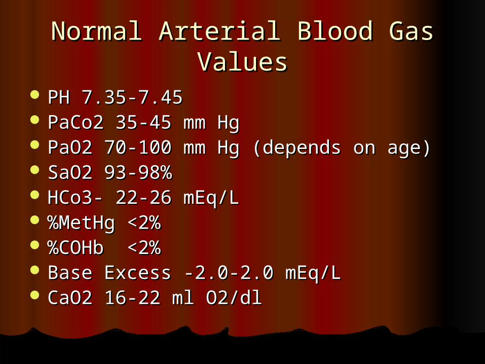
Normal Arterial Blood Gas ValuesNormal Arterial Blood Gas Values
PH 7.35-7.45PH 7.35-7.45 PaCo2 35-45 mm HgPaCo2 35-45 mm Hg PaO2 70-100 mm Hg (depends on age)PaO2 70-100 mm Hg (depends on age) SaO2 93-98%SaO2 93-98% HCo3- 22-26 mEq/LHCo3- 22-26 mEq/L %MetHg <2%%MetHg <2% %COHb <2%%COHb <2% Base Excess -2.0-2.0 mEq/LBase Excess -2.0-2.0 mEq/L CaO2 16-22 ml O2/dlCaO2 16-22 ml O2/dl
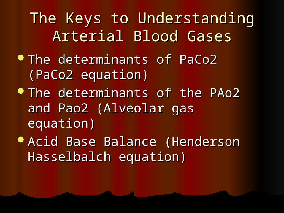
The Keys to Understanding The Keys to Understanding Arterial Blood GasesArterial Blood Gases
The determinants of PaCo2 (PaCo2 The determinants of PaCo2 (PaCo2 equation)equation)
The determinants of the PAo2 and The determinants of the PAo2 and Pao2 (Alveolar gas equation)Pao2 (Alveolar gas equation)
Acid Base Balance (Henderson Acid Base Balance (Henderson Hasselbalch equation)Hasselbalch equation)
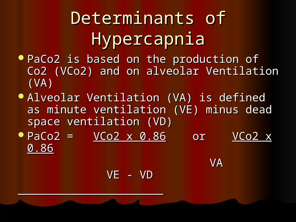
Determinants of HypercapniaDeterminants of Hypercapnia
PaCo2 is based on the production of Co2 PaCo2 is based on the production of Co2 (VCo2) and on alveolar Ventilation (VA)(VCo2) and on alveolar Ventilation (VA)
Alveolar Ventilation (VA) is defined as Alveolar Ventilation (VA) is defined as minute ventilation (VE) minus dead minute ventilation (VE) minus dead space ventilation (VD)space ventilation (VD)
PaCo2 = PaCo2 = VCo2 x 0.86VCo2 x 0.86 or or VCo2 x VCo2 x 0.860.86
VA VE - VDVA VE - VD
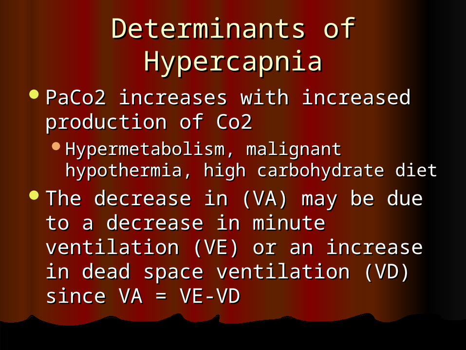
Determinants of HypercapniaDeterminants of Hypercapnia
PaCo2 increases with increased PaCo2 increases with increased production of Co2production of Co2Hypermetabolism, malignant Hypermetabolism, malignant
hypothermia, high carbohydrate diethypothermia, high carbohydrate dietThe decrease in (VA) may be due to The decrease in (VA) may be due to
a decrease in minute ventilation (VE) a decrease in minute ventilation (VE) or an increase in dead space or an increase in dead space ventilation (VD) since VA = VE-VDventilation (VD) since VA = VE-VD
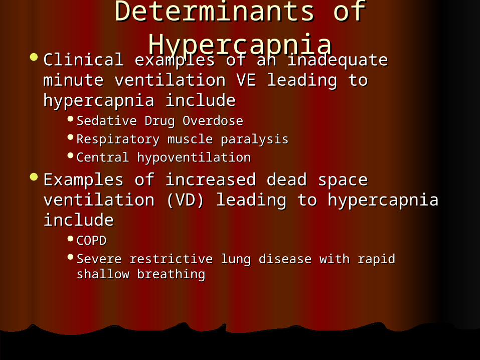
Determinants of HypercapniaDeterminants of HypercapniaClinical examples of an inadequate minute Clinical examples of an inadequate minute
ventilation VE leading to hypercapnia includeventilation VE leading to hypercapnia includeSedative Drug OverdoseSedative Drug OverdoseRespiratory muscle paralysisRespiratory muscle paralysisCentral hypoventilationCentral hypoventilation
Examples of increased dead space Examples of increased dead space ventilation (VD) leading to hypercapnia ventilation (VD) leading to hypercapnia includeinclude
COPDCOPDSevere restrictive lung disease with rapid shallow Severe restrictive lung disease with rapid shallow
breathingbreathing
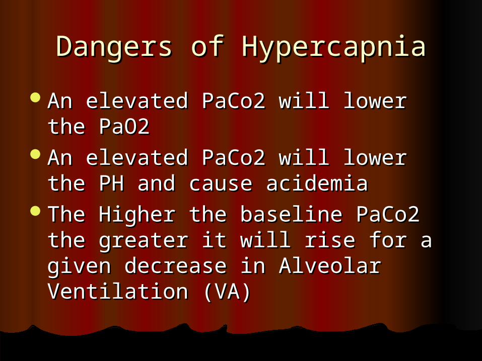
Dangers of HypercapniaDangers of Hypercapnia
An elevated PaCo2 will lower the An elevated PaCo2 will lower the PaO2 PaO2
An elevated PaCo2 will lower the PH An elevated PaCo2 will lower the PH and cause acidemiaand cause acidemia
The Higher the baseline PaCo2 the The Higher the baseline PaCo2 the greater it will rise for a given greater it will rise for a given decrease in Alveolar Ventilation (VA)decrease in Alveolar Ventilation (VA)
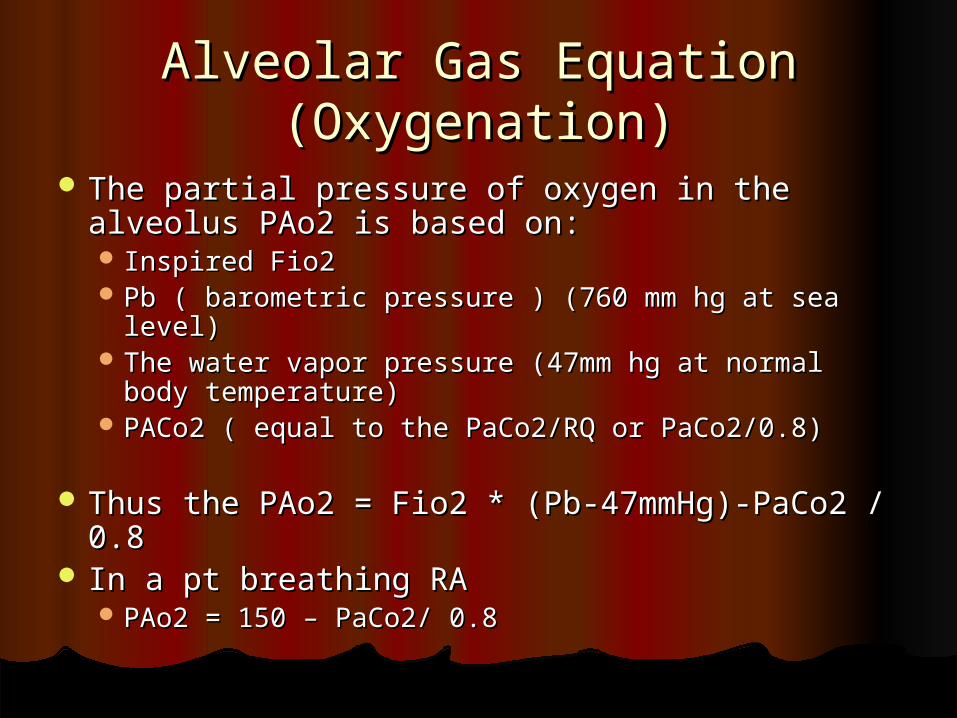
Alveolar Gas Equation Alveolar Gas Equation (Oxygenation)(Oxygenation)
The partial pressure of oxygen in the alveolus The partial pressure of oxygen in the alveolus PAo2 is based on:PAo2 is based on: Inspired Fio2Inspired Fio2 Pb ( barometric pressure ) (760 mm hg at sea level)Pb ( barometric pressure ) (760 mm hg at sea level) The water vapor pressure (47mm hg at normal The water vapor pressure (47mm hg at normal
body temperature)body temperature) PACo2 ( equal to the PaCo2/RQ or PaCo2/0.8)PACo2 ( equal to the PaCo2/RQ or PaCo2/0.8)
Thus the PAo2 = Fio2 * (Pb-47mmHg)-PaCo2 / Thus the PAo2 = Fio2 * (Pb-47mmHg)-PaCo2 / 0.80.8
In a pt breathing RAIn a pt breathing RA PAo2 = 150 – PaCo2/ 0.8PAo2 = 150 – PaCo2/ 0.8
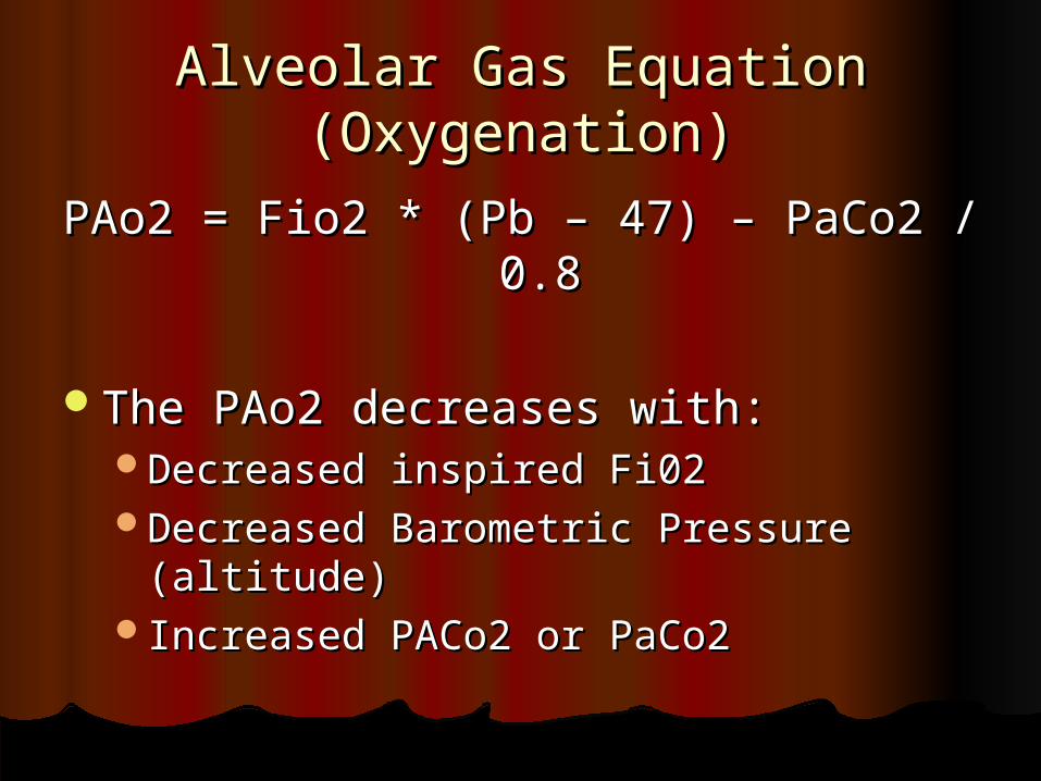
Alveolar Gas Equation Alveolar Gas Equation (Oxygenation)(Oxygenation)
PAo2 = Fio2 * (Pb – 47) – PaCo2 / 0.8PAo2 = Fio2 * (Pb – 47) – PaCo2 / 0.8
The PAo2 decreases with:The PAo2 decreases with:Decreased inspired Fi02Decreased inspired Fi02Decreased Barometric Pressure Decreased Barometric Pressure
(altitude)(altitude)Increased PACo2 or PaCo2Increased PACo2 or PaCo2
P(A-a) O2P(A-a) O2
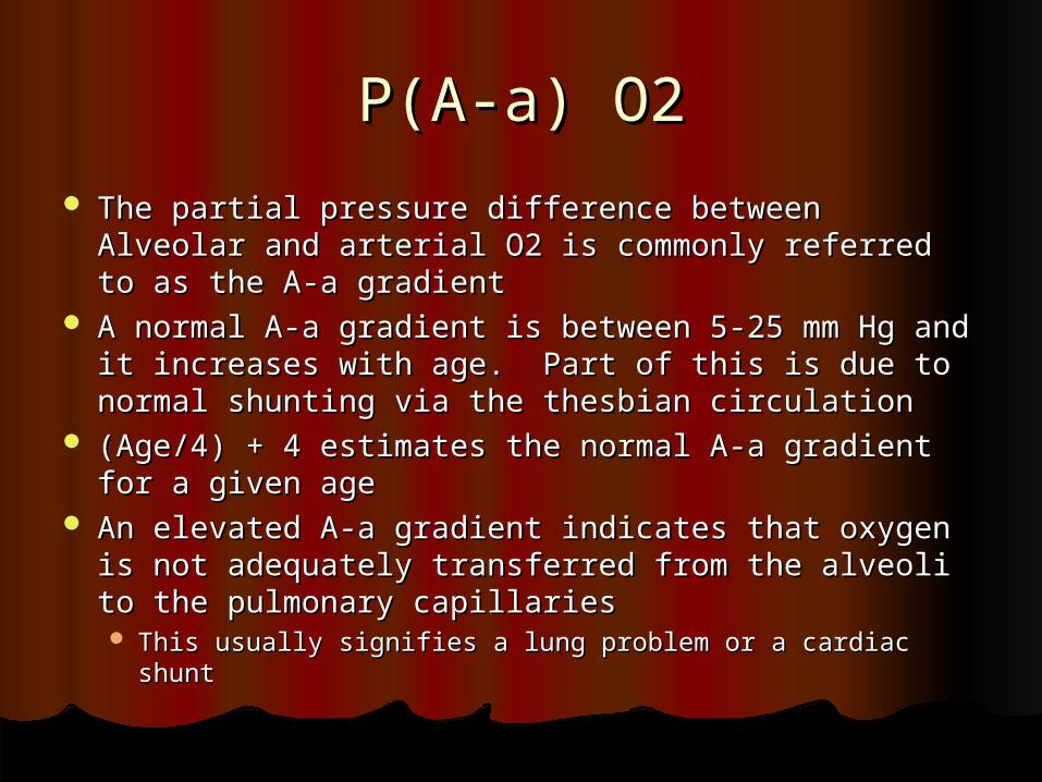
The partial pressure difference between Alveolar The partial pressure difference between Alveolar and arterial O2 is commonly referred to as the A-a and arterial O2 is commonly referred to as the A-a gradientgradient
A normal A-a gradient is between 5-25 mm Hg and A normal A-a gradient is between 5-25 mm Hg and it increases with age. Part of this is due to normal it increases with age. Part of this is due to normal shunting via the thesbian circulationshunting via the thesbian circulation
(Age/4) + 4 estimates the normal A-a gradient for (Age/4) + 4 estimates the normal A-a gradient for a given agea given age
An elevated A-a gradient indicates that oxygen is An elevated A-a gradient indicates that oxygen is not adequately transferred from the alveoli to the not adequately transferred from the alveoli to the pulmonary capillariespulmonary capillaries This usually signifies a lung problem or a cardiac shuntThis usually signifies a lung problem or a cardiac shunt
Causes of a Low PaO2Causes of a Low PaO2
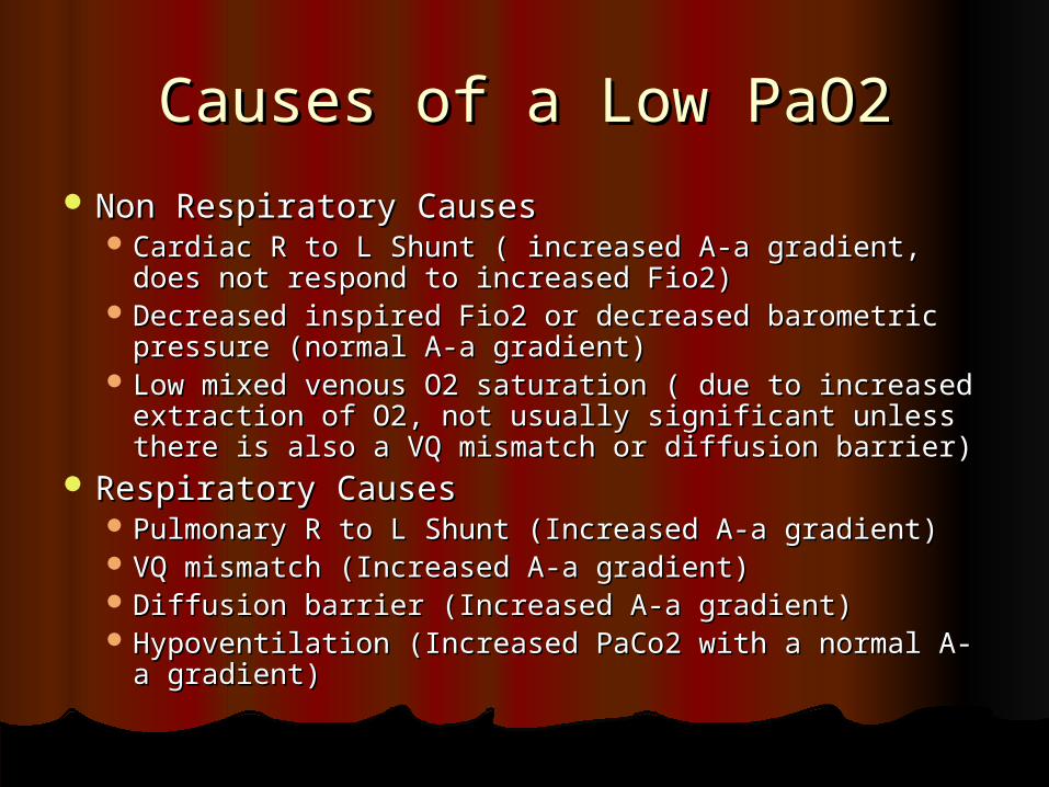
Non Respiratory CausesNon Respiratory Causes Cardiac R to L Shunt ( increased A-a gradient, does not Cardiac R to L Shunt ( increased A-a gradient, does not
respond to increased Fio2)respond to increased Fio2) Decreased inspired Fio2 or decreased barometric Decreased inspired Fio2 or decreased barometric
pressure (normal A-a gradient)pressure (normal A-a gradient) Low mixed venous O2 saturation ( due to increased Low mixed venous O2 saturation ( due to increased
extraction of O2, not usually significant unless there is extraction of O2, not usually significant unless there is also a VQ mismatch or diffusion barrier)also a VQ mismatch or diffusion barrier)
Respiratory CausesRespiratory Causes Pulmonary R to L Shunt (Increased A-a gradient)Pulmonary R to L Shunt (Increased A-a gradient) VQ mismatch (Increased A-a gradient)VQ mismatch (Increased A-a gradient) Diffusion barrier (Increased A-a gradient)Diffusion barrier (Increased A-a gradient) Hypoventilation (Increased PaCo2 with a normal A-a Hypoventilation (Increased PaCo2 with a normal A-a
gradient)gradient)
Acid Base BalanceAcid Base Balance
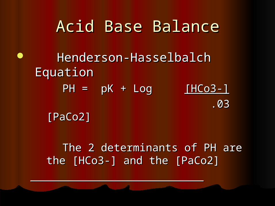
Henderson-Hasselbalch EquationHenderson-Hasselbalch Equation PH = pK + Log PH = pK + Log [HCo3-][HCo3-]
.03 [PaCo2].03 [PaCo2]
The 2 determinants of PH are the The 2 determinants of PH are the [HCo3-] and the [PaCo2][HCo3-] and the [PaCo2]
Acid Base BalanceAcid Base Balance
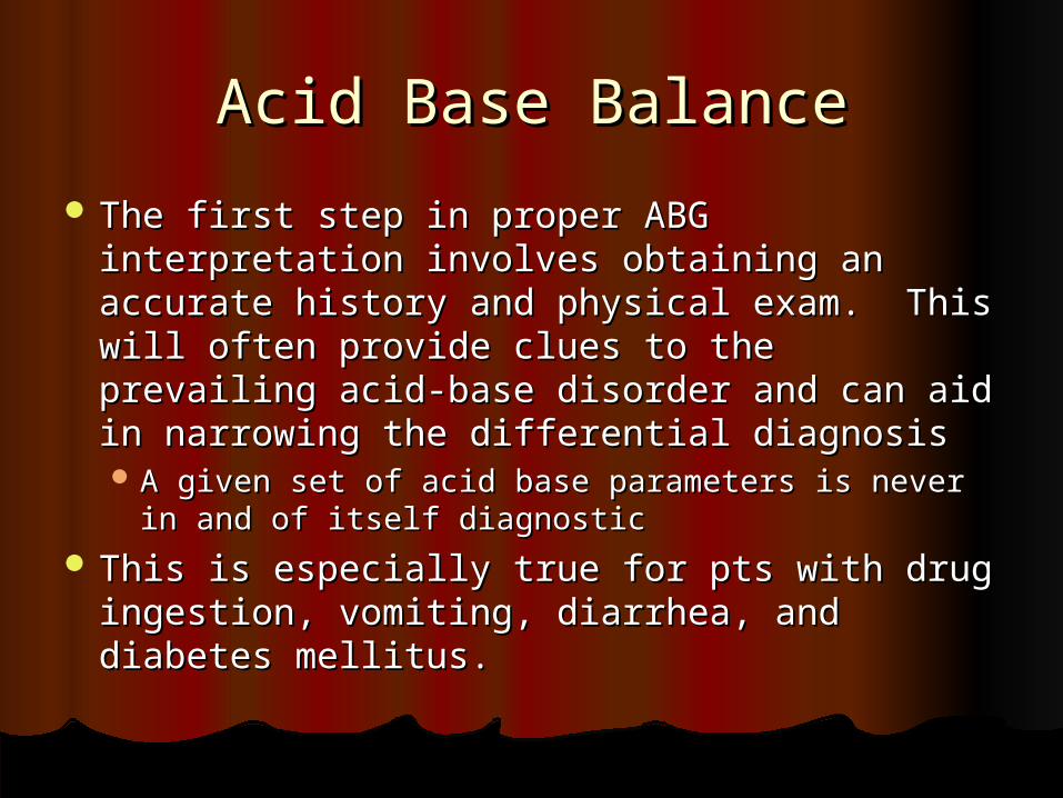
The first step in proper ABG interpretation The first step in proper ABG interpretation involves obtaining an accurate history and involves obtaining an accurate history and physical exam. This will often provide clues physical exam. This will often provide clues to the prevailing acid-base disorder and can to the prevailing acid-base disorder and can aid in narrowing the differential diagnosisaid in narrowing the differential diagnosis A given set of acid base parameters is never in A given set of acid base parameters is never in
and of itself diagnosticand of itself diagnostic This is especially true for pts with drug This is especially true for pts with drug
ingestion, vomiting, diarrhea, and diabetes ingestion, vomiting, diarrhea, and diabetes mellitus. mellitus.
Verifying the Accuracy of the Verifying the Accuracy of the DataData
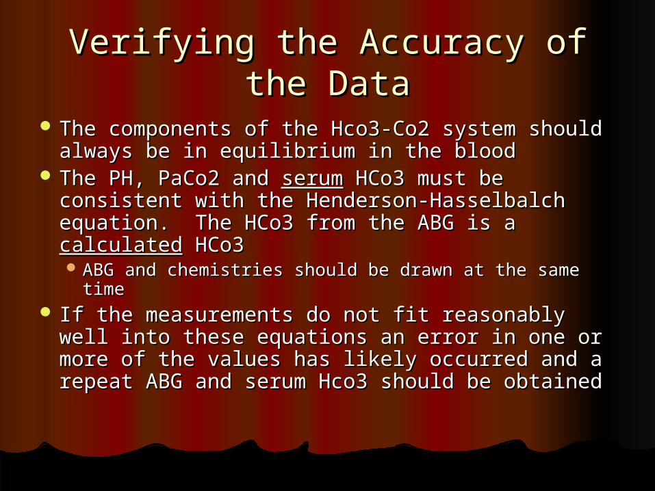
The components of the Hco3-Co2 system The components of the Hco3-Co2 system should always be in equilibrium in the bloodshould always be in equilibrium in the blood
The PH, PaCo2 and The PH, PaCo2 and serumserum HCo3 must be HCo3 must be consistent with the Henderson-Hasselbalch consistent with the Henderson-Hasselbalch equation. The HCo3 from the ABG is a equation. The HCo3 from the ABG is a calculatedcalculated HCo3 HCo3 ABG and chemistries should be drawn at the same ABG and chemistries should be drawn at the same
timetime If the measurements do not fit reasonably well If the measurements do not fit reasonably well
into these equations an error in one or more into these equations an error in one or more of the values has likely occurred and a repeat of the values has likely occurred and a repeat ABG and serum Hco3 should be obtainedABG and serum Hco3 should be obtained
Henderson EquationHenderson Equation
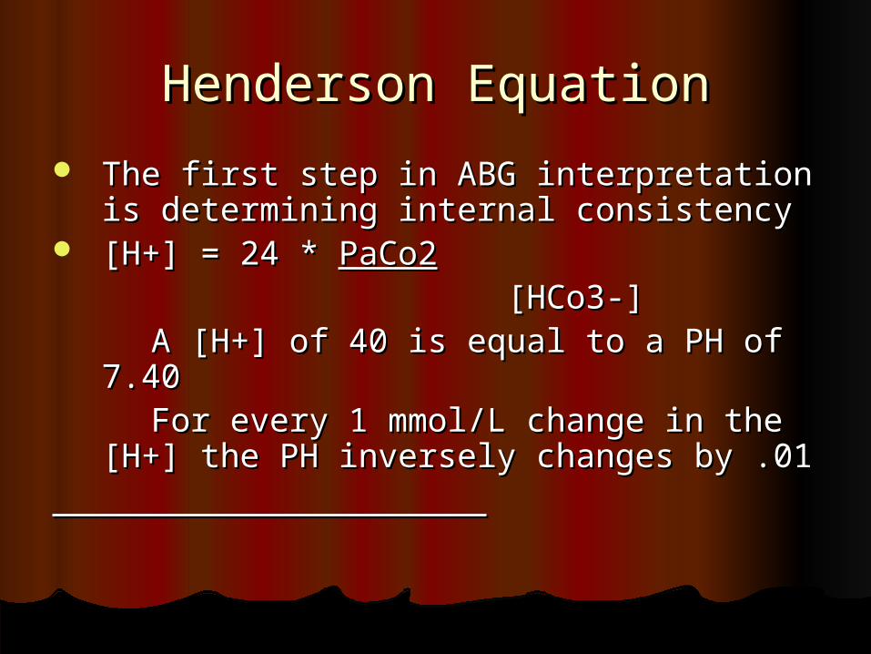
The first step in ABG interpretation is The first step in ABG interpretation is determining internal consistencydetermining internal consistency
[H+] = 24 * [H+] = 24 * PaCo2PaCo2 [HCo3-][HCo3-] A [H+] of 40 is equal to a PH of 7.40 A [H+] of 40 is equal to a PH of 7.40 For every 1 mmol/L change in the For every 1 mmol/L change in the
[H+] the PH inversely changes by .01[H+] the PH inversely changes by .01
Values for PH and corresponding Values for PH and corresponding [H+][H+]
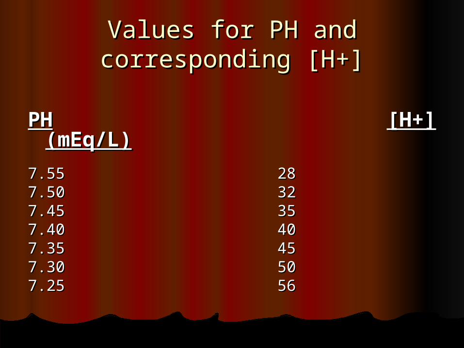
PHPH [H+] (mEq/L)[H+] (mEq/L)
7.557.55 28287.507.50 32327.457.45 35357.407.40 40407.357.35 45457.307.30 50507.257.25 5656
Determining the Serum Anion Determining the Serum Anion GapGap
The anion gap is the difference between the The anion gap is the difference between the unmeasured anions (negatively charged unmeasured anions (negatively charged molecules) and the unmeasured cations molecules) and the unmeasured cations (positively charged molecules) in the serum(positively charged molecules) in the serum
The Concentration of all anions and cations in the The Concentration of all anions and cations in the serum must balanceserum must balance
Therefore: Na + UC = [Cl + HCo3] + UATherefore: Na + UC = [Cl + HCo3] + UA Rearranged UA – UC = Na – [Cl + HCo3]Rearranged UA – UC = Na – [Cl + HCo3] Normal in most labs is 10 + or - 2Normal in most labs is 10 + or - 2 Hypoalbuminemia, hyponatremia, and increased Hypoalbuminemia, hyponatremia, and increased
[k], [mg],[ca] and [NH4] may all lower the anion [k], [mg],[ca] and [NH4] may all lower the anion gap. gap.
A decrease of the serum albumin by 50% can A decrease of the serum albumin by 50% can decrease the anion gap by 5 meq/l. decrease the anion gap by 5 meq/l.
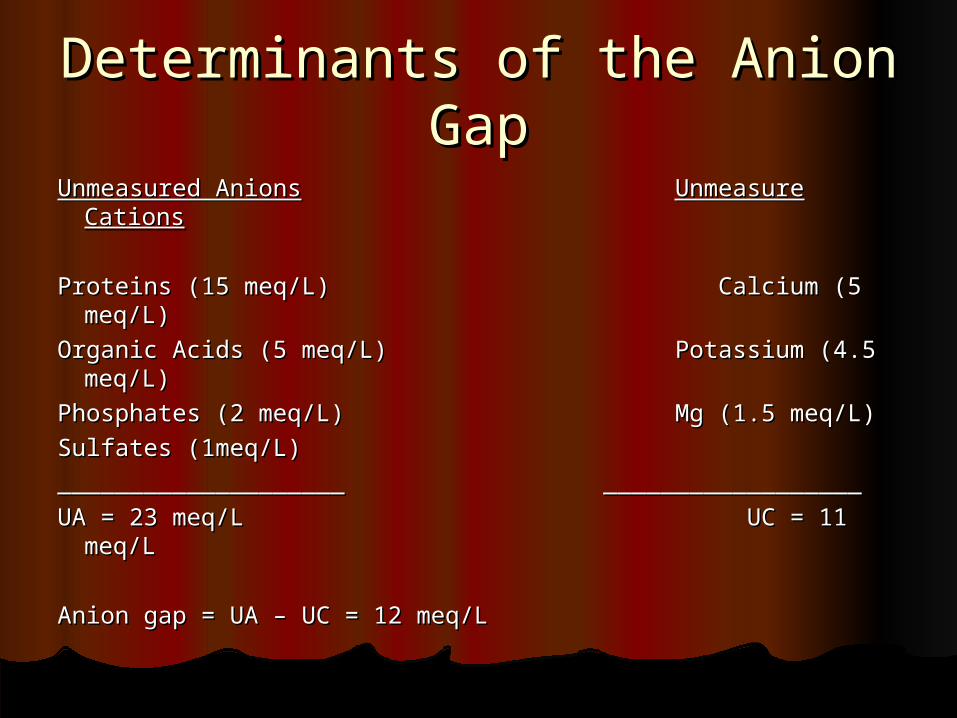
Determinants of the Anion Determinants of the Anion GapGap
Unmeasured AnionsUnmeasured Anions Unmeasure CationsUnmeasure Cations
Proteins (15 meq/L) Calcium (5 meq/L)Proteins (15 meq/L) Calcium (5 meq/L)
Organic Acids (5 meq/L) Potassium (4.5 Organic Acids (5 meq/L) Potassium (4.5 meq/L)meq/L)
Phosphates (2 meq/L) Mg (1.5 meq/L)Phosphates (2 meq/L) Mg (1.5 meq/L)
Sulfates (1meq/L)Sulfates (1meq/L)
____________________ ______________________________________ __________________
UA = 23 meq/L UC = 11 meq/LUA = 23 meq/L UC = 11 meq/L
Anion gap = UA – UC = 12 meq/LAnion gap = UA – UC = 12 meq/L
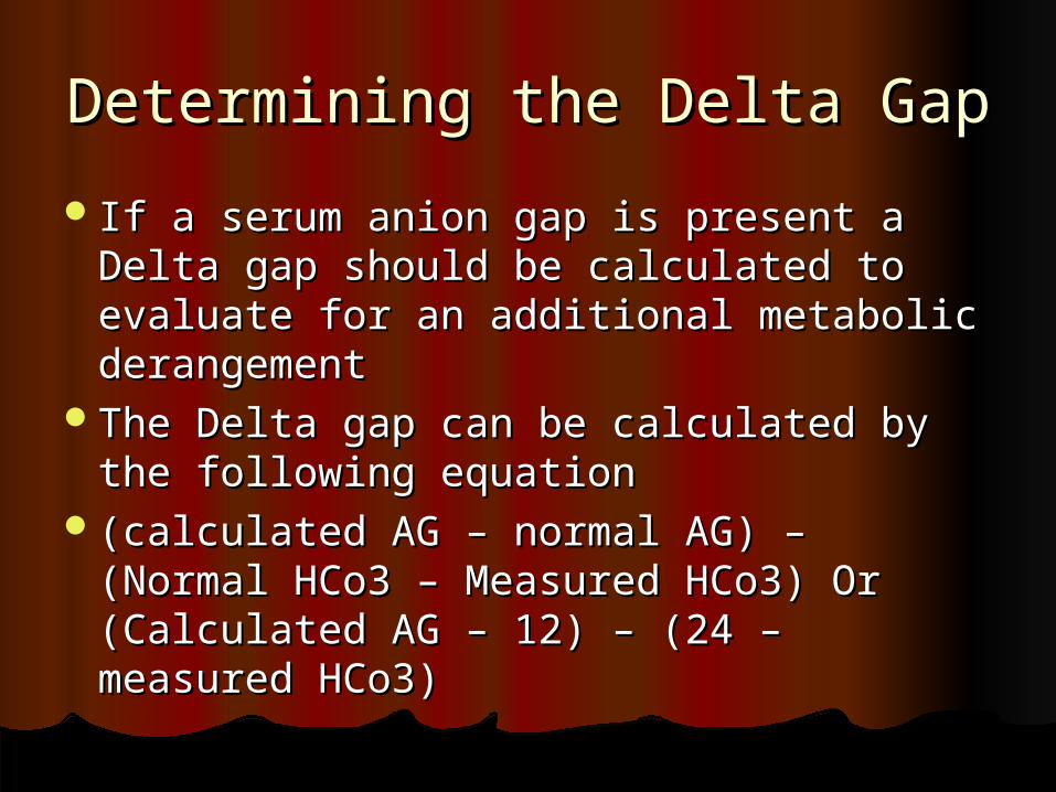
Determining the Delta GapDetermining the Delta Gap
If a serum anion gap is present a Delta If a serum anion gap is present a Delta gap should be calculated to evaluate for gap should be calculated to evaluate for an additional metabolic derangement an additional metabolic derangement
The Delta gap can be calculated by the The Delta gap can be calculated by the following equationfollowing equation
(calculated AG – normal AG) – (Normal (calculated AG – normal AG) – (Normal HCo3 – Measured HCo3) Or (Calculated HCo3 – Measured HCo3) Or (Calculated AG – 12) – (24 – measured HCo3)AG – 12) – (24 – measured HCo3)
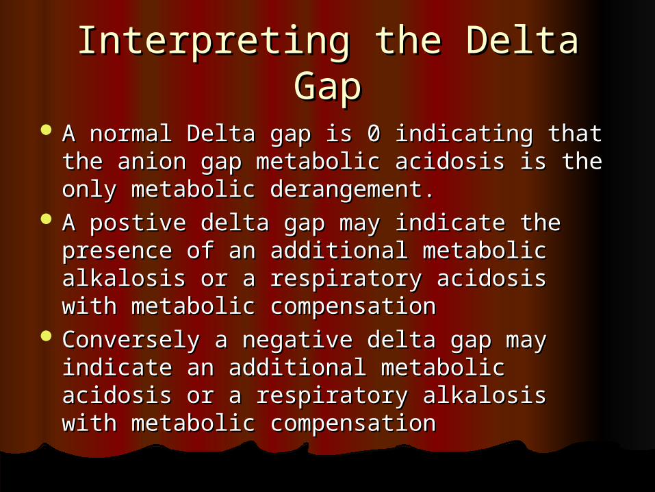
Interpreting the Delta GapInterpreting the Delta Gap
A normal Delta gap is 0 indicating that the A normal Delta gap is 0 indicating that the anion gap metabolic acidosis is the only anion gap metabolic acidosis is the only metabolic derangement. metabolic derangement.
A postive delta gap may indicate the A postive delta gap may indicate the presence of an additional metabolic alkalosis presence of an additional metabolic alkalosis or a respiratory acidosis with metabolic or a respiratory acidosis with metabolic compensationcompensation
Conversely a negative delta gap may indicate Conversely a negative delta gap may indicate an additional metabolic acidosis or a an additional metabolic acidosis or a respiratory alkalosis with metabolic respiratory alkalosis with metabolic compensationcompensation
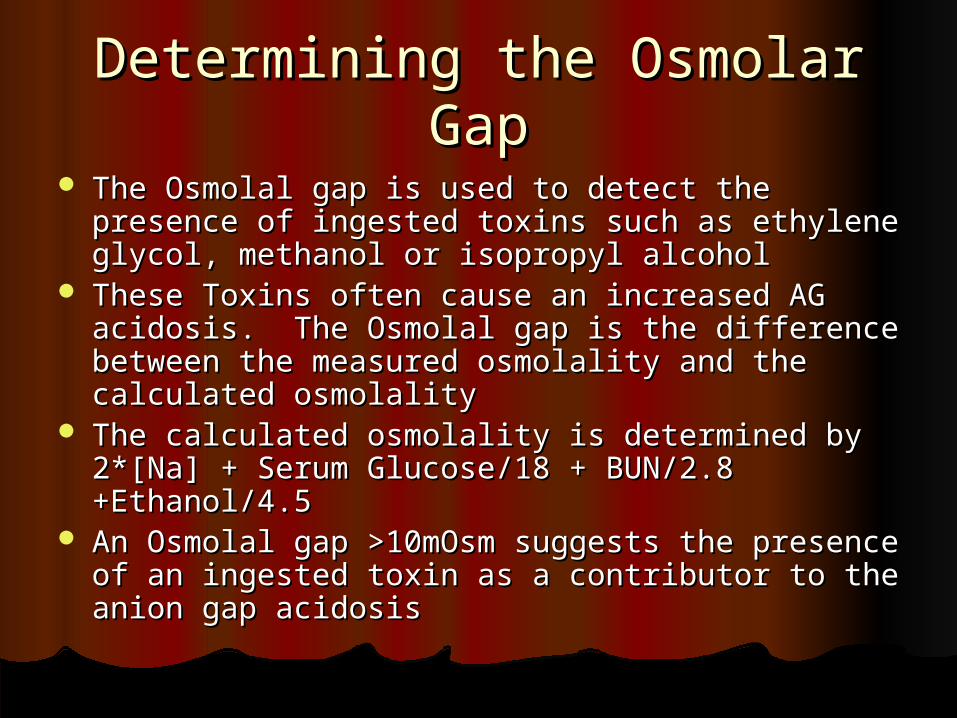
Determining the Osmolar GapDetermining the Osmolar Gap
The Osmolal gap is used to detect the presence The Osmolal gap is used to detect the presence of ingested toxins such as ethylene glycol, of ingested toxins such as ethylene glycol, methanol or isopropyl alcoholmethanol or isopropyl alcohol
These Toxins often cause an increased AG These Toxins often cause an increased AG acidosis. The Osmolal gap is the difference acidosis. The Osmolal gap is the difference between the measured osmolality and the between the measured osmolality and the calculated osmolalitycalculated osmolality
The calculated osmolality is determined by 2*[Na] The calculated osmolality is determined by 2*[Na] + Serum Glucose/18 + BUN/2.8 +Ethanol/4.5+ Serum Glucose/18 + BUN/2.8 +Ethanol/4.5
An Osmolal gap >10mOsm suggests the presence An Osmolal gap >10mOsm suggests the presence of an ingested toxin as a contributor to the anion of an ingested toxin as a contributor to the anion gap acidosisgap acidosis
Simple Acid Base Simple Acid Base AbnormalitiesAbnormalities
The term simple acid-base disorder The term simple acid-base disorder denotes the presence of a single denotes the presence of a single abnormality associated with an abnormality associated with an expected compensatory responseexpected compensatory response
The Four simple acid base disorders The Four simple acid base disorders are metabolic acidosis, metabolic are metabolic acidosis, metabolic alkalosis, respiratory acidosis (both alkalosis, respiratory acidosis (both acute and chronic) and respiratory acute and chronic) and respiratory alkalosis (acute and chronic)alkalosis (acute and chronic)
Simple Acid Base DisordersSimple Acid Base Disorders
Metabolic Acidosis – primary disturbance is Metabolic Acidosis – primary disturbance is a decrease in HCo3 with compensatory a decrease in HCo3 with compensatory hyperventilation and a decreased PaCo2. hyperventilation and a decreased PaCo2.
The Predicted PaCo2 is determined using the The Predicted PaCo2 is determined using the Winter’s equationWinter’s equation
PaCo2 = (1.5 x HCo3) + 8 + or – 2PaCo2 = (1.5 x HCo3) + 8 + or – 2 Any significant deviation from the predicted Any significant deviation from the predicted
PaCo2 signifies an additional respiratory PaCo2 signifies an additional respiratory disorderdisorder
A PaCo2 higher than predicted signifies an additional A PaCo2 higher than predicted signifies an additional Respiratory AcidosisRespiratory Acidosis
A PaCo2 lower than predicted signifies an additional A PaCo2 lower than predicted signifies an additional Respiratory AlkalosisRespiratory Alkalosis
Simple Acid Base DisordersSimple Acid Base Disorders
Metabolic Alkalosis – Primary Metabolic Alkalosis – Primary disturbance is an increase in HCo3 with disturbance is an increase in HCo3 with compensatory hypoventilation and an compensatory hypoventilation and an increased PaCo2increased PaCo2
Predicted PaCo2 = (0.7 x HCo3) + 21 + or – Predicted PaCo2 = (0.7 x HCo3) + 21 + or – 1.51.5
Any significant deviation from the predicted Any significant deviation from the predicted PaCo2 signifies an additional Respiratory PaCo2 signifies an additional Respiratory disorderdisorder
Simple Acid Base DisordersSimple Acid Base Disorders
Respiratory Acidosis – primary Respiratory Acidosis – primary derangement is an increased PaCo2 with a derangement is an increased PaCo2 with a compensatory increase in HCo3. compensatory increase in HCo3. In acute resp acidosis for every increase of 10 In acute resp acidosis for every increase of 10
mm Hg of PaCo2 the PH should drop by .08. mm Hg of PaCo2 the PH should drop by .08. Increase in [HCo3] = change PaCo2/10 + or - 3Increase in [HCo3] = change PaCo2/10 + or - 3
In Chronic resp acidosis for every 10 mm Hg In Chronic resp acidosis for every 10 mm Hg rise of the PaCo2 the PH should drop by .03 as rise of the PaCo2 the PH should drop by .03 as compensation compensation butbut not correction not correction
Increase in [HCo3] = 3.5 x change Paco2/10Increase in [HCo3] = 3.5 x change Paco2/10 Changes greater than those predicted signify Changes greater than those predicted signify
an additional metabolic disorderan additional metabolic disorder
Simple Acid Base DisordersSimple Acid Base Disorders
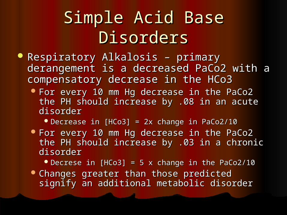
Respiratory Alkalosis – primary Respiratory Alkalosis – primary derangement is a decreased PaCo2 with a derangement is a decreased PaCo2 with a compensatory decrease in the HCo3compensatory decrease in the HCo3 For every 10 mm Hg decrease in the PaCo2 the For every 10 mm Hg decrease in the PaCo2 the
PH should increase by .08 in an acute disorderPH should increase by .08 in an acute disorderDecrease in [HCo3] = 2x change in PaCo2/10Decrease in [HCo3] = 2x change in PaCo2/10
For every 10 mm Hg decrease in the PaCo2 the For every 10 mm Hg decrease in the PaCo2 the PH should increase by .03 in a chronic disorderPH should increase by .03 in a chronic disorder
Decrese in [HCo3] = 5 x change in the PaCo2/10Decrese in [HCo3] = 5 x change in the PaCo2/10 Changes greater than those predicted signify Changes greater than those predicted signify
an additional metabolic disorderan additional metabolic disorder
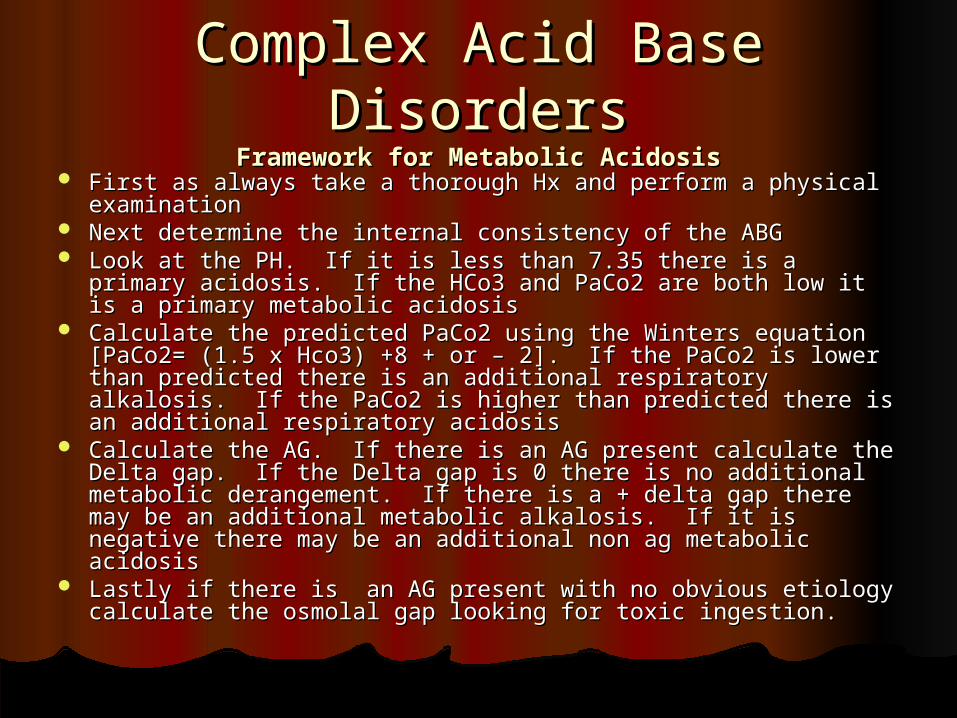
Complex Acid Base DisordersComplex Acid Base DisordersFramework for Metabolic AcidosisFramework for Metabolic Acidosis
First as always take a thorough Hx and perform a physical First as always take a thorough Hx and perform a physical examinationexamination
Next determine the internal consistency of the ABGNext determine the internal consistency of the ABG Look at the PH. If it is less than 7.35 there is a primary Look at the PH. If it is less than 7.35 there is a primary
acidosis. If the HCo3 and PaCo2 are both low it is a primary acidosis. If the HCo3 and PaCo2 are both low it is a primary metabolic acidosismetabolic acidosis
Calculate the predicted PaCo2 using the Winters equation Calculate the predicted PaCo2 using the Winters equation [PaCo2= (1.5 x Hco3) +8 + or – 2]. If the PaCo2 is lower than [PaCo2= (1.5 x Hco3) +8 + or – 2]. If the PaCo2 is lower than predicted there is an additional respiratory alkalosis. If the predicted there is an additional respiratory alkalosis. If the PaCo2 is higher than predicted there is an additional PaCo2 is higher than predicted there is an additional respiratory acidosisrespiratory acidosis
Calculate the AG. If there is an AG present calculate the Calculate the AG. If there is an AG present calculate the Delta gap. If the Delta gap is 0 there is no additional Delta gap. If the Delta gap is 0 there is no additional metabolic derangement. If there is a + delta gap there may metabolic derangement. If there is a + delta gap there may be an additional metabolic alkalosis. If it is negative there be an additional metabolic alkalosis. If it is negative there may be an additional non ag metabolic acidosismay be an additional non ag metabolic acidosis
Lastly if there is an AG present with no obvious etiology Lastly if there is an AG present with no obvious etiology calculate the osmolal gap looking for toxic ingestion. calculate the osmolal gap looking for toxic ingestion.
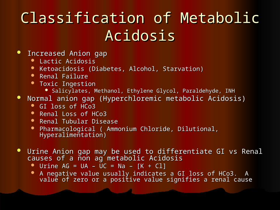
Classification of Metabolic Classification of Metabolic AcidosisAcidosis
Increased Anion gapIncreased Anion gap Lactic AcidosisLactic Acidosis Ketoacidosis (Diabetes, Alcohol, Starvation)Ketoacidosis (Diabetes, Alcohol, Starvation) Renal FailureRenal Failure Toxic IngestionToxic Ingestion
Salicylates, Methanol, Ethylene Glycol, Paraldehyde, INHSalicylates, Methanol, Ethylene Glycol, Paraldehyde, INH Normal anion gap (Hyperchloremic metabolic Acidosis)Normal anion gap (Hyperchloremic metabolic Acidosis)
GI loss of HCo3GI loss of HCo3 Renal Loss of HCo3Renal Loss of HCo3 Renal Tubular DiseaseRenal Tubular Disease Pharmacological ( Ammonium Chloride, Dilutional, Hyperalimentation)Pharmacological ( Ammonium Chloride, Dilutional, Hyperalimentation)
Urine Anion gap may be used to differentiate GI vs Renal causes of Urine Anion gap may be used to differentiate GI vs Renal causes of a non ag metabolic Acidosisa non ag metabolic Acidosis Urine AG = UA – UC = Na – [K + Cl] Urine AG = UA – UC = Na – [K + Cl] A negative value usually indicates a GI loss of HCo3. A value of zero or A negative value usually indicates a GI loss of HCo3. A value of zero or
a positive value signifies a renal causea positive value signifies a renal cause
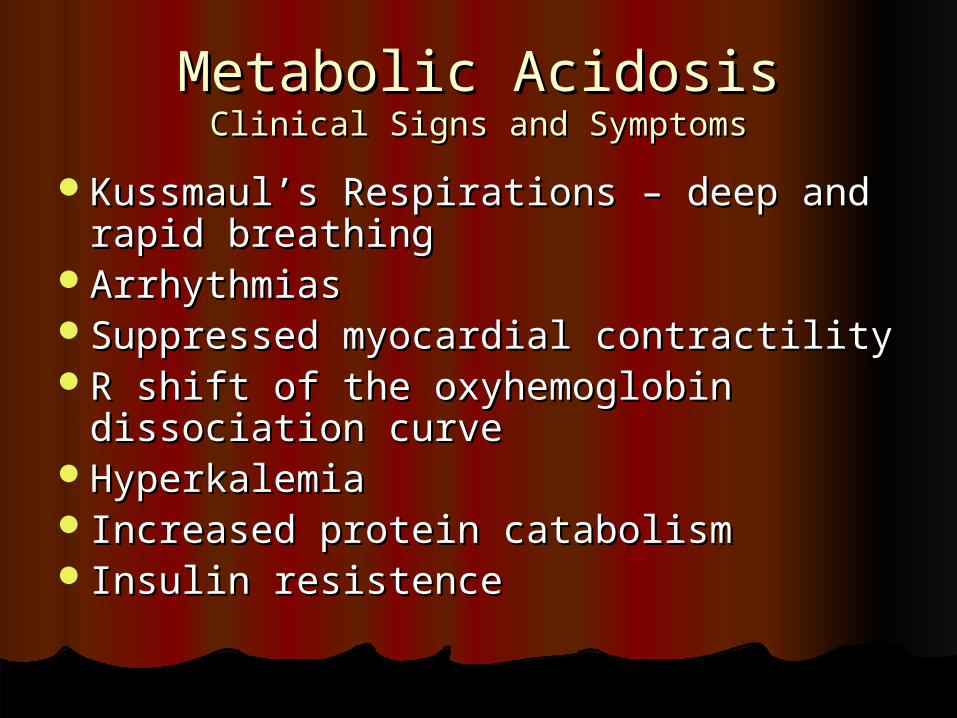
Metabolic AcidosisMetabolic AcidosisClinical Signs and SymptomsClinical Signs and Symptoms
Kussmaul’s Respirations – deep and Kussmaul’s Respirations – deep and rapid breathingrapid breathing
ArrhythmiasArrhythmiasSuppressed myocardial contractilitySuppressed myocardial contractilityR shift of the oxyhemoglobin R shift of the oxyhemoglobin
dissociation curvedissociation curveHyperkalemiaHyperkalemia Increased protein catabolismIncreased protein catabolism Insulin resistenceInsulin resistence
Metabolic AlkalosisMetabolic Alkalosis
The primary disturbance in metabolic The primary disturbance in metabolic alkalosis is an increase in HCo3 or the loss of alkalosis is an increase in HCo3 or the loss of acid. acid.
The compensatory respiratory response is a The compensatory respiratory response is a rise in PaCo2. rise in PaCo2. Calculate the predicted PaCo2 using the following Calculate the predicted PaCo2 using the following
equation: PaCo2= (0.7 x HCo3) +21 + or – 1.5. equation: PaCo2= (0.7 x HCo3) +21 + or – 1.5. If the PaCo2 is less than predicted there is an additional If the PaCo2 is less than predicted there is an additional
respiratory alkalosis. If the PaCo2 is higher than respiratory alkalosis. If the PaCo2 is higher than predicted there is an additional respiratory acidosis. predicted there is an additional respiratory acidosis.
Metabolic Alkalosis may be Hypovolemic Cl- Metabolic Alkalosis may be Hypovolemic Cl- depleted or hypervolemic Cl- expandeddepleted or hypervolemic Cl- expanded
Metabolic AlkalosisMetabolic Alkalosis The etiology of the hypovolemic Cl - depleted form includes:The etiology of the hypovolemic Cl - depleted form includes:
GI loss of H+GI loss of H+ Vomiting, Gastric suctioning, Cl- rich diarrheaVomiting, Gastric suctioning, Cl- rich diarrhea Renal loss of H+Renal loss of H+ DiureticsDiuretics Post hypercapniaPost hypercapnia High dose carbenicillinHigh dose carbenicillin
The etiology of the hypervolemic Cl-expanded form includes:The etiology of the hypervolemic Cl-expanded form includes: Primary hyperaldosteronism, hypercortisolismPrimary hyperaldosteronism, hypercortisolism ACTH excess, Hydrocortisone and mineralicorticoid excessACTH excess, Hydrocortisone and mineralicorticoid excess Renin secreting tumorRenin secreting tumor Hypokalemia, milk-alkali syndrome, Massive blood transfusionHypokalemia, milk-alkali syndrome, Massive blood transfusion
History and Urine Chloride can be helpful in differentiating the History and Urine Chloride can be helpful in differentiating the twotwo
Urine Chloride is usually less than 20 in the chloride depleted form Urine Chloride is usually less than 20 in the chloride depleted form and greater than 20 in the chloride expanded formand greater than 20 in the chloride expanded form
Metabolic AlkalosisMetabolic AlkalosisClinical Signs and SymptomsClinical Signs and Symptoms
TachycardiaTachycardiaArrhythmiasArrhythmiasObtunted mental StatusObtunted mental Status Increased risk of seizuresIncreased risk of seizuresDecreased cerebral blood flowDecreased cerebral blood flowHypocalcemiaHypocalcemiahypokalemiahypokalemia
Respiratory AcidosisRespiratory Acidosis The major disturbance in respiratory acidosis is The major disturbance in respiratory acidosis is
ineffective ventilation and or increased production ineffective ventilation and or increased production of Co2. of Co2.
In acute disorders for every increase of 10 mm Hg In acute disorders for every increase of 10 mm Hg in the PaCo2 the PH decreases by .08. in the PaCo2 the PH decreases by .08. If there is a further decrease in the PH there is an If there is a further decrease in the PH there is an
additional metabolic acidosis. Likewise if the PH is higher additional metabolic acidosis. Likewise if the PH is higher than predicted there is likely a metabolic alkalosis.than predicted there is likely a metabolic alkalosis.
In chronic respiratory acidosis for every 10 mm Hg In chronic respiratory acidosis for every 10 mm Hg increase of the PaCo2 the PH decreases by .03. increase of the PaCo2 the PH decreases by .03. Further changes signify additional metabolic Further changes signify additional metabolic derangementsderangements The Maximum Renal compensation for chronic The Maximum Renal compensation for chronic
respiratory acidosis is a HCo3 of 45. If the serum HCo3 is respiratory acidosis is a HCo3 of 45. If the serum HCo3 is greater than 45 there must be an additional metabolic greater than 45 there must be an additional metabolic alkalosisalkalosis
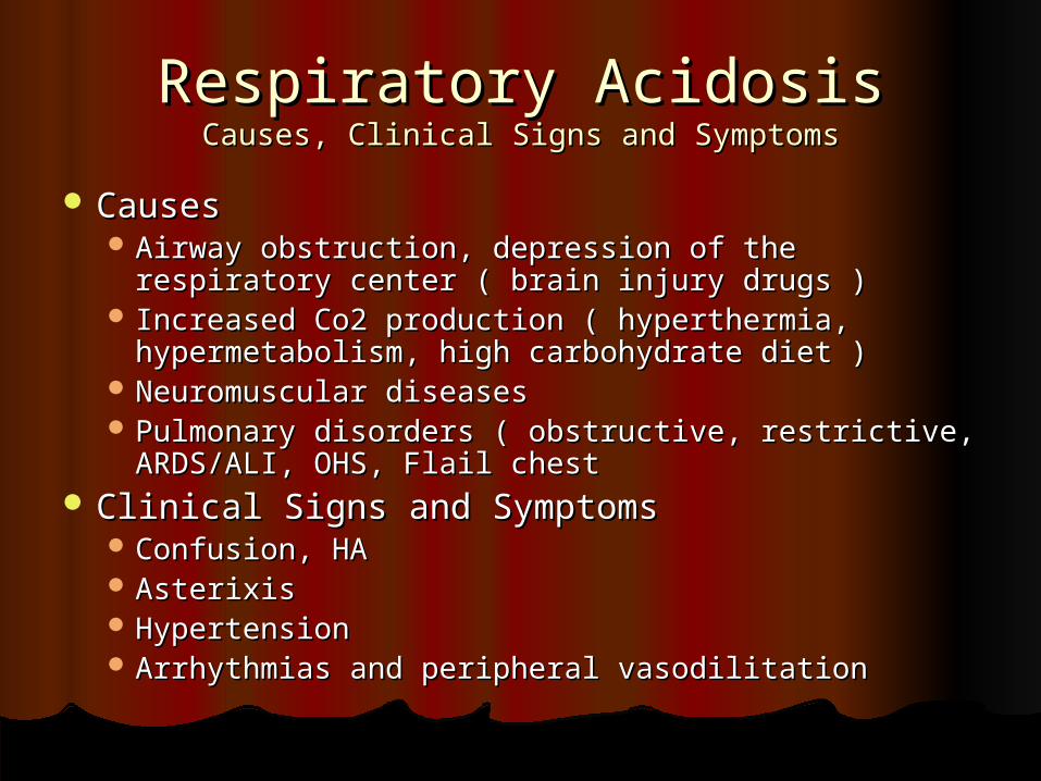
Respiratory AcidosisRespiratory AcidosisCauses, Clinical Signs and SymptomsCauses, Clinical Signs and Symptoms
CausesCauses Airway obstruction, depression of the respiratory Airway obstruction, depression of the respiratory
center ( brain injury drugs )center ( brain injury drugs ) Increased Co2 production ( hyperthermia, Increased Co2 production ( hyperthermia,
hypermetabolism, high carbohydrate diet )hypermetabolism, high carbohydrate diet ) Neuromuscular diseasesNeuromuscular diseases Pulmonary disorders ( obstructive, restrictive, Pulmonary disorders ( obstructive, restrictive,
ARDS/ALI, OHS, Flail chestARDS/ALI, OHS, Flail chest Clinical Signs and SymptomsClinical Signs and Symptoms
Confusion, HAConfusion, HA AsterixisAsterixis HypertensionHypertension Arrhythmias and peripheral vasodilitationArrhythmias and peripheral vasodilitation
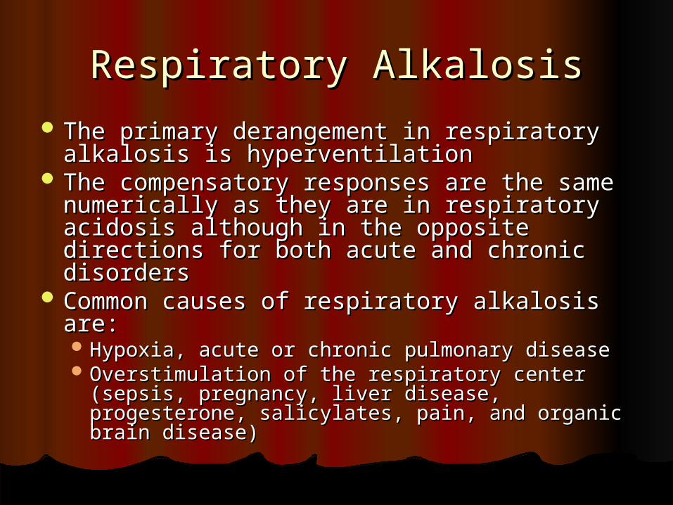
Respiratory AlkalosisRespiratory Alkalosis The primary derangement in respiratory The primary derangement in respiratory
alkalosis is hyperventilationalkalosis is hyperventilation The compensatory responses are the same The compensatory responses are the same
numerically as they are in respiratory numerically as they are in respiratory acidosis although in the opposite directions acidosis although in the opposite directions for both acute and chronic disordersfor both acute and chronic disorders
Common causes of respiratory alkalosis are:Common causes of respiratory alkalosis are: Hypoxia, acute or chronic pulmonary diseaseHypoxia, acute or chronic pulmonary disease Overstimulation of the respiratory center (sepsis, Overstimulation of the respiratory center (sepsis,
pregnancy, liver disease, progesterone, pregnancy, liver disease, progesterone, salicylates, pain, and organic brain disease)salicylates, pain, and organic brain disease)
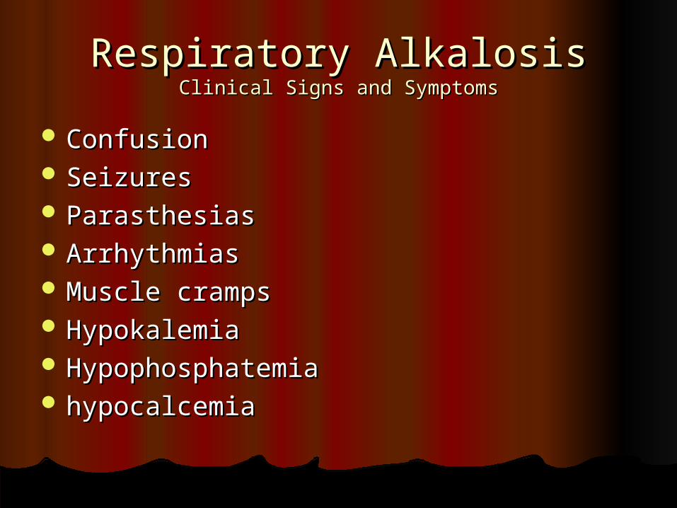
Respiratory AlkalosisRespiratory AlkalosisClinical Signs and SymptomsClinical Signs and Symptoms
ConfusionConfusion SeizuresSeizures ParasthesiasParasthesias ArrhythmiasArrhythmias Muscle crampsMuscle cramps HypokalemiaHypokalemia HypophosphatemiaHypophosphatemia hypocalcemiahypocalcemia
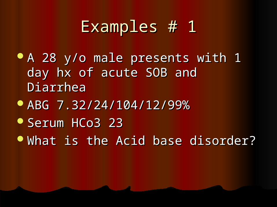
Examples # 1Examples # 1
A 28 y/o male presents with 1 day hx A 28 y/o male presents with 1 day hx of acute SOB and Diarrheaof acute SOB and Diarrhea
ABG 7.32/24/104/12/99%ABG 7.32/24/104/12/99%Serum HCo3 23Serum HCo3 23What is the Acid base disorder?What is the Acid base disorder?
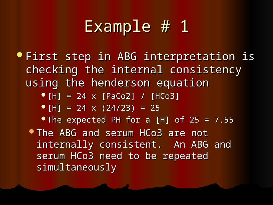
Example # 1Example # 1
First step in ABG interpretation is First step in ABG interpretation is checking the internal consistency checking the internal consistency using the henderson equationusing the henderson equation
[H] = 24 x [PaCo2] / [HCo3][H] = 24 x [PaCo2] / [HCo3][H] = 24 x (24/23) = 25[H] = 24 x (24/23) = 25The expected PH for a [H] of 25 = 7.55The expected PH for a [H] of 25 = 7.55
The ABG and serum HCo3 are not The ABG and serum HCo3 are not internally consistent. An ABG and internally consistent. An ABG and serum HCo3 need to be repeated serum HCo3 need to be repeated simultaneouslysimultaneously
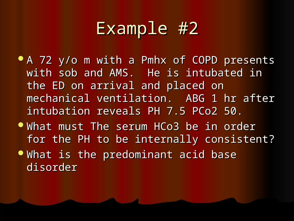
Example #2Example #2
A 72 y/o m with a Pmhx of COPD presents A 72 y/o m with a Pmhx of COPD presents with sob and AMS. He is intubated in the with sob and AMS. He is intubated in the ED on arrival and placed on mechanical ED on arrival and placed on mechanical ventilation. ABG 1 hr after intubation ventilation. ABG 1 hr after intubation reveals PH 7.5 PCo2 50. reveals PH 7.5 PCo2 50.
What must The serum HCo3 be in order What must The serum HCo3 be in order for the PH to be internally consistent?for the PH to be internally consistent?
What is the predominant acid base What is the predominant acid base disorderdisorder
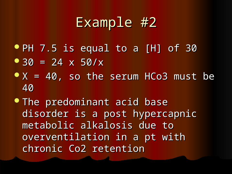
Example #2Example #2
PH 7.5 is equal to a [H] of 30PH 7.5 is equal to a [H] of 3030 = 24 x 50/x30 = 24 x 50/xX = 40, so the serum HCo3 must be X = 40, so the serum HCo3 must be
4040The predominant acid base disorder The predominant acid base disorder
is a post hypercapnic metabolic is a post hypercapnic metabolic alkalosis due to overventilation in a alkalosis due to overventilation in a pt with chronic Co2 retentionpt with chronic Co2 retention
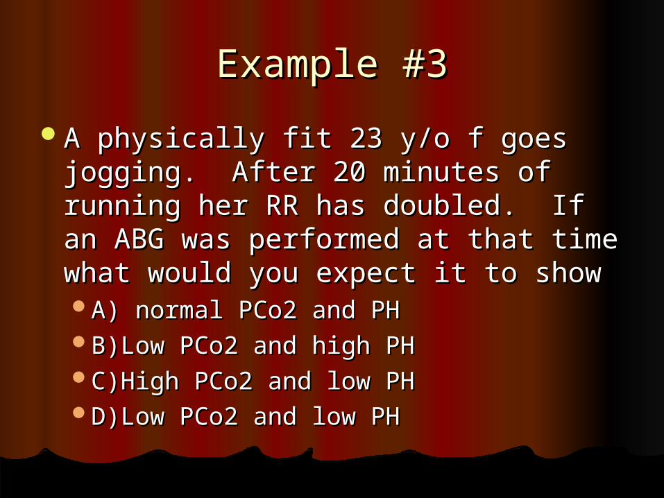
Example #3Example #3
A physically fit 23 y/o f goes jogging. A physically fit 23 y/o f goes jogging. After 20 minutes of running her RR After 20 minutes of running her RR has doubled. If an ABG was has doubled. If an ABG was performed at that time what would performed at that time what would you expect it to showyou expect it to showA) normal PCo2 and PHA) normal PCo2 and PHB)Low PCo2 and high PHB)Low PCo2 and high PHC)High PCo2 and low PHC)High PCo2 and low PHD)Low PCo2 and low PHD)Low PCo2 and low PH
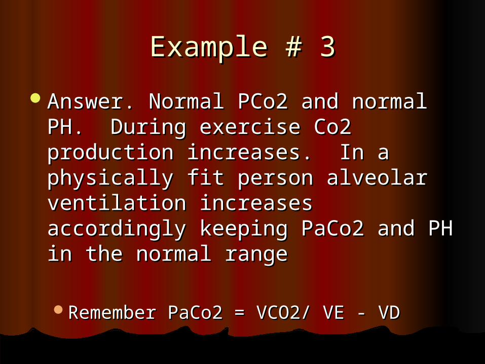
Example # 3Example # 3
Answer. Normal PCo2 and normal PH. Answer. Normal PCo2 and normal PH. During exercise Co2 production During exercise Co2 production increases. In a physically fit person increases. In a physically fit person alveolar ventilation increases alveolar ventilation increases accordingly keeping PaCo2 and PH in accordingly keeping PaCo2 and PH in the normal rangethe normal range
Remember PaCo2 = VCO2/ VE - VDRemember PaCo2 = VCO2/ VE - VD
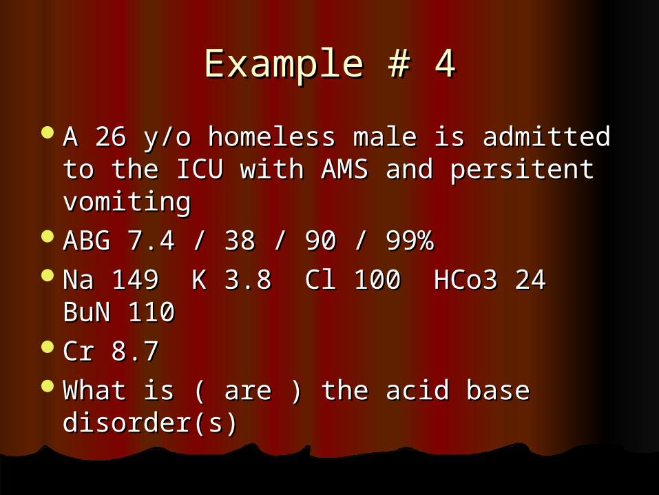
Example # 4Example # 4
A 26 y/o homeless male is admitted to A 26 y/o homeless male is admitted to the ICU with AMS and persitent the ICU with AMS and persitent vomitingvomiting
ABG 7.4 / 38 / 90 / 99%ABG 7.4 / 38 / 90 / 99%Na 149 K 3.8 Cl 100 HCo3 24 BuN Na 149 K 3.8 Cl 100 HCo3 24 BuN
110110Cr 8.7Cr 8.7What is ( are ) the acid base What is ( are ) the acid base
disorder(s)disorder(s)
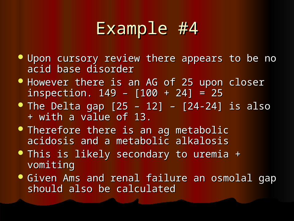
Example #4Example #4
Upon cursory review there appears to be no Upon cursory review there appears to be no acid base disorderacid base disorder
However there is an AG of 25 upon closer However there is an AG of 25 upon closer inspection. 149 – [100 + 24] = 25inspection. 149 – [100 + 24] = 25
The Delta gap [25 – 12] – [24-24] is also + The Delta gap [25 – 12] – [24-24] is also + with a value of 13. with a value of 13.
Therefore there is an ag metabolic acidosis Therefore there is an ag metabolic acidosis and a metabolic alkalosisand a metabolic alkalosis
This is likely secondary to uremia + vomitingThis is likely secondary to uremia + vomiting Given Ams and renal failure an osmolal gap Given Ams and renal failure an osmolal gap
should also be calculatedshould also be calculated
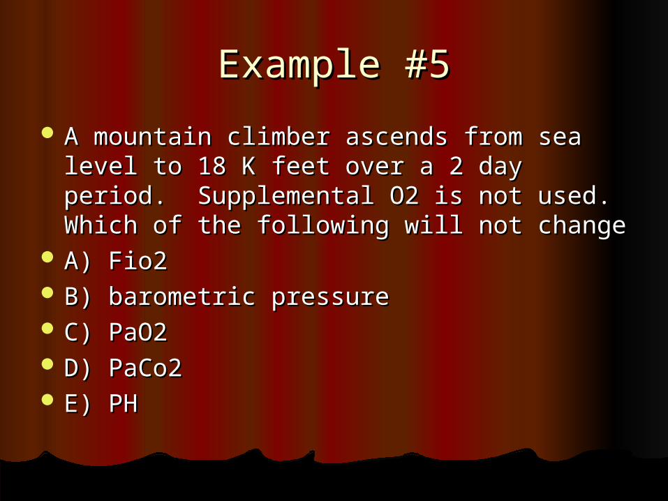
Example #5Example #5
A mountain climber ascends from sea level A mountain climber ascends from sea level to 18 K feet over a 2 day period. to 18 K feet over a 2 day period. Supplemental O2 is not used. Which of Supplemental O2 is not used. Which of the following will not changethe following will not change
A) Fio2A) Fio2 B) barometric pressureB) barometric pressure C) PaO2C) PaO2 D) PaCo2D) PaCo2 E) PHE) PH
Example # 6Example # 6
Since the early 1980’s mountain climbers Since the early 1980’s mountain climbers have climbed Mt Everest without have climbed Mt Everest without supplemental O2. How is this possible?supplemental O2. How is this possible?
Barometric Pressure at the summit is 253 Barometric Pressure at the summit is 253 mm Hg. Assume a PaCo2 of 40mm Hg. Assume a PaCo2 of 40
PAo2 = Fi02 * ( 253 – 47 ) – 40/0.8 PAo2 = Fi02 * ( 253 – 47 ) – 40/0.8 PA02 = .21 (206) – 50 = -6.7 ??PA02 = .21 (206) – 50 = -6.7 ?? If we take into account a nml A-a gradient If we take into account a nml A-a gradient
of 5 the PaO2 is –11.7?? of 5 the PaO2 is –11.7??

Example # 6Example # 6
In Fact these climbers profoundly In Fact these climbers profoundly hyperventilate and have PaCo2 hyperventilate and have PaCo2 usually < 10 mm Hg. If we plug this usually < 10 mm Hg. If we plug this into the Alveolar gas equation we get into the Alveolar gas equation we get a PAo2 of about 35 mm Hg. a PAo2 of about 35 mm Hg.
Although this is profoudly low a Although this is profoudly low a physically fit person can survive this, physically fit person can survive this, although they develop dizziness, although they develop dizziness, confusion and SOBconfusion and SOB

The ENDThe END


![HG – Precise hollow shaft solution · HG+ 300 % 200 % 100 % Torsional backlash [arcmin] Torsional rigidity [Nm/arcmin] HG+ compared to the industry standard HG+ industry standard](https://img.pdfslide.us/doc/110x75/5e48715229d361412d748168/hg-a-precise-hollow-shaft-solution-hg-300-200-100-torsional-backlash-arcmin.jpg)