Embed Size (px)
Citation preview
/ J of IMAB. 2012, vol. 18, book 3 / 349
ABDUCENS NERVE PALSY AND THROMBOSISOF THE CEREBRAL VEINS AND SINUSES - ADIAGNOSTIC PITFALL
Alexandra J. Tzoukeva1, Ara G. Kaprelyan1, Valeria Kaleva2, Chavdar Bachvarov3,Radoslav Georgiev3, Elina Peteva2.1) Department of Neurology, 2) Pediatric Department, 3) Department ofRadiology,University Hospital St Marina, Varna, Bulgaria
Journal of IMAB - Annual Proceeding (Scientific Papers) 2012, vol. 18, book 3
ABSTRACTThrombosis of the cerebral veins and sinuses is an
infrequent cerebrovascular disorder. Because the highlyvariable symptoms, recent neuroimaging plays a key role inthe diagnosis. Abducens nerve palsy as a focal neurologicaldeficit is a rare clinical manifestation in these patients. Wepresent two cases with sudden onset of diplopia andheadache.
Case 1: A 3-year old girl with B cell lymphoblasticleukemia developed bilateral abducens deficit and bilateraloptic disc edema after treatment including L-asparaginase.Thrombosis of the right jugular vein, sagittal and rightsigmoid sinuses was visualized on magnetic resonanceimaging (MRI) and magnetic resonance venography (MRV).Symptoms gradually resolved after treatment withenoxiparine and MRV demonstrated recanalization.
Case 2: A 75-year old female with medical history ofarterial hypertension presented with headache and suddenleft abduction deficit. Computerized tomography (CT) scanwas normal. MRI and MRV revealed aging brain anddisruption of venous flow at the left internal jugular vein,suspecting thrombosis. Extracranial colour duplexsonography and CT angiography proved haemodinamicequivalent of left internal jugular vein thrombosis due tosclerotic pathology of aortic arch.
Our first case illustrates the role of improvedneuroimaging techniques as the best method for diagnosisof cerebral veins and sinuses thrombosis, presenting withabducens nerve palsy. With second case the potentialneuroimaging pitfalls concerning the accurate diagnosis ofthese cerebrovascular disorders with neuro-ophthalmologicmanifestation are discussed.
Key words: abducens nerve palsy, cerebral veins andsinuses thrombosis, diagnostic pitfall
INTRODUCTIONThrombosis of the cerebral veins and sinuses is an
infrequent cerebrovascular disorder. Prothrombotic risk
factors or direct causes are often identified, e.g. oralcontraceptives, L-asparaginase, head injury, lumbarpunction, systemic or local infections (otitis, mastoiditis) (2,3, 4, 6, 8, 10, 11, 12, 15). The most frequent, but least specificclinical symptom of cerebral veins and sinus thrombosis, issevere headache. The neurologic stroke-like signs dependon the cerebral lesion’s localization, as well as the adequacyof venous collateral circulation: hemiparesis, aphasia,seizures, delirium, amnesia, mutism, coma and eye symptoms(periorbital edema, proptosis, chemosis, and paralysis of eyemovements) (5, 14). Abducens nerve palsy, as a focalneurological deficit, is a rare clinical manifestation in thesepatients (7, 9). Isolated intracranial hypertension ischaracterized by headache with diplopia due to sixth nerveinvolvement and funduscopic presentation of papilledema.Because the highly variable symptoms, recent neuroimagingplays a key role in the diagnosis. Potential diagnostic andtechnical pitfalls related to image interpretation arediscussed in the literature (1, 13). The best treatmentoptions are anticoagulation to arrest the thrombotic processand dehydration to reduce the intracranial pressure (i.e.diplopia and papilledema) (5, 14).
We present two cases with sudden onset of diplopiaand headache.
CASES REPORTCase 1.A 3-year old girl with one day headache and
horizontal diplopia was presented at our neuro-ophthalmological section. She had a medical history ofprecursor B cell lymphoblastic leukemia without CNSinvolvement. Treatment according to the ALL BFM 2000protocol, including L-asparaginase was started. A remissionwas registered. During the final of the first induction phaseshe suffered sudden headache and double vision. Neuro-ophthalmological examination showed bilateral abducensnerve palsy and funduscopy revealed bilateral optic discedema. The coagulation state showed markedly elevation infactor VIII, but without presence of thrombophilic defects;
DOI: 10.5272/jimab.2012183.349ISSN: 1312-773X (Online)
350 / J of IMAB. 2012, vol. 18, book 3 /
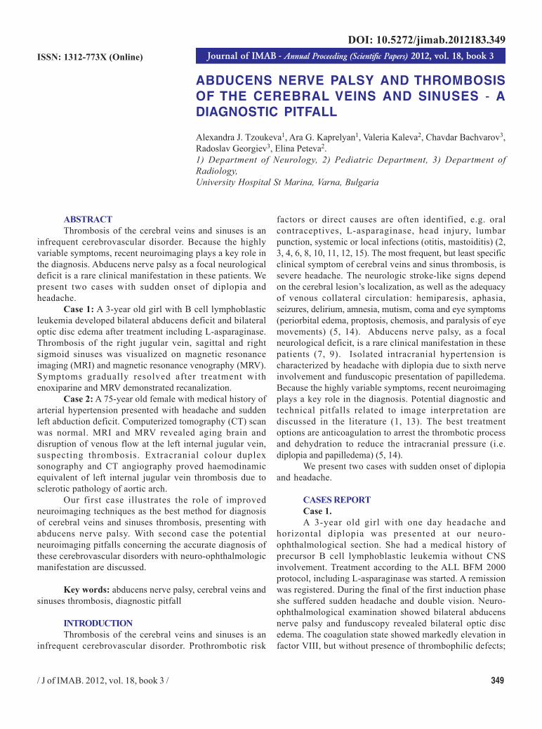
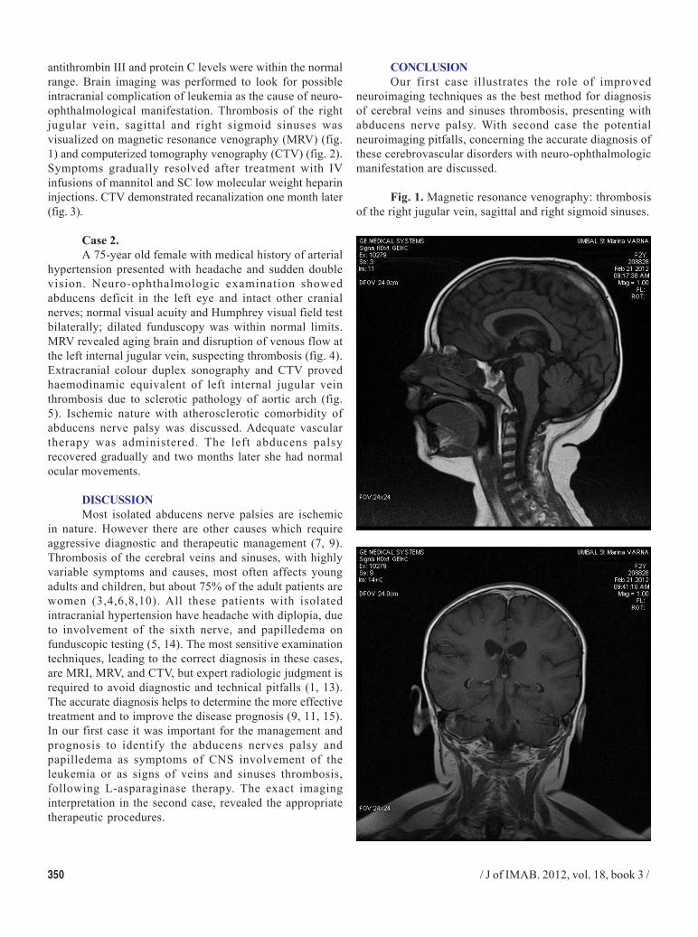
antithrombin III and protein C levels were within the normalrange. Brain imaging was performed to look for possibleintracranial complication of leukemia as the cause of neuro-ophthalmological manifestation. Thrombosis of the rightjugular vein, sagittal and right sigmoid sinuses wasvisualized on magnetic resonance venography (MRV) (fig.1) and computerized tomography venography (CTV) (fig. 2).Symptoms gradually resolved after treatment with IVinfusions of mannitol and SC low molecular weight heparininjections. CTV demonstrated recanalization one month later(fig. 3).
Case 2.A 75-year old female with medical history of arterial
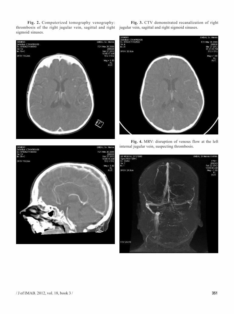
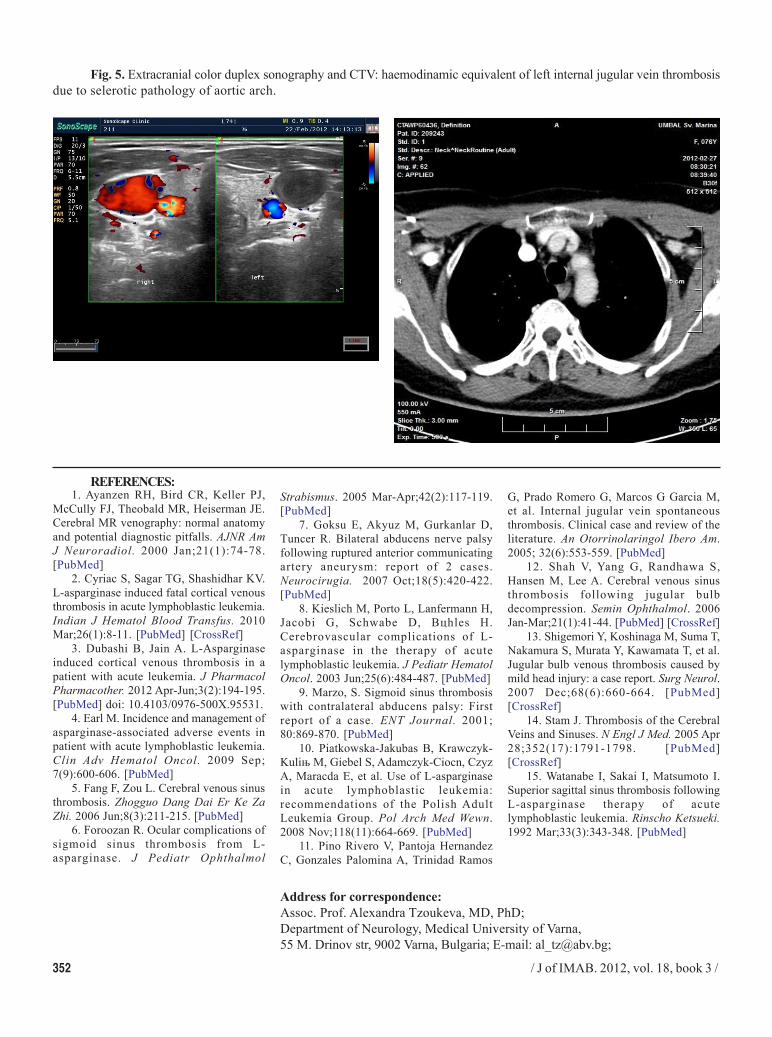
hypertension presented with headache and sudden doublevision. Neuro-ophthalmologic examination showedabducens deficit in the left eye and intact other cranialnerves; normal visual acuity and Humphrey visual field testbilaterally; dilated funduscopy was within normal limits.MRV revealed aging brain and disruption of venous flow atthe left internal jugular vein, suspecting thrombosis (fig. 4).Extracranial colour duplex sonography and CTV provedhaemodinamic equivalent of left internal jugular veinthrombosis due to sclerotic pathology of aortic arch (fig.5). Ischemic nature with atherosclerotic comorbidity ofabducens nerve palsy was discussed. Adequate vasculartherapy was administered. The left abducens palsyrecovered gradually and two months later she had normalocular movements.
DISCUSSIONMost isolated abducens nerve palsies are ischemic
in nature. However there are other causes which requireaggressive diagnostic and therapeutic management (7, 9).Thrombosis of the cerebral veins and sinuses, with highlyvariable symptoms and causes, most often affects youngadults and children, but about 75% of the adult patients arewomen (3,4,6,8,10). All these patients with isolatedintracranial hypertension have headache with diplopia, dueto involvement of the sixth nerve, and papilledema onfunduscopic testing (5, 14). The most sensitive examinationtechniques, leading to the correct diagnosis in these cases,are MRI, MRV, and CTV, but expert radiologic judgment isrequired to avoid diagnostic and technical pitfalls (1, 13).The accurate diagnosis helps to determine the more effectivetreatment and to improve the disease prognosis (9, 11, 15).In our first case it was important for the management andprognosis to identify the abducens nerves palsy andpapilledema as symptoms of CNS involvement of theleukemia or as signs of veins and sinuses thrombosis,following L-asparaginase therapy. The exact imaginginterpretation in the second case, revealed the appropriatetherapeutic procedures.
CONCLUSIONOur first case illustrates the role of improved
neuroimaging techniques as the best method for diagnosisof cerebral veins and sinuses thrombosis, presenting withabducens nerve palsy. With second case the potentialneuroimaging pitfalls, concerning the accurate diagnosis ofthese cerebrovascular disorders with neuro-ophthalmologicmanifestation are discussed.
Fig. 1. Magnetic resonance venography: thrombosisof the right jugular vein, sagittal and right sigmoid sinuses.
/ J of IMAB. 2012, vol. 18, book 3 / 351
Fig. 2. Computerized tomography venography:thrombosis of the right jugular vein, sagittal and rightsigmoid sinuses.
Fig. 3. CTV demonstrated recanalization of rightjugular vein, sagittal and right sigmoid sinuses.
Fig. 4. MRV: disruption of venous flow at the leftinternal jugular vein, suspecting thrombosis.
352 / J of IMAB. 2012, vol. 18, book 3 /
1. Ayanzen RH, Bird CR, Keller PJ,McCully FJ, Theobald MR, Heiserman JE.Cerebral MR venography: normal anatomyand potential diagnostic pitfalls. AJNR AmJ Neuroradiol. 2000 Jan;21(1):74-78.[PubMed]
2. Cyriac S, Sagar TG, Shashidhar KV.L-asparginase induced fatal cortical venousthrombosis in acute lymphoblastic leukemia.Indian J Hematol Blood Transfus. 2010Mar;26(1):8-11. [PubMed] [CrossRef]
3. Dubashi B, Jain A. L-Asparginaseinduced cortical venous thrombosis in apatient with acute leukemia. J PharmacolPharmacother. 2012 Apr-Jun;3(2):194-195.[PubMed] doi: 10.4103/0976-500X.95531.
4. Earl M. Incidence and management ofasparginase-associated adverse events inpatient with acute lymphoblastic leukemia.Clin Adv Hematol Oncol. 2009 Sep;7(9):600-606. [PubMed]
5. Fang F, Zou L. Cerebral venous sinusthrombosis. Zhogguo Dang Dai Er Ke ZaZhi. 2006 Jun;8(3):211-215. [PubMed]
6. Foroozan R. Ocular complications ofsigmoid sinus thrombosis from L-asparginase. J Pediatr Ophthalmol
Address for correspondence:Assoc. Prof. Alexandra Tzoukeva, MD, PhD;Department of Neurology, Medical University of Varna,55 M. Drinov str, 9002 Varna, Bulgaria; E-mail: [email protected];
REFERENCES:Strabismus. 2005 Mar-Apr;42(2):117-119.[PubMed]
7. Goksu E, Akyuz M, Gurkanlar D,Tuncer R. Bilateral abducens nerve palsyfollowing ruptured anterior communicatingartery aneurysm: report of 2 cases.Neurocirugia. 2007 Oct;18(5):420-422.[PubMed]
8. Kieslich M, Porto L, Lanfermann H,Jacobi G, Schwabe D, Böhles H.Cerebrovascular complications of L-asparginase in the therapy of acutelymphoblastic leukemia. J Pediatr HematolOncol. 2003 Jun;25(6):484-487. [PubMed]
9. Marzo, S. Sigmoid sinus thrombosiswith contralateral abducens palsy: Firstreport of a case. ENT Journal. 2001;80:869-870. [PubMed]
10. Piatkowska-Jakubas B, Krawczyk-Kuliœ M, Giebel S, Adamczyk-Ciocn, CzyzA, Marañda E, et al. Use of L-asparginasein acute lymphoblastic leukemia:recommendations of the Polish AdultLeukemia Group. Pol Arch Med Wewn.2008 Nov;118(11):664-669. [PubMed]
11. Pino Rivero V, Pantoja HernandezC, Gonzales Palomina A, Trinidad Ramos
G, Prado Romero G, Marcos G Garcia M,et al. Internal jugular vein spontaneousthrombosis. Clinical case and review of theliterature. An Otorrinolaringol Ibero Am.2005; 32(6):553-559. [PubMed]
12. Shah V, Yang G, Randhawa S,Hansen M, Lee A. Cerebral venous sinusthrombosis following jugular bulbdecompression. Semin Ophthalmol. 2006Jan-Mar;21(1):41-44. [PubMed] [CrossRef]
13. Shigemori Y, Koshinaga M, Suma T,Nakamura S, Murata Y, Kawamata T, et al.Jugular bulb venous thrombosis caused bymild head injury: a case report. Surg Neurol.2007 Dec;68(6):660-664. [PubMed][CrossRef]
14. Stam J. Thrombosis of the CerebralVeins and Sinuses. N Engl J Med. 2005 Apr28;352(17):1791-1798. [PubMed][CrossRef]
15. Watanabe I, Sakai I, Matsumoto I.Superior sagittal sinus thrombosis followingL-asparginase therapy of acutelymphoblastic leukemia. Rinscho Ketsueki.1992 Mar;33(3):343-348. [PubMed]
Fig. 5. Extracranial color duplex sonography and CTV: haemodinamic equivalent of left internal jugular vein thrombosisdue to selerotic pathology of aortic arch.