Embed Size (px)
Citation preview

Abdominal Compartment Syndrome
Diane Cobble M.D., FACS Professor, ETSU Dept. of Surgery
7th Annual Rural Trauma Symposium August 27, 2015

Disclosure Statement of Financial Interest
I DO NOT have a financial interest/arrangement or affiliation with one or
more organizations that could be perceived as a real or apparent
conflict of interest in the context of the subject of this presentation.

Trauma Scenario
• A 32yof was stabbed in the liver. She arrived in the ER hypotensive & unresponsive to pain. AKer she had a massive transfusion & emergent ex lap, she was admiMed to the ICU. During the next 6°, mechanical venQlaQon became progressively more difficult. Although cvp remained slightly é, her CO ê’d markedly

General Surgery Scenario
• An 87yo NH resident had severe abd pain. His care providers reported that he had recently experienced several episodes of protracted vomiQng progressing to êbp, éhr & êLOC. Transfer for tests to determine if he had sepsis was arranged & he was admiMed to ICU. Despite administraQon of mulQple fluid boluses, he remained hypotensive & anuric. ConQnuous ngt sucQoning produced large amt of green-‐brown fluid & abd CT revealed complete sbo

History
• Recognized clinically 19th century when Marey & Burt observed its assoc w/declines in respiratory fxn
• ForgoMen aKer WWI & rediscovered near end 20th century
• More recently, Kron (1984) developed simple method of accurately measuring IAP which led to beMer understanding of relaQonship btwn IAP & ACS
• 2004 group of internaQonal physicians & surgeons developed World Society of ACS (WSACS)

definiQons
• Nl IAP: 0-‐5 mmHg • IAH: sustained IAP ≥ 12mmHg • ACS: sustained IAP > 20mmHg w/new organ dysfxn
• Morbidly obese & pregnant pts can have chronically é IAP 10-‐15 mmHg w/o adverse sequelae

categories
• Primary (surgical): intra-‐abd pathology is directly & proximally responsible for the ACS
• Secondary (medical): no visible intra-‐abd injury is present but injuries outside the abd cause fluid accumulaQon
• Recurrent: ACS redevelops aKer surgical/medical tx of 1° or 2° causes

Risk factors
Primary • Major trauma • Liver transplantaQon • Ruptured AAA • Mechanical bowel obst • Retroperitoneal
hemorrhage • Postop bleeding/abd
closure under tension • Bleeding pelvic fxs
Secondary • Obesity or é BMI • Pregnancy • Major burns/sepsis • Ileus/pancreaQQs/ascites • Large vol fluid replacement • Severe intra-‐abd infexn • CAPD

Typical scenarios Abdominal injury w/ongoing intra-‐abd bleeding
A8er DCL 2° packing therefore vac closure

pathophysiology • Begins @ organ level w/direct compression
• Hollow systems (GI tract & portal-‐caval system) collapse
• Immediate effects (thrombosis or bowel wall edema) è translocaQon of bacterial products èaddiQonal fluid accumulaQon è éIAP
• Cellular level: O2 impaired è ischemia & anaerobic metabolism
• VasoacQve substances (histamine & serotonin) é endothelial permeability – Further capillary leak impairs rbc transport & ischemia worsens

Cellular hypoxia sequelae Release of cytokines FormaQon of O2 free radicals ê Cellular producQon of ATP

Systems Most Affected
• Cardiovascular • Pulmonary • Renal

CV Effects é Intrathoracic pressure compresses heart/great vessels ê CO due to ê venous return éé SVR CVP/PCWP falsely é IAH causes pressure on femoral veins è stasis è DVT. When resolved, risk of PE é
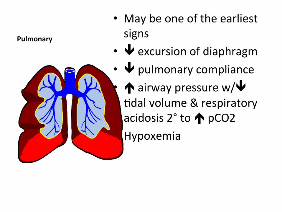
Pulmonary
• May be one of the earliest signs
• ê excursion of diaphragm • ê pulmonary compliance • é airway pressure w/ê Qdal volume & respiratory acidosis 2° to é pCO2
• Hypoxemia

Other System DysfuncQon
Renal • Oliguria è anuria 2° direct
parenchymal compression & shunQng of renal plasma flow
• é Cr may not be seen for 2-‐3d
Visceral • ê blood flow w/subsequent
intesQnal necrosis • HepaQc dysfuncQon w/ê
glucose metabolism & lactate clearance
• Gut anastomoQc breakdown

brain
• ObstrucQon of cerebral venous ouolow
• é ICP • ê CPP

Organ DysfuncQon

Grading ACs
• Grade I: 10-‐14 mmHg • Grade II: 15-‐24 mmHg • Grade III: 25-‐35 mmHg • Grade IV: >35 mmHg

Abdominal perfusion pressure
• Measure of visceral organ perfusion – Suggested as a guide by some authors
• APP = MAP-‐IAP • More accurate predictor of abd organ perfusion & more effecQve guide for resuscitaQon measures
• Ideal level is anything > 60 mmHg

app
• Cheatham et al J Trauma 2000– Concluded that APP 60 mmHg in pts w/ACS 98% sensiQve in predicQng survival compared to APP 40 mmHg being 70% sensiQve in populaQon largely comprised of trauma pts (n=144)
• Also concluded APP more accurate predictor of resuscitaQon than lactate, MAP, arterial pH, base deficit, IAP
• RetrospecQve study 5/97-‐6/99

Bladder pressure
• Can be measured intermiMently or conQnuously if foley connected to pressure transducer w/recording device
• InsQll ~25cc NS in bladder & clamp foley • 18 gauge needle inserted into aspiraQon port & aMached to pressure transducer
• Pressure obtained @ end-‐expiraQon & zeroed @ mid-‐axillary line

Measuring Bladder Pressure May not be accurate w/morbid obesity, massive ascites, pelvic hematoma, adhesions or neurogenic bladder Can use intragastric, intracolonic or IVC catheters
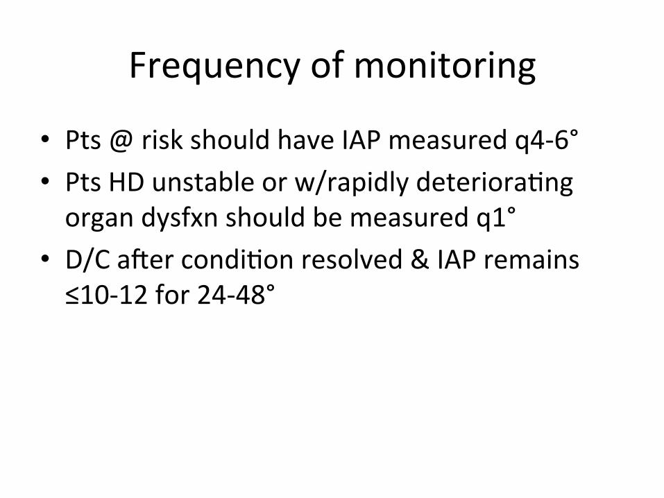
Frequency of monitoring
• Pts @ risk should have IAP measured q4-‐6° • Pts HD unstable or w/rapidly deterioraQng organ dysfxn should be measured q1°
• D/C aKer condiQon resolved & IAP remains ≤10-‐12 for 24-‐48°

epidemiology
• 5-‐15% freq in trauma ICU adm according to recent lit
• 1% gen trauma admissions

prevalence
• Malbrain et al Intensive Care Medicine 2004:– Prevalence study in 13 ICU’s involving 97 pts
• Overall rate IAH 58.8% – 65% surgical, 54.4% medical pts
• Medical pts é prevalence IAP >15 – 29.8% vs 27.5%
• Medical pts é prevalence ACS – 10.5% vs 5%

prevalence
• Reintam et al Intensive Care Medicine 2008:– 257 venQlated pts
• IAH developed in 95 pts – 60 primary, 35 secondary
• During 1st 3 days, IAP ê w/primary & éé w/secondary

Mortality rates
w/IAH • ICU mortality 37.9% • 28d mortality 48.4% • 90d mortality 53.7% • Independent risk fx for
death • Secondary ê common,
different dev course & worse outcome
w/o IAH • ICU mortality 19.1% • 28d mortality 27.8% • 90d mortality35.8%

incidence
• Vidal et al Critical Care Medicine 2008:• Studied incidence of IAP in 83 criQcally ill pts in single ICU
• 31% pts had IAH @ ICU adm & 33% developed it aKer adm
• Pts w/IAH were sicker w/é mortality rate 53% vs 27%
• IAH independent predictor of mortality – ACS developed in 10 pts w/80% mortality
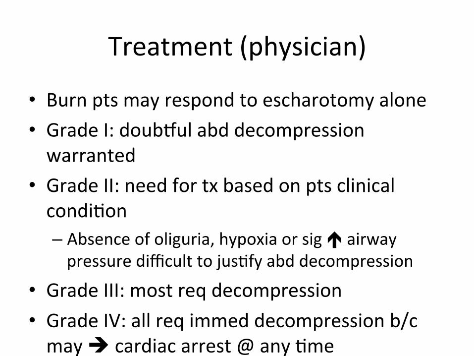
Treatment (physician)
• Burn pts may respond to escharotomy alone • Grade I: douboul abd decompression warranted
• Grade II: need for tx based on pts clinical condiQon – Absence of oliguria, hypoxia or sig é airway pressure difficult to jusQfy abd decompression
• Grade III: most req decompression • Grade IV: all req immed decompression b/c may è cardiac arrest @ any Qme

Treatment (nursing)
• Maintain patency of NGT & rectal tube if used • Monitor & record daily BM’s
– Check qd for fecal impacQon if pt unconscious, sedated or paralyzed
• Assess tolerance to enteral TF’s & adjust accordingly if residuals too high
• For pts able to eat, minimize/eliminate gas-‐producing foods
• Avoid prone posiQon & place pt in reverse Trendelenberg – Must be supine to measure bladder pressure

mortality
• Early decompression improves survival • Mortality rates range from 25-‐75% • High rate even w/tx reflects fact that condiQon affects mulQple organ systems
• Furthermore oKen a sequelae to severe injuries that independently carry high morbidity/mortality
• Assoc w/éLOS, éé costs & further dev of comorbidiQes

World society acs
• Updated consensus definiQons & clinical pracQce guidelines published in 2013 Intensive Care Medicine (prev 2006)
• PaQent (P) • IntervenQons (I) • Comparison (C) • Outcome (O)

Wsacs definiQons
IAP ~5-‐7 mmHg in criQcally ill pts • Max of 25ml NS insQlled to
determine bladder pressure
IAH • Grade I: 12-‐15 mmHg • Grade II: 16-‐20 mmHg • Grade III: 21-‐25 mmHg • Grade IV: >25 mmHg
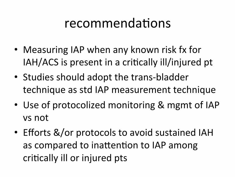
recommendaQons
• Measuring IAP when any known risk fx for IAH/ACS is present in a criQcally ill/injured pt
• Studies should adopt the trans-‐bladder technique as std IAP measurement technique
• Use of protocolized monitoring & mgmt of IAP vs not
• Efforts &/or protocols to avoid sustained IAH as compared to inaMenQon to IAP among criQcally ill or injured pts

Cont,d
• Decompressive lap in cases of overt ACS compared to strategies that do not use DL in criQcally ill adults w/ACS
• Among ICU pts w/open abdominal wounds, conscious &/or protocolized efforts be made to obtain an early or @ least same-‐hospital-‐stay abd fascial closure
• Among criQcally ill/injured pts w/open abdominal wounds, strategies uQlizing NPW therapy should be used vs not

suggesQons
• Enhanced raQo plasma/prbc’s for massive hemorrhage
• Perc catheter drainage to remove fluid when technically possible (may alleviate need for DL)
• ProphylacQc use of open abd when undergoing lap for physiologic exhausQon
• Not rouQnely using open abd for pts w/severe abd contaminaQon
• BioprostheQc meshes not rouQnely used in early closure open abd vs alt. strategies

No recommendaQons
• Use of APP • Use of diureQcs or RRT • Albumin administraQon • ProphylacQc use of open abd in non-‐trauma acute care surg pts
• Acute component separaQon technique to facilitate fascial closure

quesQons