Embed Size (px)
Citation preview
Plasminogen promotes cholesterol efflux by theABCA1 pathway
Nathalie Pamir, … , Godfrey S. Getz, Jay W. Heinecke
JCI Insight. 2017;2(15):e92176. https://doi.org/10.1172/jci.insight.92176.
Using genetic and biochemical approaches, we investigated proteins that regulatemacrophage cholesterol efflux capacity (CEC) and ABCA1-specific CEC (ABCA1 CEC), 2functional assays that predict cardiovascular disease (CVD). Macrophage CEC and theconcentration of HDL particles were markedly reduced in mice deficient in apolipoprotein A-I (APOA1) or apolipoprotein E (APOE) but not apolipoprotein A-IV (APOA4). ABCA1 CECwas markedly reduced in APOA1-deficient mice but was barely affected in mice deficient inAPOE or APOA4. High-resolution size-exclusion chromatography of plasma produced 2major peaks of ABCA1 CEC activity. The early-eluting peak, which coeluted with HDL, wasmarkedly reduced in APOA1- or APOE-deficient mice. The late-eluting peak was modestlyreduced in APOA1-deficient mice but little affected in APOE- or APOA4-deficient mice. Ion-exchange chromatography and shotgun proteomics suggested that plasminogen (PLG)accounted for a substantial fraction of the ABCA1 CEC activity in the peak not associatedwith HDL. Human PLG promoted cholesterol efflux by the ABCA1 pathway, and PLG-dependent efflux was inhibited by lipoprotein(a) [Lp(a)]. Our observations identify APOA1,APOE, and PLG as key determinants of CEC. Because PLG and Lp(a) associate withhuman CVD risk, interplay among the proteins might affect atherosclerosis by regulatingcholesterol efflux from macrophages.
Research Article Endocrinology Metabolism
Find the latest version:
http://jci.me/92176/pdf
1insight.jci.org https://doi.org/10.1172/jci.insight.92176
R E S E A R C H A R T I C L E
Conflict of interest: J. Heinecke is named as a coinventor on patents from the US Patent Office (patents are listed at the end of the supplemental material) on the use of HDL markers to predict the risk of cardiovascular disease and has served as a consultant for Kowa, Merck, Amgen, Bristol Meyer Squibb, GSK, and Pacific Biomarkers.
Submitted: December 9, 2016 Accepted: June 20, 2017 Published: August 3, 2017
Reference information: JCI Insight. 2017;2(15):e92176. https://doi.org/10.1172/jci.insight.92176.
Plasminogen promotes cholesterol efflux by the ABCA1 pathwayNathalie Pamir,1 Patrick M. Hutchins,1 Graziella E. Ronsein,1 Hao Wei,1 Chongren Tang,1 Riku Das,2 Tomas Vaisar,1 Edward Plow,2 Volker Schuster,3 Marlys L. Koschinsky,4Catherine A. Reardon,5 Richard Weinberg,6 David A. Dichek,1 Santica Marcovina,1 Godfrey S. Getz,7 and Jay W. Heinecke1
1Department of Medicine, University of Washington, Seattle, Washington, USA. 2Department of Molecular Cardiology,
Cleveland Clinic, Cleveland, Ohio, USA. 3Hospital for Children and Adolescents, Medical Faculty of Leipzig University,
Leipzig, Germany. 4Robarts Research Institute, Schulich School of Medicine and Dentistry, The University of Western
Ontario, London, Ontario, Canada. 5Ben May Research Institute, University of Chicago, Illinois, USA. 6Department of
Internal Medicine, Wake Forest School of Medicine, Winston-Salem, North Carolina, USA. 7Department of Pathology,
University of Chicago, Chicago, Illinois, USA.
IntroductionClinical and epidemiological studies show a robust, inverse association of HDL cholesterol (HDL-C) levels with cardiovascular disease (CVD) risk (1, 2). Moreover, genetically engineered deficiencies in proteins implicated in HDL metabolism markedly affect atherosclerosis in hypercholesterolemic mice (3, 4), placing HDL in the causal pathway of accelerated atherogenesis in animal models. These observations have trig-gered intense interest in targeting HDL-C for therapeutic intervention.
Levels of HDL-C do not necessarily reflect the concentrations of cardioprotective HDL particles, however. HDL is a collection of macromolecules ranging in size from less than 7 nm to more than 14 nm (5), and the cho-lesterol content of the different species can vary 3-fold. Indeed, several lines of evidence suggest that the associa-tion between HDL-C levels and CVD status is indirect and that elevating HDL-C is not necessarily therapeutic (6–12). For example, genetic variations that associate with altered HDL-C do not strongly associate with altered CVD risk (9). Trials of drugs such as cholesteryl ester transfer protein inhibitors and niacin, which elevate HDL-C by different mechanisms, have failed to reduce events in statin-treated subjects with established CVD (6–8, 10, 11). Moreover, a loss-of-function variant of scavenger receptor B1 (SCARB1) that raises HDL-C levels increases the risk of CVD in humans (13), and genetic deletion of SCARB1 in mice raises HDL-C levels and promotes atherosclerosis (14). These observations indicate that HDL-C does not necessarily reflect HDL’s cardioprotec-tive effects in either humans or mice and that it is critical to examine other functional properties of HDL.
HDL’s cardioprotective effect is attributed in part to its ability to mobilize excess cholesterol from artery wall macrophages (15–18). The first key step involves ABCA1 — an ATP-binding cassette trans-porter found in the plasma membrane of macrophages — that exports phospholipids and cholesterol from
Using genetic and biochemical approaches, we investigated proteins that regulate macrophage cholesterol efflux capacity (CEC) and ABCA1-specific CEC (ABCA1 CEC), 2 functional assays that predict cardiovascular disease (CVD). Macrophage CEC and the concentration of HDL particles were markedly reduced in mice deficient in apolipoprotein A-I (APOA1) or apolipoprotein E (APOE) but not apolipoprotein A-IV (APOA4). ABCA1 CEC was markedly reduced in APOA1-deficient mice but was barely affected in mice deficient in APOE or APOA4. High-resolution size-exclusion chromatography of plasma produced 2 major peaks of ABCA1 CEC activity. The early-eluting peak, which coeluted with HDL, was markedly reduced in APOA1- or APOE-deficient mice. The late-eluting peak was modestly reduced in APOA1-deficient mice but little affected in APOE- or APOA4-deficient mice. Ion-exchange chromatography and shotgun proteomics suggested that plasminogen (PLG) accounted for a substantial fraction of the ABCA1 CEC activity in the peak not associated with HDL. Human PLG promoted cholesterol efflux by the ABCA1 pathway, and PLG-dependent efflux was inhibited by lipoprotein(a) [Lp(a)]. Our observations identify APOA1, APOE, and PLG as key determinants of CEC. Because PLG and Lp(a) associate with human CVD risk, interplay among the proteins might affect atherosclerosis by regulating cholesterol efflux from macrophages.
2insight.jci.org https://doi.org/10.1172/jci.insight.92176
R E S E A R C H A R T I C L E
cells (15–17). Lipid-poor apolipoprotein A-I (APOA1), HDL’s major protein, is thought to be the dominant ligand for ABCA1 in vivo, but recent studies indicate that mature lipidated HDL particles also promote efflux by this pathway (13–15, 17, 19, 20).
Mouse and human studies have shown that defects in the APOA1/ABCA1 pathway are important determinants of CVD. For example, ablating the Abca1 gene in myeloid cells increases atherosclerotic lesions in hypercholesterolemic mice (21), while overexpressing human ABCA1 in myeloid cells in hyper-cholesterolemic mice retards atherogenesis (22). Importantly, humans deficient in ABCA1 (Tangier’s dis-ease) accumulate cholesterol-laden macrophages in many different tissues despite having low levels of LDL-cholesterol, providing strong evidence that ABCA1 conducts a key step in sterol excretion from these cells in humans (23).
Cholesterol efflux capacity (CEC) — the ability of human serum depleted of lipoproteins containing apolipoprotein B (APOB) to promote cholesterol efflux from macrophages — can vary markedly between different subjects despite similar levels of HDL-C and APOA1 (24). Macrophage CEC of serum HDL associated strongly and negatively with prevalent CVD status in 2 large, independent human popula-tions (25). That association was independent of HDL-C and APOA1 levels. CEC inversely associated with future cardiac events in the Dallas Heart and the EPIC-Norfolk studies (26, 27). These relationships remained highly significant after correction for other classic lipid risk factors for CVD. The assay in the Dallas Heart Study monitored cholesterol efflux capacity specifically by the ABCA1 pathway (28), while the EPIC-Norfolk Study quantified macrophage CEC (24), which involves ABCA1 and multiple other lipid transporters. Taken together, these observations provide strong evidence that macrophage CEC and ABCA1 CEC are clinically relevant metrics of HDL function that are independent of HDL-C.
The mechanisms that mediate CEC in APOB-depleted serum are poorly understood. In the current study, we used mice deficient in HDL apolipoproteins implicated in cholesterol efflux from macrophages (15, 16, 29) and analyzed plasma samples biochemically to investigate those proteins’ roles in macrophage CEC and ABCA1 CEC. Our observations indicate that APOA1, apolipoprotein E (APOE), and plasmino-gen (PLG, which is not an apolipoprotein) are key modulators of CEC, and that PLG-dependent ABCA1 CEC is inhibited by lipoprotein(a) [Lp(a)]. These findings could provide the basis for studies of factors that influence CEC — and therefore cardioprotection — in vivo.
ResultsWe quantified CEC with both serum HDL (serum depleted of lipoproteins containing APOB) and plas-ma fractionated by high-resolution size-exclusion chromatography (SEC). Because serum HDL con-tains mature HDL as well as partially lipidated and lipid-free forms of APOA1 and apolipoprotein A-IV (APOA4), fractionating the plasma by SEC allowed us to distinguish between HDL-associated and non–HDL-associated proteins that promote CEC. We complemented these studies by quantifying HDL particle concentration (defined as lipoproteins isolated by ultracentrifugation at d = 1.063–1.21 g/ml) in the differ-ent strains of mice.
Both APOA1 and APOE are important regulators of macrophage CEC, but only APOA1 promotes ABCA1 CEC. Using serum HDL from wild-type mice or mice deficient in APOA1, APOE, APOA1/APOE, or APOA4 (all in the C57BL/6 genetic background), we determined whether these proteins modulate CEC and/or HDL particle concentration. Macrophage CEC, which involves multiple lipid transporters (30, 31), was monitored with cAMP-stimulated J744 macrophages (24, 25); ABCA1 CEC, which involves only ABCA1, was quantified with BHK cells with inducible expression of human ABCA1 (32).
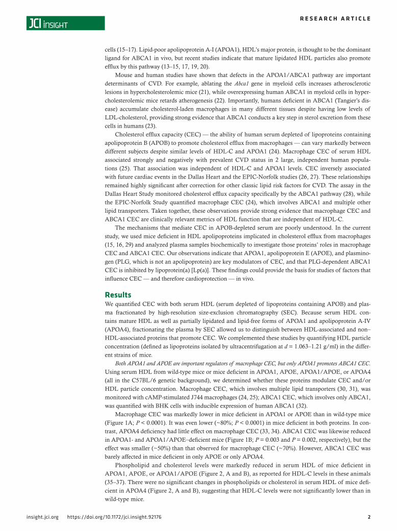
Macrophage CEC was markedly lower in mice deficient in APOA1 or APOE than in wild-type mice (Figure 1A; P < 0.0001). It was even lower (~80%; P < 0.0001) in mice deficient in both proteins. In con-trast, APOA4 deficiency had little effect on macrophage CEC (33, 34). ABCA1 CEC was likewise reduced in APOA1- and APOA1/APOE–deficient mice (Figure 1B; P = 0.003 and P = 0.002, respectively), but the effect was smaller (~50%) than that observed for macrophage CEC (~70%). However, ABCA1 CEC was barely affected in mice deficient in only APOE or only APOA4.
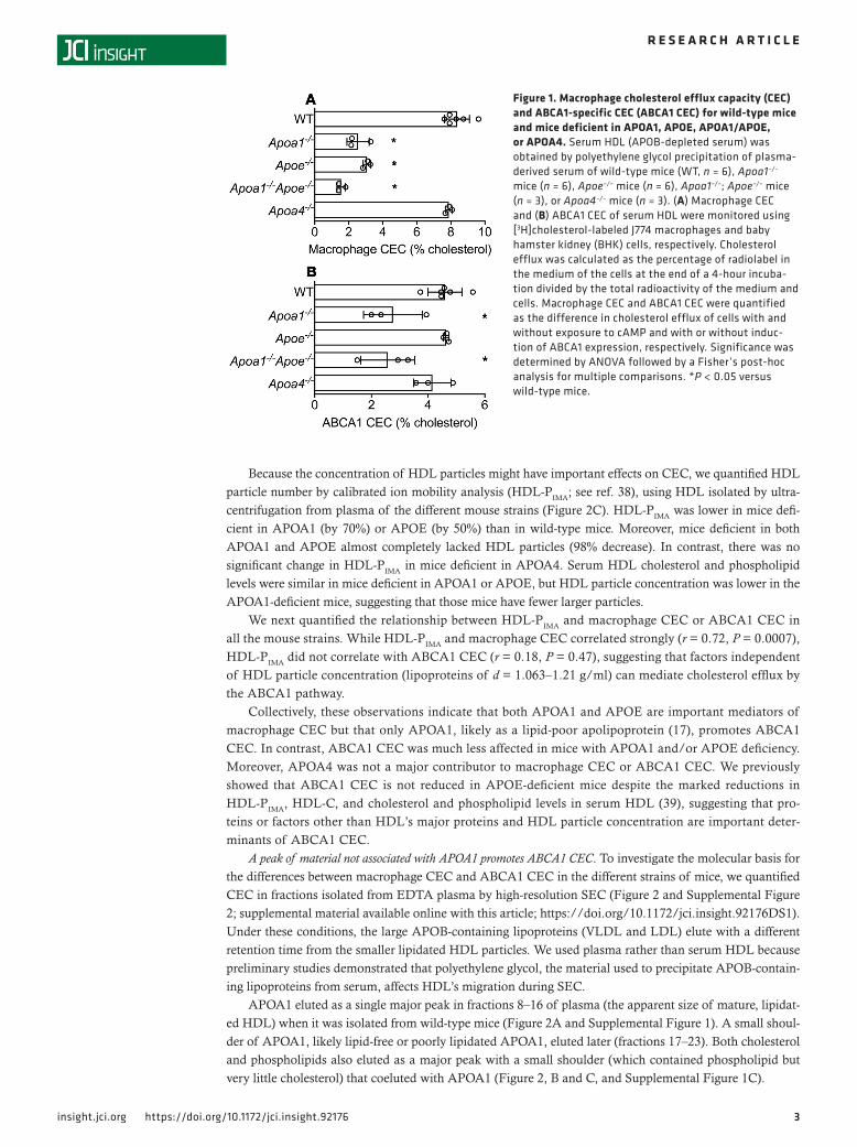
Phospholipid and cholesterol levels were markedly reduced in serum HDL of mice deficient in APOA1, APOE, or APOA1/APOE (Figure 2, A and B), as reported for HDL-C levels in these animals (35–37). There were no significant changes in phospholipids or cholesterol in serum HDL of mice defi-cient in APOA4 (Figure 2, A and B), suggesting that HDL-C levels were not significantly lower than in wild-type mice.
3insight.jci.org https://doi.org/10.1172/jci.insight.92176
R E S E A R C H A R T I C L E
Because the concentration of HDL particles might have important effects on CEC, we quantified HDL particle number by calibrated ion mobility analysis (HDL-PIMA; see ref. 38), using HDL isolated by ultra-centrifugation from plasma of the different mouse strains (Figure 2C). HDL-PIMA was lower in mice defi-cient in APOA1 (by 70%) or APOE (by 50%) than in wild-type mice. Moreover, mice deficient in both APOA1 and APOE almost completely lacked HDL particles (98% decrease). In contrast, there was no significant change in HDL-PIMA in mice deficient in APOA4. Serum HDL cholesterol and phospholipid levels were similar in mice deficient in APOA1 or APOE, but HDL particle concentration was lower in the APOA1-deficient mice, suggesting that those mice have fewer larger particles.
We next quantified the relationship between HDL-PIMA and macrophage CEC or ABCA1 CEC in all the mouse strains. While HDL-PIMA and macrophage CEC correlated strongly (r = 0.72, P = 0.0007), HDL-PIMA did not correlate with ABCA1 CEC (r = 0.18, P = 0.47), suggesting that factors independent of HDL particle concentration (lipoproteins of d = 1.063–1.21 g/ml) can mediate cholesterol efflux by the ABCA1 pathway.
Collectively, these observations indicate that both APOA1 and APOE are important mediators of macrophage CEC but that only APOA1, likely as a lipid-poor apolipoprotein (17), promotes ABCA1 CEC. In contrast, ABCA1 CEC was much less affected in mice with APOA1 and/or APOE deficiency. Moreover, APOA4 was not a major contributor to macrophage CEC or ABCA1 CEC. We previously showed that ABCA1 CEC is not reduced in APOE-deficient mice despite the marked reductions in HDL-PIMA, HDL-C, and cholesterol and phospholipid levels in serum HDL (39), suggesting that pro-teins or factors other than HDL’s major proteins and HDL particle concentration are important deter-minants of ABCA1 CEC.
A peak of material not associated with APOA1 promotes ABCA1 CEC. To investigate the molecular basis for the differences between macrophage CEC and ABCA1 CEC in the different strains of mice, we quantified CEC in fractions isolated from EDTA plasma by high-resolution SEC (Figure 2 and Supplemental Figure 2; supplemental material available online with this article; https://doi.org/10.1172/jci.insight.92176DS1). Under these conditions, the large APOB-containing lipoproteins (VLDL and LDL) elute with a different retention time from the smaller lipidated HDL particles. We used plasma rather than serum HDL because preliminary studies demonstrated that polyethylene glycol, the material used to precipitate APOB-contain-ing lipoproteins from serum, affects HDL’s migration during SEC.
APOA1 eluted as a single major peak in fractions 8–16 of plasma (the apparent size of mature, lipidat-ed HDL) when it was isolated from wild-type mice (Figure 2A and Supplemental Figure 1). A small shoul-der of APOA1, likely lipid-free or poorly lipidated APOA1, eluted later (fractions 17–23). Both cholesterol and phospholipids also eluted as a major peak with a small shoulder (which contained phospholipid but very little cholesterol) that coeluted with APOA1 (Figure 2, B and C, and Supplemental Figure 1C).
Figure 1. Macrophage cholesterol efflux capacity (CEC) and ABCA1-specific CEC (ABCA1 CEC) for wild-type mice and mice deficient in APOA1, APOE, APOA1/APOE, or APOA4. Serum HDL (APOB-depleted serum) was obtained by polyethylene glycol precipitation of plasma-derived serum of wild-type mice (WT, n = 6), Apoa1–/– mice (n = 6), Apoe–/– mice (n = 6), Apoa1–/–; Apoe–/– mice (n = 3), or Apoa4–/– mice (n = 3). (A) Macrophage CEC and (B) ABCA1 CEC of serum HDL were monitored using [3H]cholesterol-labeled J774 macrophages and baby hamster kidney (BHK) cells, respectively. Cholesterol efflux was calculated as the percentage of radiolabel in the medium of the cells at the end of a 4-hour incuba-tion divided by the total radioactivity of the medium and cells. Macrophage CEC and ABCA1 CEC were quantified as the difference in cholesterol efflux of cells with and without exposure to cAMP and with or without induc-tion of ABCA1 expression, respectively. Significance was determined by ANOVA followed by a Fisher’s post-hoc analysis for multiple comparisons. *P < 0.05 versus wild-type mice.
4insight.jci.org https://doi.org/10.1172/jci.insight.92176
R E S E A R C H A R T I C L E
We next quantified the CEC of fractions isolated by SEC from the 5 strains of mice (Figure 2 and Supplemental Figure 2). The fractions of wild-type mouse plasma that pro-moted macrophage CEC eluted as a single major peak of material that coeluted with the apparent size of HDL (Figure 2A and Supple-mental Figure 2A). In striking contrast, plas-ma exhibited 2 major peaks of ABCA1 CEC activity. One coeluted with the apparent size of HDL, while the other eluted later and had an apparent size smaller than that of HDL (Figure 2B and Supplemental Figure 2B).
To determine the role of APOA1 in CEC, we fractionated plasma from mice deficient in the protein (Figure 2, C and D, and Supplemental Material). Macrophage CEC was reduced by 70% in the peak of material that coeluted with the size of HDL. Macrophage CEC was also smaller in the small shoulder of the early-eluting peak of activity, suggesting that lipid-free APOA1 promotes cholesterol efflux.
ABCA1 CEC was also reduced (~50%) in the major peak of material that coeluted with the size of HDL. However, there was only a modest reduction of ABCA1 CEC in the late-eluting peak of material (Figure 2D). Taken together, these observations indicate that HDL particles containing APOA1 are major media-tors of macrophage CEC but that proteins or other factors not associated with APOA1 are also important contributors to ABCA1 CEC.
We next investigated the effects of APOE deficiency and APOA1/APOE deficiency on macrophage CEC and ABCA1 CEC (Figure 2, E–H, and Supplemental Figure 2). The peak with macrophage CEC activity that coeluted with the apparent size of HDL was approximately 30% smaller in the APOE-deficient mice than in the wild-type mice. ABCA1 CEC activity was lower in the peak of material with the apparent size of HDL but was not decreased in the late-eluting material (Figure 2, E and F, and Supplemental Figure 2, E and F). These observations show that lipoproteins with the apparent size of HDL that contain APOE can also promote ABCA1 CEC. In contrast, APOE deficiency had no effect on the ABCA1 CEC activity of the material whose apparent size was smaller than that of mature HDL.
When we fractionated plasma from the APOA1/APOE–deficient mice by SEC (Figure 2G and Sup-plemental Figure 2G), the activity in the peak with the apparent size of HDL was markedly lower (90%) than in the wild-type mice. The peak also had little ABCA1 CEC activity (Figure 2H and Supplemen-tal Figure 2H), suggesting that HDL containing APOA1 and/or APOE promotes efflux mainly by the ABCA1 pathway. However, the ABCA1 CEC activity of the late-eluting peak was only modestly lower than the activity in the late-eluting peak of the wild-type mice (Figure 2H and Supplemental Figure 2H). Collectively, these observations indicate that HDL particles containing APOA1 and/or APOE account
Figure 2. Plasma phospholipid level, plasma cholesterol level, and HDL particle concentration (HDL-PIMA) of wild-type mice and mice deficient in APOA1, APOE, APOA1/APOE, or APOA4. Phos-pholipid (A) and total cholesterol (B) levels were quantified biochemically. (C) HDL-PIMA was deter-mined by calibrated ion mobility analysis using glucose oxidase as the standard. Significance was determined by ANOVA followed by Fisher’s post-hoc analysis for multiple comparisons. *P < 0.05 versus wild-type mice (WT).
5insight.jci.org https://doi.org/10.1172/jci.insight.92176
R E S E A R C H A R T I C L E
almost quantitatively for macrophage CEC, but that other proteins or factors with an apparent molecular size smaller than HDL are major contributors to the ABCA1 CEC activity of plasma.
Albumin does not account for the late-eluting peak of ABCA1 CEC activity. Albumin and the late-eluting peak of material with ABCA1 CEC activity exhibited similar apparent sizes on SEC. However, albumin promotes shuttling of cholesterol from cells to medium when the medium is supplemented with high concentrations of exogenous cholesterol acceptor such as LDL, phospholipid vesicles, or red blood cells (40). Two lines of evidence suggest that albumin was not responsible for the late-eluting peak of ABCA1 CEC activity we observed in plasma fractionated by SEC. First, we do not include exogenous cholesterol acceptors in our CEC assays, and in our hands albumin does not promote cholesterol efflux by BHK cells expressing ABCA1.
Second, when we subjected mouse plasma of wild-type mice or APOA1-deficient mice to antibody-based affinity chromatography, albumin was quantitatively removed from the fractions containing the late-eluting ABCA1 CEC activity (Figure 3 and Supplemental Figure 3). However, HDL particles were also almost quantitatively lost from the albumin-depleted material, likely because of removal of APOA1. Previous work has shown that APOA1 coisolates with albumin during 4 independent methods of bio-chemical fractionation, including affinity isolation with antibodies (41). Consistent with these data, the peak of HDL-associated macrophage CEC was also markedly reduced. In striking contrast, the ABCA1 CEC activity of the late-eluting peak was reduced by only approximately 50%, likely because of the loss of poorly lipidated APOA1.
Together, these observations strongly suggest that albumin is not a major contributor to the ABCA1 CEC activity we observe in the late-eluting peak of material.
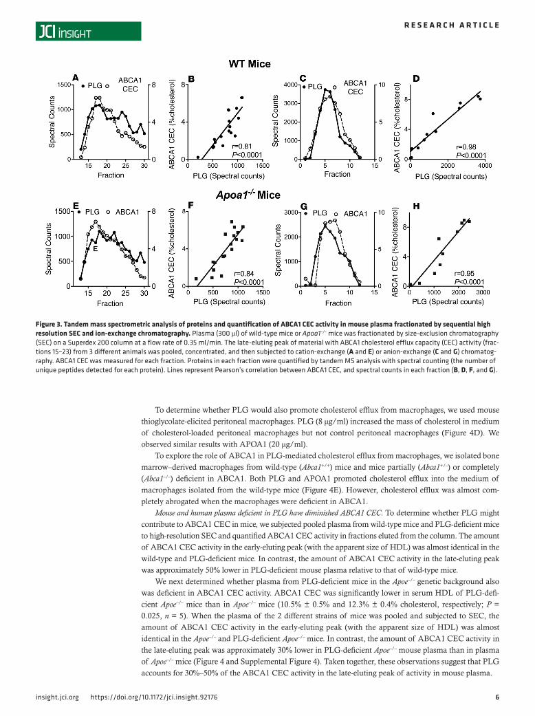
PLG correlates with ABCA1 CEC in plasma subjected to orthogonal chromatography-mass spectrometry. We used orthogonal chromatography-mass spectrometry to identify non–HDL-associated proteins whose elu-tion patterns correlated with those of ABCA1 CEC. Plasma from wild-type mice or APOA1-deficient mice were subjected to high-resolution SEC, and each fraction was monitored for ABCA1 CEC. Fractions from the late-eluting peak of material with ABCA1 CEC activity were combined and then separated by cation-exchange (Figure 4, A and E) or anion-exchange (Figure 4, C and G) chromatography; each fraction was again assessed for ABCA1 CEC activity. The active fractions were analyzed with shotgun proteomics (42), and the relative concentrations of proteins (quantified as spectral counts, the number of unique peptides detected by MS/MS (43) in each fraction were correlated with ABCA1 CEC activity.
Hierarchical clustering assessed by Euclidean distance analysis revealed an array of proteins linked to ABCA1 CEC in the fractions obtained by each of the 2 methods. However, the proteins in the 2 clusters were distinct, and albumin was not in either cluster. Only 1 protein — plasminogen (PLG) — correlated strongly with ABCA1 CEC in both clusters. Moreover, only PLG correlated with ABCA1 CEC activity when plasma was first fractionated by SEC and then by cation-exchange (Figure 4, B and F) or anion-exchange (Figure 4, D and H) chromatography (cation exchange, r = 0.81 and r = 0.84 for wild-type and Apoa1–/– mice, respectively; anion exchange, r = 0.98 and r = 0.95 for wild-type and Apoa1–/– mice, respectively).
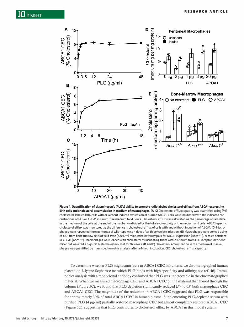
PLG stimulates cholesterol efflux from macrophages by the ABCA1 pathway. We exposed BHK cells (with and without inducible expression of human ABCA1), macrophages (with and without cholesterol loading), and bone marrow–derived macrophages (with and without expression of ABCA1) to human PLG (Figure 5) to determine whether the protein would promote cholesterol efflux from cells. SDS-PAGE analysis with Coomassie blue staining indicated that material with the apparent MW of PLG accounted for greater than 95% of the protein in the PLG preparation.
Stimulation by PLG of ABCA1-specific cholesterol efflux from BHK cells was half-maximal at approx-imately 0.7 μg/ml and plateaued at 3 μg/ml (Figure 4A). In contrast, PLG failed to promote cholesterol efflux from BHK cells expressing ABCG1 (Supplemental Figure 6) (44). Although PLG is a serine pro-tease, its ABCA1 activity was not affected by phenylmethylsulfonyl fluoride (PMSF), which covalently modifies reactive serine residues (45). Cholesterol accumulation in the medium of cells exposed to 1 μg/ml PLG was linear for 2 hours but then gradually decreased (Figure 4B). In contrast, higher concentrations of APOA1 were required for half-maximal and maximal ABCA1-specific cholesterol efflux from BHK cells (Figure 4C): at 5 μg/ml APOA1, the amount of cholesterol in the medium of cells was approximately 30% higher than when cells were exposed to PLG. These results indicate that PLG stimulates cholesterol efflux from BHK cells that express lower concentrations of human ABCA1 than APOA1 but that APOA1 can promote greater total efflux than PLG in this model system.
6insight.jci.org https://doi.org/10.1172/jci.insight.92176
R E S E A R C H A R T I C L E
To determine whether PLG would also promote cholesterol efflux from macrophages, we used mouse thioglycolate-elicited peritoneal macrophages. PLG (8 μg/ml) increased the mass of cholesterol in medium of cholesterol-loaded peritoneal macrophages but not control peritoneal macrophages (Figure 4D). We observed similar results with APOA1 (20 μg/ml).
To explore the role of ABCA1 in PLG-mediated cholesterol efflux from macrophages, we isolated bone marrow–derived macrophages from wild-type (Abca1+/+) mice and mice partially (Abca1+/–) or completely (Abca1–/–) deficient in ABCA1. Both PLG and APOA1 promoted cholesterol efflux into the medium of macrophages isolated from the wild-type mice (Figure 4E). However, cholesterol efflux was almost com-pletely abrogated when the macrophages were deficient in ABCA1.
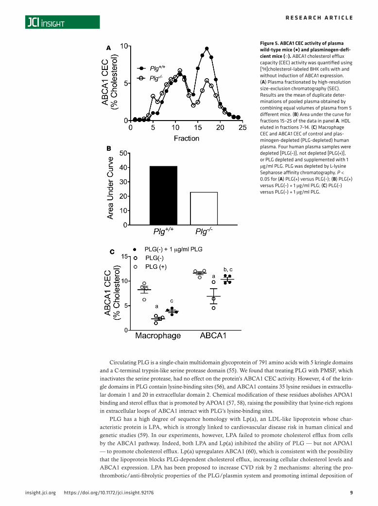
Mouse and human plasma deficient in PLG have diminished ABCA1 CEC. To determine whether PLG might contribute to ABCA1 CEC in mice, we subjected pooled plasma from wild-type mice and PLG-deficient mice to high-resolution SEC and quantified ABCA1 CEC activity in fractions eluted from the column. The amount of ABCA1 CEC activity in the early-eluting peak (with the apparent size of HDL) was almost identical in the wild-type and PLG-deficient mice. In contrast, the amount of ABCA1 CEC activity in the late-eluting peak was approximately 50% lower in PLG-deficient mouse plasma relative to that of wild-type mice.
We next determined whether plasma from PLG-deficient mice in the Apoe–/– genetic background also was deficient in ABCA1 CEC activity. ABCA1 CEC was significantly lower in serum HDL of PLG-defi-cient Apoe–/– mice than in Apoe–/– mice (10.5% ± 0.5% and 12.3% ± 0.4% cholesterol, respectively; P = 0.025, n = 5). When the plasma of the 2 different strains of mice was pooled and subjected to SEC, the amount of ABCA1 CEC activity in the early-eluting peak (with the apparent size of HDL) was almost identical in the Apoe–/– and PLG-deficient Apoe–/– mice. In contrast, the amount of ABCA1 CEC activity in the late-eluting peak was approximately 30% lower in PLG-deficient Apoe–/– mouse plasma than in plasma of Apoe–/– mice (Figure 4 and Supplemental Figure 4). Taken together, these observations suggest that PLG accounts for 30%–50% of the ABCA1 CEC activity in the late-eluting peak of activity in mouse plasma.
Figure 3. Tandem mass spectrometric analysis of proteins and quantification of ABCA1 CEC activity in mouse plasma fractionated by sequential high resolution SEC and ion-exchange chromatography. Plasma (300 μl) of wild-type mice or Apoa1–/– mice was fractionated by size-exclusion chromatography (SEC) on a Superdex 200 column at a flow rate of 0.35 ml/min. The late-eluting peak of material with ABCA1 cholesterol efflux capacity (CEC) activity (frac-tions 15–23) from 3 different animals was pooled, concentrated, and then subjected to cation-exchange (A and E) or anion-exchange (C and G) chromatog-raphy. ABCA1 CEC was measured for each fraction. Proteins in each fraction were quantified by tandem MS analysis with spectral counting (the number of unique peptides detected for each protein). Lines represent Pearson’s correlation between ABCA1 CEC, and spectral counts in each fraction (B, D, F, and G).
7insight.jci.org https://doi.org/10.1172/jci.insight.92176
R E S E A R C H A R T I C L E
To determine whether PLG might contribute to ABCA1 CEC in humans, we chromatographed human plasma on L-lysine Sepharose (to which PLG binds with high specificity and affinity; see ref. 46). Immu-noblot analysis with a monoclonal antibody confirmed that PLG was undetectable in the chromatographed material. When we measured macrophage CEC and ABCA1 CEC on the material that flowed through the column (Figure 5C), we found that PLG depletion significantly reduced (P < 0.05) both macrophage CEC and ABCA1 CEC. The magnitude of the reduction in ABCA1 CEC suggested that PLG was responsible for approximately 30% of total ABCA1 CEC in human plasma. Supplementing PLG-depleted serum with purified PLG (4 μg/ml) partially restored macrophage CEC but almost completely restored ABCA1 CEC (Figure 5C), suggesting that PLG contributes to cholesterol efflux by ABCA1 in this model system.
Figure 4. Quantification of plasminogen’s (PLG’s) ability to promote radiolabeled cholesterol efflux from ABCA1-expressing BHK cells and cholesterol accumulation in medium of macrophages. (A–C) Cholesterol efflux capacity was quantified using [3H]cholesterol-labeled BHK cells with or without induced expression of human ABCA1. Cells were incubated with the indicated con-centrations of PLG or APOA1 in serum-free medium for 4 hours. Cholesterol efflux was calculated as the percentage of radiolabel in the medium of the cells at the end of the incubation divided by the total radioactivity of the medium and cells. ABCA1-specific cholesterol efflux was monitored as the difference in cholesterol efflux of cells with and without induction of ABCA1. (D) Macro-phages were harvested from peritonea of wild-type mice 4 days after thioglycolate injection. (E) Macrophages were derived using M-CSF from bone marrow cells of wild-type (Abca1+/+) mice, mice heterozygous for ABCA1 expression (Abca1+/–), or mice deficient in ABCA1 (Abca1–/–). Macrophages were loaded with cholesterol by incubating them with 2% serum from LDL receptor–deficient mice that were fed a high-fat high-cholesterol diet for 16 weeks. (D and E) Cholesterol accumulation in the medium of macro-phages was quantified by mass spectrometric analysis after a 4-hour incubation. CEC, cholesterol efflux capacity.
8insight.jci.org https://doi.org/10.1172/jci.insight.92176
R E S E A R C H A R T I C L E
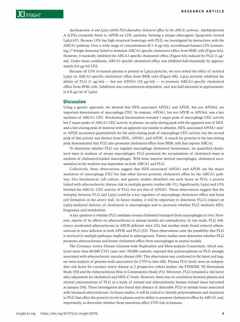
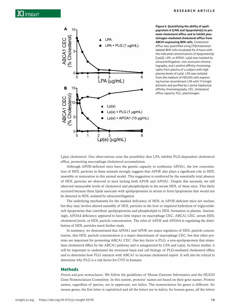
Apolipoprotein A and Lp(a) inhibit PLG-dependent cholesterol efflux by the ABCA1 pathway. Apolipoprotein A (LPA) covalently binds to APOB on LDL particles, forming a unique atherogenic lipoprotein termed Lp(a) (47). Because LPA has high structural homology with PLG, we investigated its interactions with the ABCA1 pathway. Over a wide range of concentrations (0.1–6 μg/ml), recombinant human LPA (contain-ing 17 kringle domains) failed to stimulate ABCA1-specific cholesterol efflux from BHK cells (Figure 6A). However, it markedly inhibited the ABCA1-specific cholesterol efflux (Figure 6A) induced by PLG (1 μg/ml). Under those conditions, ABCA1-specific cholesterol efflux was inhibited half-maximally by approxi-mately 0.6 μg/ml LPA.
Because all LPA in human plasma is present in Lp(a) particles, we next tested the effect of isolated Lp(a) on ABCA1-specific cholesterol efflux from BHK cells (Figure 6B). Lp(a) potently inhibited the ability of PLG (1 μg/ml) — but not APOA1 (10 μg/ml) — to promote ABCA1-specific cholesterol efflux from BHK cells. Inhibition was concentration-dependent, and was half-maximal at approximate-ly 0.8 μg/ml of Lp(a).
DiscussionUsing a genetic approach, we showed that HDL-associated APOA1 and APOE, but not APOA4, are important determinants of macrophage CEC. In contrast, APOA1, but not APOE or APOA4, was a key mediator of ABCA1 CEC. Biochemical fractionation revealed 1 major peak of macrophage CEC activity but 2 major peaks of ABCA1 CEC activity in plasma: an early-eluting peak with the apparent size of HDL and a late-eluting peak of material with an apparent size similar to albumin. HDL-associated APOA1 and/or APOE accounted quantitatively for the early-eluting peak of macrophage CEC activity, but the second peak of late activity was distinct from HDL, APOA1, and APOE. A search for proteins in the late-eluting peak demonstrated that PLG also promotes cholesterol efflux from BHK cells that express ABCA1.
To determine whether PLG can regulate macrophage cholesterol homeostasis, we quantified choles-terol mass in medium of mouse macrophages. PLG promoted the accumulation of cholesterol mass in medium of cholesterol-loaded macrophages. With bone marrow–derived macrophages, cholesterol accu-mulation in the medium was dependent on both ABCA1 and PLG.
Collectively, these observations suggest that HDL-associated APOA1 and APOE are the major mediators of macrophage CEC but that other factors promote cholesterol efflux by the ABCA1 path-way. Our biochemical, cell culture, and genetic studies identified one such factor as PLG, a protein linked with atherosclerotic disease risk in multiple genetic studies (48–51). Significantly, Lp(a) and LPA blocked the ABCA1 CEC activity of PLG, but not that of APOA1. These observations suggest that the interplay between PLG and Lp(a) could be a key regulator of macrophage cholesterol efflux and foam cell formation in the artery wall. In future studies, it will be important to determine PLG’s impact on Lp(a)-mediated delivery of cholesterol to macrophages and to ascertain whether PLG mediates HDL biogenesis and metabolism.
A key question is whether PLG mediates reverse cholesterol transport from macrophages in vivo. How-ever, reports of its effects on atherosclerosis in animal models are contradictory. In one study, PLG defi-ciency accelerated atherosclerosis in APOE-deficient mice (52), but another study found reduced athero-sclerosis in mice deficient in both APOE and PLG (53). These observations raise the possibility that PLG is involved in multiple pathways implicated in atherogenesis. Future studies must determine whether PLG promotes atherosclerosis and boosts cholesterol efflux from macrophages in animal models.
The Coronary Artery Disease Genome-wide Replication and Meta-analysis Consortium, which ana-lyzed more than 60,000 CVD cases and 130,000 controls, reported that polymorphisms in PLG strongly associated with atherosclerotic vascular disease (49). This observation was confirmed in the latest and larg-est meta-analysis of genome-wide association for CVD to date (48). Plasma PLG levels were an indepen-dent risk factor for coronary artery disease in 2 prospective cohort studies: the FINRISK ‘92 Hemostasis Study (50) and the Atherosclerosis Risk in Communities Study (51). Moreover, PLG remained a risk factor after adjustment for cholesterol and HDL-C levels. However, there was no correlation between plasma and intimal concentrations of PLG in a study of normal and atherosclerotic human intimal tissue harvested at autopsy (54). Those investigators also found that absence of detectable PLG in intimal tissue associated with increased atherosclerosis. In future studies, it will be critical to identify polymorphisms and mutations in PLG that affect the protein’s levels in plasma and its ability to promote cholesterol efflux by ABCA1 and, importantly, to determine whether those mutations affect CVD risk in humans.
9insight.jci.org https://doi.org/10.1172/jci.insight.92176
R E S E A R C H A R T I C L E
Circulating PLG is a single-chain multidomain glycoprotein of 791 amino acids with 5 kringle domains and a C-terminal trypsin-like serine protease domain (55). We found that treating PLG with PMSF, which inactivates the serine protease, had no effect on the protein’s ABCA1 CEC activity. However, 4 of the krin-gle domains in PLG contain lysine-binding sites (56), and ABCA1 contains 35 lysine residues in extracellu-lar domain 1 and 20 in extracellular domain 2. Chemical modification of these residues abolishes APOA1 binding and sterol efflux that is promoted by APOA1 (57, 58), raising the possibility that lysine-rich regions in extracellular loops of ABCA1 interact with PLG’s lysine-binding sites.
PLG has a high degree of sequence homology with Lp(a), an LDL-like lipoprotein whose char-acteristic protein is LPA, which is strongly linked to cardiovascular disease risk in human clinical and genetic studies (59). In our experiments, however, LPA failed to promote cholesterol efflux from cells by the ABCA1 pathway. Indeed, both LPA and Lp(a) inhibited the ability of PLG — but not APOA1 — to promote cholesterol efflux. Lp(a) upregulates ABCA1 (60), which is consistent with the possibility that the lipoprotein blocks PLG-dependent cholesterol efflux, increasing cellular cholesterol levels and ABCA1 expression. LPA has been proposed to increase CVD risk by 2 mechanisms: altering the pro-thrombotic/anti-fibrolytic properties of the PLG/plasmin system and promoting intimal deposition of
Figure 5. ABCA1 CEC activity of plasma wild-type mice (●) and plasminogen-defi-cient mice (○). ABCA1 cholesterol efflux capacity (CEC) activity was quantified using [3H]cholesterol-labeled BHK cells with and without induction of ABCA1 expression. (A) Plasma fractionated by high-resolution size-exclusion chromatography (SEC). Results are the mean of duplicate deter-minations of pooled plasma obtained by combining equal volumes of plasma from 5 different mice. (B) Area under the curve for fractions 15–25 of the data in panel A. HDL eluted in fractions 7–14. (C) Macrophage CEC and ABCA1 CEC of control and plas-minogen-depleted (PLG-depleted) human plasma. Four human plasma samples were depleted [PLG(–)], not depleted [PLG(+)], or PLG depleted and supplemented with 1 μg/ml PLG. PLG was depleted by L-lysine Sepharose affinity chromatography. P < 0.05 for (A) PLG(+) versus PLG(–); (B) PLG(+) versus PLG(–) + 1 μg/ml PLG; (C) PLG(–) versus PLG(–) + 1 μg/ml PLG.
1 0insight.jci.org https://doi.org/10.1172/jci.insight.92176
R E S E A R C H A R T I C L E
Lp(a) cholesterol. Our observations raise the possibility that LPA inhibits PLG-dependent cholesterol efflux, promoting macrophage cholesterol accumulation.
Although APOE-deficient mice have the genetic capacity to synthesize APOA1, the low concentra-tion of HDL particles in these animals strongly suggests that APOE also plays a significant role in HDL assembly or maturation in this animal model. This suggestion is reinforced by the essentially total absence of HDL particles we observed in mice lacking both APOE and APOA1. Despite this anomaly, we still observed measurable levels of cholesterol and phospholipids in the serum HDL of these mice. This likely occurred because these lipids associate with apolipoproteins in serum to form lipoproteins that would not be detected in HDL isolated by ultracentrifugation.
The underlying mechanisms for the marked deficiency of HDL in APOE-deficient mice are unclear, but they may involve altered assembly of HDL particles in the liver or impaired hydrolysis of triglyceride-rich lipoproteins that contribute apolipoproteins and phospholipid to HDL formation in plasma. Interest-ingly, APOA4 deficiency appeared to have little impact on macrophage CEC, ABCA1 CEC, serum HDL cholesterol levels, or HDL particle concentration. The roles of APOE and APOA4 in regulating the distri-bution of HDL particles merit further study.
In summary, we demonstrated that APOA1 and APOE are major regulators of HDL particle concen-tration, that HDL particle concentration is a major determinant of macrophage CEC, but that other pro-teins are important for promoting ABCA1 CEC. One key factor is PLG, a non-apolipoprotein that stimu-lates cholesterol efflux by the ABCA1 pathway and is antagonized by LPA and Lp(a). In future studies, it will be important to understand the structural basis and cell biology of PLG-mediated cholesterol efflux and to determine how PLG interacts with ABCA1 to increase cholesterol export. It will also be critical to determine why PLG is a risk factor for CVD in humans.
MethodsProtein and gene nomenclature. We follow the guidelines of Mouse Genome Informatics and the HUGO Gene Nomenclature Committee. In this system, proteins’ names are based on their gene names. Protein names, regardless of species, are in uppercase, not italics. The nomenclature for genes is different: for mouse genes, the first letter is capitalized and all the letters are in italics; for human genes, all the letters
Figure 6. Quantifying the ability of apoli-poprotein A (LPA) and lipoprotein(s) to pro-mote cholesterol efflux and to inhibit plas-minogen-mediated cholesterol efflux from ABCA1-expressing BHK cells. Cholesterol efflux was quantified using [3H]cholesterol-labeled BHK cells incubated for 4 hours with the indicated concentrations of lipoprotein(a) [Lp(a)], LPA, or APOA1. Lp(a) was isolated by ultracentrifugation, size-exclusion chroma-tography, and L-proline affinity chromatog-raphy from plasma of a subject with high plasma levels of Lp(a). LPA was isolated from the medium of HEK293 cells express-ing human recombinant LPA with 17 kringle domains and purified by L-lysine Sepharose affinity chromatography. CEC, cholesterol efflux capacity; PLG, plasminogen.
1 1insight.jci.org https://doi.org/10.1172/jci.insight.92176
R E S E A R C H A R T I C L E
are capitalized and in italics. For example, the gene name for human apolipoprotein(a) is LPA and the protein name is LPA.
Mice. Studies were approved by the Animal Care and Use committees of the University of Washing-ton, University of Chicago, and Wake Forest University. Transgenic mice were housed (3–5 per cage) in a specific pathogen–free barrier facility (22°C) with a 12-hour light/dark cycle with free access to food and water. All the strains were fed a low-fat diet (Wayne Rodent BLOX 8604; Harlan Teklad Laboratory) until they were 8–16 weeks old. Mice fasted for 4 hours in the morning were bled from the retro-orbital sinus into tubes containing EDTA (final concentration 1 mM), and killed by isoflurane inhalation. Plasma samples were collected and stored at −80°C until analysis.
Plasma lipids. Phospholipids (Wako Diagnostics) and cholesterol levels (Invitrogen) were determined biochemically.
SEC. Plasma (300 μl) was fractionated on a Superdex 200 column (24 ml column volume) at a 0.35 ml/min flow rate with PBS, and 0.5-ml fractions were collected.
Ion-exchange chromatography. The late-eluting peak of material with ABCA1 activity (4.5 ml) isolated by SEC from 2 pooled plasma samples was concentrated to a final volume of 0.6 ml with 3,000 MW cut-off filters. After passage over a 10DG desalting column (GE Healthcare) equilibrated with buffer A (20 mM Tris-HCl, pH 8.0 for MonoQ and 20 mM 2-[N-morpholino] ethanesulfonic acid (MES), pH 6.0 for MonoS) or buffer B (20 mM Tris-HCl, 1.0 M NaCl, pH 8.0 for MonoQ and 20 mM MES, 1.0 M NaCl, pH 6.0 for MonoS ), the concentrated material was loaded onto a MonoQ 5/50GL or MonoS 5/50L column (24 ml volume) at a flow rate of 0.35 ml/min (61). After the column was washed with 100 ml of buffer A, it was eluted with a linear gradient of 100 ml buffer B, and 0.5-ml fractions were collected.
Lp(a) isolation. Plasma from 2 subjects with high levels of Lp(a) was subjected to density gradient ultra-centrifugation followed by chromatography on L-proline (62).
Recombinant LPA. Seventeen-kringle LPA expressed by human embryonic kidney cells (63) was isolated from conditioned medium by chromatography on L-lysine Sepharose. Protein purity was assessed by SDS-PAGE and silver staining under nonreducing and reducing conditions.
PLG depletion. Human plasma samples were passed through an L-lysine Sepharose column equilibrated with 0.1 M NaHCO3 at pH 8.9 (46). PLG was quantified in samples before and after depletion by immu-noblot analysis with a rabbit polyclonal antibody against PLG (Santa Cruz Biotechnology).
Bone marrow–derived macrophages. Bone marrow harvested from femurs and tibias of 16-week-old Abca1+/+, Abca1+/–, and Abca1–/– mice was gently suspended in RPMI medium (Sigma-Aldrich). Cells were collected by centrifugation (300 g, 5 minutes), plated (2 × 10⁶ cells) in 24-well plates, and cultured for 7 days in RPMI supplemented with 10% fetal bovine serum, 1% penicillin, and 30 ng/ml mouse recombinant M-CSF. Medium was replaced on days 4 and 6. Cells were loaded with cholesterol by incubating them overnight in RPMI medium supplemented with 2% plasma harvested from LDL recep-tor–deficient mice fed a high-fat diet for 16 weeks (64–66).
Peritoneal macrophages. Cells were harvested from the peritoneum of C57BL/6J mice 4 days after injection of 1 ml of thioglycolate (Sigma-Aldrich) (67). Cells were washed with PBS, plated, and allowed to adhere for 2 hours at 37°C in serum-free high-glucose DMEM (Sigma-Aldrich). Nonadherent cells were removed by 3 washes with PBS. Cells were loaded with cholesterol as described for bone marrow–derived macrophages.
Cholesterol mass. Following the addition of [2H7]cholesterol (Sigma-Aldrich) as internal standard and then saponification, lipids were extracted from cell-conditioned medium with hexane and dried under nitrogen gas. Total cholesterol levels were determined after derivatization by LC-MS/MS (68). Free cho-lesterol was measured with the same procedure without saponification; cholesteryl ester was calculated as total cholesterol minus free cholesterol.
Cholesterol efflux. CEC was assessed with J774 macrophages labeled with [3H]cholesterol and stimulated with a cAMP analogue, as described by de la Llera-Moya et al. (24). Efflux by the ABCA1 pathway was measured with BHK cells containing mifepristone-inducible human ABCA1 that were radiolabeled with [3H]cholesterol and 1 mg/ml fatty acid–free albumin (69). Efflux of [3H]cholesterol was measured after a 4-hour incubation in medium with or without serum depleted of apoB (2.8% v/v), human APOA1 (Mil-lipore), human PLG (R&D Systems), Lp(a), and LPA. Cholesterol efflux was calculated as the percentage of radiolabel in the medium of the cells at the end of the incubation divided by the total radioactivity of the medium and cells. ABCA1-specific cholesterol efflux was monitored as the difference in cholesterol efflux of cells with and without induction of ABCA1.
1 2insight.jci.org https://doi.org/10.1172/jci.insight.92176
R E S E A R C H A R T I C L E
Liquid chromatography-electrospray ionization tandem mass spectrometric (LC-ESI-MS/MS) analysis. Pro-teins (1 μg precipitated) were solubilized with 0.1% RapiGest (Waters) in 200 mM ammonium bicarbon-ate, reduced with dithiothreitol, alkylated with iodoacetamide, and digested with trypsin (1:20, w/w protein; Promega) for 3 hours at 37°C. After a second aliquot of trypsin (1:20, w/w protein) was added, samples were incubated overnight at 37°C. After RapiGest was removed by acid hydrolysis, samples were dried and stored at −20°C until analysis. Prior to analysis, samples were reconstituted in 5% aceto-nitrile, 0.1% formic acid (42, 70).
Tryptic digests (1 μg protein) were injected onto a C18 trap column, eluted onto an analyti-cal reverse-phase column (0.15 × 150 mm, Magic C18AQ, 5 μm, 200 A; Michrom Bioresources Inc.), and separated on a Paradigm MS4B HPLC (Michrom Bioresources Inc.) (42, 70). Peptides were introduced into the gas phase by electrospray ionization, and positive-ion mass spectra were acquired with an Orbitrap Mass Spectrometer (Thermo Fisher Scientific), using data-dependent acquisition (1 MS survey scan followed by MS/MS scans of the 8 most abundant ions in the survey scan). An exclusion window of 45 seconds was used after 2 acquisitions of the same precursor ion with a mass window 400–2,000 m/z.
Protein identification. MS/MS spectra were matched against the mouse International Protein Index (IPI) database (mouse v.3.54, Jan 2009), using the SEQUEST (v 2.7) search engine with fixed Cys carbamidomethylation and variable Met oxidation modifications. The mass tolerance for precursor ions was 2.5 Da; SEQUEST default tolerance was accepted for product ions. SEQUEST results were further validated with PeptideProphet and ProteinProphet (71, 72), using an adjusted probability of 0.90 or higher for peptides and 0.95 or higher for proteins, giving an estimated false discovery rate of 5%. Each charge state of a peptide was considered a unique identification.
We used the gene names for the proteins identified in HDL to eliminate the redundant identifications of isoforms and protein fragments frequently found in protein databases used in proteomic analysis (73). This approach also permits cross-referencing of proteins from different species.
Protein quantification. Proteins were quantified using spectral counts — the total number of MS/MS spectra detected for a protein. Spectral counting of HDL proteins strongly agrees with the targeted proteomic method, selected reaction monitoring (43, 74–77). Proteins considered for analysis had to be detected in 3 or more analyses with 2 or more unique peptides. When MS/MS spectra could not differ-entiate between protein isoforms, the isoform with the most unique peptides was used for further analy-sis. Spectral counts for each protein normalized to total spectral counts for peptides from each sample were used to calculate a spectral index. We used the index to compare relative protein composition (43).
HDL-PIMA. HDL-PIMA was quantified by calibrated ion mobility analysis (38). HDL peak areas were converted into aqueous particle concentrations, using glucose oxidase calibration curves. The method yields a stoichiometry of APOA1 and sizes and relative abundances of HDL subspecies that are in excel-lent agreement with those determined by nondenaturing gradient gel electrophoresis and analytical ultra-centrifugation (38, 78).
Statistics. Data are the mean ± SD. Differences among groups were assessed with ANOVA with Tukey’s post-hoc analysis to correct for multiple comparisons. Linear correlations were assessed with Pearson’s product-moment coefficient. All reported P values are 2-tailed, with a P value less than 0.05 indicating sta-tistical significance. Analyses were performed with Prism (GraphPad) and R GNU software.
Author contributionsNP designed studies, conducted experiments, analyzed data, and helped prepare the manuscript. PMH con-ducted experiments. GER acquired data. HW conducted experiments and acquired data. CT provided reagents. RD, EP, and RW provided mice. TV designed studies and acquired data. VS provided human samples. MLK provided 17 kringle recombinant Apo(a). CAR and GSG provided mice and helped prepare the manuscript. DAD designed studies and helped prepare the manuscript. SM provided reagents, designed studies, and helped prepare the manuscript. JWH designed and analyzed studies and helped prepare the manuscript.
AcknowledgmentsThis work was supported by awards from the National Institutes of Health (HL112625, HL108897, P30 DK17047, P01 HL092969, T32HL007828, HL076491). We thank Marlys Koschinsky for providing recombi-nant 17-kringle LPA.
1 3insight.jci.org https://doi.org/10.1172/jci.insight.92176
R E S E A R C H A R T I C L E
Address correspondence to: Nathalie Pamir, Knight Cardiovascular Institute, Oregon Health and Science University, 3181 S.W. Sam Jackson Park Road, Portland, Oregon 97239, USA. Phone: 503.346.1568; Email: [email protected].
1. Gordon DJ, Rifkind BM. High-density lipoprotein--the clinical implications of recent studies. N Engl J Med. 1989;321(19):1311–1316. 2. Wilson PW, Abbott RD, Castelli WP. High density lipoprotein cholesterol and mortality. The Framingham Heart Study. Arterio-
sclerosis. 1988;8(6):737–741. 3. Tall AR, Costet P, Wang N. Regulation and mechanisms of macrophage cholesterol efflux. J Clin Invest. 2002;110(7):899–904. 4. Getz GS, Reardon CA. Animal models of atherosclerosis. Arterioscler Thromb Vasc Biol. 2012;32(5):1104–1115. 5. Hutchins PM, Heinecke JW. Cholesterol efflux capacity, macrophage reverse cholesterol transport and cardioprotective HDL.
Curr Opin Lipidol. 2015;26(5):388–393. 6. AIM-HIGH Investigators, et al. Niacin in patients with low HDL cholesterol levels receiving intensive statin therapy. N Engl J
Med. 2011;365(24):2255–2267. 7. HPS2-THRIVE Collaborative Group, et al. Effects of extended-release niacin with laropiprant in high-risk patients. N Engl J
Med. 2014;371(3):203–212. 8. Schwartz GG, et al. Effects of dalcetrapib in patients with a recent acute coronary syndrome. N Engl J Med. 2012;367(22):2089–2099. 9. Rader DJ, Hovingh GK. HDL and cardiovascular disease. Lancet. 2014;384(9943):618–625. 10. Kingwell BA, Chapman MJ, Kontush A, Miller NE. HDL-targeted therapies: progress, failures and future. Nat Rev Drug Discov.
2014;13(6):445–464. 11. van Capelleveen JC, Brewer HB, Kastelein JJ, Hovingh GK. Novel therapies focused on the high-density lipoprotein particle.
Circ Res. 2014;114(1):193–204. 12. Vickers KC, Remaley AT. HDL and cholesterol: life after the divorce? J Lipid Res. 2014;55(1):4–12. 13. Zanoni P, et al. Rare variant in scavenger receptor BI raises HDL cholesterol and increases risk of coronary heart disease.
Science. 2016;351(6278):1166–1171. 14. Zhang S, et al. Diet-induced occlusive coronary atherosclerosis, myocardial infarction, cardiac dysfunction, and premature death in
scavenger receptor class B type I-deficient, hypomorphic apolipoprotein ER61 mice. Circulation. 2005;111(25):3457–3464. 15. Rader DJ, Alexander ET, Weibel GL, Billheimer J, Rothblat GH. The role of reverse cholesterol transport in animals and
humans and relationship to atherosclerosis. J Lipid Res. 2009;50 Suppl:S189–S194. 16. Tall AR, Yvan-Charvet L, Terasaka N, Pagler T, Wang N. HDL, ABC transporters, and cholesterol efflux: implications for the
treatment of atherosclerosis. Cell Metab. 2008;7(5):365–375. 17. Oram JF, Heinecke JW. ATP-binding cassette transporter A1: a cell cholesterol exporter that protects against cardiovascular dis-
ease. Physiol Rev. 2005;85(4):1343–1372. 18. Rye KA, Barter PJ. Regulation of high-density lipoprotein metabolism. Circ Res. 2014;114(1):143–156. 19. Rothblat GH, Phillips MC. High-density lipoprotein heterogeneity and function in reverse cholesterol transport. Curr Opin
Lipidol. 2010;21(3):229–238. 20. Bashtovyy D, Jones MK, Anantharamaiah GM, Segrest JP. Sequence conservation of apolipoprotein A-I affords novel insights
into HDL structure-function. J Lipid Res. 2011;52(3):435–450. 21. van Eck M, et al. Leukocyte ABCA1 controls susceptibility to atherosclerosis and macrophage recruitment into tissues. Proc Natl
Acad Sci USA. 2002;99(9):6298–6303. 22. Van Eck M, et al. Macrophage ATP-binding cassette transporter A1 overexpression inhibits atherosclerotic lesion progression in
low-density lipoprotein receptor knockout mice. Arterioscler Thromb Vasc Biol. 2006;26(4):929–934. 23. Ferrans VJ, Fredrickson DS. The pathology of Tangier disease. A light and electron microscopic study. Am J Pathol.
1975;78(1):101–158. 24. de la Llera-Moya M, Drazul-Schrader D, Asztalos BF, Cuchel M, Rader DJ, Rothblat GH. The ability to promote efflux via
ABCA1 determines the capacity of serum specimens with similar high-density lipoprotein cholesterol to remove cholesterol from macrophages. Arterioscler Thromb Vasc Biol. 2010;30(4):796–801.
25. Khera AV, et al. Cholesterol efflux capacity, high-density lipoprotein function, and atherosclerosis. N Engl J Med. 2011;364(2):127–135.
26. Rohatgi A, et al. HDL cholesterol efflux capacity and incident cardiovascular events. N Engl J Med. 2014;371(25):2383–2393. 27. Saleheen D, et al. Association of HDL cholesterol efflux capacity with incident coronary heart disease events: a prospective
case-control study. Lancet Diabetes Endocrinol. 2015;3(7):507–513. 28. Sankaranarayanan S, et al. A sensitive assay for ABCA1-mediated cholesterol efflux using BODIPY-cholesterol. J Lipid Res.
2011;52(12):2332–2340. 29. Remaley AT, Schumacher UK, Stonik JA, Farsi BD, Nazih H, Brewer HB. Decreased reverse cholesterol transport from Tangier
disease fibroblasts. Acceptor specificity and effect of brefeldin on lipid efflux. Arterioscler Thromb Vasc Biol. 1997;17(9):1813–1821. 30. Wang X, et al. Macrophage ABCA1 and ABCG1, but not SR-BI, promote macrophage reverse cholesterol transport in vivo.
J Clin Invest. 2007;117(8):2216–2224. 31. Yvan-Charvet L, et al. Combined deficiency of ABCA1 and ABCG1 promotes foam cell accumulation and accelerates athero-
sclerosis in mice. J Clin Invest. 2007;117(12):3900–3908. 32. Shao B, et al. Myeloperoxidase impairs ABCA1-dependent cholesterol efflux through methionine oxidation and site-specific
tyrosine chlorination of apolipoprotein A-I. J Biol Chem. 2006;281(14):9001–9004. 33. Fournier N, et al. Human ApoA-IV overexpression in transgenic mice induces cAMP-stimulated cholesterol efflux from J774
macrophages to whole serum. Arterioscler Thromb Vasc Biol. 2000;20(5):1283–1292. 34. von Eckardstein A, et al. Lipoproteins containing apolipoprotein A-IV but not apolipoprotein A-I take up and esterify cell-
derived cholesterol in plasma. Arterioscler Thromb Vasc Biol. 1995;15(10):1755–1763.
1 4insight.jci.org https://doi.org/10.1172/jci.insight.92176
R E S E A R C H A R T I C L E
35. Plump AS, Breslow JL. Apolipoprotein E and the apolipoprotein E-deficient mouse. Annu Rev Nutr. 1995;15:495–518. 36. Voyiaziakis E, Goldberg IJ, Plump AS, Rubin EM, Breslow JL, Huang LS. ApoA-I deficiency causes both hypertriglyceridemia
and increased atherosclerosis in human apoB transgenic mice. J Lipid Res. 1998;39(2):313–321. 37. Linton MF, Atkinson JB, Fazio S. Prevention of atherosclerosis in apolipoprotein E-deficient mice by bone marrow transplanta-
tion. Science. 1995;267(5200):1034–1037. 38. Hutchins PM, et al. Quantification of HDL particle concentration by calibrated ion mobility analysis. Clin Chem.
2014;60(11):1393–1401. 39. Pamir N, et al. Proteomic analysis of HDL from inbred mouse strains implicates APOE associated with HDL in reduced cho-
lesterol efflux capacity via the ABCA1 pathway. J Lipid Res. 2016;57(2):246–257. 40. Sankaranarayanan S, de la Llera-Moya M, Drazul-Schrader D, Phillips MC, Kellner-Weibel G, Rothblat GH. Serum albumin
acts as a shuttle to enhance cholesterol efflux from cells. J Lipid Res. 2013;54(3):671–676. 41. Gundry RL, Fu Q, Jelinek CA, Van Eyk JE, Cotter RJ. Investigation of an albumin-enriched fraction of human serum and its
albuminome. Proteomics Clin Appl. 2007;1(1):73–88. 42. Vaisar T, et al. Shotgun proteomics implicates protease inhibition and complement activation in the antiinflammatory properties
of HDL. J Clin Invest. 2007;117(3):746–756. 43. Ronsein GE, et al. Parallel reaction monitoring (PRM) and selected reaction monitoring (SRM) exhibit comparable linearity,
dynamic range and precision for targeted quantitative HDL proteomics. J Proteomics. 2015;113:388–399. 44. Vaughan AM, Oram JF. ABCG1 redistributes cell cholesterol to domains removable by high density lipoprotein but not by lipid-
depleted apolipoproteins. J Biol Chem. 2005;280(34):30150–30157. 45. Sekar V, Hageman JH. Specificity of the serine protease inhibitor, phenylmethylsulfonyl fluoride. Biochem Biophys Res Commun.
1979;89(2):474–478. 46. Deutsch DG, Mertz ET. Plasminogen: purification from human plasma by affinity chromatography. Science.
1970;170(3962):1095–1096. 47. Marcovina SM, Koschinsky ML. Lipoprotein(a) as a risk factor for coronary artery disease. Am J Cardiol. 1998;82(12A):57U–66U. 48. Nikpay M, et al. A comprehensive 1,000 Genomes-based genome-wide association meta-analysis of coronary artery disease.
Nat Genet. 2015;47(10):1121–1130. 49. CARDIoGRAMplusC4D Consortium, et al. Large-scale association analysis identifies new risk loci for coronary artery disease.
Nat Genet. 2013;45(1):25–33. 50. Rajecki M, Pajunen P, Jousilahti P, Rasi V, Vahtera E, Salomaa V. Hemostatic factors as predictors of stroke and cardiovascular
diseases: the FINRISK ‘92 Hemostasis Study. Blood Coagul Fibrinolysis. 2005;16(2):119–124. 51. Folsom AR, Aleksic N, Park E, Salomaa V, Juneja H, Wu KK. Prospective study of fibrinolytic factors and incident coronary
heart disease: the Atherosclerosis Risk in Communities (ARIC) Study. Arterioscler Thromb Vasc Biol. 2001;21(4):611–617. 52. Xiao Q, et al. Plasminogen deficiency accelerates vessel wall disease in mice predisposed to atherosclerosis. Proc Natl Acad Sci
USA. 1997;94(19):10335–10340. 53. Kremen M, et al. Plasminogen mediates the atherogenic effects of macrophage-expressed urokinase and accelerates atheroscle-
rosis in apoE-knockout mice. Proc Natl Acad Sci USA. 2008;105(44):17109–17114. 54. Smith EB, Ashall C. Fibrinolysis and plasminogen concentration in aortic intima in relation to death following myocardial
infarction. Atherosclerosis. 1985;55(2):171–186. 55. Petersen TE, Martzen MR, Ichinose A, Davie EW. Characterization of the gene for human plasminogen, a key proenzyme in
the fibrinolytic system. J Biol Chem. 1990;265(11):6104–6111. 56. Xue Y, Bodin C, Olsson K. Crystal structure of the native plasminogen reveals an activation-resistant compact conformation.
J Thromb Haemost. 2012;10(7):1385–1396. 57. Nagao K, Kimura Y, Ueda K. Lysine residues of ABCA1 are required for the interaction with apoA-I. Biochim Biophys Acta.
2012;1821(3):530–535. 58. Sorci-Thomas MG, Thomas MJ. Microdomains, inflammation, and atherosclerosis. Circ Res. 2016;118(4):679–691. 59. Emerging Risk Factors Collaboration, et al. Lipoprotein(a) concentration and the risk of coronary heart disease, stroke, and
nonvascular mortality. JAMA. 2009;302(4):412–423. 60. Sharma M, Von Zychlinski-Kleffmann A, Porteous CM, Jones GT, Williams MJ, McCormick SP. Lipoprotein (a) upregulates
ABCA1 in liver cells via scavenger receptor-B1 through its oxidized phospholipids. J Lipid Res. 2015;56(7):1318–1328. 61. Gherardi E, Gray J, Stoker M, Perryman M, Furlong R. Purification of scatter factor, a fibroblast-derived basic protein that
modulates epithelial interactions and movement. Proc Natl Acad Sci USA. 1989;86(15):5844–5848. 62. Marcovina SM, et al. Use of a reference material proposed by the International Federation of Clinical Chemistry and Laborato-
ry Medicine to evaluate analytical methods for the determination of plasma lipoprotein(a). Clin Chem. 2000;46(12):1956–1967. 63. Romagnuolo R, Marcovina SM, Boffa MB, Koschinsky ML. Inhibition of plasminogen activation by apo(a): role of carboxyl-
terminal lysines and identification of inhibitory domains in apo(a). J Lipid Res. 2014;55(4):625–634. 64. Lutz MB, et al. An advanced culture method for generating large quantities of highly pure dendritic cells from mouse bone mar-
row. J Immunol Methods. 1999;223(1):77–92. 65. Inaba K, et al. Generation of large numbers of dendritic cells from mouse bone marrow cultures supplemented with granulo-
cyte/macrophage colony-stimulating factor. J Exp Med. 1992;176(6):1693–1702. 66. Pamir N, et al. Granulocyte/macrophage colony-stimulating factor-dependent dendritic cells restrain lean adipose tissue expan-
sion. J Biol Chem. 2015;290(23):14656–14667. 67. Becker L, et al. Unique proteomic signatures distinguish macrophages and dendritic cells. PLoS One. 2012;7(3):e33297. 68. Honda A, et al. Highly sensitive quantification of key regulatory oxysterols in biological samples by LC-ESI-MS/MS. J Lipid
Res. 2009;50(2):350–357. 69. Shao B, et al. Acrolein impairs ATP binding cassette transporter A1-dependent cholesterol export from cells through site-specif-
ic modification of apolipoprotein A-I. J Biol Chem. 2005;280(43):36386–36396. 70. Cheung MC, Vaisar T, Han X, Heinecke JW, Albers JJ. Phospholipid transfer protein in human plasma associates with proteins
linked to immunity and inflammation. Biochemistry. 2010;49(34):7314–7322.
1 5insight.jci.org https://doi.org/10.1172/jci.insight.92176
R E S E A R C H A R T I C L E
71. Keller A, Nesvizhskii AI, Kolker E, Aebersold R. Empirical statistical model to estimate the accuracy of peptide identifications made by MS/MS and database search. Anal Chem. 2002;74(20):5383–5392.
72. Nesvizhskii AI, Keller A, Kolker E, Aebersold R. A statistical model for identifying proteins by tandem mass spectrometry. Anal Chem. 2003;75(17):4646–4658.
73. Rappsilber J, Mann M. What does it mean to identify a protein in proteomics? Trends Biochem Sci. 2002;27(2):74–78. 74. Liu H, Sadygov RG, Yates JR. A model for random sampling and estimation of relative protein abundance in shotgun pro-
teomics. Anal Chem. 2004;76(14):4193–4201. 75. Old WM, et al. Comparison of label-free methods for quantifying human proteins by shotgun proteomics. Mol Cell Proteomics.
2005;4(10):1487–1502. 76. Fu X, et al. Spectral index for assessment of differential protein expression in shotgun proteomics. J Proteome Res.
2008;7(3):845–854. 77. Hoofnagle AN, Becker JO, Oda MN, Cavigiolio G, Mayer P, Vaisar T. Multiple-reaction monitoring-mass spectrometric assays
can accurately measure the relative protein abundance in complex mixtures. Clin Chem. 2012;58(4):777–781. 78. Rosenson RS, et al. HDL measures, particle heterogeneity, proposed nomenclature, and relation to atherosclerotic cardiovascu-
lar events. Clin Chem. 2011;57(3):392–410.