Embed Size (px)
Citation preview

ExcEllEcEncE in cosmEtic DEntistry
� TheJournalofCosmeticDentistry SpecialEdition
What’s Next?Fellow AGD Members,
I just want to take a couple of minutes to invite you to sample this mini-version of our Journal of Cosmetic Dentistry. In some ways, this may serve as an introduction to many of you to the American Academy of Cosmetic Dentistry(AACD).
As AACD President this year, I am in the enviable position of helping build bridges between two organizations that have each made significant contributions to my career. I confess, I am a continuing
education “junkie.” I have been a member of the Academy of General Dentistry since 1974. I joined just to get my CE tracked by someone official in case I ever got sued. Then, one year when I was a little negligent about seeking courses, I got my AGD reminder that I had a “promise” to fulfill in meeting my requirements of membership. It was a wake-up call. It reminded me that we never can afford to let ourselves stop learning. I have never needed another reminder. I remember when I got my AGD Fellowship and how proud I was (and my Dad, too, who was also a Fellow in the AGD). I remember asking myself “What’s Next?”
That was the same year as my first AACD conference. Suddenly I was exposed to the greatest cosmetic dentistry get-together the world had ever seen. I was mesmerized. I was challenged. And, I was a little intimidated to see work that beautiful being done by mere mortals. Soon I knew what was next.
I accepted my own challenge and went barreling after AACD’s Accreditation. Alas, I failed at my first attempt, and it made me want it even more. I took every cosmetic dentistry course in sight. And I improved. I learned to see what had been right in front of my eyes. I learned to discern what was cosmetic. I gained the ability to picture what I wanted to occur and the hands to make it happen. And, I eventually passed Accreditation. It was the single greatest accomplishment of my dental life.
After the dust settled, and my ego returned to a little closer to normal, I remember asking myself that age-old question once again, “What’s Next?”
Mastertrack. I was fortunate to get involved with a great bunch of dentists and for five years we met twice a year. We studied. We pushed each other. We taught each other. We showed off to each other. We reached out in new directions. And eventually, I became a Master in the AGD.
Again I asked, “What’s Next?” This time it was to become involved in the teaching of cosmetic dentistry and leadership in the AACD. It has been a wonderful journey.
As I reflect, as President of the AACD, on the similarities of our two organizations, I see that they both are focused on the education of not only dental professionals, but also the public. I see that they both have a foundation of continual continuing education. I see them both presenting challenges to our skills to make us improve. I see them both serving as reminders of the goal to serve our patients with the maximum skill, knowledge, care and integrity. And I see them both as sources of professional camaraderie.
When the next time comes for you to ask yourself, “What’s Next?” please give serious consideration to joining the American Academy of Cosmetic Dentistry, of enhancing your knowledge and skills in this exponentially growing field, and perhaps of even attaining the AACD credential of Accreditation. This is a continuing journey of knowledge we have embarked on in dentistry. Come celebrate it with us.
Dr. Marty Zase
Accredited Member and President American Academy of Cosmetic Dentistry
Master, Academy of General Dentistry
Message froM the aaCD PresiDent
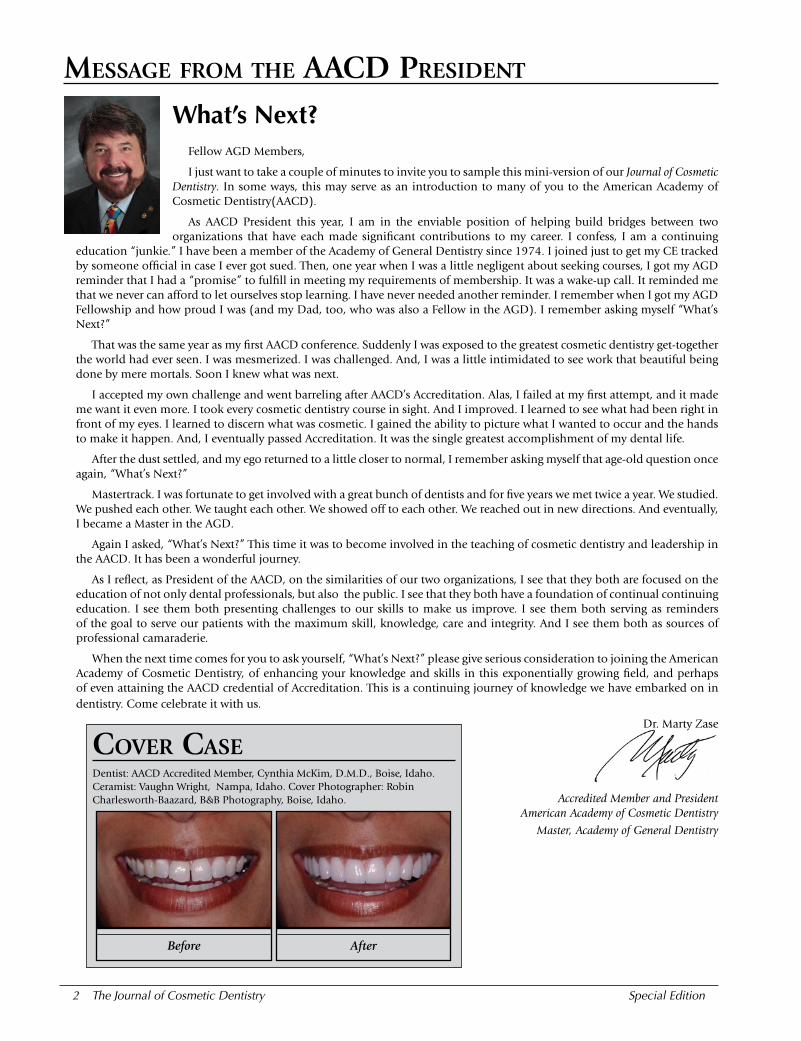
Cover CaseDentist: AACD Accredited Member, Cynthia McKim, D.M.D., Boise, Idaho. Ceramist: Vaughn Wright, Nampa, Idaho. Cover Photographer: Robin Charlesworth-Baazard, B&B Photography, Boise, Idaho.
Before After
SpecialEdition TheJournalofCosmeticDentistry �
IntroductIon
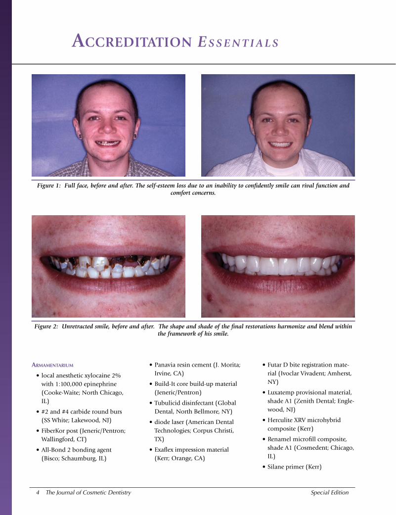
The cosmetic dentist is faced with a wide array of challenges, ranging from the patient who wishes to change an acceptable smile to an outstand-ing smile; to the other end of the spectrum, wherein a patient is faced with a severe esthetic deformity such that smiling is simply not an option. The loss of self-esteem due to an inability to smile confidently can rival function and comfort concerns in severity1,2 (Figs 1 & 2).
His previous dental visits had only made him feel worse about his smile.
clInIcal HIstory
The patient was a 23-year-old male with a history of poor dental care and frequent consumption of soft drinks. Teeth #2 and #31 were carious below the level of the bone. There was generalized gingival inflammation but no clinical bone loss. There were no signs or symptoms of temporomandibular disease. The patient reported that his mother has not seen him smile since he was 10. He often wore a nose ring to detract from the appearance of his teeth. His previous dental visits had only made him feel worse about his smile, as some dentists and staff members had derided him for having such poor dental health.
dIagnosIs and treatment Plan
The diagnosis consisted of the following:
• caries, some severe, on teeth ##2–15, ##18–21, and ##28–31
• gingivitis
• abscessed teeth #2 and #31
• inadequate home care
• poor diet.
The treatment plan included home care instruction; dietary education; porcelain restorations at ##5–12; composite resin restorations at #3 and #4, #13 and #14, ##18–21, ##28–30; and extraction of #2 and #31.
Accreditation Case Report, Case Type I: Six or More Indirect Restorations
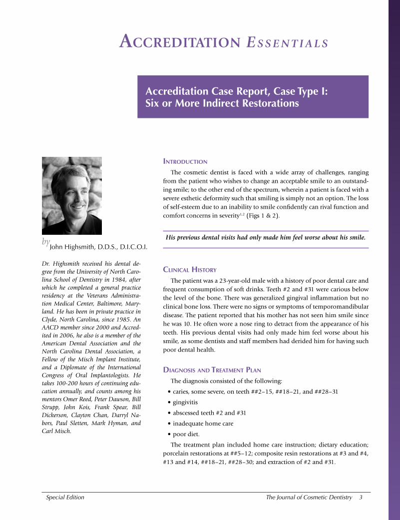
Dr. Highsmith received his dental de-gree from the University of North Caro-lina School of Dentistry in 1984, after which he completed a general practice residency at the Veterans Administra-tion Medical Center, Baltimore, Mary-land. He has been in private practice in Clyde, North Carolina, since 1985. An AACD member since 2000 and Accred-ited in 2006, he also is a member of the American Dental Association and the North Carolina Dental Association, a Fellow of the Misch Implant Institute, and a Diplomate of the International Congress of Oral Implantologists. He takes 100-200 hours of continuing edu-cation annually, and counts among his mentors Omer Reed, Peter Dawson, Bill Strupp, John Kois, Frank Spear, Bill Dickerson, Clayton Chan, Darryl Na-bors, Paul Sletten, Mark Hyman, and Carl Misch.
byJohn Highsmith, D.D.S., D.I.C.O.I.
aCCreDitation E s s E n t i a l s
� TheJournalofCosmeticDentistry SpecialEdition
aCCreDitation E s s E n t i a l s
armamentarIum
• local anesthetic xylocaine 2% with 1:100,000 epinephrine (Cooke-Waite; North Chicago, IL)
• #2 and #4 carbide round burs (SS White; Lakewood, NJ)
• FiberKor post (Jeneric/Pentron; Wallingford, CT)
• All-Bond 2 bonding agent (Bisco; Schaumburg, IL)
• Panavia resin cement (J. Morita; Irvine, CA)
• Build-It core build-up material (Jeneric/Pentron)
• Tubulicid disinfectant (Global Dental, North Bellmore, NY)
• diode laser (American Dental Technologies; Corpus Christi, TX)
• Exaflex impression material (Kerr; Orange, CA)
• Futar D bite registration mate-rial (Ivoclar Vivadent; Amherst, NY)
• Luxatemp provisional material, shade A1 (Zenith Dental; Engle-wood, NJ)
• Herculite XRV microhybrid composite (Kerr)
• Renamel microfill composite, shade A1 (Cosmedent; Chicago, IL)
• Silane primer (Kerr)
Figure 1: Full face, before and after. The self-esteem loss due to an inability to confidently smile can rival function and comfort concerns.
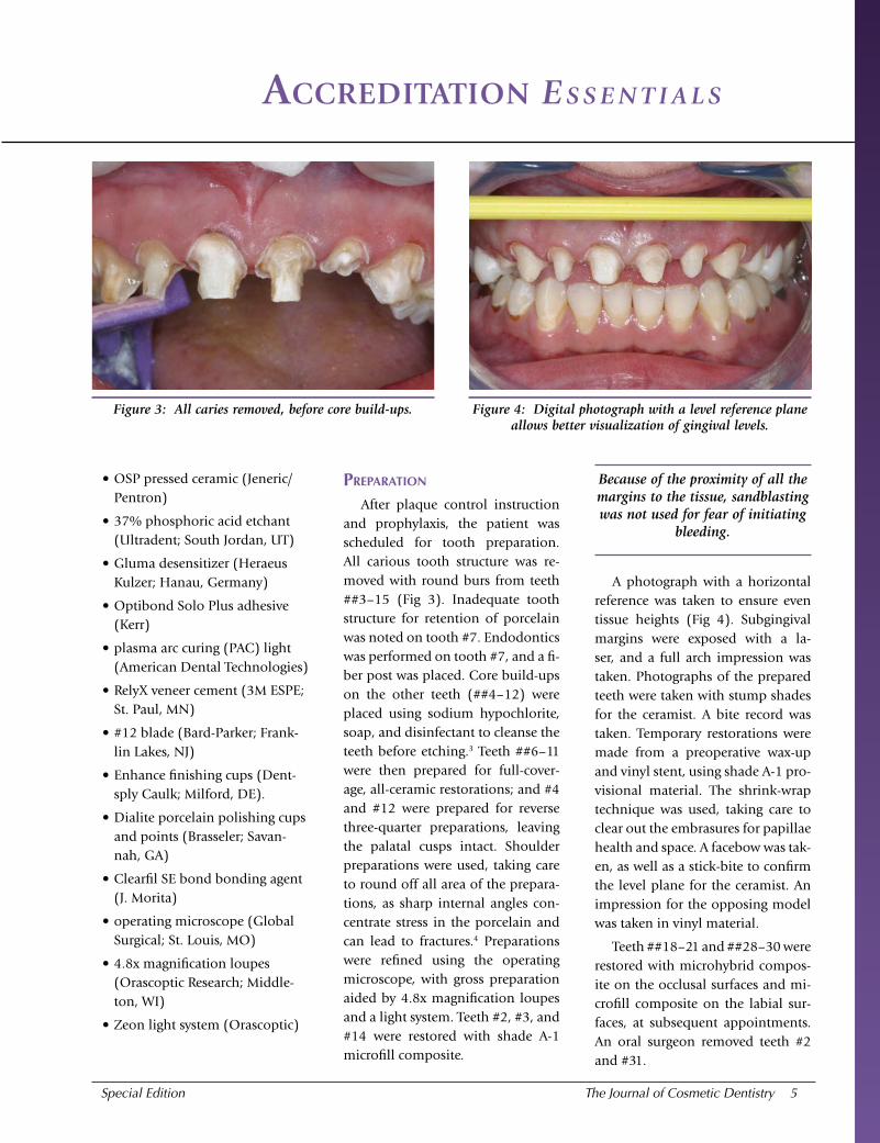
Figure 2: Unretracted smile, before and after. The shape and shade of the final restorations harmonize and blend within the framework of his smile.
SpecialEdition TheJournalofCosmeticDentistry �
aCCreDitation E s s E n t i a l s
• OSP pressed ceramic (Jeneric/Pentron)
• 37% phosphoric acid etchant (Ultradent; South Jordan, UT)
• Gluma desensitizer (Heraeus Kulzer; Hanau, Germany)
• Optibond Solo Plus adhesive (Kerr)
• plasma arc curing (PAC) light (American Dental Technologies)
• RelyX veneer cement (3M ESPE; St. Paul, MN)
• #12 blade (Bard-Parker; Frank-lin Lakes, NJ)
• Enhance finishing cups (Dent-sply Caulk; Milford, DE).
• Dialite porcelain polishing cups and points (Brasseler; Savan-nah, GA)
• Clearfil SE bond bonding agent (J. Morita)
• operating microscope (Global Surgical; St. Louis, MO)
• 4.8x magnification loupes (Orascoptic Research; Middle-ton, WI)
• Zeon light system (Orascoptic)
PreParatIon
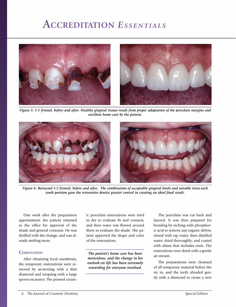
After plaque control instruction and prophylaxis, the patient was scheduled for tooth preparation. All carious tooth structure was re-moved with round burs from teeth ##3–15 (Fig 3). Inadequate tooth structure for retention of porcelain was noted on tooth #7. Endodontics was performed on tooth #7, and a fi-ber post was placed. Core build-ups on the other teeth (##4–12) were placed using sodium hypochlorite, soap, and disinfectant to cleanse the teeth before etching.3 Teeth ##6–11 were then prepared for full-cover-age, all-ceramic restorations; and #4 and #12 were prepared for reverse three-quarter preparations, leaving the palatal cusps intact. Shoulder preparations were used, taking care to round off all area of the prepara-tions, as sharp internal angles con-centrate stress in the porcelain and can lead to fractures.4 Preparations were refined using the operating microscope, with gross preparation aided by 4.8x magnification loupes and a light system. Teeth #2, #3, and #14 were restored with shade A-1 microfill composite.
Because of the proximity of all the margins to the tissue, sandblasting was not used for fear of initiating
bleeding.
A photograph with a horizontal reference was taken to ensure even tissue heights (Fig 4). Subgingival margins were exposed with a la-ser, and a full arch impression was taken. Photographs of the prepared teeth were taken with stump shades for the ceramist. A bite record was taken. Temporary restorations were made from a preoperative wax-up and vinyl stent, using shade A-1 pro-visional material. The shrink-wrap technique was used, taking care to clear out the embrasures for papillae health and space. A facebow was tak-en, as well as a stick-bite to confirm the level plane for the ceramist. An impression for the opposing model was taken in vinyl material.
Teeth ##18–21 and ##28–30 were restored with microhybrid compos-ite on the occlusal surfaces and mi-crofill composite on the labial sur-faces, at subsequent appointments. An oral surgeon removed teeth #2 and #31.
Figure 3: All caries removed, before core build-ups. Figure 4: Digital photograph with a level reference plane allows better visualization of gingival levels.
� TheJournalofCosmeticDentistry SpecialEdition
aCCreDitation E s s E n t i a l s
One week after the preparation appointment, the patient returned to the office for approval of the shade and general contours. He was thrilled with the change, and was al-ready smiling more.
cementatIon
After obtaining local anesthesia, the temporary restorations were re-moved by sectioning with a thin diamond and torquing with a large spoon excavator. The pressed ceram-
ic porcelain restorations were tried in dry to evaluate fit and contacts, and then water was flowed around them to evaluate the shade. The pa-tient approved the shape and color of the restorations.
The patient’s home care has been meticulous, and the change in his outlook on life has been extremely rewarding for everyone involved.
The porcelain was cut back and layered. It was then prepared for bonding by etching with phosphor-ic acid to remove any organic debris; rinsed with tap water, then distilled water; dried thoroughly; and coated with silane that includes resin. The restorations were dried with a gentle air stream.
The preparations were cleansed of all temporary material before the try in, and the teeth abraded gen-tly with a diamond to create a new
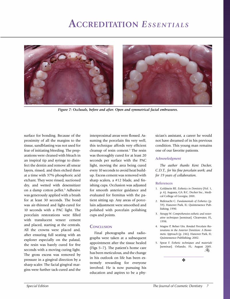
Figure 5: 1:1 frontal, before and after. Healthy gingival tissues result from proper adaptation of the porcelain margins and excellent home care by the patient.
Figure 6: Retracted 1:2 frontal, before and after. The combination of acceptable gingival levels and suitable intra-arch tooth position gave the restorative dentist greater control in creating an ideal final result.
SpecialEdition TheJournalofCosmeticDentistry �
aCCreDitation E s s E n t i a l s
surface for bonding. Because of the proximity of all the margins to the tissue, sandblasting was not used for fear of initiating bleeding. The prep-arations were cleaned with bleach in an inspiral tip and syringe to disin-fect the dentin and remove all smear layers, rinsed, and then etched three at a time with 37% phosphoric acid etchant. They were rinsed, suctioned dry, and wetted with desensitizer on a damp cotton pellet.5 Adhesive was generously applied with a brush for at least 30 seconds. The bond was air-thinned and light-cured for 10 seconds with a PAC light. The porcelain restorations were filled with translucent veneer cement and placed, starting at the centrals. All the crowns were placed and, after ensuring full seating with an explorer especially on the palatal, the resin was barely cured for five seconds with a moving curing light. The gross excess was removed by pressure in a gingival direction by a sharp scaler. The facial gingival mar-gins were further tack-cured and the
interproximal areas were flossed. As-suming the porcelain fits very well, this technique affords very efficient cleanup of resin cement.3 The resin was thoroughly cured for at least 20 seconds per surface with the PAC light, moving the area being cured every 10 seconds to avoid heat build-up. Excess cement was removed with sharp scalers, a #12 blade, and fin-ishing cups. Occlusion was adjusted for smooth anterior guidance and evaluated for fremitus with the pa-tient sitting up. Any areas of porce-lain adjustment were smoothed and polished with porcelain polishing cups and points.
conclusIon
Final photographs and radio-graphs were taken at a subsequent appointment after the tissue healed (Figs 5–7). The patient’s home care has been meticulous, and the change in his outlook on life has been ex-tremely rewarding for everyone involved. He is now pursuing his education and aspires to be a phy-
sician’s assistant, a career he would not have dreamed of in his previous condition. This young man remains one of our favorite patients.
Acknowledgment
The author thanks Kent Decker, C.D.T., for his fine porcelain work; and for 19 years of collaboration.
References1. Goldstein RE. Esthetics in Dentistry (Vol. 1,
p. 6). Augusta, GA: B.C. Decker Inc., Medi-cal College of Georgia; 2001.
2. Rufenacht C. Fundamentals of Esthetics (p. 59). Hanover Park, IL: Quintessence Pub-lishing; 1990.
3. Strupp W. Comprehensive esthetic and restor-ative techniques [seminar]. Clearwater, FL, 1998.
4. Magne P, Belser Urs. Bonded Porcelain Res-torations in the Anterior Dentition: A Biomi-metic Approach (p. 246). Hanover Park, IL: Quintessence Publishing; 2002.
5. Spear F. Esthetic techniques and materials [seminar]. Orlando, FL; August 2001.
______________________v
Figure 7: Occlusals, before and after. Open and symmetrical facial embrasures.
� TheJournalofCosmeticDentistry SpecialEdition
aCCreDitation E s s E n t i a l s
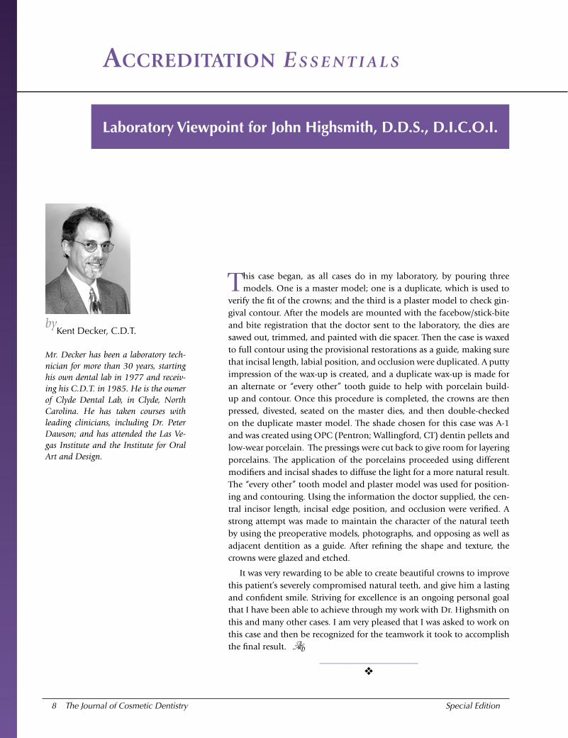
Laboratory Viewpoint for John Highsmith, D.D.S., D.I.C.O.I.
Mr. Decker has been a laboratory tech-nician for more than 30 years, starting his own dental lab in 1977 and receiv-ing his C.D.T. in 1985. He is the owner of Clyde Dental Lab, in Clyde, North Carolina. He has taken courses with leading clinicians, including Dr. Peter Dawson; and has attended the Las Ve-gas Institute and the Institute for Oral Art and Design.
byKent Decker, C.D.T.
This case began, as all cases do in my laboratory, by pouring three models. One is a master model; one is a duplicate, which is used to
verify the fit of the crowns; and the third is a plaster model to check gin-gival contour. After the models are mounted with the facebow/stick-bite and bite registration that the doctor sent to the laboratory, the dies are sawed out, trimmed, and painted with die spacer. Then the case is waxed to full contour using the provisional restorations as a guide, making sure that incisal length, labial position, and occlusion were duplicated. A putty impression of the wax-up is created, and a duplicate wax-up is made for an alternate or “every other” tooth guide to help with porcelain build-up and contour. Once this procedure is completed, the crowns are then pressed, divested, seated on the master dies, and then double-checked on the duplicate master model. The shade chosen for this case was A-1 and was created using OPC (Pentron; Wallingford, CT) dentin pellets and low-wear porcelain. The pressings were cut back to give room for layering porcelains. The application of the porcelains proceeded using different modifiers and incisal shades to diffuse the light for a more natural result. The “every other” tooth model and plaster model was used for position-ing and contouring. Using the information the doctor supplied, the cen-tral incisor length, incisal edge position, and occlusion were verified. A strong attempt was made to maintain the character of the natural teeth by using the preoperative models, photographs, and opposing as well as adjacent dentition as a guide. After refining the shape and texture, the crowns were glazed and etched.
It was very rewarding to be able to create beautiful crowns to improve this patient’s severely compromised natural teeth, and give him a lasting and confident smile. Striving for excellence is an ongoing personal goal that I have been able to achieve through my work with Dr. Highsmith on this and many other cases. I am very pleased that I was asked to work on this case and then be recognized for the teamwork it took to accomplish the final result.
______________________v
Accreditationin the
AmericAn AcAdemy of cosmetic dentistry®
Confidence, Credibility, Excellence
Ex
cE
ll
En
cE
inc
os
mE
tic
DE
nt
ist
ry
“I wanted to be the best I could possibly be in cosmetic dentistry. Accreditation gave me the motivation and direction to get there.”
~ AACD Accredited Fellow Dr. Michael Sesemann, Omaha, Nebraska
now it’s your turnvisit www.AAcd.com or cAll 800.543.9220
ignite your cAreer by eArning
the highest designAtion in cosmetic dentistry-
10 TheJournalofCosmeticDentistry SpecialEdition
aCCreDitation E s s E n t i a l s
Dr. John Highsmith submitted a Case Type I, Six or More In-direct Restorations, with a very dramatic end result. Despite
the fact that the pretreatment images revealed a patient in con-siderable esthetic distress, there were basic factors that inherently favored this case. Dentofacially the case was a good candidate for Accreditation consideration. The soft tissue health and architecture presented few challenges, and there were no problematic function-al issues. Intra- as well as inter-arch tooth positions were close to ideal. Lastly, the lower arch displayed few challenges that were out-side of the restorative dentist’s control.
The postoperative images exhibited a beautiful result. There were, however, relatively minor issues that nevertheless compro-mised the end result, and should be mentioned. The dental midline appeared to be slightly canted, with a resultant lack of symmetry between the central incisors in width and outline form. Addition-ally, the axial inclinations of the lateral incisors were not mirror images of one another. These issues did not in any way jeopardize the case in terms of Accreditation.
In summary, this case was successful because the esthetic crite-ria vital to passing an Accreditation case were clearly adhered to. Proper case selection allowed for the final outcome to be solidly in the zone of excellence. The lessons learned from Dr. Highsmith’s prior unsuccessful submission were more than amply demonstrat-ed with this case.
______________________v
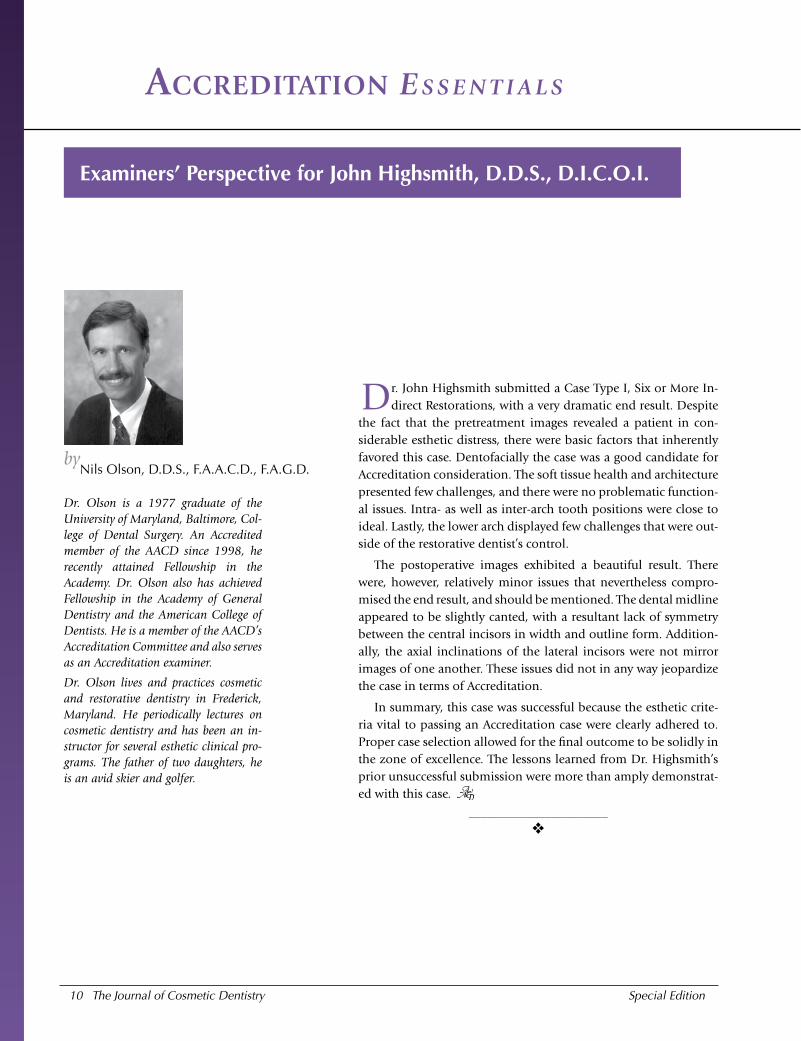
Dr. Olson is a 1977 graduate of the University of Maryland, Baltimore, Col-lege of Dental Surgery. An Accredited member of the AACD since 1998, he recently attained Fellowship in the Academy. Dr. Olson also has achieved Fellowship in the Academy of General Dentistry and the American College of Dentists. He is a member of the AACD’s Accreditation Committee and also serves as an Accreditation examiner.
Dr. Olson lives and practices cosmetic and restorative dentistry in Frederick, Maryland. He periodically lectures on cosmetic dentistry and has been an in-structor for several esthetic clinical pro-grams. The father of two daughters, he is an avid skier and golfer.
byNils Olson, D.D.S., F.A.A.C.D., F.A.G.D.
Examiners’ Perspective for John Highsmith, D.D.S., D.I.C.O.I.
ExcEllEncEin cosmEtic DEntistry
2007AmericAn AcAdemy of cosmetic dentistry
23rd AnnuAl scientific session • AtlAntA, GeorGiA • mAy 15 - 19, 2007
The World’s Premier Cosmetic Dental CE Event!
For more than 20 years, leading cosmetic dental professionals from around the world have gathered at the American Academy of Cosmetic Dentistry’s Annual Scientific Session to enhance their clinical skills, enrich their professional lives, and elevate their practices to new heights. 2007’s AACD conference in Atlanta promises to be the Academy’s biggest and best to date.
Highlights of ExcEllEncE in cosmEtic DEntistry 2007include:
• Leading Educators from Multiple Disciplines within Cosmetic Dentistry
• Integrated Learning Opportunities for the Entire Dental Team
• In-Depth Hands-on Training in Clinical Workshops
• Action-Packed, Hi-Tech Exhibit Hall
• AACD’s Charitable Foundation Celebration of Smiles Event starring Kenny Rogers!
View an up-to-date list of educators and register online at www.aacd.com or call (800) 543-9220 to request a complete Pre-Conference Guide.
AACD’s advance registration discount expires on April 19, 2007.
Beat the rush and register today!
PRSRT STDU.S. PoSTage
Pa i dMaDiSon, WiPeRMiT no.
2583
american academy of Cosmetic dentistry®
5401 World dairy driveMadison, Wi 53718
Return Service Requested
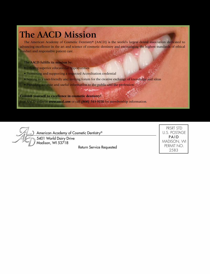
The AACD MissionThe American Academy of Cosmetic Dentistry® (AACD) is the world’s largest dental association dedicated to
advancing excellence in the art and science of cosmetic dentistry and encouraging the highest standards of ethical conduct and responsible patient care.
The AACD fulfills its mission by:
• Offering superior educational opportunities
• Promoting and supporting a respected Accreditation credential
• Serving as a user-friendly and inviting forum for the creative exchange of knowledge and ideas
• Providing accurate and useful information to the public and the profession
Commit yourself to excellence in cosmetic dentistry!
Join AACD today at www.aacd.com or call (800) 543-9220 for membership information.