Embed Size (px)
Citation preview

10.1110/ps.041148605Access the most recent version at doi: 2005 14: 257-269 Protein Sci.
Wolfgang Baumeister
A voyage to the inner space of cells
References
http://www.proteinscience.org/cgi/content/full/14/1/257#otherarticlesArticle cited in:
http://www.proteinscience.org/cgi/content/full/14/1/257#ReferencesThis article cites 102 articles, 23 of which can be accessed free at:
serviceEmail alerting
click heretop right corner of the article or Receive free email alerts when new articles cite this article - sign up in the box at the
Notes
http://www.proteinscience.org/subscriptions/ go to: Protein ScienceTo subscribe to
© 2005 Cold Spring Harbor Laboratory Press
Cold Spring Harbor Laboratory Press on April 14, 2008 - Published by www.proteinscience.orgDownloaded from

AWARD ADDRESS
A voyage to the inner space of cells
WOLFGANG BAUMEISTERDepartment of Structural Biology, Max-Planck-Institute of Biochemistry, 82152 Martinsried, Germany
Introduction
The title I have chosen for my personal recollections de-scribes, in a nutshell, the direction my scientific endeavorsfrom the time of my Ph.D. thesis, which I began in 1970, tothe present day. Over the years, I have changed fields anumber of times. There were periods when I was preoccu-pied with the development of methods; at other times, thefocus was on biological problems. Science can be advancedby new hypotheses about how things work, which can betested and proven right or wrong, and by new methodswhich enable us to tackle questions that we were unable toaddress with the existing methods. Or, as Richard Feynmanput it, “Science means, sometimes, a special method offinding things out. Sometimes it means the body of knowl-edge arising from the things found out. It may also mean thenew things you can do when you have found something out,or the actual doing of new things. This last field is usuallycalled technology. . . .” (R.P. Feynman in the John DanzLectures, 1963 [Feynman 1998]).
Apprenticeship with great freedom
After graduating from the University of Bonn in late 1969,I joined the Institute of Biophysics and Electron Microscopyat the University of Düsseldorf in January 1970. The direc-tor of the Institute at the time was Helmut Ruska, whobecame my Ph.D. supervisor. Helmut Ruska, a medical doc-tor, was the younger brother of Ernst Ruska, the electricalengineer who, in 1932, at the age of 26, had published hiscalculations on the theoretical resolving power of an elec-tron microscope and, in the face of strong skepticism, hadcompleted the development of a commercial instrument by1939 (Ruska 1979). Rarely has a scientific instrument hadsuch an impact on so many branches of science, and yet it
took more than 50 years before Ernst Ruska was rewardedwith the Nobel Prize in Physics in 1986 for his fundamen-tal work in electron optics and his design of the first electronmicroscope. Helmut Ruska, who was very close to hisbrother, realized immediately the potential of such an in-strument for the biomedical sciences, in particular the visu-alization of hitherto invisible infectious agents and for ul-trastructural studies of cells (Fig. 1). Helmut Ruska playeda very important role in the early days of electron micros-copy, not only by raising awareness and support—his clini-cal mentor at the Charité in Berlin, Richard Siebeck, be-came a decisive advocate at a critical time—but also by hisachievements in the visualization of viruses, bacteria, andblood cells (for review, see Kruger et al. 2000; for relevantreferences, see also Ruska 1979).
At the time I joined Helmut Ruska’s laboratory, the per-formance of transmission electron microscopes had reacheda level that allowed the imaging of single heavy atoms.Several groups in Europe, the United States, and Japan triedto take advantage of this capability and to use heavy atomsas site-specific labels, e.g., for mapping the bases in strandsof DNA. In the same vein, Helmut Ruska gave me the taskof exploring the use of heavy atom labels to study mem-brane topology. I decided to begin with well-defined modelmembranes before tackling membranes of biological rel-evance. I never got that far! Using Langmuir–Blodgett tech-niques, I prepared monomolecular layers at the water–airinterface and transferred them under precisely controlledconditions to specimen supports, but when I exposed mycarefully designed lipid layers to the electron beam, theyfaded away before I was able to take a picture. Occasion-ally, I obtained images of remnants of them with the heavyatoms coalesced into clusters. Eventually, with an unusuallyradiation-resistant organometallic compound of no rel-evance to biology, thorium-hexafluoracetylacetonate, I suc-ceeded in obtaining images showing a heavy atom patternthat was consistent with my design plan (Baumeister andHahn 1972).
Helmut Ruska was preoccupied with administrative du-ties during my time as a graduate student in his laboratoryand, as a consequence, his supervision of me was very ca-
Reprint requests to: Wolfgang Baumeister, Department of StructuralBiology, Max-Planck-Institute of Biochemistry, Am Klopferspitz 18,82152 Martinsried, Germany; e-mail: [email protected]; fax:+49-89-8578-2641.
Article and publication are at http://www.proteinscience.org/cgi/doi/10.1110/ps.041148605.
Protein Science (2005), 14:257–269. Published by Cold Spring Harbor Laboratory Press. Copyright © 2005 The Protein Society 257
Cold Spring Harbor Laboratory Press on April 14, 2008 - Published by www.proteinscience.orgDownloaded from
sual. Nevertheless, he was very supportive and he gave meall the resources I needed for my work. In late August 1973,only a few months after receiving my Ph.D., Helmut Ruskadied after a short illness.
I had offers from other places, but decided to stay inDüsseldorf, and since it took several years until a successorfor Helmut Ruska was found, I enjoyed complete freedomduring my postdoctoral years. Thanks to benevolent review-ers, I obtained my first grant in 1974 and I began to work onradiation damage—the electron microscopist’s greatest foe.I used a variety of methods for a quantitative assessment ofradiation damage in lipids and proteins under the conditionsencountered in electron microscopy (Baumeister et al. 1976;Hahn et al. 1976). My hope was that a better understandingof the underlying radiation chemistry might enable us tofind a remedy—a vain hope as it turned out (Baumeister1978).
Heading into new directions
Having realized that I was on an unproductive path, I had tochange direction. While the electron microscopy commu-nity in Germany with its strong tradition in electron opticswas preoccupied with “high resolution,” others, driven morestrongly by their desire to obtain insights into biomoleculararchitectures, took more pragmatic approaches. Already in1968, De Rosier and Klug had formulated the principles forthe three-dimensional reconstruction of objects from pro-jection images and applied them to the tail of bacteriophageT4, taking advantage of its helical symmetry (De Rosier andKlug 1968). When working with periodic or repetitivestructures, one can minimize radiation damage by underex-posing the samples; the information is retrieved from thestatistically noisy images by averaging over many identicalstructures. Averaging can be performed by direct superpo-sition (see, e.g., Markham et al. 1964) or by Fourier trans-form filtering (see, e.g., De Rosier and Klug 1972) using
both optical and digital methods (Aebi et al. 1973). Usingthe aforementioned stratagem and applying it to unstained,glucose-embedded purple membrane, Henderson and Un-win succeeded in 1975 in obtaining a 7 Å structure of bac-teriorhodopsin (Henderson and Unwin 1975), which be-came the paradigm of a membrane protein structure.
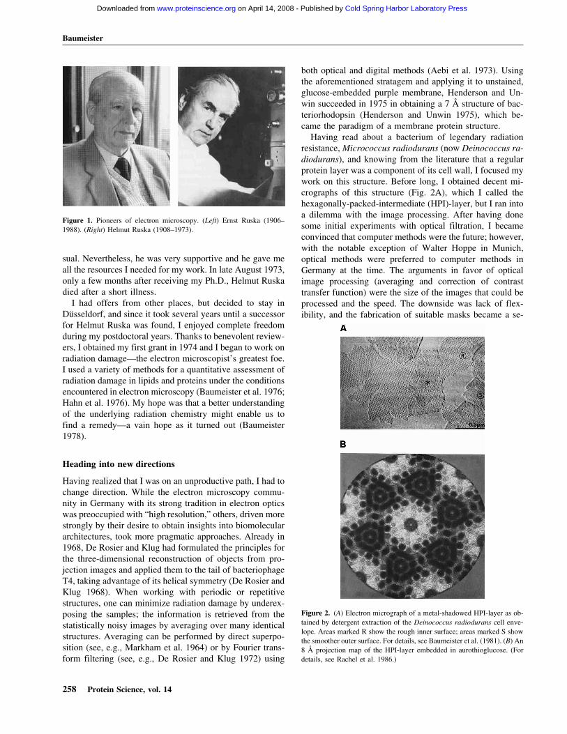
Having read about a bacterium of legendary radiationresistance, Micrococcus radiodurans (now Deinococcus ra-diodurans), and knowing from the literature that a regularprotein layer was a component of its cell wall, I focused mywork on this structure. Before long, I obtained decent mi-crographs of this structure (Fig. 2A), which I called thehexagonally-packed-intermediate (HPI)-layer, but I ran intoa dilemma with the image processing. After having donesome initial experiments with optical filtration, I becameconvinced that computer methods were the future; however,with the notable exception of Walter Hoppe in Munich,optical methods were preferred to computer methods inGermany at the time. The arguments in favor of opticalimage processing (averaging and correction of contrasttransfer function) were the size of the images that could beprocessed and the speed. The downside was lack of flex-ibility, and the fabrication of suitable masks became a se-
Figure 1. Pioneers of electron microscopy. (Left) Ernst Ruska (1906–1988). (Right) Helmut Ruska (1908–1973).
Figure 2. (A) Electron micrograph of a metal-shadowed HPI-layer as ob-tained by detergent extraction of the Deinococcus radiodurans cell enve-lope. Areas marked R show the rough inner surface; areas marked S showthe smoother outer surface. For details, see Baumeister et al. (1981). (B) An8 Å projection map of the HPI-layer embedded in aurothioglucose. (Fordetails, see Rachel et al. 1986.)
Baumeister
258 Protein Science, vol. 14
Cold Spring Harbor Laboratory Press on April 14, 2008 - Published by www.proteinscience.orgDownloaded from

rious bottleneck. Since I had neither access to the necessaryinfrastructure nor the know-how for computer-based pro-cessing, I started a collaboration with Olaf Kübler at theETH in Zürich who was in an inverse position: He had thesoftware and the hardware that was needed but no data. Inthe following years, we made substantial progress in eluci-dating the molecular architecture of the D. radiodurans cellenvelope (Baumeister and Kübler 1978; Kübler andBaumeister 1978; Baumeister et al. 1981, 1982).
In 1979, I organized a meeting entitled “Electron Micros-copy at Molecular Dimensions” held at Burg Gemen nearMünster, Germany, which in retrospect became quite influ-ential (see, e.g., Deisenhofer and Michel 1989). Besidesstate-of-the-art applications, it covered many developmentsin technology from image recording and processing to low-temperature electron microscopy (EM), or strategies formaking regular 2-D arrays (Baumeister and Vogell 1980).The intrinsic disorder in the 2-D protein arrays limits theresolution one can attain by Fourier filtering, and during theGemen meeting, I became convinced that there are betterways of dealing with imperfect 2-D crystals. The emergingmethods for averaging of single molecules offered an alter-native, and I decided to join forces with Joachim Frank. Afew months later when I visited him in Albany, we exploredthe application of correlation-based averaging to the micro-graphs of the HPI-layer, but to our disappointment we failedto obtain meaningful results during this short period of time.A year later, when I spent several months at the CavendishLaboratory in Cambridge, England, working with OwenSaxton, we were able to overcome the problems and ob-tained the first correlation-averaged images of the HPI-layerwith a significantly improved resolution. In trying to get thiswork published, we faced unusual problems; it took severalrounds of reviewing and several steps down the ladder of(journal) prestige, until our manuscript was finally pub-lished (Saxton and Baumeister 1982). In retrospect, how-ever, it is gratifying to see that more than 20 years later, thispaper is still cited frequently and, in the guise of “latticeunbending” (Baldwin et al. 1988) our strategem for over-coming the limitations due to lattice disorder became part ofthe standard repertoire used for processing images of 2-Dcrystals. Shortly thereafter, we applied correlation averag-ing to Scanning Transmission Electron Microscopy (STEM)images of unstained preparations of the HPI-layer and ob-tained the first quantitative mass maps (Engel et al. 1982),the beginning of a long-standing and successful collabora-tion with Andreas Engel at the Biocenter in Basel, Switzer-land.
The first decade in Martinsried: Studying proteinarchitecture on prokaryotic cell surfaces
At the beginning of 1982, I moved to the Max-Planck-Institute of Biochemistry where, after a short overlap pe-riod, I was appointed successor of Walter Hoppe. Hoppe
was a microscopist-turned-X-ray crystallographer of re-markable originality and theoretical ability but with limitedinterest in the practical aspects of structural biology. Nohumble man, he insisted that his “nonconventional” ap-proach to the structural analysis of individual macromol-ecules, as he liked to call it, was superior to other strategies(for review, see Hoppe 1983).
I remained unconvinced, and felt that the clever tactics of“single particle analysis” as pioneered by Joachim Frank, aformer student of Hoppe, and Marin van Heel at the Fritz-Haber-Institute in Berlin held greater promise. Not only didtheir approach greatly simplify data acquisition, the combi-nation of intelligent image classification procedures and ex-tensive averaging had the great advantage of yielding sig-nificant and interpretable structural data (for a recent re-view, see Frank 2002). In all fairness, I must add that inspite of a fierce public dispute I had with Walter Hoppe afew years earlier (Baumeister and Hahn 1975; Hoppe et al.1975) and divergent views on the course to take, he was,in general, supportive when I arrived in Martinsried andbegan to set up my laboratory. We continued our work withthe HPI-layer; a 3-D model was generated in due courseand, using cryomicroscopy, an 8 Å projection map was alsoobtained (Baumeister et al. 1986; Rachel et al. 1986;Fig. 2B).
With the plentiful resources now at our disposal, we notonly extended our structural studies to several other bacte-rial surface layers, we also widened our repertoire of meth-ods. Our comparative structural studies revealed some com-mon architectural principles (Baumeister et al. 1986, 1988)and sequence analyses led to the identification of new mo-tifs (Peters et al. 1987, 1989; Lupas et al. 1994) such as theS-layer homology domain (for a recent review, see Engel-hardt and Peters 1998) but the biological function of S-layers remained an enigma. Intuitively, I still feel that theremust be some function beyond mediating adhesion to ani-mate or inanimate surfaces or protecting underlying com-ponents of the cell envelope, but this remains pure specu-lation.
Colleagues in Martinsried (Wolfram Zillig) and in Re-gensburg (Karl-Otto Stetter) introduced me to the excitingworld of extremophiles. Most hyperthermophiles belong tothe archaeal domain of life where (glyco)protein surfacelayers are common. They represent the main macromolecu-lar component of the cell envelope and are intimately asso-ciated with the plasma membrane. Some show a high degreeof order and have a role in maintaining and possibly deter-mining cell shape (Wildhaber and Baumeister 1987; Phippset al. 1991b) while others form poorly ordered and flexiblesurface networks on pleomorphic cells (Wildhaber et al.1987; Peters et al. 1995). In spite of their apparent diversity,archaeal surface layers have some common structural prin-ciples: A stalk of variable length (10–70 nm) emanates froma membrane-anchoring domain and connects to a highly
Award Address
www.proteinscience.org 259
Cold Spring Harbor Laboratory Press on April 14, 2008 - Published by www.proteinscience.orgDownloaded from
variable (filiform or bulky) domain that forms a canopy-likelayer by means of end-to-end contacts enclosing a quasi-periplasmic space (Baumeister and Lembcke 1992). Aperiplasmic space of unusual width and maintained by arod-shaped spacer protein (Omp �) is also found in thehyperthermophilic ancestral bacterium Thermotoga mari-tima (Engel et al. 1992; Lupas et al. 1995).
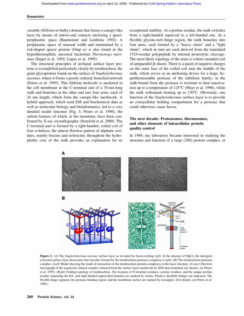
The structural principles of archaeal surface layer pro-teins is exemplified particularly clearly by tetrabrachion, thegiant glycoprotein found on the surface of Staphylothermusmarinus, where it forms a poorly ordered, branched network(Peters et al. 1995). This filiform molecule is anchored inthe cell membrane at the C-terminal end of a 70-nm-longstalk and branches at the other end into four arms, each of24 nm length, which form the canopy-like meshwork. Ahybrid approach, which used EM and biochemical data aswell as molecular biology and bioinformatics, led to a verydetailed model structure (Fig. 3; Peters et al. 1996), thesalient features of which, in the meantime, have been con-firmed by X-ray crystallography (Stetefeld et al. 2000). TheC-terminal part is formed by a right-handed, coiled coil offour �-helices; the almost flawless pattern of aliphatic resi-dues, mainly leucine and isoleucine, throughout the hydro-phobic core of the stalk provides an explanation for its
exceptional stability. At a proline residue, the stalk switchesfrom a right-handed supercoil to a left-handed one. At aflexible glycine-rich hinge region, the stalk branches intofour arms, each formed by a “heavy chain” and a “lightchain”, which in turn are each derived from the translated1524-residue polypeptide by internal proteolytic cleavage.The most likely topology of the arms is a three-stranded coilof antiparallel �-sheets. There is a patch of negative chargeson the outer face of the coiled coil near the middle of thestalk, which serves as an anchoring device for a large, hy-perthermostable protease of the subtilisin family; in thestalk-bound form the protease is resistant to heat inactiva-tion up to a temperature of 125°C (Mayr et al. 1996), whilethe stalk withstands heating up to 130°C. Obviously, onefunction of the Staphylothermus surface layer is to providean extracellular holding compartment for a protease thatcould otherwise cause havoc.
The next decade: Proteasomes, thermosomes,and other elements of intracellular proteinquality control
In 1989, my laboratory became interested in studying thestructure and function of a large (20S) protein complex, at
Figure 3. (A) The Staphylothermus marinus surface layer as revealed by freeze-etching (left). In the absence of MgCl2 the detergentextracted surface layer dissociates into micelles formed by the tetrabrachion-protease complexes (right). (B) The tetrabrachion-proteasecomplex. (Left) Model showing the mode of interaction of the tetrabrachion-protein complexes in the layer structure. (Center) Electronmicrograph of the negatively stained complex released from the surface layer meshwork by SDS-heat treatment (for details, see Peterset al. 1995). (Right) Folding topology of tetrabrachion. The location of N-terminal residues, cysteine residues, and the unique prolineresidue separating the left- and right-handed supercoiled domains are marked by circles. Putative disulfide bridges are indicated. Theflexible hinge segment, the protease-binding region, and the membrane anchor are marked by rectangles. (For details, see Peters et al.1996.)
Baumeister
260 Protein Science, vol. 14
Cold Spring Harbor Laboratory Press on April 14, 2008 - Published by www.proteinscience.orgDownloaded from

the time known as the multicatalytic proteinase (Dahlmannet al. 1989). Already in 1980, a large, multisubunit proteasehad been isolated and characterized (Hase et al. 1980; Wilkand Orlowski 1980; Orlowski and Wilk 1981). Initially, themulticatalytic proteinase was believed to be composed of3–5 subunits, ranging from 24 kDa to 28 kDa in size; itdisplayed three distinct proteolytic activities (trypsin-like,chymotrypsin-like, and peptidylglutamylpeptide-hydrolyz-ing) when assays were performed with small synthetic pep-tides, and it was noted that the integrity of the 20S complexwas essential for all proteolytic activities. Attempts weremade to assign specific activities to distinct subunits, but inspite of the efforts of many groups, the nature of the activesites remained enigmatic. Along a different line, a particlenamed “prosome” was under intensive investigation in themid-1980’s (for review, see Scherrer et al. 1990). Reminis-cent in size and subunit composition of the multicatalyticprotease complex, it appeared to be associated with RNAand it was suggested to have a role in the regulation of geneexpression. In 1988, it was established beyond doubt thatthe prosome and the multicatalytic proteinase complex wereone and the same particle (Arrigo et al. 1988; Falkenburg etal. 1988) and the name “proteasome” was coined by AlfredL. Goldberg (Harvard Medical School) to highlight its onlyestablished function, the proteolytic one, and its complexstructure. In the following years, evidence began to accu-mulate that the 20S proteasome was part of an even largercomplex, the 26S proteasome, which was implicated in theATP-dependent degradation of ubiquitin-conjugated pro-teins (Eytan et al. 1989; Driscoll and Goldberg 1990; Re-chsteiner et al. 1993).
By 1990, the 20S proteasome was structurally rather fea-tureless and its subunit composition and stoichiometry wereill-defined. Reports that proteasomes could undergochanges in subunit composition during development (Haassand Kloetzel 1989) made its structural analysis a dauntingchallenge, since structural methods rely, in one guise oranother, on averaging and, therefore, on homogeneouspreparations of molecules. This led us to search for pro-teasomes of hopefully simpler subunit composition in pro-karyotic cells. While our initial attempts to find protea-somes in bacteria were unsuccessful, we found them in thearchaeon Thermoplasma acidophilum (Dahlmann et al.1989). The Thermoplasma proteasome turned out to be verysimilar in size and shape to proteasomes from eukaryoticcells, but much simpler in subunit composition; it com-prises only two subunits, � (25.8 kDa) and � (22.3 kDa).The two subunits have significant sequence similarity, sug-gesting that they arose from a common ancestor via geneduplication (Zwickl et al. 1991, 1992a). Due to its relativesimplicity, the ensuing years saw the Thermoplasma pro-teasome play a pivotal role in elucidating the structure andenzymatic mechanism of this intriguing protein degradationmachine.
In 1991, a first, three-dimensional structure of the Ther-moplasma proteasome was obtained by EM single particleanalysis, showing with remarkable clarity the organizationof the barrel-shaped complex with its tripartite inner com-partment (Hegerl et al. 1991). Immunoelectron microscopystudies allowed us to assign the �-subunits to the two outerrings of the barrel, and the �-subunits to the inner rings(Grziwa et al. 1991). Mass measurements by STEM helpedus to establish the stoichiometry (�7�7�7�7), and metaldecoration studies of proteasome crystals (not yet goodenough for high resolution X-ray crystallography) clearlyrevealed the symmetry of the 20S complex. The structuralmodel we put forward on the basis of these data stood thetest of time and it recurred in all proteasomes, eukaryoticand prokaryotic (Pühler et al. 1992).
Another important advance was the expression of fullyassembled and functional 20S proteasomes in Escherichiacoli (Zwickl et al. 1992b; Fig. 4A). It not only allowed us toperform systematic mutagenesis studies aimed at identify-ing the active site, it also greatly facilitated the growth ofcrystals diffracting to high resolution (Jap et al. 1993). In1995, the crystal structure analysis was completed in a col-laboration with the group of Robert Huber (Löwe et al.1995; Fig. 4B). The long-sought catalytic nucleophile of the20S proteasome, the N-terminal threonine of the mature�-subunit was identified independently and almost simulta-neously by site-directed mutagenesis and crystal structureanalysis (Löwe et al. 1995; Seemüller et al. 1995). As an-ticipated from their sequence similarity the (noncatalytic) �-and the (catalytic) �-type subunits showed the same fold: afour-layer � + � structure with two antiparallel five-stranded �-sheets, flanked on one side by two, and on theother side by three �-helices. In the �-type subunits, the�-sheet sandwich is closed at one end by four hairpin loopsand opens at the opposite end to form the active-site cleft;the cleft is oriented toward the inner surface of the centralcavity. In the �-type subunits, an additional helix formed byan N-terminal extension crosses the top of the �-sheet sand-wich and fills this cleft. Initially, the proteasome fold wasbelieved to be unique; however, it turned out to be commonto a new superfamily of proteins referred to as Ntn (N-terminal nucleophile) hydrolases (Brannigan et al. 1995).Beyond the common fold, members of this family share themechanisms of the nucleophilic attack and self-processing(for reviews, see Baumeister et al. 1998; Dodson and Wlo-dawer 1998; Seemüller et al. 2001; Zwickl et al. 2002).
The crystal structure revealed that access to the innercavity that harbors the active sites is controlled by fourconstrictions. The constrictions in the �-rings which giveaccess to the two “antechambers” are narrow and partiallyobstructed, while the constrictions which regulate access tothe central cavity are wider. We were able to show withNanogold-labeled substrates, visible in electron micro-graphs, that polypeptides indeed enter the proteasome via
Award Address
www.proteinscience.org 261
Cold Spring Harbor Laboratory Press on April 14, 2008 - Published by www.proteinscience.orgDownloaded from

the orifice at the center of the �-rings. Bulky additions to thepolypeptide chain, such as a gold cluster, prevent passageinto the interior, suggesting that the discrimination betweenfolded and unfolded substrates is based on a size-exclusionmechanism (Wenzel and Baumeister 1995). Thus the 20Sproteasome is a molecular nano-compartment that confinesthe proteolytic reaction to its interior and sequesters it fromthe crowded environment of the cell. Interestingly, forma-tion of the active sites by the posttranslational removal of
the propeptides of the �-subunits (Seemüller et al. 1996) iscoupled to the assembly of the 20S proteasome in such amanner that activation is delayed until the assembly is com-plete (for review, see Seemüller et al. 2001). This led us topropose the concept of self-compartmentalization as a regu-latory principle (Lupas et al. 1997; Baumeister et al. 1998).
As mentioned earlier, it began to transpire in the early1990s that the 20S proteasome of eukaryotes associates withregulatory complexes, in an ATP-dependent manner, toform the 26S proteasome. Now it is firmly established thatthis 2.5 MDa complex altogether comprising more than 30different subunits acts downstream in the ubiquitin–protea-some pathway and is the central player in intracellular pro-teolysis. Proteins destined for degradation are marked bycovalent attachment of Ub chains, which mediate recogni-tion by the 26S proteasome (for recent reviews, see Hershkoand Ciechanover 1998; Voges et al. 1999). In 1993, we wereable to provide the first detailed description of the 26Scomplex, based on electron microscopy and image analysis(Peters et al. 1993). The averages showing the regulatory(19S) particles attached to one or both ends of the 20Sproteasome core particle (the “dragon-head” or “doubledragon-head” motif) became the classical textbook imagesof the 26S proteasome. Since then, however, progress hasbeen embarrassingly slow; the notorious instability of thecomplex and its dynamics have made it very difficult toachieve more than gradual improvements of the structuralmodel (Glickmann et al. 1998; Walz et al. 1998; Hölzl et al.2000). While it is clear that the role of the 19S regulatorycomplexes is the preparation of substrates for degradation inthe 20S core particle—involving the recognition of ubiqui-tinated substrates, the removal of the polyubiquitin chains,the unfolding of substrates, and assistance in translocationacross the gates of the 20S complex—the precise topologyand role of the 19S subunits is hitherto only dimly under-stood (Zwickl et al. 1999).
In 1991, we found, in a serendipitous manner, a novelATPase complex. During the lysis of accidentally heat-shocked Pyridictium cells on electron microscopy grids, amassive release of toroidal particles composed of thestacked octameric rings was observed (Phipps et al. 1991a).Not only the shape, but also the heat-shock induction of thiscomplex were reminiscent of the GroEL/Hsp60 family, andtherefore raised the possibility that it represented an ar-chaeal chaperonin. Subsequently, we named it “thermo-some” to highlight its heat induction and extreme thermo-stability (Phipps et al. 1993). Independently, a closely re-lated complex (TF55) was discovered in the laboratory ofArt Horwich in Yale (Trent et al. 1991). The thermosome orTF55 were the first representatives of the Group II chaper-onins found in archaea and in the eukaryotic cytosol. Themain structural feature distinguishing the Group II from theGroup I chaperonins is, in the absence of a co-chaperonin,a built-in lid provided by the protrusions of the apical do-
Figure 4. The 20S proteasome from Thermoplasma acidophilum. (A)Electron micrograph of recombinant 20S proteasomes in vitreous ice.(B,top left) Structure of the 20S proteasome in surface representation,low-pass filtered to 1 nm resolution. The �- and �-subunits are located inthe outer and the inner rings, respectively. (Top right) The same structurecut open along the sevenfold axis to display the inner compartments withthe active sites of the �-subunits in the central chamber marked in red.(Bottom left and right) Similar fold of the �- (left) and �- subunits (right).Both subunits contain a sandwich of two, five-stranded antiparallel�-sheets flanked by helices. (For details, see Löwe et al. 1995; Zwickl etal. 2002.)
Baumeister
262 Protein Science, vol. 14
Cold Spring Harbor Laboratory Press on April 14, 2008 - Published by www.proteinscience.orgDownloaded from
mains which can seal the folding chamber by an iris-typeclosure mechanism (Klumpp et al. 1997; Gutsche et al.1999).
In 1996, in our quest for a more comprehensive under-standing of the protein quality control machinery in Ther-moplasma we found a fascinating, large proteolytic com-plex that works in conjunction with an array of aminopep-tidases (Tamura et al. 1996). In view of the shape of thehexamer, we named it “tricorn protease”; soon thereafter wewere able to show that tricorn protease exists in the cell asa giant icosahedral complex of approximately 15 MDa,which in addition to its peptide-cleaving activity, appears toserve as an organizing center for the more downstream el-ements of the protein degradation pathway (Walz et al.1997). Tricorn protease converts the oligo-peptides (typi-cally about 8 amino acid residues) released by the protea-some into smaller (2–4 residue) peptides which are de-graded further by aminopeptidases (Tamura et al. 1998).These findings stimulated the search for “functional ho-mologs” of tricorn protease in eukaryotic cells; one of thecandidates is tripeptidylpeptidase (TPP) II, another giantprotein complex with an intriguing structure (Geier et al.1999; Rockel et al. 2002).
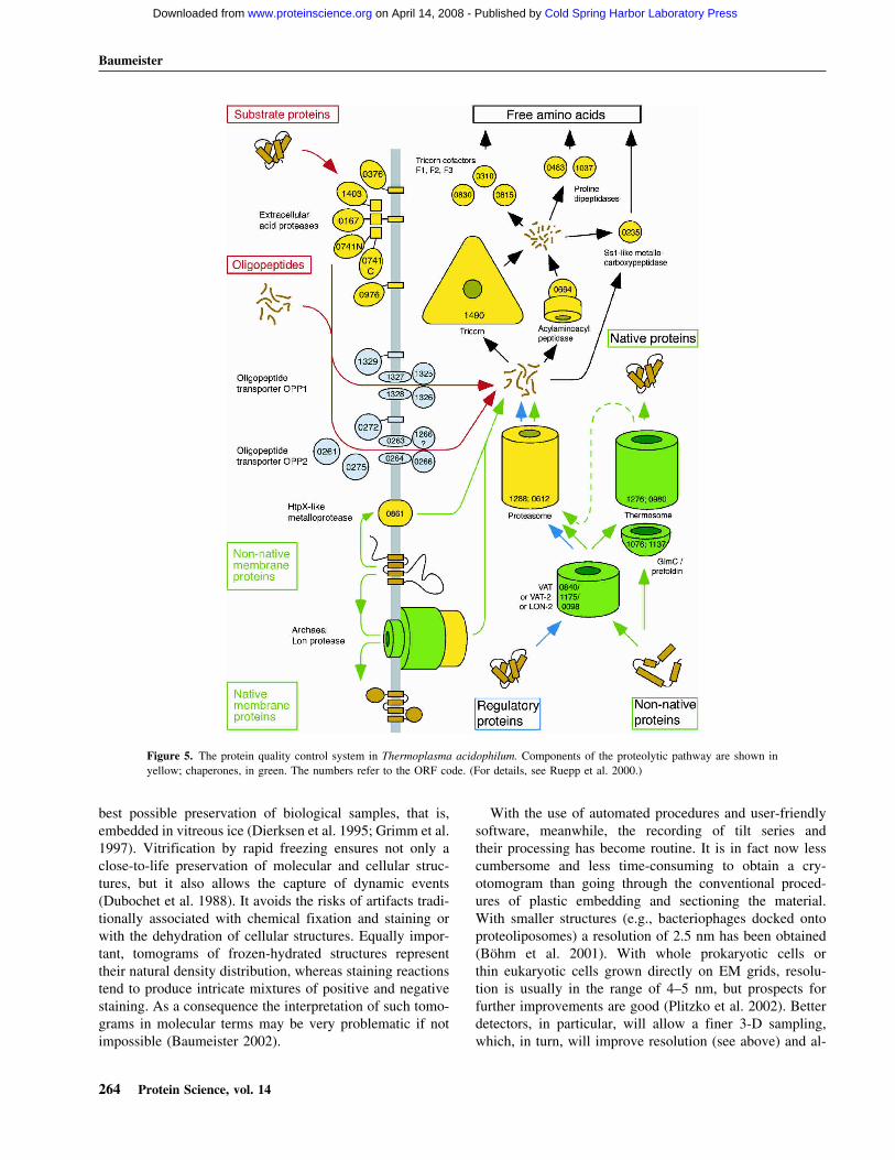
In 2000, we completed the sequencing of the genome ofThermoplasma acidophilum, an endeavor we had under-taken with modest resources (Ruepp et al. 2000). It not onlyserved to further establish Thermoplasma as a model systemfor studying cellular protein quality control, it also providedthe platform for a very ambitious project, namely the map-ping of its cellular proteome by cryoelectron tomography;this, in turn, can be expected to shed new light on thepathways of intracellular protein quality control (Fig. 5).
The latest frontier: Charting molecular landscapesinside cells by cryoelectron tomography
The foundations of electron tomography were laid alreadyin the late 1960s. In their landmark paper, De Rosier andKlug outlined very clearly and in general terms the prin-ciples of 3D reconstruction from electron micrographs (DeRosier and Klug 1968). Being aware of the practical prob-lems in recording 3-D data sets, they took advantage of thehelical symmetry of the bacteriophage T4 tail in a verypragmatic manner. Walter Hoppe, guided by his back-ground in X-ray crystallography, also realized the potentialof 3-D electron microscopy. Diverging from the approachestaken by most others, he focused on the development ofmethods suitable for studying individual structures (“Crys-tallography of crystals consisting of a single unit cell,”Hoppe 1978). In fact, his group presented as early as 1974a 3-D reconstruction of single fatty acid synthetase mol-ecules obtained by tomography (Hoppe et al. 1974). Asmentioned earlier, the “brute force” approach they used pro-voked some criticism. Besides doubts that negative staining
can portray details of the underlying structure to the reso-lution they claimed, the main concern was the enormouselectron dose to which the specimen was exposed duringrecording of the data. There was much discussion in thefollowing years as to whether it might ultimately be possibleto do electron tomography with acceptable electron doses.Also in 1968, R.G. Hart at the Lawrence Livermore Labo-ratory published a paper entitled “Electron microscopy ofunstained biological material: The polytropic montage” (Hart1968). Despite its vision, the Hart paper had negligible im-pact. For a vision to materialize, timing is a crucial element; ifit is too early, the necessary technologies might not yet exist.
The key problem in electron tomography, which formany years was a formidable obstacle and a deterrent, is toreconcile two requirements that are in conflict with eachother: To obtain a reconstruction that is detailed and largelyundistorted, one has to collect data over as wide a tilt rangeas possible with increments as small as possible (for review,see Baumeister et al. 1999). At the same time, the electrondose must be minimized. Above a critical dose, the speci-men undergoes structural degradation that, in the worstcase, can render a reconstruction meaningless. In principle,one could fractionate the dose over as many projections asan optimized tilt geometry might require. However, there isa practical limitation; the signal-to-noise ratio of the 2-Dimages has to be sufficient to permit their accurate align-ment by cross-correlation. This problem is further aggra-vated by the far-from-perfect mechanical accuracy of thetilting devices that causes image shifts and changes of focus.Therefore, following each change of tilt angle, the specimen(or its image) has to be realigned and refocused. Doing thismanually and with minimal exposure to the electron beam isutterly impossible.
In the late 1980s when computer-controlled electron mi-croscopes and large-area charge-coupled device (CCD)cameras became available, we saw an opportunity to auto-mate tomographic data acquisition (Typke 1991; Dierksenet al. 1992, 1995; Koster et al. 1992). This made the re-cording of data sets not only less cumbersome, but first andforemost it allowed the cumulative electron dose to be keptwithin tolerable limits. The fraction of the dose that is spenton overhead (search, recentering, [auto-]focusing) can bekept as low as 3% of the total dose; in other words, almostall electrons are used for gaining information (Koster et al.1997). As is evident from the recent resurgence, this haschanged the perspectives of electron tomography in a mostprofound manner; electron tomography had been used fromtime to time for ultrastructural studies, mostly of plasticembedded biological material, but it has gathered momen-tum only recently.
As demonstrated originally with “phantom cells,” that is,lipid vesicles encapsulating specific sets of macromol-ecules, automated tomography in a “low-dose mode” hasenabled us to combine the potential of 3-D imaging with the
Award Address
www.proteinscience.org 263
Cold Spring Harbor Laboratory Press on April 14, 2008 - Published by www.proteinscience.orgDownloaded from
best possible preservation of biological samples, that is,embedded in vitreous ice (Dierksen et al. 1995; Grimm et al.1997). Vitrification by rapid freezing ensures not only aclose-to-life preservation of molecular and cellular struc-tures, but it also allows the capture of dynamic events(Dubochet et al. 1988). It avoids the risks of artifacts tradi-tionally associated with chemical fixation and staining orwith the dehydration of cellular structures. Equally impor-tant, tomograms of frozen-hydrated structures representtheir natural density distribution, whereas staining reactionstend to produce intricate mixtures of positive and negativestaining. As a consequence the interpretation of such tomo-grams in molecular terms may be very problematic if notimpossible (Baumeister 2002).
With the use of automated procedures and user-friendlysoftware, meanwhile, the recording of tilt series andtheir processing has become routine. It is in fact now lesscumbersome and less time-consuming to obtain a cry-otomogram than going through the conventional proced-ures of plastic embedding and sectioning the material.With smaller structures (e.g., bacteriophages docked ontoproteoliposomes) a resolution of 2.5 nm has been obtained(Böhm et al. 2001). With whole prokaryotic cells orthin eukaryotic cells grown directly on EM grids, resolu-tion is usually in the range of 4–5 nm, but prospects forfurther improvements are good (Plitzko et al. 2002). Betterdetectors, in particular, will allow a finer 3-D sampling,which, in turn, will improve resolution (see above) and al-
Figure 5. The protein quality control system in Thermoplasma acidophilum. Components of the proteolytic pathway are shown inyellow; chaperones, in green. The numbers refer to the ORF code. (For details, see Ruepp et al. 2000.)
Baumeister
264 Protein Science, vol. 14
Cold Spring Harbor Laboratory Press on April 14, 2008 - Published by www.proteinscience.orgDownloaded from

low tomography to enter the realm of molecular resolution(2–3 nm).
Even at the present practical level of resolution, cryoto-mograms of organelles or cells contain an imposing amountof information. They are, essentially, 3-D images of entireproteomes, and they should ultimately enable us to map thespatial relationships of the full complement of macromol-ecules in an unperturbed cellular context; however, newstrategies and innovative image analysis techniques areneeded for “mining” this information. Retrieving it is con-fronted with two major problems: Cryotomograms are “con-taminated” by residual noise, and they are distorted by miss-ing data—in spite of optimized image acquisition schemes.Moreover, the cytoplasm of most cells is densely packed(“crowded”) with molecules literally touching each other. Itis therefore often impossible to perform a segmentation andto extract features, based on visual inspection of the tomo-grams. Denoising procedures (Frangakis and Hegerl 2001)can facilitate the visualization of features, but advancedpattern recognition techniques are needed for detecting andidentifying specific macromolecules by their respectivestructural signatures.
The most powerful method for improving the signal-to-noise ratio is averaging. Although averaging can obviouslynot be applied to tomograms of pleomorphic structures in afirst instance, such tomograms may nevertheless containrepetitive elements which can be extracted in silico, and thesubtomograms containing them can be subjected to classi-fication and averaging. These averages can be used subse-quently for replacing the original data in the tomograms,resulting in “synthetic” tomograms with a locally improvedsignal-to-noise ratio. This strategy was used, for example, toobtain a density map of whole Herpes simplex virions(Grünewald et al. 2003).
In spite of the low signal-to-noise ratio of tomograms,continuous structures such as membranes of cytoskeletalfilaments are easy to recognize. Cryotomograms of Dictyo-stelium discoideum cells grown directly on carbon supportfilms have provided unprecedented insights into the orga-nization of actin filaments in an unperturbed cellular envi-ronment (Medalia et al. 2002). The tomograms show, on thelevel of individual filaments, their modes of interaction (iso-tropic networks, bundles, etc.), they allow us to determinethe branching angles precisely (in 3-D), and they reveal thestructure of membrane attachment sites. For the quantitativeanalysis of large data sets, as is needed for extracting sta-tistically significant quantitative data, it will be necessary todevelop algorithms for automated segmentation, to establishconnectivity of filaments in noisy data sets—a notoriouslydifficult problem—and measure structural parameters offilaments (Fig. 6A).
Cryoelectron tomography enables us to obtain images ofsingle macromolecules inside intact cells as is exemplifiedby Figure 6B, which shows a single 26S proteasome within
the cytoplasm of a Dictyostelium cell. Although in this casethe detection and identification was facilitated by the largesize (∼2.5 MDa) and the peculiar shape of this complex, itindicates that a molecular signature-based approach to map-ping cellular proteomes should become feasible.
Alternatively, one could envisage strategies for introduc-ing electron-dense labels marking the spatial distribution ofthe molecules of interest. Such an approach, however,would no longer be noninvasive—unless it is based entirelyon genetic manipulations—and it would be difficult, if notimpossible, to achieve quantitative detection. Moreover, itis hard to imagine how this approach could be parallelizedsuch that it becomes a high-throughput technology capableof mapping entire proteomes. For every molecule of interestit would be necessary to repeat the whole procedure, thelabeling of cells, the recording of tilt series, and the tomo-graphic reconstruction. Even if this could be accomplished,it would be a daunting challenge to interrelate the individualmaps and to reveal the structure of molecular networks,owing to the stochastic nature of cellular supramolecular
Figure 6. Cryoelectron tomography of Dictyostelium discoideum cell. (A)Visualization of the actin network and cytoplasmic complexes in a Dic-tyostelium cell grown directly on an EM grid and embedded in vitreous ice(for details, see Medalia et al. 2002). (B) Visualization of a 26S proteasomewithin an intact Dictyostelium cell. (Left) Slice from a tomogram. Domi-nant features are ribosomes, some of them attached to the endoplasmicreticulum (lower left corner), and actin filaments. The encircled particle isa 26S proteasome. (Right) enlarged contour plot of the single (unaveraged)26S proteasome (projection of a stack of slices from tomogram).
Award Address
www.proteinscience.org 265
Cold Spring Harbor Laboratory Press on April 14, 2008 - Published by www.proteinscience.orgDownloaded from

architecture. Therefore, there is a strong incentive to exploitthe information content of cryotomograms by means of in-telligent pattern recognition algorithms. With this approach,a tomogram needs to be produced only once, and it is theninterpreted in a sequential manner in terms of its moleculararchitecture. The strategy we are pursuing is “templatematching” (Böhm et al. 2000; Frangakis et al. 2002). Pro-vided that high- or medium-resolution structures of the mac-romolecules of interest are available, they can be used for asystematic interrogation of the tomograms (Fig. 7). Imagesimulations have shown that template matching is indeed afeasible approach for identifying macromolecules in“noisy” tomograms. Experimental studies with “phantomcells”, i.e., lipid vesicles encapsulating known sets of pro-teins provide a means of validating the results of the tem-plate matching (Fig. 8). At the present resolution of 4–5 nm,only very large complexes (ribosomes, 26S proteasomes)can be mapped with high fidelity (>95%); an improvementin resolution to 2 nm will allow the mapping of medium-sized complexes (∼200–400 kDa, depending on shape).While tomograms with a resolution of 2 nm are a realisticprospect, major technical innovations will be required to gobeyond.
Once the challenges of obtaining a sufficiently good reso-lution are met, the next challenge will be to create compre-hensive libraries of templates. A whole array of methodscan be used to this end. Worldwide structural genomicsefforts will increase the pace with which high resolutionstructures of domains, subunits, and larger molecular enti-ties become available and eventually provide a comprehen-sive structural dictionary. In integrative hybrid approaches,
combining information gathered by a variety of techniques,computational methods will play a crucial role (for a recentreview, see Sali et al. 2003). EM-based “single” particleanalysis will undoubtedly become a major player in furnish-ing medium-resolution (∼1 nm) structures of complexes.Currently, this technique is slow and cumbersome, but greatstrides have been made in recent years toward improvingthroughput by automating data acquisition and analysis(Carragher et al. 2004).
With cryoelectron tomography providing 3-D images atmolecular resolution of cells in a close-to-life state, and with
Figure 7. Strategy for the detection and identification of macromolecules in cellular volumes. Because of the crowded nature of cellsand the high noise levels in tomograms (left), an interactive segmentation and feature extraction is, in most cases, not feasible. Itrequires automated pattern recognition techniques to exploit the rich information content of such tomograms. An approach that has beendemonstrated to work is based on the recognition of the structural signature (size, shape) of molecules by template matching. Templatesof the macromolecules under scrutiny are obtained by high- or medium-resolution techniques. Theses templates are then used to searchthe volume of the tomograms (Vin) systematically for matching structures by cross-correlation. The tomogram has to be scanned forall possible Eulerian angles around three different axes, with templates of all the different protein structures in which one is interested.The search is computationally demanding, but can be parallelized efficiently. The output information (Vout) is a set of coordinates thatdescribes the positions and orientation of all the molecules found in the tomogram. (For details, see Frangakis et al. 2002.)
Figure 8. Mapping molecular landscapes by pattern recognition. Volume-rendered representation of an ice-embedded “phantom cell” containingthermosomes (blue) and 20S proteasomes (yellow) with a 1:1 molar ratio.The two protein species were identified by template matching and arerepresented by averages derived from the tomogram. (For details, see Fran-gakis et al. 2002.)
Baumeister
266 Protein Science, vol. 14
Cold Spring Harbor Laboratory Press on April 14, 2008 - Published by www.proteinscience.orgDownloaded from
the availability of image analysis tools for interpreting thetomograms, we are poised now to integrate structural infor-mation gathered at multiple levels—from atoms to cells—into pseudoatomic maps of organelles or cells. The movefrom proteomics parts lists to precise maps of supramolecu-lar landscapes will provide unprecedented insights into thenetwork structures that underlie higher cellular functionsand the structural principles that orchestrate them.
Epilogue
An epilogue is the place for reflections and also for ac-knowledgments. I have deliberately changed fields a fewtimes. In doing so, the decision to leave a field was usuallymore difficult than the decision to embark on a new one.After working on a problem for a significant period of time,one becomes emotionally attached to it or even obsessed byit, but there is also an element of convenience: One knowsthe field with all of its ramifications, and one becomesestablished and is recognized by his/her peers. On the otherhand, a change of fields can be rejuvenating. One is lessinhibited by the knowledge of problems or obstacles, andmore willing to take new approaches. By looking at a prob-lem from a different angle, new opportunities arise and it isoften the interface between fields and disciplines where thesparks fly. I believe, for example, that our work in cyoelec-tron tomography will eventually enable us to address prob-lems in intracellular protein quality control in a new man-ner.
The work I have described in this essay would not havebeen accomplished without the support and the great effortsof many coworkers and colleagues. I have been fortunate towork with generations of talented and motivated studentsand postdoctoral fellows and I greatly enjoyed the collabo-ration with fine colleagues, with several of them over longperiods of time to this day. I mentioned a few of them in themain text, but for the sake of the space it was impossible toacknowledge them all; the names of most of them appear ascoauthors in the list of references. I wish to thank them all.Finally, I had the privilege to work in environments thatwere very supportive and allowed me to undertake the pro-jects I liked to do, irrespective of the chances of success.
References
Aebi, U., Smith, P.R., Dubochet, J., Henry, C., and Kellenberger, E. 1973. Astudy of the structure of the T-layer of Bacillus brevis. J. Supramol. Struct.1: 498–522.
Arrigo, A.P., Tanaka, K., Goldberg, A.L., and Welch, W.J. 1988. Identity of the19S “prosome” particle with the large multifunctional protease complex ofmammalian cells (the proteasome). Nature 331: 192–194.
Baldwin, J.M., Henderson, R., Beckman, E., and Zemlin, F. 1988. Images ofpurple membrane at 2.8 Å resolution obtained by cryo-electron microscopy.J. Mol. Biol. 202: 585–591.
Baumeister, W. 1978. Biological horizons in molecular microscopy. Eur. J. CellBiol. 17: 246–297.
———. 2002. Electron tomography: Towards visualizing the molecular orga-nization of the cytoplasm. Curr. Opin. Struct. Biol. 12: 679–684.
Baumeister, W. and Hahn, M. 1972. Electron microscopy of thorium atoms inmonomolecular layers. Nature 241: 445–447.
———. 1975. Relevance of three-dimensional reconstructions of stain distri-butions for structural analysis of biomolecules. Hoppe-Seyler’s Z. Physiol.Chem. 356: 1313–1316.
Baumeister, W. and Kübler, O. 1978. Topographic study of the cell surface ofMicrococcus radiodurans. Proc. Natl. Acad. Sci. 75: 5525–5528.
Baumeister, W. and Lembcke, G. 1992. Structural features of archaebacterialcell envelopes. J. Bioenerg. Biomembr. 24: 567–575.
Baumeister, W. and Vogell, W. 1980. Electron microscopy at molecular di-mensions. Springer Verlag, Berlin.
Baumeister, W., Fringeli, U.P., Hahn, M., Kopp, F., and Seredynski, J. 1976.Radiation-damage in tripalmitin layers studied by means of infrared spec-troscopy and electron-microscopy. Biophys. J. 16: 791–810.
Baumeister, W., Kübler, O., and Zingsheim, H.P. 1981. The structure of the cellenvelope of Micrococcus radiodurans as revealed by metal shadowing anddecoration. J. Ultrastruct. Res. 75: 60–71.
Baumeister, W., Karrenberg, F., Rachel, R., Engel, A., Ten Heggeler, B., andSaxton, W.O. 1982. The major cell envelope protein of Micrococcus ra-diodurans (R1): structural and chemical characterization. Eur. J. Biochem.725: 535–544.
Baumeister, W., Barth, M., Hegerl, R., Guckenberger, R., Hahn, M., and Sax-ton, W.O. 1986. Three-dimensional structure of the regular surface layer(HPI-layer) of Deinococcus radiodurans. Appendix: W.O. Saxton and W.Baumeister: Principles of organization in S layers. J. Mol. Biol. 187: 241–253.
Baumeister, W., Wildhaber, I., and Engelhardt, H. 1988. Bacterial surface pro-teins. Some structural, functional and evolutionary aspects. Biophys. Chem.29: 39–49.
Baumeister, W., Walz, J., Zühl, F., and Seemüller, E. 1998. The proteasome:Paradigm of a self-compartmentalizing protease. Cell 92: 367–380.
Baumeister, W., Grimm, R., and Walz, J. 1999. Electron tomography of mol-ecules and cells. Trends Cell Biol. 9: 81–85.
Böhm, J., Frangakis, A., Hegerl, R., Nickell, S., Typke, D., and Baumeister, W.2000. Toward detecting and identifying macromolecules in a cellular con-text: Template matching applied to electron tomograms. Proc. Natl. Acad.Sci. 97: 14245–14250.
Böhm, J., Lambert, O., Frangakis, A., Letellier, L., Baumeister, W., and Rigaud,J.L. 2001. FhuA-mediated phage genome transfer into liposomes: A cryo-electron tomography study. Curr. Biol. 11: 1168–1175.
Brannigan, J.A., Dodson, G., Duggleby, H.J., Moody, P.C.E., Smith, J.L., Tom-chick, D.R., and Murzin, A.G. 1995. A protein catalytic framework with anN-terminal nucleophile is capable of self-activation. Nature 378: 416–419.
Carragher, B., Fellmann, D., Guerra, F., Milligan, R.A., Mouche, F., Pulokas, J.,Sheehan, B., Quispe, J., Suloway, C., Zhu, Y., et al. 2004. Rapid routinestructure determination of macromolecular assemblies using electron mi-croscopy: Current progress and further challenges. J. Synchrotron Radiat.11: 83–85.
Dahlmann, B., Kopp, F., Kuehn, L., Niedel, B., Pfeifer, G., Hegerl, R., andBaumeister, W. 1989. The multicatalytic proteinase (prosome) is ubiquitousfrom eukaryotes to archaebacteria. FEBS Lett. 251: 125–131.
De Rosier, D.J. and Klug, A. 1968. Reconstruction of three dimensional struc-tures from electron micrographs. Nature 217: 130–134.
———. 1972. Structure of the tubular variants of the head of bacteriophage T4(polyheads). J. Mol. Biol. 65: 469–488.
Deisenhofer, J. and Michel, H. 1989. The photosynthetic reaction center fromthe purple bacterium rhodopseudomonas viridis (Nobel lecture). Angew.Chemie 28: 872–892.
Dierksen, K., Typke, D., Hegerl, R., Koster, A.J., and Baumeister, W. 1992.Towards automatic electron tomography. Ultramicroscopy 40: 71–87.
Dierksen, K., Typke, D., Hegerl, R., Walz, J., Sackmann, E., and Baumeister,W. 1995. Three-dimensional structure of lipid vesicles embedded in vitre-ous ice and investigated by automated electron tomography. Biophys. J. 68:1416–1422.
Dodson, G. and Wlodawer, A. 1998. Catalytic triads and their relatives. TrendsBiochem. Sci. 23: 347–352.
Driscoll, J. and Goldberg, A.L. 1990. The proteasome (multicatalytic protease)is a component of the 1500 kDa proteolytic complex which degrades ubiq-uitin-conjugated proteins. J. Biol. Chem. 265: 4789–4792.
Dubochet, J., Adrian, M., Chang, J.J., Homo, J.C., Lepault, J., McDowall, A.W.,and Schulz, P. 1988. Cryo-electron microscopy of vitrified specimens. Q.Rev. Biophys. 21: 129–228.
Engel, A., Baumeister, W., and Saxton, W.O. 1982. Mass mapping of a protein
Award Address
www.proteinscience.org 267
Cold Spring Harbor Laboratory Press on April 14, 2008 - Published by www.proteinscience.orgDownloaded from

complex with the scanning transmission electron microscope. Proc. Natl.Acad. Sci. 79: 4050–4054.
Engel, A.M., Cejka, Z., Lupas, A., Lottspeich, F., and Baumeister, W. 1992.Isolation and cloning of Omp �, a coiled-coil protein spanning the periplas-mic space of the ancestral eubacterium Thermotoga maritima. EMBO J. 11:4369–4378.
Engelhardt, H. and Peters, J. 1998. Structural research on surface layers: A focuson stability, surface layer homology domains, and surface layer-cell wallinteractions. J. Struct. Biol. 124: 276–302.
Eytan, E., Ganoth, D., Armon, T., and Hershko, A. 1989. ATP-dependent in-corporation of 20S protease into the 26S complex that degrades proteinsconjugated to ubiquitin. Proc. Natl. Acad. Sci. 86: 7751–7755.
Falkenburg, P.E., Haass, C., Kloetzel, P.M., Niedel, B., Kopp, F., Kuehn, L.,and Dahlmann, B. 1988. Drosophila small cytoplasmic 19S ribonucleopro-tein is homologous to the rat multicatalytic proteinase. Nature 331: 190–192.
Feynman, R.P. 1998. The meaning of it all. Addison-Wesley, Reading, MA.Frangakis, A. and Hegerl, R. 2001. Noise reduction in electron tomographic
reconstructions using nonlinear anisotropic diffusion. J. Struct. Biol. 135:239–250.
Frangakis, A., Böhm, J., Förster, F., Nickell, S., Nicastro, D., Typke, D., Hegerl,R., and Baumeister, W. 2002. Identification of macromolecular complexesin cryoelectron tomograms of phantom cells. Proc. Natl. Acad. Sci. 99:14153–14158.
Frank, J. 2002. Single-particle imaging of macromolecules by cryo-electronmicroscopy. Annu. Rev. Biomol. Struct. 31: 303–319.
Geier, E., Pfeifer, G., Wilm, M., Lucchiari-Hartz, M., Baumeister, W., Eich-mann, K., and Niedermann, G. 1999. A giant protease with potential tosubstitute for some functions of the proteasome. Science 283: 978–981.
Glickman, M.H., Rubin, D.M., Coux, O., Wefes, I., Pfeifer, G., Cejka, Z.,Baumeister, W., Fried, V.A., and Finley, D. 1998. A subcomplex of theproteasome regulatory particle required for ubiquitin-conjugate degradationand related to the COP9-signalosome and elF3. Cell 94: 615–623.
Grimm, R., Bärmann, M., Häckl, W., Typke, D., Sackmann, E., and Baumeister,W. 1997. Energy filtered electron tomography of ice-embedded actin andvesicles. Biophys. J. 72: 482–489.
Grünewald, K., Desai, P., Winkler, D.C., Heymann, J.B., Belnap, D.M.,Baumeister, W., and Steven, A.C. 2003. Three-dimensional structure ofHerpes simplex virus from cryo-electron tomography. Science 302: 1396–1398.
Grziwa, A., Baumeister, W., Dahlmann, B., and Kopp, F. 1991. Localization ofsubunits in proteasomes from Thermoplasma acidophilum by immunoelec-tron microscopy. FEBS Lett. 290: 186–190.
Gutsche, I., Essen, L.-O., and Baumeister. W. 1999. Group II chaperonins: NewTRiC(k)s and turns of a protein folding machine. J. Mol. Biol. 293: 295–312.
Haass, C. and Kloetzel, P.M. 1989. The Drosophila proteasome undergoeschanges in its subunit pattern during development. Exp. Cell Res. 180:243–252.
Hahn, M., Seredynski, J., and Baumeister, W. 1976. Inactivation of catalasemonolayers by irradiation with 100 keV electrons. Proc. Natl. Acad. Sci. 73:823–827.
Hart, R.G. 1968. Electron microscopy of unstained biological material: Thepolytropic montage. Science 159: 1464–1467.
Hase, J., Kobashi, K., Nakai, N., Mitsui, K., Iwata, K., and Takadera, T. 1980.The quaternary structure of carp muscle alkaline protease. Biochim. Bio-phys. Acta 611: 205–213.
Hegerl, R., Pfeifer, G., Pühler, G., Dahlmann, B., and Baumeister, W. 1991. Thethree-dimensional structure of proteasomes from Thermoplasma acidophi-lum as determined by electron microscopy using random conical tilting.FEBS Lett. 283: 117–121.
Henderson, R. and Unwin, P.N.T. 1975. Three-dimensional model of purplemembrane obtained by electron microscopy. Nature 257: 28–32.
Hershko, A. and Ciechanover, A. 1998. The ubiquitin system. Ann. Rev. Bio-chem. 67: 425–479.
Hölzl, H., Kapelari, B., Kellermann, J., Seemüller, E., Sümegi, M., Udvardy, A.,Medalia, O., Sperling, J., Müller, J.A., Engel, A., et al. 2000. The regulatorycomplex of Drosophila melangolaster 26S proteasomes: Subunit composi-tion and localization of a deubiquitylating enzyme. J. Cell Biol. 150: 119–129.
Hoppe, W. 1978. Three-dimensional electron microscopy of individual struc-tures: Crystallography of “crystals” consisting of a single unit cell. Chem.Scripta 79: 227–243.
———. 1983. Electron diffraction with the transmission electron microscope asa phase-determining diffractometer—From spatial frequency filtering to the
three-dimensional structure analysis of ribosomes. Angew. Chem. Int. Ed.Engl. 22: 456–485.
Hoppe, W., Gassmann, J., Hunsmann, N., Schramm, H.J., and Sturm, M. 1974.Three-dimensional reconstruction of individual negatively stained yeastfatty-acid synthetase molecules from tilt series in the electron microscope.H.-S. Z. Physiol. Chem. 355: 1483–1487.
———. 1975. Comments on the paper “Relevance of three-dimensional recon-structions of stain distributions for structural analysis of biomolecules.”Hoppe-Seyler’s Z. Physiol. Chem. 356: 1317–1320.
Jap, B., Pühler, G., Lücke, H., Typke, D., Löwe, J., Stock, D., Huber, R., andBaumeister, W. 1993. Preliminary x-ray crystallography study of the pro-teasome from Thermoplasma acidophilum. J. Mol. Biol. 234: 881–884.
Klumpp, M., Baumeister, W., and Essen, L.-O. 1997. Structure of the substratebinding domain of the thermosome, an archaeal group II chaperonin. Cell91: 263–270.
Koster, A.J., Chen, H., Sedat, J.W., and Agard, D.A. 1992. Automated micros-copy for electron tomography. Ultramicroscopy 46: 207–227.
Koster, A.J., Grimm, R., Typke, D., Hegerl, R., Stoschek, A., Walz, J., andBaumeister, W. 1997. Perspectives of molecular and cellular electron to-mography. J. Struct. Biol. 120: 276–308.
Kruger, D.H., Schneck, P., and Gelderblom, H.R. 2000. Helmut Ruska and thevisualization of viruses. Lancet 355: 1713–1717.
Kübler, O. and Baumeister, W. 1978. The structure of periodic cell wall com-ponent (HPI-layer) of Micrococcus radiodurans. Eur. J. Cell Biol. 17: 1–9.
Löwe, J., Stock, D., Jap, B., Zwickl, P., Baumeister, W., and Huber, R. 1995.Crystal structure of the 20S proteasome from the archaeon T. acidophilumat 3.4 Å resolution. Science 268: 533–539.
Lupas, A., Engelhardt, H., Peters, J., Santarius, U., Volker, S., and Baumeister,W. 1994. Domain structure of the Acetogenium kivui surface layer revealedby electron crystallography and sequence analysis. J. Bacteriol. 176: 1224–1233.
Lupas, A., Müller, S., Goldie, K., Engel, A.M., Engel, A., and Baumeister, W.1995. Model structure of the Omp � rod, a parallel four-stranded coiled coilfrom the hyperthermophilic eubacterium Thermotoga maritima. J. Mol.Biol. 248: 180–189.
Lupas, A., Flanagan, J.M., Tamura, T., and Baumeister, W. 1997. Self-com-partmentalizing proteases. Trends Biochem. Sci. 22: 399–404.
Markham, R., Hitchborn, J.H., Hills, G.J., and Frey, S. 1964. The anatomy ofthe tobacco mosaic virus. Virology 22: 342–359.
Mayr, J., Lupas, A., Kellermann, J., Eckerskorn, C., Baumeister, W., and Peters,J. 1996. A hyperthermostable protease of the subtilisin family bound to thesurface layer of the archaeon Staphylothermus marinus. Curr. Biol. 6: 739–749.
Medalia, O., Weber, I., Frangakis, A., Nicastro, D., Gerisch, G., and Baumeis-ter, W. 2002. Macromolecular architecture in eukaryotic cells visualized bycryoelectron tomography. Science 298: 1209–1213.
Orlowski, M. and Wilk, S. 1981. A multicatalytic protease complex from pitu-itary that forms enkephalin and enkephalin containing peptides. Biochim.Biophys. Res. Commun. 101: 814–822.
Peters, J., Peters, M., Lottspeich, F., Schäfer, W., and Baumeister, W. 1987.Nucleotide sequence analysis of the gene encoding the Deinococcus radio-durans surface protein, derived amino acid sequence and complementaryprotein chemical studies. J. Bacteriol. 169: 5216–5223.
Peters, J., Peters, M., Lottspeich, F., and Baumeister, W. 1989. S-layer proteingene of Acetogenium kivui: Cloning and expression in Escherichia coli anddetermination of the nucleotide sequence. J. Bacteriol. 171: 6307–6315.
Peters, J.-M., Cejka, Z., Harris, J.R., Kleinschmidt, J.A., and Baumeister, W.1993. Structural features of the 26S proteasome complex. J. Mol. Biol. 234:932–937.
Peters, J., Nitsch, M., Kühlmorgen, B., Golbik, R., Lupas, A., Kellermann, J.,Engelhardt, H., Pfander, J.-P., Müller, S., Goldie, K., et al. 1995. Tetrabra-chion: A filamentous archaebacterial surface protein assembly of unusualstructure and extreme stability. J. Mol. Biol. 245: 385–401.
Peters, J., Baumeister, W., and Lupas, A. 1996. Hyperthermostable surface layerprotein tetrabrachion from the archaebacterium Staphylothermus marinus:Evidence for the presence of a right-handed coiled coil derived from theprimary structure. J. Mol. Biol. 257: 1031–1041.
Phipps, B.M., Hoffmann, A., Stetter, K.O., and Baumeister, W. 1991a. A novelATPase complex selectively accumulated upon heat shock is a major cel-lular component of thermophilic archaebacteria. EMBO J. 10: 1711–1722.
Phipps, B.M., Huber, R., and Baumeister, W. 1991b. The cell envelope of thehyperthermophilic archaebacterium Pyrobaculum organotrophum consistsof two regularly arrayed protein layers: Three-dimensional structure of theouter layer. Mol. Microbiol. 5: 253–265.
Phipps, B.M., Typke, D., Hegerl, R., Volker, S., Hoffmann, A., Stetter, K.O.,
Baumeister
268 Protein Science, vol. 14
Cold Spring Harbor Laboratory Press on April 14, 2008 - Published by www.proteinscience.orgDownloaded from

and Baumeister, W. 1993. Structure of a molecular chaperone from a ther-mophilic archaebacterium. Nature 361: 475–477.
Plitzko, J., Frangakis, A.S., Nickell, S., Förster, F., Gross, A., and Baumeister,W. 2002. In vivo veritas: Electron cryotomography of cells. Trends Bio-technol. 20: S40–S44.
Pühler, G., Weinkauf, S., Bachmann, L., Müller, S., Engel, A., Hegerl, R., andBaumeister, W. 1992. Subunit stoichiometry and three-dimensional arrange-ment in proteasomes from Thermoplasma acidophilum. EMBO J. 11: 1607–1616.
Rachel, R., Jakubowski, U., Tietz, H., Hegerl, R., and Baumeister, W. 1986.Projected structure of the surface protein of Deinococcus radiodurans de-termined to 8 Å resolution by cryomicroscopy. Ultramicroscopy 20: 305–316.
Rechsteiner, M., Hoffman, L., and Dubiel, W. 1993. The multicatalytic and 26Sproteases. J. Biol. Chem. 268: 6065–6068.
Rockel, B., Peters, J., Kühlmorgen, B., Glaeser, R.M., and Baumeister, W.2002. A giant protease with a twist: The TPP II complex from Drosophilastudied by electron microscopy. EMBO J. 21: 5979–5984.
Ruepp, A., Graml, W., Santos-Martinez, M.-L., Koretke, K.K., Volker, C.,Mewes, H.W., Frishman, D., Stocker, S., Lupas, A.N., and Baumeister, W.2000. The genome sequence of the thermoacidophilic scavenger Thermo-plasma acidophilum. Nature 407: 508–513.
Ruska, E. 1979. Die frühe Entwicklung der Elektronenlinsen und der Elek-tronenmikroskopie. Acta Hist. Leopoldina 12: 7–136.
Sali, A., Glaeser, R., Earnest, T., and Baumeister, W. 2003. From words toliterature in structural proteomics. Nature 422: 216–225.
Saxton, W.O. and Baumeister, W. 1982. The correlation averaging of a regu-larly arranged bacterial cell envelope protein. J. Microsc. 127: 127–138.
Scherrer, K., Nothwang, H.G., Silva, P., Bey, F., Olin-Coux, M., Huesca, M.,Coux, O., Arcangeletti, C., Chezzi, C., Buri, J.F., et al. 1990. The prosomes:Molecular and cellular biology. Mol. Biol. Rep. 14: 75.
Seemüller, E., Lupas, A., Stock, D., Löwe, J., Huber, R., and Baumeister, W.1995. Proteasome from Thermoplasma acidophilum: A threonine protease.Science 268: 579–582.
Seemüller, E., Lupas, A., and Baumeister, W. 1996. Autocatalytic processing ofthe 20S proteasome. Nature 382: 486–470.
Seemüller, E., Zwickl, P., and Baumeister, W. 2001. Self-processing of subunitsof the proteasome. In Co- and posttranslational proteolysis of proteins, Theenzymes, Vol. XXII, 3rd ed. (eds. R.E. Dalbey and D.S. Sigman), pp. 335–371. Academic Press.
Stetefeld, J., Jenny, M., Schulthess, T., Landwehr, R., Engel, J., and Kammerer,R.A. 2000. Crystal structure of a naturally occurring parallel right-handedcoiled coil tetramer. Nat. Struct. Biol. 7: 772–775.
Tamura, T., Tamura, N., Cejka, Z., Hegerl, R., Lottspeich, F., and Baumeister,W. 1996. Tricorn protease—The core of a modular proteolytic system.Science 274: 1385–1389.
Tamura, N., Lottspeich, F., Baumeister, W., and Tamura, T. 1998. The role of
tricorn protease in its aminopeptidase-interacting factors in cellular proteindegradation. Cell 95: 637–648.
Trent, J.D., Nimmesgern, E., Wall, J.S., Hartl, F.U., and Horwich, A.L. 1991. Amolecular chaperone from a thermophilic archaebacterium is related to theeukaryotic protein t-complex polypeptide-1. Nature 354: 490–493.
Typke, D., Dierksen, K., and Baumeister, W. 1991. Automatic electron tomog-raphy. In Proceedings of the 49th Annual Meeting of the Electron Micros-copy Society of America (ed. W. Bailey), pp. 544–545. San Francisco Press,San Francisco, CA.
Voges, D., Zwickl, P., and Baumeister, W. 1999. The 26S proteasome: Amolecular machine designed for controlled proteolysis. Ann. Rev. Biochem.68: 1015–1068.
Walz, J., Tamura, T., Tamura, N., Grimm, R., Baumeister, W., and Koster, A.J.1997. Tricorn protease exists as an icosahedral supermolecule in vivo. Mol.Cell 1: 59–65.
Walz, J., Erdmann, A., Kania, M., Typke, D., Koster, A.J., and Baumeister, W.1998. 26S proteasome structure revealed by three-dimensional electron mi-croscopy. J. Struct. Biol. 121: 19–29.
Wenzel, T. and Baumeister, W. 1995. Conformational constraints in proteindegradation by the 20S proteasome. Nat. Struct. Biol. 2: 199–204.
Wildhaber, I. and Baumeister, W. 1987. The cell envelope of Thermoproteustenax: Three-dimensional structure of the surface layer and its role in shapemaintenance. EMBO J. 6: 1475–1480.
Wildhaber, I., Santarius, U., and Baumeister, W. 1987. The three-dimensionalstructure of the surface protein of Desulfurococcus mobilis. J. Bacteriol.169: 5563–5568.
Wilk, S. and Orlowski, M. 1980. Cation-sensitive neutral endopeptidase: Iso-lation and specificity of the bovine pituitary enzyme. J. Neurochem. 35:1172–1182.
Zwickl, P., Lottspeich, F., Dahlmann, B., and Baumeister, W. 1991. Cloningand sequencing of the gene encoding the large (�-) subunit of proteasomefrom Thermoplasma acidophilum. FEBS Lett. 278: 217–221.
Zwickl, P., Grziwa, A., Pühler, G., Dahlmann, B., Lottspeich, F., and Baumeis-ter, W. 1992a. Primary structure of the Thermoplasma proteasome and itsimplications for the structure, function and evolution of the multicatalyticproteinase. Biochemistry 31: 964–972.
Zwickl, P., Lottspeich, F., and Baumeister, W. 1992b. Expression of functionalThermoplasma acidophilum proteasomes in Escherichia coli. FEBS Lett.312: 157–160.
Zwickl, P., Voges, D., and Baumeister, W. 1999. The proteasome: A macro-molecular assembly designed for controlled proteolysis. Philos. T. Roy. Soc.B. 354: 1501–1511.
Zwickl, P., Seemüller, E., Kapelari, B., and Baumeister, W. 2002. The protea-some: A supramolecular assembly designed for controlled proteolysis. InProtein folding in the cell (eds. F.M. Richards et al.), pp. 187–222. Aca-demic Press, San Diego, CA.
Award Address
www.proteinscience.org 269
Cold Spring Harbor Laboratory Press on April 14, 2008 - Published by www.proteinscience.orgDownloaded from