Embed Size (px)
Citation preview
SC I ENCE ADVANCES | R E S EARCH ART I C L E
PALEONTOLOGY
1Bereich Paläontologie, Steinmann-Institut für Geologie, Mineralogie und Paläontologie,Universität Bonn, Nussallee 8, 53115 Bonn, Germany. 2OsakaMuseumof Natural History,Nagai Park 1-23, Higashi-Sumiyoshi-ku, Osaka 546-0034, Japan. 3Division ofMaterials andManufacturing Science, Graduate School of Engineering, Osaka University, 2-1 Yamada-Oka,Suita, Osaka 565-0871, Japan. 4UMR 7179 CNRS/Muséum National d’Histoire Naturelle,Départment Adaptations du Vivant, 57 rue Cuvier CP-55, 75005 Paris, France. 5Atmo-sphere andOcean Research Institute, University of Tokyo, 5-1-5 Kashiwanoha, Kashiwa-shi,Chiba 277-8564, Japan. 6Dinosaur Institute, Natural History Museum of Los AngelesCounty, 900 Exposition Boulevard, Los Angeles, CA 90007, USA.*Present address: Biosphere-Geosphere Science, Okayama University of Science,Ridai-cho 1-1, Kita-ku, Okayama 700-0005, Japan.†Corresponding author. Email: [email protected]
Wintrich et al., Sci. Adv. 2017;3 : e1701144 13 December 2017
Copyright © 2017
The Authors, some
rights reserved;
exclusive licensee
American Association
for the Advancement
of Science. No claim to
original U.S. Government
Works. Distributed
under a Creative
Commons Attribution
NonCommercial
License 4.0 (CC BY-NC).
Dow
nloade
A Triassic plesiosaurian skeleton and bone histologyinform on evolution of a unique body planTanja Wintrich,1 Shoji Hayashi,2,3* Alexandra Houssaye,4 Yasuhisa Nakajima,5 P. Martin Sander1,6†
Secondary marine adaptation is a major pattern in amniote evolution, accompanied by specific bone histologicaladaptations. In the aftermath of the end-Permian extinction, diverse marine reptiles evolved early in the Triassic.Plesiosauria is the most diverse and one of the longest-lived clades of marine reptiles, but its bone histology is leastknown among themajor marine amniote clades. Plesiosaurians had a unique and puzzling body plan, sporting fourevenly shaped pointed flippers and (inmost clades) a small head on a long, stiffened neck. The flippers were used ashydrofoils in underwater flight. A wide temporal, morphological, and morphometric gap separates plesiosauriansfrom their closest relatives (basal pistosaurs, Bobosaurus). For nearly two centuries, plesiosaurians were thought toappear suddenly in the earliest Jurassic after the end-Triassic extinctions.Wedescribe the first Triassic plesiosaurian,from the Rhaetian of Germany, and compare its long bone histology to that of later plesiosaurians sampled for thisstudy. The new taxon is recovered as a basal member of the Pliosauridae, revealing that diversification of plesiosaurianswas a Triassic event and that several lineages must have crossed into the Jurassic. Plesiosaurian histology is strikinglyuniform and different from stem sauropterygians. Histology suggests the concurrent evolution of fast growth and anelevatedmetabolic rate as an adaptation to cruising andefficient foraging in theopen sea. Thenew specimen corroboratesthe hypothesis that open ocean life of plesiosaurians facilitated their survival of the end-Triassic extinctions.
d fro
on June 10, 2018http://advances.sciencemag.org/
m
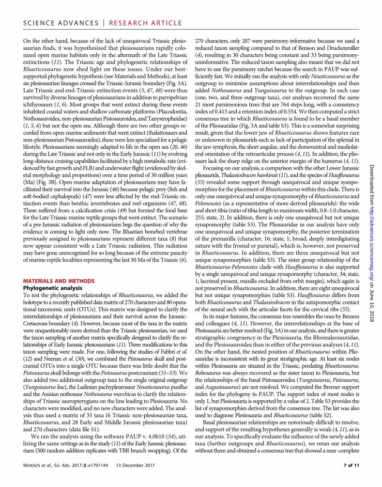
INTRODUCTIONTriassic sauropterygians exhibit great morphological and body size dis-parity andhighly varied feeding and locomotor adaptations (1) reflectedin their bone microstructure (2). Nearly all stem sauropterygians arefound in coastal or platform deposits of the Tethys Sea. Plesiosaurians,on the other hand, are recorded globally from open-water deposits andhave a strikingly uniform bauplan (variation primarily residing in skullsize and neck length) (3–6), with all four limbsmodified into hydrofoilsused in some kind of underwater flight (7). The great similarity betweenforelimbs andhindlimbs is unique among tetrapods. After nearly 300 yearsof frequent discoveries of plesiosaurian skeletons from the Early Jurassic tothe end of the Cretaceous, we here report the first plesiosaurian skeletonfrom the Triassic (Figs. 1 and 2 and figs. S1 to S5). The find pertains to anew, small-bodied taxon,Rhaeticosaurusmertensi gen. et sp. nov., from theRhaetian of Germany (Fig. 1). Previously, isolated vertebrae from theRhaetian bonebeds of England had been assigned to Plesiosauria (8),and a partial, poorly preserved, and undiagnostic sauropterygian skeletonfrom theRussianArctic (9) hadhinted at aTriassic origin of the clade, butthese finds remain inconclusive (8).
RESULTSSystematic paleontologyReptilia Linnaeus, 1758Diapsida Osborn, 1903Plesiosauria de Blainville, 1835
Phylogenetic definitionWe offer the following apomorphy-based definition of Plesiosauria:Sauropterygians with a short, wide trunk–bearing four flippers of evenstructure and subequal size, the flippers consisting of long, straight propo-dials combinedwith very short and dorsoventrally flattened zeugopodials.
DiagnosisPlesiosauria is diagnosed (seeMaterials andMethods) by two unique andunambiguous synapomorphies: tooth enamel surface, striations present(character, 106; state, 0; see comment in table S2); orientation of cervicalzygapophyses, dorsomedially facing (128, 1). An unambiguous but notunique synapomorphy is as follows: dorsal half of ilium, subequal anteriorand posterior expansion (174, 0).
Rhaeticosaurus mertensi gen. et sp. nov.
EtymologyThe genus name is based on rhaeticus, latinized adjectivemeaning “fromthe Rhaetian stage,” and sauros (Greek), meaning lizard or saurian. Thespecific epithet honors the discoverer of the holotype,MichaelMertens ofSchwaney, Westphalia, Germany.
Holotype specimenLWL-Museum für Naturkunde (Münster, Germany), LWL-MFN P 64047.
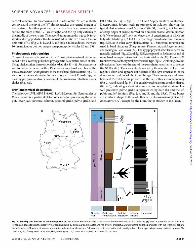
Locality and horizonClay pit #3 of Lücking brick company, 1 km north of the village ofBonenburg, city of Warburg, North Rhine-Westphalia, Germany(Fig. 1A). The specimen derives from Rhaetian dark marine mudstonesof the Exter Formation, 21 m in the section below the Triassic-Jurassicboundary and about 3.5mbelowabonebed containing a vertebrate faunaof Rhaetian age (Fig. 1B and table S1).
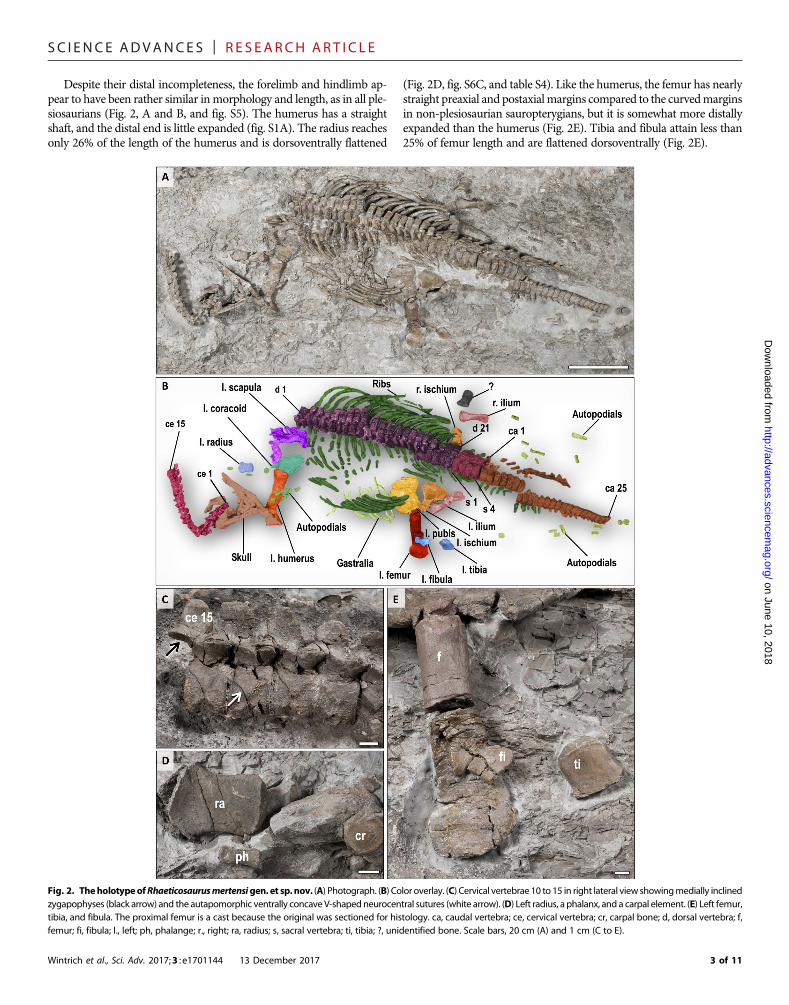
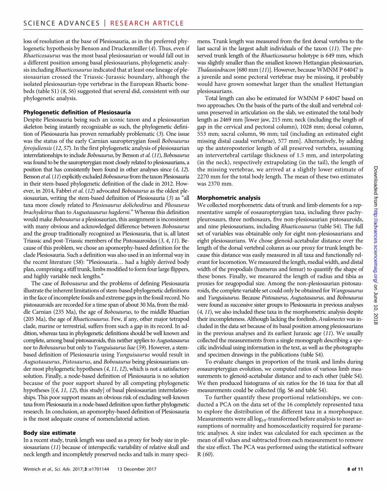
DiagnosisSmall-bodied plesiosaurian with an estimated total length of 237 cm(Fig. 2, A and B). The new taxon has two autapomorphies (Fig. 2C):a modified V-shaped neurocentral suture in the anterior and middle
1 of 11
SC I ENCE ADVANCES | R E S EARCH ART I C L E
Dow
nloaded from
cervical vertebrae. In Rhaeticosaurus, the sides of the “V” are ventrallyconcave, and the tip of the “V” almost reaches the ventral margin ofthe centrum. In other plesiosaurians with a V-shaped neurocentralsuture, the sides of the “V” are straight, and the tip only extends tothemiddle of the centrum. The second autapomorphy is greatly fore-shortened zeugopodials with a humerus/radius ratio of 3.8 and a femur/tibia ratio of 4.3 (Fig. 2, B, D, and E, and table S4). In addition, there are10 unambiguous but not unique synapomorphies (tables S2 and S3).
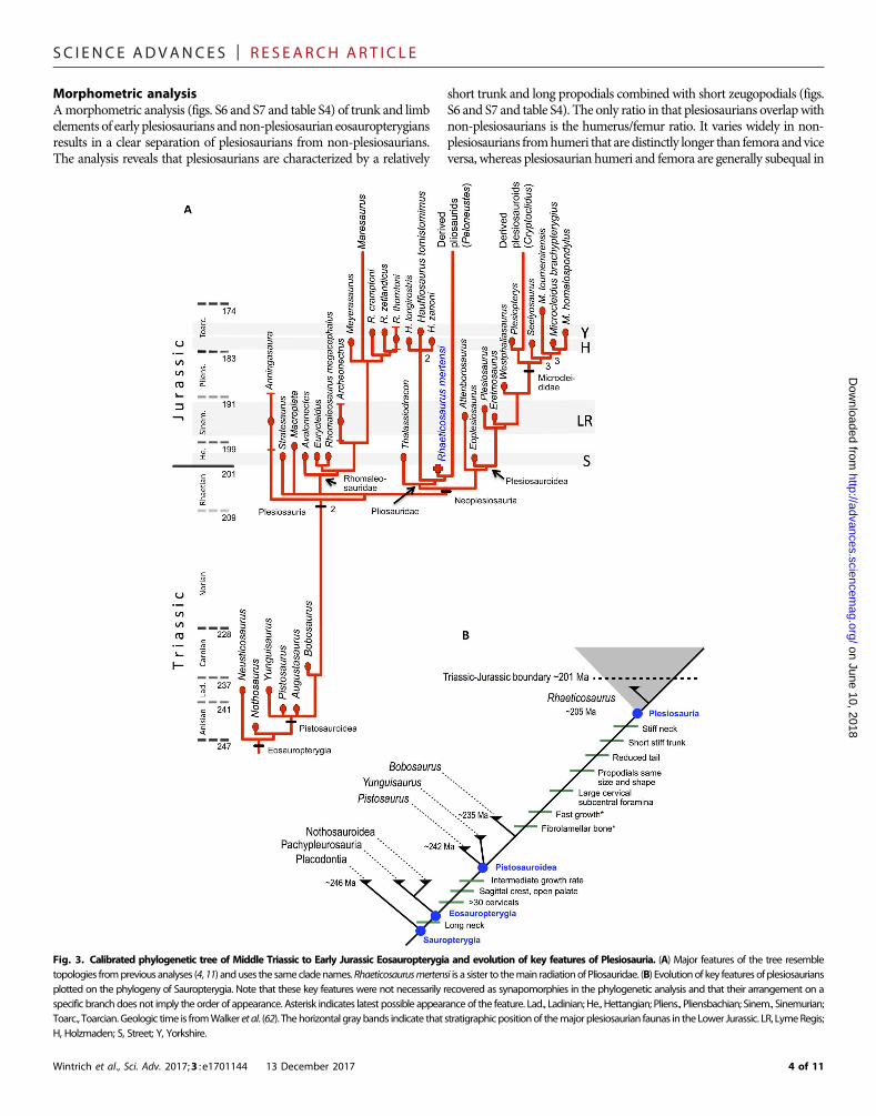
Phylogenetic relationshipsToassess the systematic position of theTriassic plesiosaurian skeleton,wecoded it for a recently published phylogenetic data matrix aimed at clar-ifying plesiosaurian interrelationships (data file S1) (4). Rhaeticosauruswas found to be nested within Plesiosauria as a basal member of thePliosauridae, withAnningasaura as themost basal plesiosaurian (Fig. 3A).As a consequence, six nodes in the cladogram are of Triassic age, in-dicating pre-Jurassic diversification of plesiosaurians into their majorclades (Fig. 3A).
Brief anatomical descriptionThe holotype (LWL-MFN P 64047, LWL-Museum für Naturkunde) ofRhaeticosaurus is a partial skeleton of a subadult preserving the occi-put, lower jaw, vertebral column, pectoral girdle, pelvic girdle, and
Wintrich et al., Sci. Adv. 2017;3 : e1701144 13 December 2017
left limbs (see Fig. 2, figs. S1 to S4, and Supplementary AnatomicalDescriptions). Several teeth are preserved in isolation, showing thetypical plesiosaurian enamel “striations” (fig. S1, B and C), which consistof sharp ridges of enamel formed on a smooth enamel dentin junction(10). We estimate ≥37 neck vertebrae, the 15 anteriormost of which arefully articulated (Fig. 2,A toC).There are large paired subcentral foramina(fig. S2E), as in other early plesiosaurians (11). Subcentral foramina aresmall in basal pistosaurs (Yunguisaurus, Pistosaurus, and Augustasaurus)and lacking in Bobosaurus (12). The zygapophyseal articular surfaces aremedially inclined (Fig. 2C and fig. S2B), as opposed to Bobosaurus and allmore basal sauropterygians that have horizontal facets (12). There are 21trunk vertebrae of the typical plesiosaurian type (fig. S3),with single roundrib articular facets on the end of the prominent transverse processes(fig. S3, B andC). These are entirely formedby theneural arch. The trunkregion is short and appears stiff because of the tight articulation of thedorsal centra and the width of the rib cage. There are four sacral verte-brae, and 25 vertebrae are preserved in the tail, with a few more missing(Fig. 2, A and B, and fig. S4). The caudal vertebral centra are disk-shaped(fig. S4B), indicating a short tail compared to non-plesiosaurians. Thewell-preserved pelvic girdle is represented by both ilia and the leftpubis and left ischium (Fig. 2, A and B, and fig. S3A). These bonesare similar in shape to those of other early plesiosaurians (11) and toBobosaurus (12), except for the ilium that is stouter in the latter.
on June 10, 2018http://advances.sciencem
ag.org/
Fig. 1. Locality and horizon of the new species. (A) Location of Bonenburg clay pit in eastern North Rhine-Westphalia, Germany. (B) Measured section of the Norian toHettangian deposits with the discovery horizon (indicated by plesiosaurian silhouette and red arrows) of Rhaeticosaurus mertensi and the bonebeds with the Triassic vertebratefauna. Horizons of lowermost Jurassic ammonites indicated by silhouettes. Colors of the rock types in the main stratigraphic column approximate colors in fresh outcrop. Cly,claystone; Fss, fine-grained sandstone; Het., Hettangian; L. J., Lower Jurassic; Md, mudstone; Slt, siltstone.
2 of 11
SC I ENCE ADVANCES | R E S EARCH ART I C L E
Despite their distal incompleteness, the forelimb and hindlimb ap-pear to have been rather similar inmorphology and length, as in all ple-siosaurians (Fig. 2, A and B, and fig. S5). The humerus has a straightshaft, and the distal end is little expanded (fig. S1A). The radius reachesonly 26% of the length of the humerus and is dorsoventrally flattened
Wintrich et al., Sci. Adv. 2017;3 : e1701144 13 December 2017
(Fig. 2D, fig. S6C, and table S4). Like the humerus, the femur has nearlystraight preaxial and postaxialmargins compared to the curvedmarginsin non-plesiosaurian sauropterygians, but it is somewhat more distallyexpanded than the humerus (Fig. 2E). Tibia and fibula attain less than25% of femur length and are flattened dorsoventrally (Fig. 2E).
on June 10, 2018http://advances.sciencem
ag.org/D
ownloaded from
Fig. 2. TheholotypeofRhaeticosaurusmertensigen. et sp. nov. (A) Photograph. (B) Color overlay. (C) Cervical vertebrae 10 to 15 in right lateral view showingmedially inclinedzygapophyses (black arrow) and the autapomorphic ventrally concave V-shapedneurocentral sutures (white arrow). (D) Left radius, a phalanx, and a carpal element. (E) Left femur,tibia, and fibula. The proximal femur is a cast because the original was sectioned for histology. ca, caudal vertebra; ce, cervical vertebra; cr, carpal bone; d, dorsal vertebra; f,femur; fi, fibula; l., left; ph, phalange; r., right; ra, radius; s, sacral vertebra; ti, tibia; ?, unidentified bone. Scale bars, 20 cm (A) and 1 cm (C to E).
3 of 11
SC I ENCE ADVANCES | R E S EARCH ART I C L E
Morphometric analysisAmorphometric analysis (figs. S6 and S7 and table S4) of trunk and limbelements of early plesiosaurians andnon-plesiosaurian eosauropterygiansresults in a clear separation of plesiosaurians from non-plesiosaurians.The analysis reveals that plesiosaurians are characterized by a relatively
Wintrich et al., Sci. Adv. 2017;3 : e1701144 13 December 2017
short trunk and long propodials combined with short zeugopodials (figs.S6 and S7 and table S4). The only ratio in that plesiosaurians overlapwithnon-plesiosaurians is the humerus/femur ratio. It varies widely in non-plesiosaurians fromhumeri that are distinctly longer than femora andviceversa, whereas plesiosaurian humeri and femora are generally subequal in
on June 10, 2018http://advances.sciencem
ag.org/D
ownloaded from
Fig. 3. Calibrated phylogenetic tree of Middle Triassic to Early Jurassic Eosauropterygia and evolution of key features of Plesiosauria. (A) Major features of the tree resembletopologies fromprevious analyses (4,11) anduses the samecladenames.Rhaeticosaurusmertensi is a sister to themain radiationof Pliosauridae. (B) Evolutionof key features ofplesiosauriansplotted on the phylogeny of Sauropterygia. Note that these key features were not necessarily recovered as synapomorphies in the phylogenetic analysis and that their arrangement on aspecific branch does not imply the order of appearance. Asterisk indicates latest possible appearance of the feature. Lad., Ladinian; He., Hettangian; Pliens., Pliensbachian; Sinem., Sinemurian;Toarc., Toarcian. Geologic time is fromWalker et al. (62). Thehorizontal graybands indicate that stratigraphicpositionof themajor plesiosaurian faunas in theLower Jurassic. LR, LymeRegis;H, Holzmaden; S, Street; Y, Yorkshire.
4 of 11
SC I ENCE ADVANCES | R E S EARCH ART I C L E
on June 10, 2018http://advances.sciencem
ag.org/D
ownloaded from
length (fig. S6E and table S4). For all five ratios that separate plesiosau-rians from non-plesiosaurians, Rhaeticosaurus always plotted amongplesiosaurians and Bobosaurus among non-plesiosaurians. We alsocomputed the humerus/tibia ratio as an additional proxy for relativezeugopodial length because these are the only major limb bones pre-served in Bobosaurus. Rhaeticosaurus again shows a plesiosaurian val-ue, and Bobosaurus shows a non-plesiosaurian one (table S4).
In a principal component analysis (PCA) (fig. S7), the twomain axesexplain 78.1% of the variance (61.0 and 17.1%, respectively). Note thathumerus and femur length covary. This is also the case for radius andtibia length and glenoid-acetabular distance that vary antagonistically todistal femur width. All variables contribute to the first axis, althoughglenoid-acetabular distance, radius length, tibia length, and distal femurwidth do so to a greater extent. The first axis discriminates plesiosaur-ians from non-plesiosaurian sauropterygians (fig. S7). In the analysis,plesiosaurians show a proportionally shorter radius and tibia, a shorterglenoid-acetabular distance, and a greater distal width of the femur and,to a lesser extent, the humerus. Conversely, humerus and femur arerelatively longer in plesiosaurians than in non-plesiosaurians, as alreadyseen in the ratio histograms (fig. S6). However, there is marked vari-ation in relative humerus and femur lengthwithin each group, as shownby the distribution of the taxa along the second axis that is essentiallydriven by these two variables (and humerus distal width, but to a lesserextent).
Locomotion and feedingThe anatomical features and proportions of Rhaeticosaurus have func-tional implications, particularly for locomotion and feeding. The thinintervertebral cartilages and tall neural spines (Fig. 2C and fig. S2)would have restricted dorsal neck movement, and the thin inter-vertebral cartilage and medially inclined cervical zygapophyses (asopposed to the horizontally oriented ones of non-plesiosaurians) wouldhave restricted lateral neck movements, suggesting a markedly in-flexible neck. The forelimbs and hindlimbs of Rhaeticosaurus onlydiffer from later plesiosaurians by having a less expanded distal end ofthe propodials, but they nevertheless must have functioned as stiffhydrofoils, as in all plesiosaurians (7). The extremely short and widezeugopodium and the straight stylopodial shaft are the major featuresthat set Rhaeticosaurus limbs apart from the otherwise similar limbs ofsome non-plesiosaurians, such as Yunguisaurus, in that propulsion byaxial undulation still played an important role. The lack of a tightmosaic of zeugopodial and carpal/tarsal bones (3) in Rhaeticosaurusdoes not argue against the limbs functioning as hydrofoils because roundcarpals and tarsals are also seen in many Early Jurassic plesiosaurianswith undoubtedly hydrofoil limbs. Only in later plesiosaurians is therea tight mosaic of zeugopodial and carpal/tarsal bones (3). The shorttail corroborates paraxial locomotion.
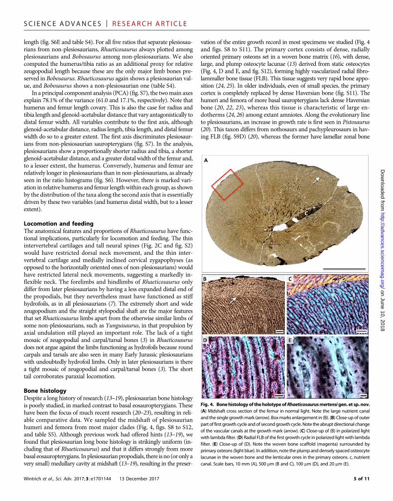
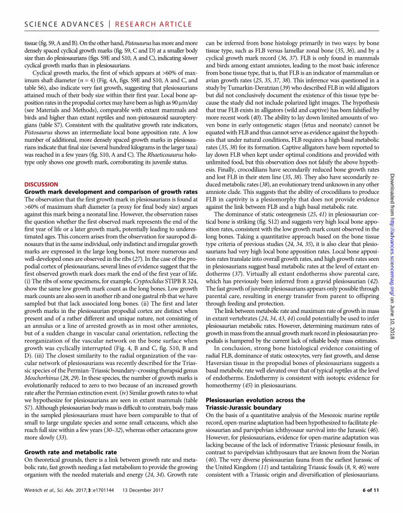
Bone histologyDespite a long history of research (13–19), plesiosaurian bone histologyis poorly studied, in marked contrast to basal eosauropterygians. Thesehave been the focus of much recent research (20–23), resulting in reli-able comparative data. We sampled the midshaft of plesiosaurianhumeri and femora from most major clades (Fig. 4, figs. S8 to S12,and table S5). Although previous work had offered hints (13–19), wefound that plesiosaurian long bone histology is strikingly uniform (in-cluding that of Rhaeticosaurus) and that it differs strongly from morebasal eosauropterygians. In plesiosaurianpropodials, there is no (or only avery small) medullary cavity at midshaft (13–19), resulting in the preser-
Wintrich et al., Sci. Adv. 2017;3 : e1701144 13 December 2017
vation of the entire growth record in most specimens we studied (Fig. 4and figs. S8 to S11). The primary cortex consists of dense, radiallyoriented primary osteons set in a woven bone matrix (16), with dense,large, and plump osteocyte lacunae (13) derived from static osteocytes(Fig. 4, D and E, and fig. S12), forming highly vascularized radial fibro-lammaller bone tissue (FLB). This tissue suggests very rapid bone appo-sition (24, 25). In older individuals, even of small species, the primarycortex is completely replaced by dense Haversian bone (fig. S11). Thehumeri and femora of more basal sauropterygians lack dense Haversianbone (20, 22, 23), whereas this tissue is characteristic of large en-dotherms (24, 26) among extant amniotes. Along the evolutionary lineto plesiosaurians, an increase in growth rate is first seen in Pistosaurus(20). This taxon differs from nothosaurs and pachypleurosaurs in hav-ing FLB (fig. S9D) (20), whereas the former have lamellar zonal bone
Fig. 4. Bonehistologyof theholotypeofRhaeticosaurusmertensigen. et sp. nov.(A) Midshaft cross section of the femur in normal light. Note the large nutrient canaland the single growthmark (arrow). Boxmarks enlargement in (B). (B) Close-upof outerpart of first growth cycle andof secondgrowth cycle. Note the abrupt directional changeof the vascular canals at the growth mark (arrow). (C) Close-up of (B) in polarized lightwith lambda filter. (D) Radial FLB of the first growth cycle in polarized lightwith lambdafilter. (E) Close-up of (D). Note the woven bone scaffold (magenta) surrounded byprimary osteons (light blue). In addition, note the plump anddensely spaced osteocytelacunae in the woven bone and the lenticular ones in the primary osteons. c, nutrientcanal. Scale bars, 10 mm (A), 500 mm (B and C), 100 mm (D), and 20 mm (E).
5 of 11
SC I ENCE ADVANCES | R E S EARCH ART I C L E
tissue (fig. S9,A andB).On theother hand,Pistosaurushasmore andmoredensely spaced cyclical growth marks (fig. S9, C and D) at a smaller bodysize than do plesiosaurians (figs. S9E and S10, A and C), indicating slowercyclical growth marks than in plesiosaurians.
Cyclical growth marks, the first of which appears at >60% of max-imum shaft diameter (n = 4) (Fig. 4A, figs. S9E and S10, A and C, andtable S6), also indicate very fast growth, suggesting that plesiosauriansattained much of their body size within their first year. Local bone ap-position rates in the propodial cortexmayhave been as high as 90mm/day(see Materials and Methods), comparable with extant mammals andbirds and higher than extant reptiles and non-pistosauroid sauroptery-gians (table S7). Consistent with the qualitative growth rate indicators,Pistosaurus shows an intermediate local bone apposition rate. A lownumber of additional, more densely spaced growth marks in plesiosau-rians indicate that final size (several hundred kilograms in the larger taxa)was reached in a few years (fig. S10, A and C). The Rhaeticosaurus holo-type only shows one growth mark, corroborating its juvenile status.
on June 10, 2018http://advances.sciencem
ag.org/D
ownloaded from
DISCUSSIONGrowth mark development and comparison of growth ratesThe observation that the first growth mark in plesiosaurians is found at>60% of maximum shaft diameter (a proxy for final body size) arguesagainst this mark being a neonatal line. However, the observation raisesthe question whether the first observed mark represents the end of thefirst year of life or a later growth mark, potentially leading to underes-timated ages. This concern arises from the observation for sauropod di-nosaurs that in the same individual, only indistinct and irregular growthmarks are expressed in the large long bones, but more numerous andwell-developed ones are observed in the ribs (27). In the case of the pro-podial cortex of plesiosaurians, several lines of evidence suggest that thefirst observed growth mark does mark the end of the first year of life.(i) The ribs of some specimens, for example, Cryptoclidus STIPB R 324,show the same low growth mark count as the long bones. Low growthmark counts are also seen in another rib and one gastral rib that we havesampled but that lack associated long bones. (ii) The first and latergrowth marks in the plesiosaurian propodial cortex are distinct whenpresent and of a rather different and unique nature, not consisting ofan annulus or a line of arrested growth as in most other amniotes,but of a sudden change in vascular canal orientation, reflecting thereorganization of the vascular network on the bone surface whengrowth was cyclically interrupted (Fig. 4, B and C, fig. S10, B andD). (iii) The closest similarity to the radial organization of the vas-cular network of plesiosaurians was recently described for the Trias-sic species of the Permian-Triassic boundary–crossing therapsid genusMoschorhinus (28, 29). In these species, the number of growth marks isevolutionarily reduced to zero to two because of an increased growthrate after the Permian extinction event. (iv) Similar growth rates towhatwe hypothesize for plesiosaurians are seen in extant mammals (tableS7). Although plesiosaurian bodymass is difficult to constrain, bodymassin the sampled plesiosaurians must have been comparable to that ofsmall to large ungulate species and some small cetaceans, which alsoreach full size within a few years (30–32), whereas other cetaceans growmore slowly (33).
Growth rate and metabolic rateOn theoretical grounds, there is a link between growth rate and meta-bolic rate, fast growth needing a fast metabolism to provide the growingorganism with the needed materials and energy (24, 34). Growth rate
Wintrich et al., Sci. Adv. 2017;3 : e1701144 13 December 2017
can be inferred from bone histology primarily in two ways: by bonetissue type, such as FLB versus lamellar zonal bone (35, 36), and by acyclical growth mark record (36, 37). FLB is only found in mammalsand birds among extant amniotes, leading to the most basic inferencefrom bone tissue type, that is, that FLB is an indicator of mammalian oravian growth rates (25, 35, 37, 38). This inference was questioned in astudy by Tumarkin-Deratzian (39) who described FLB in wild alligatorsbut did not conclusively document the existence of this tissue type be-cause the study did not include polarized light images. The hypothesisthat true FLB exists in alligators (wild and captive) has been falsified bymore recent work (40). The ability to lay down limited amounts of wo-ven bone in early ontogenetic stages (fetus and neonate) cannot beequatedwith FLB and thus cannot serve as evidence against the hypoth-esis that under natural conditions, FLB requires a high basal metabolicrates (35, 38) for its formation. Captive alligators have been reported tolay down FLB when kept under optimal conditions and provided withunlimited food, but this observation does not falsify the above hypoth-esis. Finally, crocodilians have secondarily reduced bone growth ratesand lost FLB in their stem line (35, 38). They also have secondarily re-ducedmetabolic rates (38), an evolutionary trend unknown in any otheramniote clade. This suggests that the ability of crocodilians to produceFLB in captivity is a plesiomorphy that does not provide evidenceagainst the link between FLB and a high basal metabolic rate.
The dominance of static osteogenesis (25, 41) in plesiosaurian cor-tical bone is striking (fig. S12) and suggests very high local bone appo-sition rates, consistent with the low growth mark count observed in thelong bones. Taking a quantitative approach based on the bone tissuetype criteria of previous studies (24, 34, 35), it is also clear that plesio-saurians had very high local bone apposition rates. Local bone apposi-tion rates translate into overall growth rates, and high growth rates seenin plesiosaurians suggest basal metabolic rates at the level of extant en-dotherms (37). Virtually all extant endotherms show parental care,which has previously been inferred from a gravid plesiosaurian (42).The fast growth of juvenile plesiosaurians appears only possible throughparental care, resulting in energy transfer from parent to offspringthrough feeding and protection.
The link betweenmetabolic rate andmaximumrate of growth inmassin extant vertebrates (24, 34, 43, 44) could potentially be used to inferplesiosaurian metabolic rates. However, determining maximum rates ofgrowth inmass from the annual growthmark record in plesiosaurian pro-podials is hampered by the current lack of reliable body mass estimates.
In conclusion, strong bone histological evidence consisting ofradial FLB, dominance of static osteocytes, very fast growth, and denseHaversian tissue in the propodial bones of plesiosaurians suggests abasal metabolic rate well elevated over that of typical reptiles at the levelof endotherms. Endothermy is consistent with isotopic evidence forhomeothermy (45) in plesiosaurians.
Plesiosaurian evolution across theTriassic-Jurassic boundaryOn the basis of a quantitative analysis of the Mesozoic marine reptilerecord, open-marine adaptation had been hypothesized to facilitate ple-siosaurian and parvipelvian ichthyosaur survival into the Jurassic (46).However, for plesiosaurians, evidence for open-marine adaptation waslacking because of the lack of informative Triassic plesiosaur fossils, incontrast to parvipelvian ichthyosaurs that are known from the Norian(46). The very diverse plesiosaurian fauna from the earliest Jurassic ofthe United Kingdom (11) and tantalizing Triassic fossils (8, 9, 46) wereconsistent with a Triassic origin and diversification of plesiosaurians.
6 of 11
SC I ENCE ADVANCES | R E S EARCH ART I C L E
http://advances.sciencema
Dow
nloaded from
On the other hand, because of the lack of unequivocal Triassic plesio-saurian finds, it was hypothesized that plesiosaurians rapidly colo-nized open marine habitats only in the aftermath of the Late Triassicextinctions (11). The Triassic age and phylogenetic relationships ofRhaeticosaurus now shed light on these issues. Under our best-supported phylogenetic hypothesis (seeMaterials andMethods), at leastsix plesiosaurian lineages crossed the Triassic-Jurassic boundary (Fig. 3A).Late Triassic and end-Triassic extinction events (5, 47, 48) were thussurvived by diverse lineages of plesiosaurians in addition to parvipelvianichthyosaurs (1, 6). Most groups that went extinct during these eventsinhabited coastal waters and shallow carbonate platforms (Placodontia,Nothosauroidea,non-plesiosaurianPistosauroidea, andTanystropheidae)(1, 5, 6) but not the open sea. Although there are two other groups re-corded from open-marine sediments that went extinct (thalattosaurs andnon-plesiosaurianPistosauroidea), thesewere less specialized for a pelagiclifestyle. Plesiosaurians seemingly adapted to life in the open sea (20, 46)during the LateTriassic andnot only in the Early Jurassic (11) by evolvinglong-distance cruising capabilities facilitated by a highmetabolic rate (evi-dencedby fast growthandFLB) andunderwater flight (evidencedby skel-etal morphology and proportions) over a time period of 30 million years(Ma) (Fig. 3B). Open-marine adaptation of plesiosaurians may have fa-cilitated their survival into the Jurassic (46) because pelagic prey (fish andsoft-bodied cephalopods) (47) were less affected by the end-Triassic ex-tinction events than benthic invertebrates and reef organisms (47, 48).These suffered from a calcification crisis (49) but formed the food basefor the Late Triassicmarine reptile groups that went extinct. The scenarioof a pre-Jurassic radiation of plesiosaurians begs the question of why theevidence is coming to light only now. The Rhaetian bonebed vertebraepreviously assigned to plesiosaurians represent different taxa (8) thatnow appear consistent with a Late Triassic radiation. This radiationmay have gone unrecognized for so long because of the extreme paucityofmarine reptile localities representing the last 30Ma of the Triassic (6).
on June 10, 2018g.org/
MATERIALS AND METHODSPhylogenetic analysisTo test the phylogenetic relationships of Rhaeticosaurus, we added theholotype to a recently publisheddatamatrix of 270 characters and 80opera-tional taxonomic units (OTUs). This matrix was designed to clarify theinterrelationships of plesiosaurians and their survival across the Jurassic-Cretaceous boundary (4). However, because most of the taxa in the matrixwere unquestionably more derived than the Triassic plesiosaurian, we usedthe taxon sampling of another matrix specifically designed to clarify the re-lationships of Early Jurassic plesiosaurians (11). Three modifications to thistaxon sampling were made. For one, following the studies of Fabbri et al.(12) and Neenan et al. (50), we combined the Pistosaurus skull and post-cranial OTUs into a single OTU because there was little doubt that thePistosaurus skull belongswith thePistosaurus postcranium (51–53).Wealso added two additional outgroup taxa to the single original outgroup(Yunguisaurus liae), the Ladinian pachypleurosaurNeusticosaurus pusillusand the Anisian nothosaurNothosaurus marchicus to clarify the relation-ships of Triassic sauropterygians on the line leading to Plesiosauria. Nocharacters weremodified, and no new characters were added. The anal-ysis thus used a matrix of 35 taxa (6 Triassic non-plesiosaurian taxa,Rhaeticosaurus, and 28 Early and Middle Jurassic plesiosaurian taxa)and 270 characters (data file S1).
We ran the analysis using the software PAUP v. 4.0b10 (54), uti-lizing the same settings as in the study (11) of the Early Jurassic plesiosau-rians (500 random addition replicates with TBR branch swapping). Of the
Wintrich et al., Sci. Adv. 2017;3 : e1701144 13 December 2017
270 characters, only 207 were parsimony-informative because we used areduced taxon sampling compared to that of Benson and Druckenmiller(4), resulting in 30 characters being constant and 33 being parsimony-uninformative. The reduced taxon sampling also meant that we did nothave to use the parsimony ratchet because the search in PAUP was suf-ficiently fast. We initially ran the analysis with onlyNeusticosaurus as theoutgroup to minimize assumptions about interrelationships and thenadded Nothosaurus and Yunguisaurus to the outgroup. In each case(one, two, and three outgroup taxa), our analyses recovered the same21 most parsimonious trees that are 764 steps long, with a consistencyindex of 0.415 and a retention index of 0.554.We then computed a strictconsensus tree in which Rhaeticosaurus is found to be a basal memberof the Pliosauridae (Fig. 3A and table S3). This is a somewhat surprisingresult, given that the lower jaw of Rhaeticosaurus shows features rareor unknown in pliosaurids such as lack of participation of the splenial inthe jaw symphysis, the short angular, and the dorsoventral andmediolat-eral orientation of the retroarticular process (4, 11). In addition, the plio-saurs lack the sharp ridge on the anterior margin of the humerus (4, 11).
Focusing on our analysis, a comparison with the other Lower Jurassicpliosaurids,Thalassiodracon hawkinsii (11), and the species ofHauffiosaurus(55) revealed some support through unequivocal and unique syanpo-morphies for the placement ofRhaeticosauruswithin this clade. There isonly one unequivocal and unique synapomorphy ofRhaeticosaurus andPeloneustes (as a representative of more derived pliosaurids): the wideand short tibia (ratio of tibia length tomaximumwidth, 0.8–1.0 character,255; state, 2). In addition, there is only one unequivocal but not uniquesynapomorphy (table S3). The Pliosauridae in our analysis have onlyone unequivocal and unique synapomorphy, the posterior terminationof the premaxilla (character, 16; state, 1; broad, deeply interdigitatingsuture with the frontal or parietal), which is, however, not preservedin Rhaeticosaurus. In addition, there are three unequivocal but notunique synapomorphies (table S3). The sister group relationship of theRhaeticosaurus-Peloneustes clade with Hauffiosaurus is also supportedby a single unequivocal and unique synapomorphy (character, 34; state,1; lacrimal present, maxilla excluded from orbit margin), which again isnot preserved inRhaeticosaurus. In addition, there are eight unequivocalbut not unique synapomorphies (table S3). Hauffiosaurus differs fromboth Rhaeticosaurus and Thalassiodracon in the autapomorphic contactof the neural arch with the articular facets for the cervical ribs (55).
In itsmajor features, the consensus tree resembles the ones by Bensonand colleagues (4, 11). However, the interrelationships at the base ofPlesiosauria are better resolved (Fig. 3A) inour analysis, and there is greaterstratigraphic congruency in the Plesiosauria, the Rhomaleosauridae,and the Plesiosauroidea than in either of the previous analyses (4, 11).On the other hand, the nested position of Rhaeticosaurus within Plio-sauridae is inconsistent with its great stratigraphic age. At least six nodeswithin Plesiosauria are situated in the Triassic, predating Rhaeticosaurus.Bobosaurus was always recovered as the sister taxon to Plesiosauria, butthe relationships of the basal Pistosauroidea (Yunguisaurus, Pistosaurus,and Augustasaurus) are not resolved. We computed the Bremer supportindex for the phylogeny in PAUP. The support index of most nodes isonly 1, but Plesiosauria is supported by a value of 2. Table S3 provides thelist of synapomorphies derived from the consensus tree. The list was alsoused to diagnose Plesiosauria and Rhaeticosaurus (table S2).
Basal plesiosaurian relationships are notoriously difficult to resolve,and support of the resulting hypotheses generally is weak (4, 11), as inour analysis. To specifically evaluate the influence of the newly addedtaxa (further outgroups and Rhaeticosaurus), we reran our analysiswithout themandobtained a consensus tree that showed anear-complete
7 of 11
SC I ENCE ADVANCES | R E S EARCH ART I C L E
on June 10, 2018http://advances.sciencem
ag.org/D
ownloaded from
loss of resolution at the base of Plesiosauria, as in the preferred phy-logenetic hypothesis by Benson and Druckenmiller (4). Thus, even ifRhaeticosaurus was the most basal plesiosaurian or would fall out ina different position among basal plesiosaurians, phylogenetic analy-sis including Rhaeticosaurus indicated that at least one lineage of ple-siosaurian crossed the Triassic-Jurassic boundary, although theisolated plesiosaurian-type vertebrae in the European Rhaetic bone-beds (table S1) (8, 56) suggested that several did, consistent with ourphylogenetic analysis.
Phylogenetic definition of PlesiosauriaDespite Plesiosauria being such an iconic taxon and a plesiosaurianskeleton being instantly recognizable as such, the phylogenetic defini-tion of Plesiosauria has proven remarkably problematic (3). One issuewas the status of the early Carnian sauropterygian fossil Bobosaurusforojuliensis (12, 57). In the first phylogenetic analysis of plesiosaurianinterrelationships to include Bobosaurus, by Benson et al. (11), Bobosauruswas found to be the sauropterygianmost closely related to plesiosaurians, aposition that has consistently been found in other analyses since (4, 12).Benson etal. (11) explicitly excludedBobosaurus fromthe taxonPlesiosauriain their stem-based phylogenetic definition of the clade in 2012. How-ever, in 2014, Fabbri et al. (12) advocated Bobosaurus as the oldest ple-siosaurian, writing the stem-based definition of Plesiosauria (3) as “alltaxa more closely related to Plesiosaurus dolichodirus and Pliosaurusbrachydeirus than to Augustasaurus hagdorni.”Whereas this definitionwouldmake Bobosaurus a plesiosaurian, this assignment is inconsistentwith many obvious and acknowledged difference between Bobosaurusand the group traditionally recognized as Plesiosauria, that is, all latestTriassic and post-Triassic members of the Pistosauroidea (3, 4, 11). Be-cause of this problem, we chose an apomorphy-based definition for theclade Plesiosauria. Such a definition was also used in an informal way inthe recent literature (58): “Plesiosauria… had a highly derived bodyplan, comprising a stiff trunk, limbsmodified to form four large flippers,and highly variable neck lengths.”
The case of Bobosaurus and the problems of defining Plesiosauriaillustrate the inherent limitations of stem-based phylogenetic definitionsin the face of incomplete fossils and extreme gaps in the fossil record.Nopistosauroids are recorded for a time span of about 30Ma, from themid-dle Carnian (235 Ma), the age of Bobosaurus, to the middle Rhaetian(205 Ma), the age of Rhaeticosaurus. Few, if any, other major tetrapodclade, marine or terrestrial, suffers from such a gap in its record. In ad-dition, whereas taxa in phylogenetic definitions should be well known andcomplete, among basal pistosauroids, this neither applies toAugustasaurusnor to Bobosaurus but only toYunguisaurus liae (59). However, a stem-based definition of Plesiosauria using Yunguisaurus would result inAugustasaurus, Pistosaurus, and Bobosaurus being plesiosaurians un-der most phylogenetic hypotheses (4, 11, 12), which is not a satisfactorysolution. Finally, a node-based definition of Plesiosauria is no solutionbecause of the poor support shared by all competing phylogenetichypotheses [(4, 11, 12), this study] of basal plesiosaurian interrelation-ships. This poor support means an obvious risk of excluding well-knowntaxa fromPlesiosauria in a node-based definitionupon further phylogeneticresearch. In conclusion, an apomorphy-based definition of Plesiosauriais the most adequate course of nomenclatorial action.
Body size estimateIn a recent study, trunk length was used as a proxy for body size in ple-siosaurians (11) because of interspecific variability of relative skull andneck length and incompletely preserved necks and tails in many speci-
Wintrich et al., Sci. Adv. 2017;3 : e1701144 13 December 2017
mens. Trunk length was measured from the first dorsal vertebra to thelast sacral in the largest adult individuals of the taxon (11). The pre-served trunk length of the Rhaeticosaurus holotype is 649 mm, whichwas slightly smaller than the smallest known Hettangian plesiosaurian,Thalassiodracon [680 mm (11)]. However, becauseWMNMP 64047 isa juvenile and some pectoral vertebrae may be missing, it probablywould have grown somewhat larger than the smallest Hettangianplesiosaurians.
Total length can also be estimated for WMNM P 64047 based ontwo approaches. On the basis of the parts of the skull and vertebral col-umn preserved in articulation on the slab, we estimated the total bodylength as 2469 mm [lower jaw, 215 mm; neck (including the length ofgap in the cervical and pectoral column), 1028 mm; dorsal column,553 mm; sacral column, 96 mm; tail (including an estimated eightmissing distal caudal vertebrae), 577 mm]. Alternatively, by addingup the anteroposterior length of all preserved vertebra, assumingan intervertebral cartilage thickness of 1.5 mm, and interpolating(in the neck), respectively extrapolating (in the tail), the length ofthe missing vertebrae, we arrived at a slightly lower estimate of2270 mm for the total body length. The mean of these two estimateswas 2370 mm.
Morphometric analysisWe collected morphometric data of trunk and limb elements for a rep-resentative sample of eosauropterygian taxa, including three pachy-pleurosaurs, three nothosaurs, five non-plesiosaurian pistosauroids,and nine plesiosaurians, including Rhaeticosaurus (table S4). The fullset of variables was obtainable only for eight non-plesiosaurians andeight plesiosaurians. We chose glenoid-acetabular distance over thelength of the dorsal vertebral column as our proxy for trunk length be-cause this distance was easily measured in all taxa and functionally rel-evant for locomotion.Wemeasured the length,medial width, and distalwidth of the propodials (humerus and femur) to quantify the shape ofthese bones. Finally, we measured the length of radius and tibia asproxies for zeugopodial size. Among the non-plesiosaurian pistosau-roids, the complete variable set could only be obtained forWangosaurusand Yunguisaurus. Because Pistosaurus, Augustasaurus, and Bobosauruswere found as successive sister groups to Plesiosauria in previous analyses(4, 11), we also included these taxa in the morphometric analysis despitetheir incompleteness. Although lacking the forelimb,Avalonecteswas in-cluded in the data set because of its basal position among plesiosauriansin the previous analyses and its earliest Jurassic age (11). We usuallycollected the measurements from a single monograph describing a spe-cific individual using information in the text, as well as the photographsand specimen drawings in the publications (table S4).
To evaluate changes in proportion of the trunk and limbs duringeosauropterygian evolution, we computed ratios of various limb mea-surements to glenoid-acetabular distance and to each other (table S4).We then produced histograms of six ratios for the 16 taxa for that allmeasurements could be collected (fig. S6 and table S4).
To further quantify these proportional relationships, we con-ducted a PCA on the data set of the 16 completely represented taxato explore the distribution of the different taxa in a morphospace.Measurements were all log10-transformed before analysis tomeet as-sumptions of normality and homoscedasticity required for parame-tric analyses. A size index was calculated for each specimen as themean of all values and subtracted from eachmeasurement to removethe size effect. The PCA was performed using the statistical softwareR (60).
8 of 11
SC I ENCE ADVANCES | R E S EARCH ART I C L E
on June 10, 2018http://advances.sciencem
ag.org/D
ownloaded from
Histological analysisAs noted in themain text, it was puzzling that the uniqueness of plesiosau-rian bone histology was not recognized before, despite the long history ofresearch. Reasons were insufficiently constrained samples in terms of tax-onomy [undiagnostic samples (13, 16, 17)], insufficient clade coverage(13–17), anatomy [lack of identification of skeletal element (13, 14)],ontogenetic stage [old individuals, in which the peculiar primary FLBhad been replaced by secondary Haversian bone (13, 14, 16, 17)], andplane of section [plane of section in long bones offset from nutrient ca-nal (13, 14, 16–19)]. A case in point is the 19th century work byKiprijanoff(13) on Russian marine reptiles that described all manner of histologyand microanatomy, correctly figuring ichthyosaur microanatomy andplesiosaurian dental histology in the finest detail, but bypassing plesio-saurian microanatomy, only schematically illustrating propodial crosssection of seemingly old individuals.
We obtained samples of plesiosaurian propodials across the tree(table S5), representing the widest taxonomic coverage in plesiosaurianhistologic studies so far. All individuals except for the Rhaeticosaurusholotype and the Japanese elasmosaur were osteologically mature.The humeri and femora were sectioned by two cuts spaced about 5 mmapart across the mid-diaphysis, with the two cuts preferentially enclosingthe inner terminus of the nutrient canal because this indicates the site ofearliest bone growth (61). The location of the nutrient canal wasdetermined either visually or by computed tomography (CT) scanningof the specimens (fig. S8) using the GE phoenix v|tome|x s240 scannerat the Division of Paleontology of the Steinmann Institute, University ofBonn, Germany. Using the 240-kV tube of the scanner, scan parameterswere 200 kV, 200 mA, and a voxel size of 142 mm. CT scanning is impor-tant because in same bones, the nutrient canal does not extend radiallyfrom the center to the surface but deviates proximally. Thus, a sectionplaced at the nutrient foramenmay be located proximal to the centerof growth, leading to erroneous interpretations in previous studies(14, 17).
Themid-diaphysial slice of bone was then processed into a standardpetrographic thin section 50 to 80 mm in thickness. The sections wereobserved under a Leica DM2500LP polarizing microscope, and digitalphotomicrographs were taken with a Leica DFC420 color cameramounted on this microscope and edited using the 2007 Leica IMAGEACCESSEASYLAB7 software.Overview imageswere obtainedwith anEpson V750 high-resolution scanner. The terminology followed thestudy of Francillon-Vieillot et al. (24).
A useful proxy for growth rate is the maximum local bone apposi-tion rate in the femur cortex, reflecting the increase in thickness of thisbone (35). Maximum local bone apposition rate was expressed in mi-crometers per day and was determined in extant and extinct species inthe midshaft region following established protocols (35). Four of thenine plesiosaurian specimens histologically sampled were suitable forthis analysis (table S6) because they had a complete growth recordand at least one postnatal growth mark preserved. One specimen, thejuvenile elasmosaur OMNH MV 85, did not show any growth marks,and the remaining four (table S5) were completely remodeled, obliter-ating the growth mark record. Note that both humeri and femora wereincluded in this analysis because they are of nearly the same length(fig. S6) and show the same histology in plesiosaurians, unlike in theamniotes previously analyzed (35). We slightly modified the protocolby measuring apposition along the nutrient canal that represents a ho-mologous location, at least in plesiosaurians, and is close to the region ofthe thickest cortex (Fig. 4 and figs. S8 to S10).Wemeasured the corticalthickness from the center of the bone (the inner terminus of the nutrient
Wintrich et al., Sci. Adv. 2017;3 : e1701144 13 December 2017
canal, which is well preserved in plesiosaurians because of the lack ofmedullary resorption) to the first growthmarks inmillimeters.We thendivided this value by 730, accounting for the 365 days in a year, plus ahypothetical gestation period of equal length [365 days as in many dol-phin species (33)] to cover the prenatal part of the cortex. Thisprocedure was necessary because a neonatal line could not reliably bedetected. Whereas the resulting local bone apposition rates (table S6)are only estimates, the margin of error is insignificant in the compara-tive context (local bone apposition rates in reptiles versusmammals andbirds; table S7). Even if we assume an unrealistically short gestation pe-riod of 50 days or an unrealistically long one of 500 days (table S6), localbone apposition rates donot overlapwith those of extant reptiles (table S7).We were also aware of the higher number of days per year in the geo-logic past but felt that the error of a few days introduced this way wasnegligible. On the basis of Amprino’s rule (35), we assumed that pre-natal and postnatal bone apposition rate were very similar because ofthe uniformity of the primary bone from the onset of osteogenesis inthe embryo to the first postnatal growth mark. For estimating the rela-tive size of the plesiosaurians at the end of their first year, we also usedcortical thickness along the nutrient canal.
Note that the maximum local bone apposition rate also depends onbody size at the time of fastest growth (35). Our plesiosaurian data weredifficult to correct for size because of the difficulty of determining plesi-osaurian bodymass, but the size effect was overridden by the general pat-tern, with the endotherms (birds, mammals) showing apposition ratesmuch higher, mostly an order of magnitude, than the ectotherms inthe sample (lizards, turtles, a crocodile). A case in point is the crocodiledata point (Crocodilus niloticus). This taxon is in the same body massrange as plesiosaurians but grows at only somewhat more than half therate of the slowest plesiosaurian and only a 10th of the rate of the fastestplesiosaurian (table S7).
SUPPLEMENTARY MATERIALSSupplementary material for this article is available at http://advances.sciencemag.org/cgi/content/full/3/12/e1701144/DC1Supplementary Anatomical Descriptionsfig. S1. The holotype of R. mertensi gen. et sp. nov.fig. S2. The holotype of R. mertensi gen. et sp. nov., anterior cervical vertebral column.fig. S3. The holotype of R. mertensi gen. et sp. nov.fig. S4. The holotype of R. mertensi gen. et sp. nov.fig. S5. Reconstruction of the skeleton of R. mertensi gen. et sp. nov. based on the availablemeasurements and proportions.fig. S6. Selected skeletal proportions in Eosauropterygia.fig. S7. Principal component analysis of trunk and limb measurements in Eosauropterygia.fig. S8. Examples of CT scans of plesiosaurian long bones used in locating the nutrient canalbefore sectioning.fig. S9. Evolution of long bone histology in Triassic Eosauropterygia.fig. S10. Long bone histology of Jurassic and Cretaceous Plesiosauria.fig. S11. Long bone histology of a mature Middle Jurassic plesiosaurian.fig. S12. Long bone histology of the holotype of R. mertensi gen. et sp. nov. in longitudinalsection.table S1. Faunal list of bonebed above plesiosaurian discovery horizon.table S2. Unambiguous but not unique synapomorphies diagnosing R. mertensi gen. et sp.nov. in addition to the two autapomorphies.table S3. List of synapomorphies from phylogenetic analysis.table S4. Measurements and proportions in the trunk and limbs of Eosauropterygia.table S5. List of histological samples.table S6. Local bone apposition rate to the end of the first year and relative body size at theend of the first year in selected sauropterygians.table S7. Comparison of local bone apposition rates in the femur of selected amniotescompared to local bone apposition rates in the humeri and femora of plesiosaurians.data file S1. Character matrix in NEXUS format for phylogenetic analysis described in Materialsand Methods.References (63–75)
9 of 11
SC I ENCE ADVANCES | R E S EARCH ART I C L E
on June 10, 2018http://advances.sciencem
ag.org/D
ownloaded from
REFERENCES AND NOTES1. N. P. Kelley, N. D. Pyenson, Evolutionary innovation and ecology in marine tetrapods from
the Triassic to the Anthropocene. Science 348, aaa3716 (2015).2. A. Houssaye, Bone histology of aquatic reptiles: What does it tell us about secondary
adaptation to an aquatic life? Biol. J. Linn. Soc. 108, 3–21 (2013).3. H. F. Ketchum, R. B. J. Benson, Global interrelationships of Plesiosauria (Reptilia,
Sauropterygia) and the pivotal role of taxon sampling in determining the outcome ofphylogenetic analyses. Biol. Rev. 85, 361–392 (2010).
4. R. B. J. Benson, P. S. Druckenmiller, Faunal turnover of marine tetrapods during theJurassic–Cretaceous transition. Biol. Rev. 89, 1–23 (2014).
5. N. P. Kelley, R. Motani, D.-y. Jiang, O. Rieppel, L. Schmitz, Selective extinction of Triassicmarine reptiles during long-term sea-level changes illuminated by seawater strontiumisotopes. Palaeogeogr. Palaeoclimatol. Palaeoecol. 400, 9–16 (2014).
6. N. Bardet, J. Falconnet, V. Fischer, A. Houssaye, S. Jouve, X. Pereda Suberbiola,A. Pérez-García, J.-C. Rage, P. Vincent, Mesozoic marine reptile palaeobiogeography inresponse to drifting plates. Gondw. Res. 26, 869–887 (2014).
7. S. Liu, A. S. Smith, Y. Gu, J. Tan, C. K. Liu, G. Turk, Computer simulations imply forelimb-dominated underwater flight in plesiosaurs. PLOS Comput. Biol. 11, e1004605 (2015).
8. G. W. Storrs, Fossil vertebrate faunas of the British Rhaetian (latest Triassic). Zool. J. Linn.Soc. 112, 217–259 (1994).
9. A. G. Sennikov, M. S. Arkhangelsky, On a typical Jurassic sauropterygian from the UpperTriassic of Wilczek Land (Franz Josef Land, Arctic Russia). Paleont. J. 44, 567–572 (2010).
10. P. M. Sander, The microstructure of reptilian tooth enamel: Terminology, function, andphylogeny. Münchner Geowiss. Abh. 38, 1–102 (1999).
11. R. B. J. Benson, M. Evans, P. S. Druckenmiller, High diversity, low disparity and small bodysize in plesiosaurs (Reptilia, Sauropterygia) from the Triassic–Jurassic boundary.PLOS ONE 7, e31838 (2012).
12. M. Fabbri, F. M. Dalla Vecchia, A. Cau, New information on Bobosaurus forojuliensis(Reptilia: Sauropterygia): Implications for plesiosaurian evolution. Hist. Biol. 26, 661–669(2014).
13. A. V. Kiprijanoff, Studien fiber die fossilen Reptilien Russlands. Mém. Acad. Imp. Sci. St.Petersbourg 7, 1–144 (1881–1883).
14. Ł. Fostowicz-Frelik, A. Gaździcki, Anatomy and histology of plesiosaur bones fromthe Late Cretaceous of Seymour Island, Antarctic Peninsula. Palaeontol. Pol. 60, 7–32(2001).
15. L. Salgado, M. S. Fernandez, M. Talevi, Observaciones histologicas en reptiles marinos(Elasmosauridae y Mosasauridae) del Cretacico Tardio de Patagonia y Antartida.Ameghiniana 44, 513–523 (2007).
16. J. Wiffen, V. de Buffrénil, A. de Ricqlès, J.-M. Mazin, Ontogenetic evolution of bonestructure in Late Cretaceous Plesiosauria from New Zealand. Geobios 28, 625–640 (1995).
17. L. Liebe, J. H. Hurum, Gross internal structure and microstructure of plesiosaur limbbones from the Late Jurassic, central Spitsbergen. Nor. J. Geol. 92, 285–309 (2012).
18. J. P. O’Gorman, M. Talevi, M. S. Fernández, Osteology of a perinatal aristonectine(Plesiosauria; Elasmosauridae). Antarct. Sci. 29, 61–72 (2017).
19. L. Ossa-Fuentes, R. A. Otero, D. Rubilar-Rogers, Microanatomy and osteohistology of ajuvenile elasmosaurid plesiosaur from the Upper Maastrichtian of Marambio (Seymour)Island, Antarctica. Bol. Museo Nacional Hist. Natural Chile 66, 149–160 (2017).
20. A. Krahl, N. Klein, P. M. Sander, Evolutionary implications of the divergent long bonehistologies of Nothosaurus and Pistosaurus (Sauropterygia, Triassic). BMC Evol. Biol.13, 123 (2013).
21. A. Houssaye, P. M. Sander, N. Klein, Adaptive patterns in aquatic amniote bonemicroanatomy—More complex than previously thought. Integr. Comp. Biol.56, 1349–1369 (2016).
22. N. Klein, P. M. Sander, A. Krahl, T. M. Scheyer, A. Houssaye, Diverse aquatic adaptations inNothosaurus spp. (Sauropterygia)—Inferences from humeral histology andmicroanatomy. PLOS ONE 11, e0158448 (2016).
23. N. Klein, E. M. Griebeler, Bone histology, microanatomy, and growth of the nothosauroidSimosaurus gaillardoti (Sauropterygia) from the Upper Muschelkalk of southern Germany/Baden-Württemberg. C. R. Palevol 15, 142–162 (2016).
24. H. Francillon-Vieillot, V. de Buffrénil, J. Castanet, J. Géraudie, F. J. Meunier, J. Y. Sire,L. Zylberberg, A. de Ricquès, Microstructure and mineralization of vertebrate skeletaltissues, in Skeletal Biomineralization: Patterns, Processes and Evolutionary Trends, J. G.Carter, Ed. (Van Nostrand Reinhold, 1990), vol. 1, pp. 471–530.
25. K. Stein, E. Prondvai, Rethinking the nature of fibrolamellar bone: An integrativebiological revision of sauropod plexiform bone formation. Biol. Rev. Camb. Philos. Soc.89, 24–47 (2014).
26. J. D. Currey, The many adaptations of bone. J. Biomech. 36, 1487–1495 (2003).27. K. Waskow, P. M. Sander, Growth record and histological variation in the dorsal ribs of
Camarasaurus sp. (Sauropoda). J. Vertebr. Paleontol. 34, 852–869 (2014).28. A. K. Huttenlocker, J. Botha-Brink, Body size and growth patterns in the therocephalian
Moschorhinus kitchingi (Therapsida: Eutheriodontia) before and after the end-Permianextinction in South Africa. Paleobiology 39, 253–277 (2013).
Wintrich et al., Sci. Adv. 2017;3 : e1701144 13 December 2017
29. J. Botha-Brink, D. Codron, A. K. Huttenlocker, K. D. Angielczyk, M. Ruta, Breedingyoung as a survival strategy during Earth’s greatest mass extinction. Sci. Rep. 6, 24053(2016).
30. M. Köhler, N. Marín-Moratalla, X. Jordana, R. Aanes, Seasonal bone growth and physiologyin endotherms shed light on dinosaur physiology. Nature 487, 358–361 (2012).
31. A. S. Barreto, F. C. W. Rosas, Comparative growth analysis of two populations ofPontoporia blainvillei on the Brazilian coast. Mar. Mamm. Sci. 22, 644–653 (2006).
32. S. Murphy, E. Rogan, External morphology of the short-beaked common dolphin,Delphinus delphis: Growth, allometric relationships and sexual dimorphism. Acta Zool.87, 315–329 (2006).
33. A. Berta, Whales, Dolphins, and Porpoises. A Natural History and Species Guide (Universityof Chicago Press, 2015), 288 pp.
34. L. Montes, N. Le Roy, M. Perret, V. de Buffrenil, J. Castanet, J. Cubo, Relationships betweenbone growth rate, body mass and resting metabolic rate in growing amniotes: Aphylogenetic approach. Biol. J. Linn. Soc. 92, 63–76 (2007).
35. J. Cubo, N. Le Roy, C. Martinez-Maza, L. Montes, Paleohistological estimation of bonegrowth rate in extinct archosaurs. Paleobiology 38, 335–349 (2012).
36. A. H. Lee, K. Huttenlocker, K. Padian, H. N. Woodward, Analysis of growth rates, in BoneHistology of Fossil Tetrapods: Advancing Methods, Analysis, and Interpretation, K. Padian,E.-T. Lamm, Eds. (University of California Press, 2013), pp. 217–251.
37. K. Padian, K. Stein, Evolution of growth rates and their implications, in Bone Histology ofFossil Tetrapods: Advancing Methods, Analysis, and Interpretation, K. Padian, E.-T. Lamm,Eds. (University of California Press, 2013), pp. 253–264.
38. L. J. Legendre, G. Guénard, J. Botha-Brink, J. Cubo, Palaeohistological evidence forancestral high metabolic rate in archosaurs. Syst. Biol. 65, 989–996 (2016).
39. A. R. Tumarkin-Deratzian, Fibrolamellar bone in wild adult Alligator mississippiensis.J. Herpetol. 41, 341–345 (2007).
40. H. N. Woodward, J. R. Horner, J. O. Farlow, Quantification of intraskeletal histovariability inAlligator mississippiensis and implications for vertebrate osteohistology. PeerJ 2, e422(2014).
41. E. Prondvai, K. H. W. Stein, A. de Ricqlès, J. Cubo, Development-based revision of bonetissue classification: The importance of semantics for science. Biol. J. Linn. Soc. 112,799–816 (2014).
42. F. R. O’Keefe, L. M. Chiappe, Viviparity and K-selected life history in a Mesozoic marineplesiosaur (Reptilia, Sauropterygia). Science 333, 870–873 (2011).
43. J. Werner, E. M. Griebeler, Allometries of maximum growth rate versus body mass atmaximum growth indicate that non-avian dinosaurs had growth rates typical of fastgrowing ectothermic sauropsids. PLOS ONE 9, e88834 (2014).
44. J. M. Grady, B. J. Enquist, E. Dettweiler-Robinson, N. A. Wright, F. A. Smith, Evidence formesothermy in dinosaurs. Science 344, 1268–1272 (2014).
45. A. Bernard, C. Lécuyer, P. Vincent, R. Amiot, N. Bardet, E. Buffetaut, G. Cuny, F. Fourel,F. Martineau, J.-M. Mazin, A. Prieur, Regulation of body temperature by some Mesozoicmarine reptiles. Science 328, 1379–1382 (2010).
46. R. B. J. Benson, R. J. Butler, J. Lindgren, A. S. Smith, Mesozoic marine tetrapod diversity:Mass extinctions and temporal heterogeneity in geological megabiases affectingvertebrates. Proc. R. Soc. B 277, 829–834 (2010).
47. L. H. Tanner, S. G. Lucas, M. G. Chapman, Assessing the record and causes of Late Triassicextinctions. Earth Sci. Rev. 65, 103–139 (2004).
48. M. S. Hodges, G. D. Stanley Jr., North American coral recovery after the end-Triassic massextinction, New York Canyon, Nevada, USA. GSA Today 25, 4–7 (2015).
49. M. Hautmann, M. J. Benton, A. Tomasových, Catastrophic ocean acidification at theTriassic-Jurassic boundary. N. Jb. Geol. Paläont. Abh. 249, 119–127 (2008).
50. J. M. Neenan, N. Klein, T. M. Scheyer, European origin of placodont marine reptiles andthe evolution of crushing dentition in Placodontia. Nat. Commun. 4, 1621 (2013).
51. H.-D. Sues, Postcranial skeleton of Pistosaurus and interrelationships of the Sauropterygia(Diapsida). Zool. J. Linn. Soc. 90, 109–131 (1987).
52. P. M. Sander, O. C. Rieppel, H. Bucher, A new pistosaurid (Reptilia: Sauropterygia) from theMiddle Triassic of Nevada and its implications for the origin of plesiosaurs. J. Vertebr.Paleontol. 17, 526–533 (1997).
53. O. Rieppel, P. M. Sander, G. W. Storrs, The skull of the pistosaur Augustasaurus from theMiddle Triassic of northwestern Nevada. J. Vertebr. Paleontol. 22, 577–592 (2002).
54. D. L. Swofford, PAUP*. Phylogenetic Analysis Using Parsimony (*and Other Methods).Version 4.0b10 (Sinauer Associates, 2002).
55. R. B. J. Benson, H. F. Ketchum, L. F. Noé, M. Gómez-Pérez, New information onHauffiosaurus (Reptilia, Plesiosauria) based on a new species from the Alum ShaleMember (Lower Toarcian: Lower Jurassic) of Yorkshire, UK. Palaeontology 54, 547–571(2011).
56. V. Fischer, H. Cappetta, P. Vincent, G. Garcia, S. Goolaerts, J. E. Martin, D. Roggero,X. Valentin, Ichthyosaurs from the French Rhaetian indicate a severe turnover across theTriassic–Jurassic boundary. Naturwissenschaften 101, 1027–1040 (2014).
57. F. M. Dalla Vecchia, A new sauropterygian reptile with plesiosaurian affinity from the LateTriassic of Italy. Riv. Ital. Paleontol. Strat. 112, 207–225 (2006).
10 of 11
SC I ENCE ADVANCES | R E S EARCH ART I C L E
http://advances.scienceD
ownloaded from
58. R. B. J. Benson, M. Evans, M. A. Taylor, The anatomy of Stratesaurus (Reptilia, Plesiosauria)from the lowermost Jurassic of Somerset, United Kingdom. J. Vertebr. Paleontol. 35,e933739 (2015).
59. T. Sato, L.-J. Zhao, X.-C. Wu, C. Li, A new specimen of the Triassic pistosauroidYunguisaurus, with implications for the origin of Plesiosauria (Reptilia, Sauropterygia).Palaeontology 57, 55–76 (2014).
60. R Development Core Team, A language and environment for statistical computing (RFoundation for Statistical Computing, 2014); www.r-project.org/.
61. Y. Nakajima, R. Hirayama, H. Endo, Turtle humeral microanatomy and its relationship tolifestyle. Biol. J. Linn. Soc. 112, 719–734 (2014).
62. J. D. Walker, J. W. Geissman, S. A. Bowring, L. E. Babcock, Compilers, Geologic time scalev. 4.0 (Geological Society of America, 2012).
63. P. M. Sander, T. Wintrich, A. H. Schwermann, R. Kindlimann, Die paläontologischeGrabung in der Rhät-Lias-Tongrube der Fa. Lücking bei Warburg-Bonenburg (Kr. Höxter)im Frühjahr 2015. Geol. Paläont.Westf. 88, 11–37 (2016).
64. O. Rieppel, A new pachypleurosaur (Reptilia: Sauropterygia) from the Middle Triassic ofMonte San Giorgio, Switzerland. Philos. Trans. R. Soc. Lond. B Biol. Sci. 323, 1–73 (1989).
65. P. M. Sander, The pachypleurosaurids (Reptilia: Nothosauria) from the Middle Triassic ofMonte San Giorgio (Switzerland) with the description of a new species. Philos. Trans.R. Soc. Lond. B Biol. Sci. 325, 561–666 (1989).
66. O. Rieppel, R. Wild, A revision of the genus Nothosaurus (Reptilia: Sauropterygia) from theGermanic Triassic, with comments on the status of Chonchiosaurus clavatus. FieldianaGeol. New Ser. 34, 1–82 (1996).
67. O. Rieppel, Handbook of Paleoherpetology/Sauropterygia I.: Placodontia,Pachypleurosauria, Nothosauroidea, Pistosauroidea (Friedrich Pfeil, 2000), 134 pp.
68. L.-T. Ma, D.-Y. Jiang, O. Rieppel, R. Motani, A. Tintori, A new pistosauroid (Reptilia,Sauropterygia) from the late Ladinian Xingyi marine reptile level, southwestern China.J. Vert. Paleontol. 35, e881832 (2015).
69. G. Geissler, Ueber neue Saurier-Funde aus dem Muschelkalk von Bayreuth. Z. Dtsch. Geol.Ges. 47, 331–355 (1895).
70. W. D. Conybeare, Additional notices on the fossil genera Ichthyosaurus and Plesiosaurus.Trans. Geol. Soc. Lond. Ser. 2, 103–123 (1822).
71. L. Schwermann, P. M. Sander, Osteologie und Phylogenie von Westphaliasaurussimonsensii: Ein neuer Plesiosauride (Sauropterygia) aus dem Unteren Jura(Pliensbachium) von Sommersell (Kreis Höxter), Nordrhein-Westfalen, Deutschland. Geol.Paläont.Westf. 79, 1–56 (2011).
72. F. R. O’Keefe, Preliminary description and phylogenetic position of a new plesiosaur (Reptilia:Sauropterygia) from the Toarcian of Holzmaden, Germany. J. Paleo. 78, 973–988 (2004).
Wintrich et al., Sci. Adv. 2017;3 : e1701144 13 December 2017
73. A. S. Smith, P. Vincent, A new genus of pliosaur (Reptilia: Sauropterygia) from the LowerJurassic of Holzmaden, Germany. Palaeontology 53, 1049–1063 (2010).
74. A. S. Smith, R. B. J. Benson, Osteology of Rhomaleosaurus thorntoni (Sauropterygia,Rhomaleosauridae) from the Lower Jurassic (Toarcian) of Northamptonshire, England.Monogr. Palaeontogr. Soc. 168, 1–40 (2014).
75. N. Klein, Long bone histology of Sauropterygia from the Lower Muschelkalk of theGermanic Basin provides unexpected implications for phylogeny. PLOS ONE 5, e11613(2010).
Acknowledgments: Access to the Bonenburg clay pit was granted by J. Thater of LückingZiegelwerke. A. Hendricks and D. Grzegorczyk provided access to the specimen. Histologicalsampling was permitted and facilitated by E. Maxwell and L. Chiappe. We thank the staff at theLWL-Museum für Naturkunde for the preparation of the specimen and O. Dülfer for thehistological thin sections. We thank M. Aberhan, R. Bussert, and P. E. Olsen for providing themeasured sections for Fig. 1B. We acknowledge R. O’Keefe and T. Sato for the insightful discussionand R. Kindlimann for the help with Rhaetian chondrichthyan fossil identification. We thankthe reviewers for their insightful suggestions for the improvement of the manuscript. We alsothank T. Oda for contributing fig. S2. Funding: Funding was provided by the LWL-Museum fürNaturkunde, Münster, Germany, the German Research Foundation (grant no. SA 469/47-1) and theJapan Society for the Promotion of Science (project nos. 27/6594 and 26800270). Authorcontributions: T.W. and P.M.S. designed and performed the morphological work and conductedthe phylogenetic analysis. P.M.S. and A.H. conducted the morphometric analysis. All authorsdesigned the histological part of the study, contributed the histological samples, and cooperatedin their interpretation. P.M.S. and T.W. wrote the manuscript, with contributions from all otherauthors. Competing interests: The authors declare that they have no competing interests. Dataand materials availability: All data needed to evaluate the conclusions in the paper arepresent in the paper and/or the Supplementary Materials. Additional data related to this papermay be requested from the authors.
Submitted 4 April 2017Accepted 16 November 2017Published 13 December 201710.1126/sciadv.1701144
Citation: T. Wintrich, S. Hayashi, A. Houssaye, Y. Nakajima, P. M. Sander, A Triassic plesiosaurianskeleton and bone histology inform on evolution of a unique body plan. Sci. Adv. 3, e1701144(2017).
m
11 of 11
on June 10, 2018ag.org/
planA Triassic plesiosaurian skeleton and bone histology inform on evolution of a unique body
Tanja Wintrich, Shoji Hayashi, Alexandra Houssaye, Yasuhisa Nakajima and P. Martin Sander
DOI: 10.1126/sciadv.1701144 (12), e1701144.3Sci Adv
ARTICLE TOOLS http://advances.sciencemag.org/content/3/12/e1701144
MATERIALSSUPPLEMENTARY http://advances.sciencemag.org/content/suppl/2017/12/11/3.12.e1701144.DC1
REFERENCES
http://advances.sciencemag.org/content/3/12/e1701144#BIBLThis article cites 67 articles, 9 of which you can access for free
PERMISSIONS http://www.sciencemag.org/help/reprints-and-permissions
Terms of ServiceUse of this article is subject to the
registered trademark of AAAS.is aScience Advances Association for the Advancement of Science. No claim to original U.S. Government Works. The title
York Avenue NW, Washington, DC 20005. 2017 © The Authors, some rights reserved; exclusive licensee American (ISSN 2375-2548) is published by the American Association for the Advancement of Science, 1200 NewScience Advances
on June 10, 2018http://advances.sciencem
ag.org/D
ownloaded from


![Histology Slides - mediconotes.commediconotes.com/freenotes/basic/histology_laboratory_slides.pdf[Histology] Histology Slides MedicoNotes provides real laboratory Histological slides](https://img.pdfslide.us/doc/110x75/5ae110e87f8b9a5a668e6aa3/histology-slides-histology-histology-slides-mediconotes-provides-real-laboratory.jpg)