Embed Size (px)
Citation preview
Copyright © 2008 Pearson Education, Inc., publishing as Pearson Benjamin Cummings
PowerPoint® Lecture Presentations for
BiologyEighth Edition
Neil Campbell and Jane Reece
Lectures by Chris Romero, updated by Erin Barley with contributions from Joan Sharp
Chapter 6
A Tour of the Cell
Overview: The Fundamental Units of Life
• All organisms are made of cells
• The cell is the simplest collection of matter
that can live
• Cell structure is correlated to cellular function
• All cells are related by their descent from
earlier cells
Copyright © 2008 Pearson Education, Inc., publishing as Pearson Benjamin Cummings
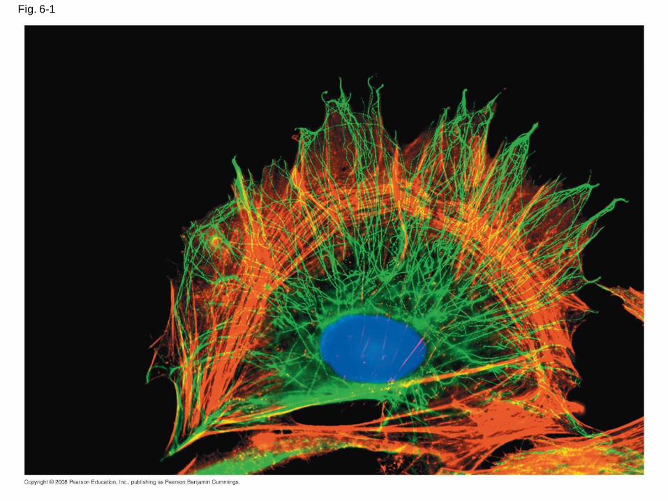
Fig. 6-1
Concept 6.1: To study cells, biologists use microscopes and the tools of biochemistry
• Though usually too small to be seen by the
unaided eye, cells can be complex
Copyright © 2008 Pearson Education, Inc., publishing as Pearson Benjamin Cummings
Microscopy
• Scientists use microscopes to visualize cells
too small to see with the naked eye
• In a light microscope (LM), visible light
passes through a specimen and then through
glass lenses, which magnify the image
Copyright © 2008 Pearson Education, Inc., publishing as Pearson Benjamin Cummings
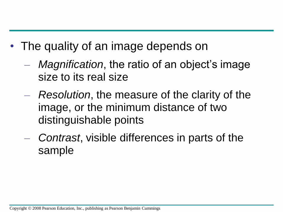
• The quality of an image depends on
– Magnification, the ratio of an object’s image size to its real size
– Resolution, the measure of the clarity of the image, or the minimum distance of two distinguishable points
– Contrast, visible differences in parts of the sample
Copyright © 2008 Pearson Education, Inc., publishing as Pearson Benjamin Cummings
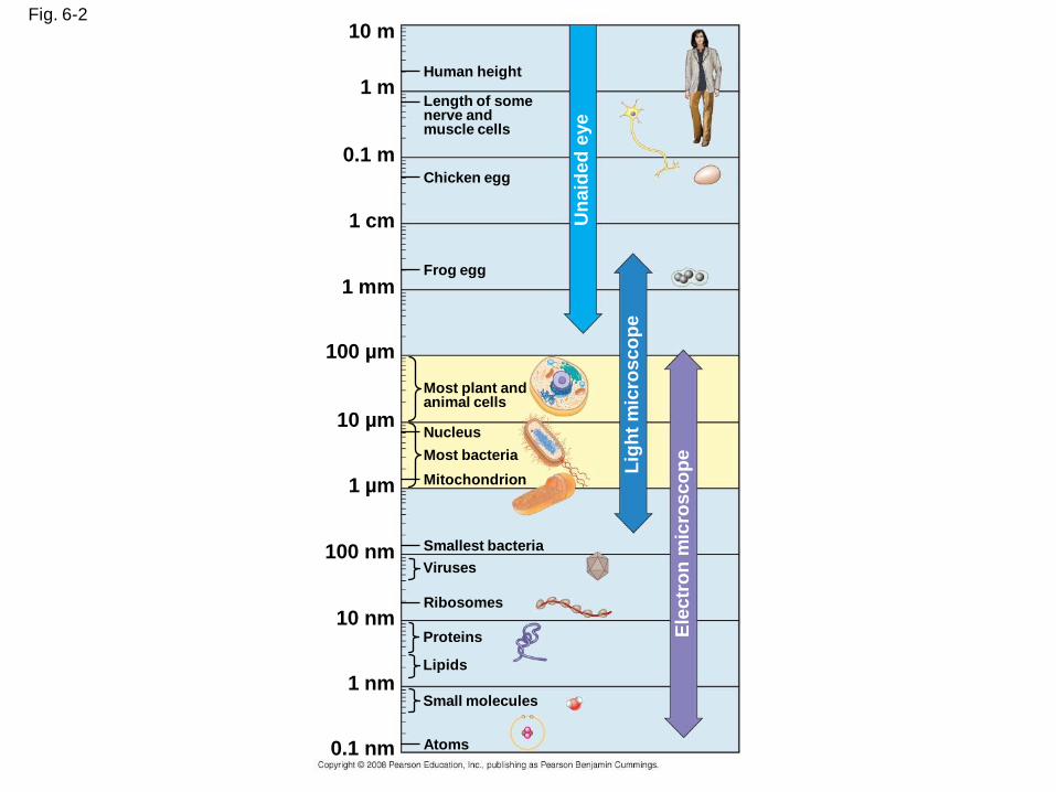
Fig. 6-210 m
1 m
0.1 m
1 cm
1 mm
100 µm
10 µm
1 µm
100 nm
10 nm
1 nm
0.1 nm Atoms
Small molecules
Lipids
Proteins
Ribosomes
Viruses
Smallest bacteria
Mitochondrion
Nucleus
Most bacteria
Most plant and animal cells
Frog egg
Chicken egg
Length of some nerve and muscle cells
Human height
Un
aid
ed
eye
Lig
ht
mic
rosco
pe
Ele
ctr
on
mic
ros
co
pe
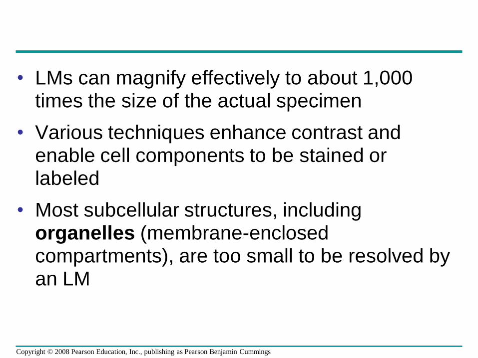
• LMs can magnify effectively to about 1,000 times the size of the actual specimen
• Various techniques enhance contrast and enable cell components to be stained or labeled
• Most subcellular structures, including organelles (membrane-enclosed compartments), are too small to be resolved by an LM
Copyright © 2008 Pearson Education, Inc., publishing as Pearson Benjamin Cummings
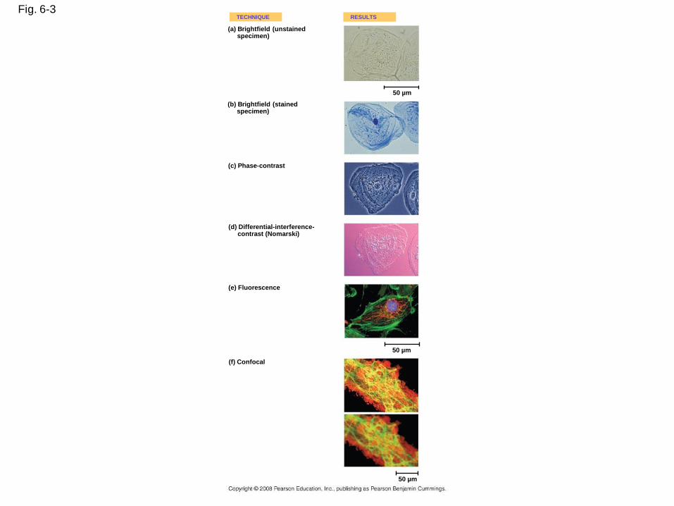
Fig. 6-3TECHNIQUE RESULTS
(a) Brightfield (unstainedspecimen)
(b) Brightfield (stainedspecimen)
50 µm
(c) Phase-contrast
(d) Differential-interference-contrast (Nomarski)
(e) Fluorescence
(f) Confocal
50 µm
50 µm
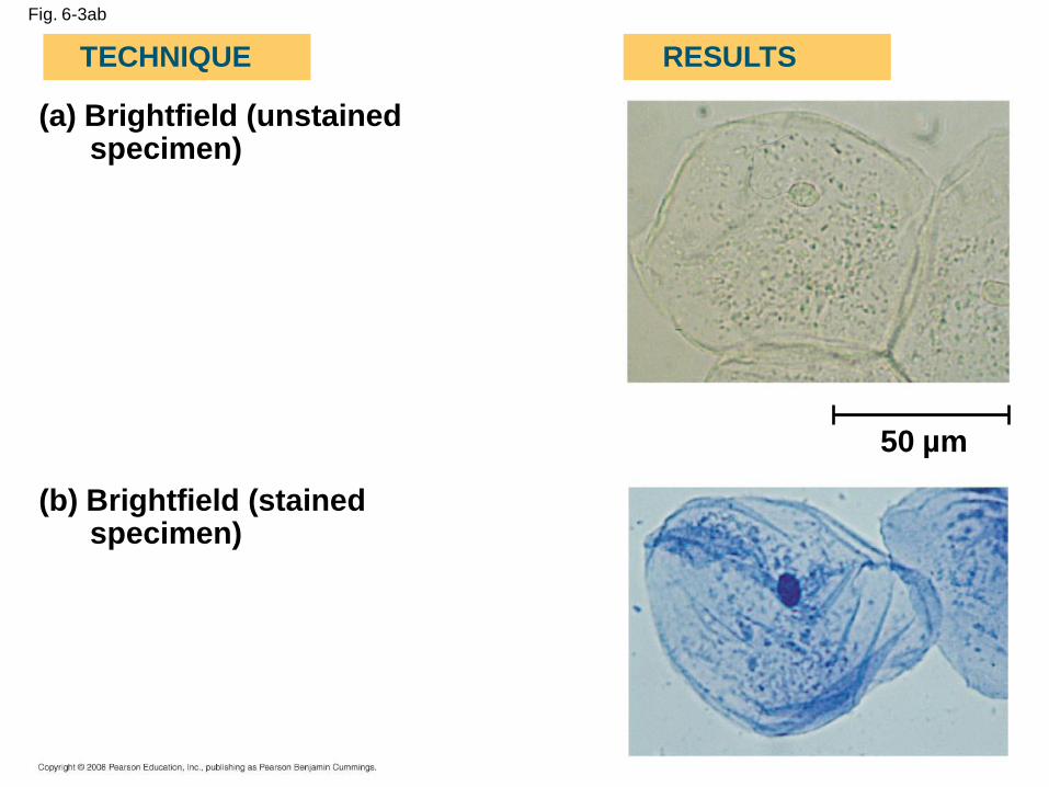
Fig. 6-3ab
(a) Brightfield (unstainedspecimen)
(b) Brightfield (stainedspecimen)
TECHNIQUE RESULTS
50 µm
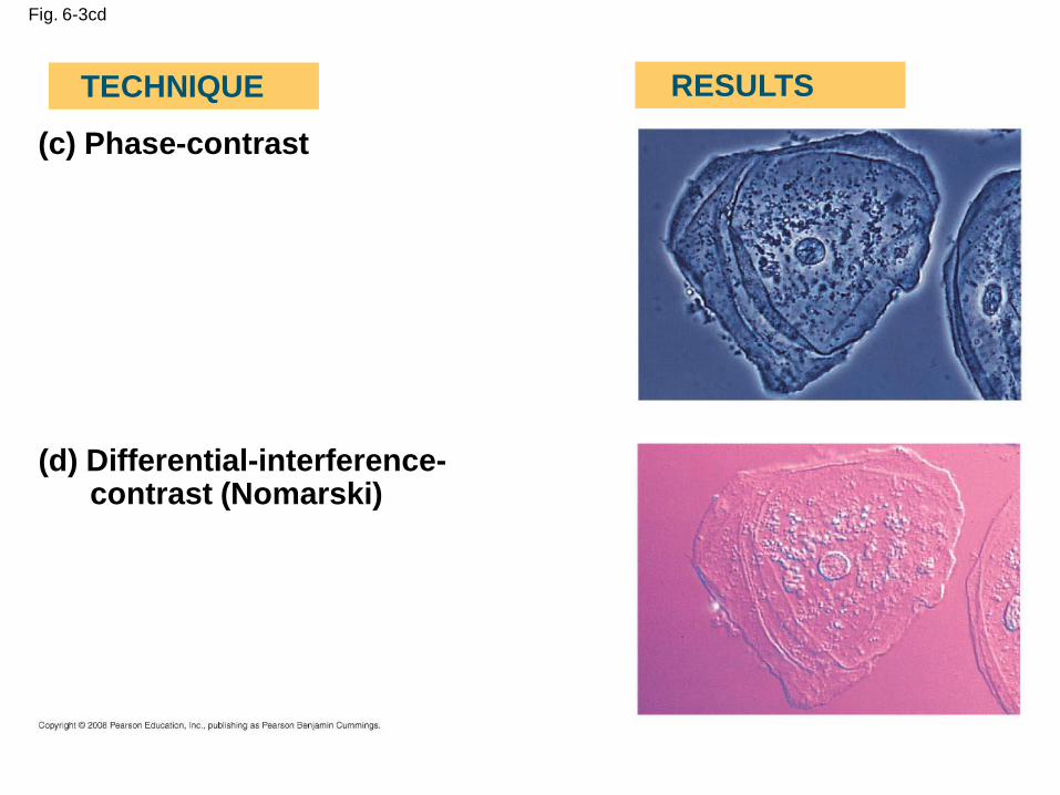
Fig. 6-3cd
(c) Phase-contrast
(d) Differential-interference-contrast (Nomarski)
TECHNIQUE RESULTS
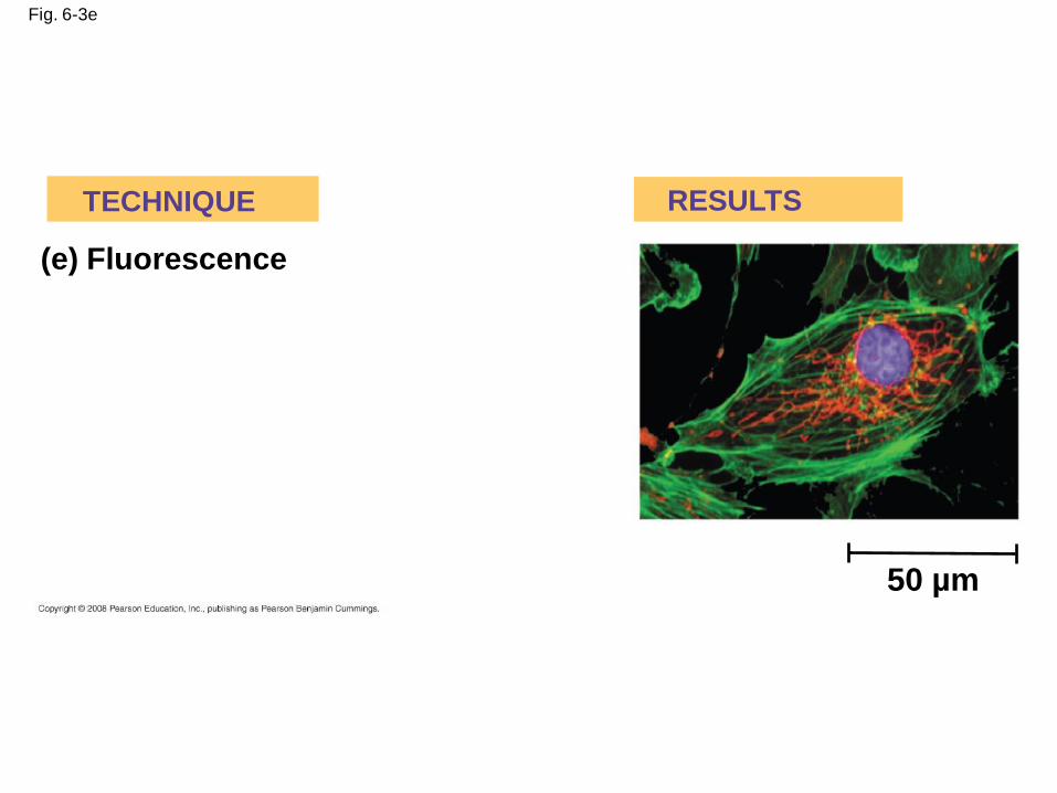
Fig. 6-3e
(e) Fluorescence
TECHNIQUE RESULTS
50 µm
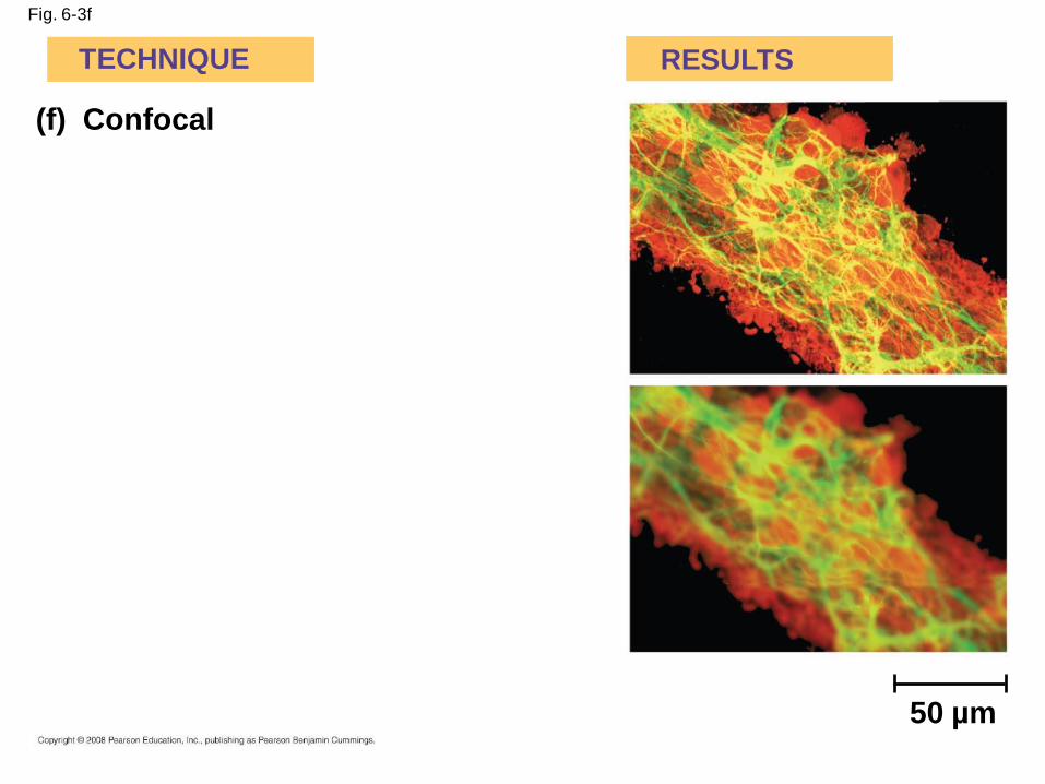
Fig. 6-3f
(f) Confocal
TECHNIQUE RESULTS
50 µm
• Two basic types of electron microscopes
(EMs) are used to study subcellular structures
• Scanning electron microscopes (SEMs)
focus a beam of electrons onto the surface of a
specimen, providing images that look 3-D
• Transmission electron microscopes (TEMs)
focus a beam of electrons through a specimen
• TEMs are used mainly to study the internal
structure of cells
Copyright © 2008 Pearson Education, Inc., publishing as Pearson Benjamin Cummings
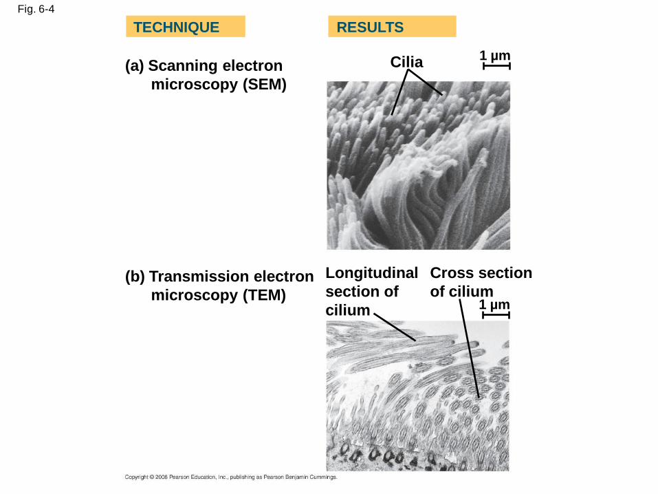
Fig. 6-4
(a) Scanning electron
microscopy (SEM)
TECHNIQUE RESULTS
(b) Transmission electron
microscopy (TEM)
Cilia
Longitudinal
section of
cilium
Cross section
of cilium1 µm
1 µm
Cell Fractionation
• Cell fractionation takes cells apart and
separates the major organelles from one
another
• Ultracentrifuges fractionate cells into their
component parts
• Cell fractionation enables scientists to
determine the functions of organelles
• Biochemistry and cytology help correlate cell
function with structure
Copyright © 2008 Pearson Education, Inc., publishing as Pearson Benjamin Cummings
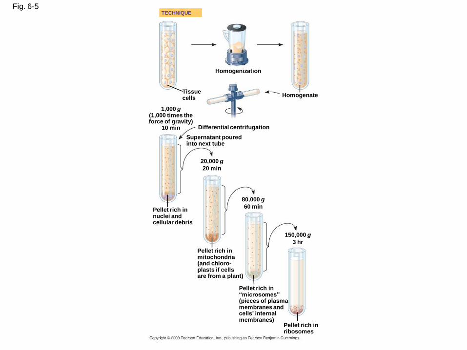
Fig. 6-5
Homogenization
TECHNIQUE
HomogenateTissuecells
1,000 g(1,000 times theforce of gravity)
10 min Differential centrifugation
Supernatant pouredinto next tube
20,000 g
20 min
80,000 g
60 minPellet rich innuclei andcellular debris
Pellet rich inmitochondria(and chloro-plasts if cellsare from a plant)
Pellet rich in“microsomes”(pieces of plasmamembranes andcells’ internalmembranes)
150,000 g
3 hr
Pellet rich inribosomes
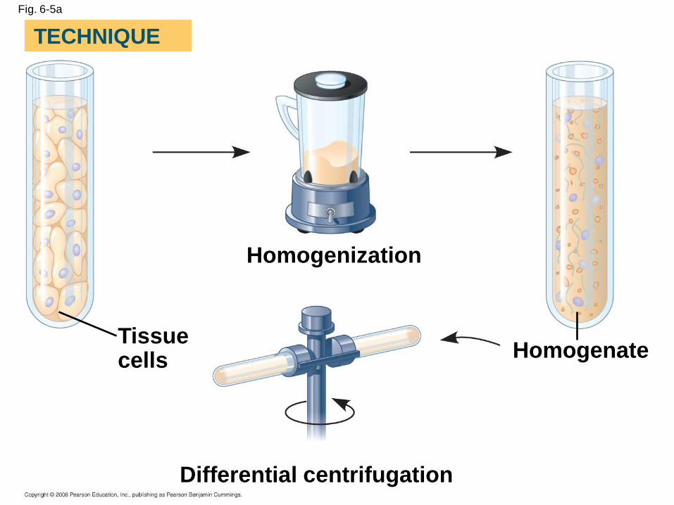
Fig. 6-5a
Homogenization
Homogenate
Differential centrifugation
Tissuecells
TECHNIQUE
Fig. 6-5b
1,000 g(1,000 times the force of gravity)
10 min
Supernatant poured into next tube
20,000 g
20 min
80,000 g
60 min
150,000 g
3 hr
Pellet rich in nuclei and cellular debris
Pellet rich in mitochondria (and chloro-plasts if cellsare from a plant)
Pellet rich in “microsomes” (pieces of plasmamembranes and cells’ internal membranes) Pellet rich in
ribosomes
TECHNIQUE (cont.)
Concept 6.2: Eukaryotic cells have internal membranes that compartmentalize their functions
• The basic structural and functional unit of every
organism is one of two types of cells:
prokaryotic or eukaryotic
• Only organisms of the domains Bacteria and
Archaea consist of prokaryotic cells
• Protists, fungi, animals, and plants all consist of
eukaryotic cells
Copyright © 2008 Pearson Education, Inc., publishing as Pearson Benjamin Cummings
Comparing Prokaryotic and Eukaryotic Cells
• Basic features of all cells:
– Plasma membrane
– Semifluid substance called cytosol
– Chromosomes (carry genes)
– Ribosomes (make proteins)
Copyright © 2008 Pearson Education, Inc., publishing as Pearson Benjamin Cummings
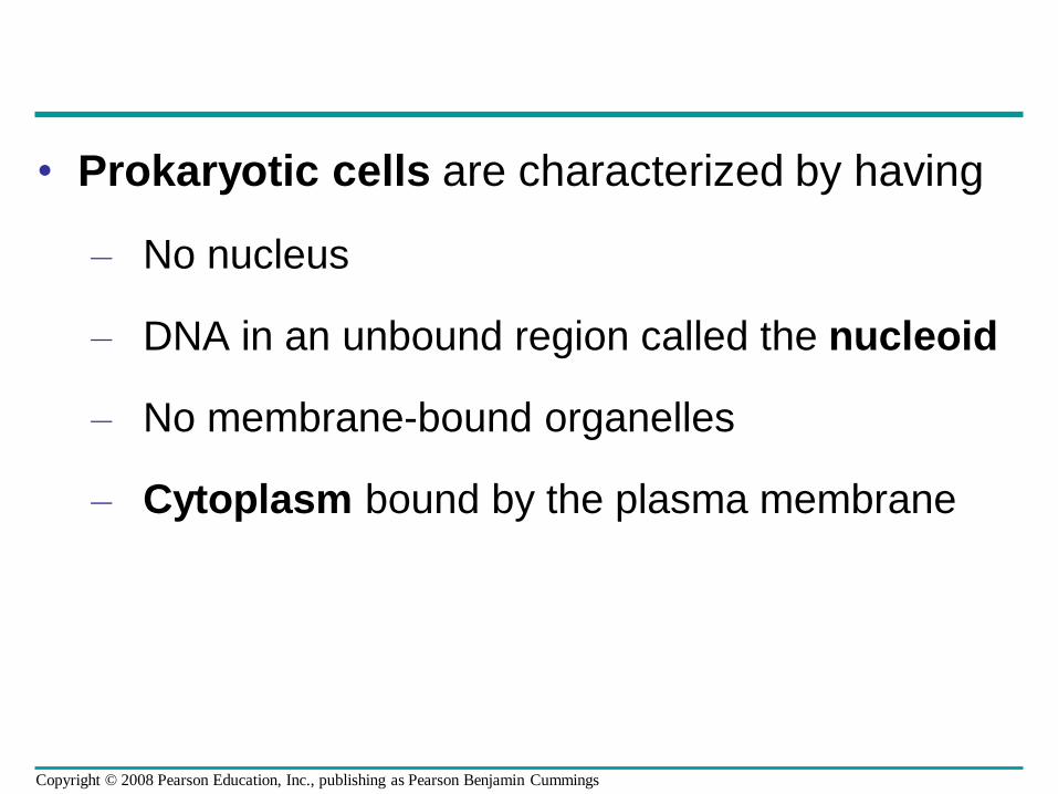
• Prokaryotic cells are characterized by having
– No nucleus
– DNA in an unbound region called the nucleoid
– No membrane-bound organelles
– Cytoplasm bound by the plasma membrane
Copyright © 2008 Pearson Education, Inc., publishing as Pearson Benjamin Cummings
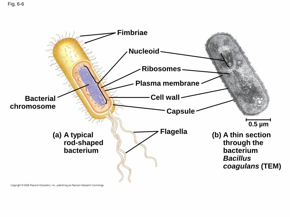
Fig. 6-6
Fimbriae
Nucleoid
Ribosomes
Plasma membrane
Cell wall
Capsule
Flagella
Bacterialchromosome
(a) A typical rod-shaped bacterium
(b) A thin section through the bacterium Bacillus coagulans (TEM)
0.5 µm
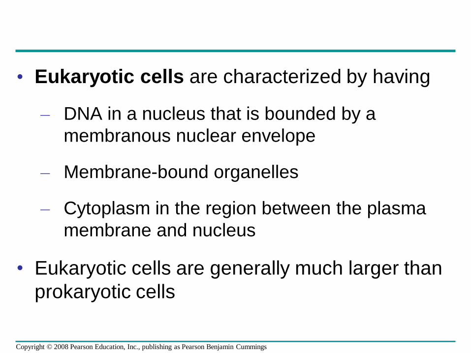
• Eukaryotic cells are characterized by having
– DNA in a nucleus that is bounded by a
membranous nuclear envelope
– Membrane-bound organelles
– Cytoplasm in the region between the plasma
membrane and nucleus
• Eukaryotic cells are generally much larger than
prokaryotic cells
Copyright © 2008 Pearson Education, Inc., publishing as Pearson Benjamin Cummings
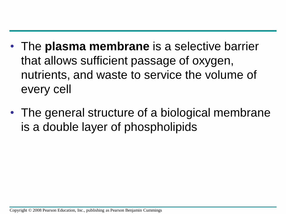
• The plasma membrane is a selective barrier
that allows sufficient passage of oxygen,
nutrients, and waste to service the volume of
every cell
• The general structure of a biological membrane
is a double layer of phospholipids
Copyright © 2008 Pearson Education, Inc., publishing as Pearson Benjamin Cummings
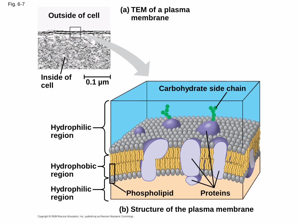
Fig. 6-7
TEM of a plasmamembrane
(a)
(b) Structure of the plasma membrane
Outside of cell
Inside ofcell 0.1 µm
Hydrophilicregion
Hydrophobicregion
Hydrophilicregion
Phospholipid Proteins
Carbohydrate side chain
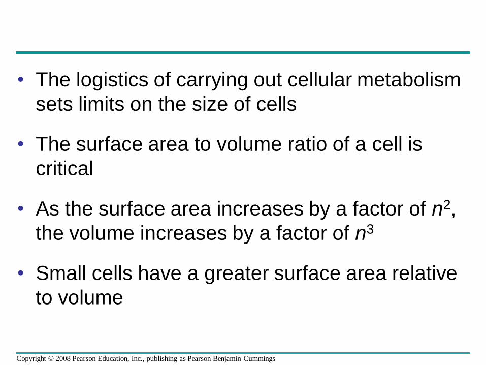
• The logistics of carrying out cellular metabolism
sets limits on the size of cells
• The surface area to volume ratio of a cell is
critical
• As the surface area increases by a factor of n2,
the volume increases by a factor of n3
• Small cells have a greater surface area relative
to volume
Copyright © 2008 Pearson Education, Inc., publishing as Pearson Benjamin Cummings
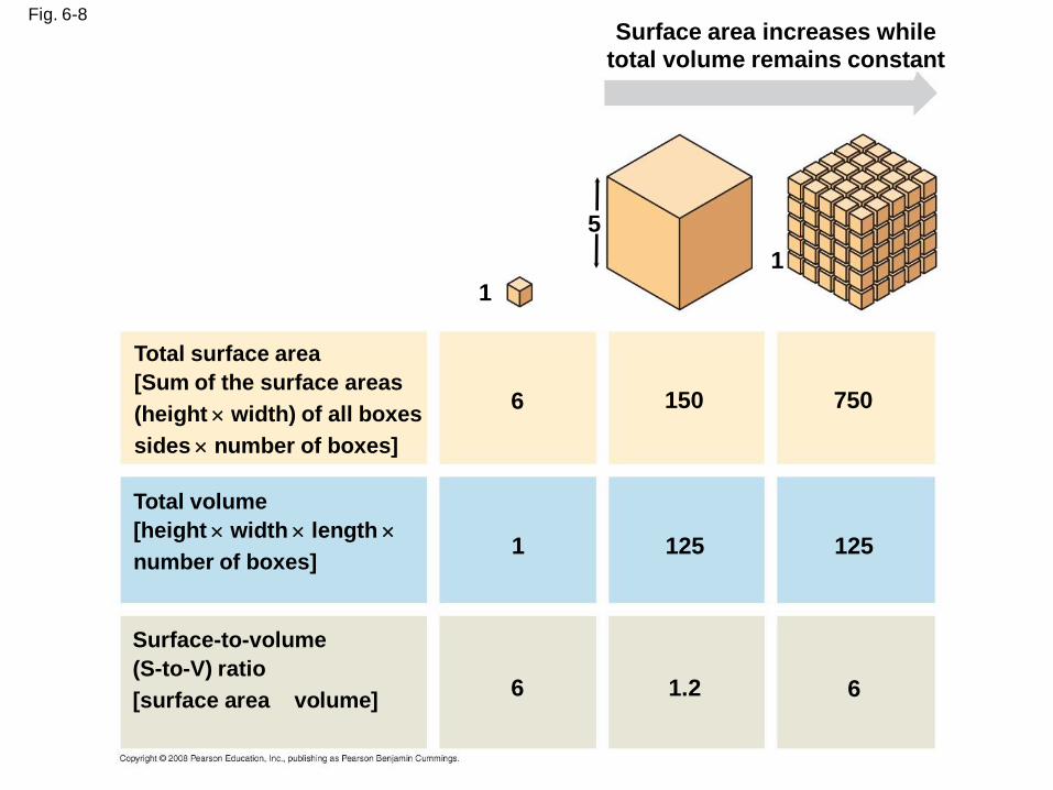
Fig. 6-8Surface area increases while
total volume remains constant
5
1
1
6 150 750
125 1251
6 61.2
Total surface area
[Sum of the surface areas
(height width) of all boxes
sides number of boxes]
Total volume
[height width length
number of boxes]
Surface-to-volume
(S-to-V) ratio
[surface area volume]
A Panoramic View of the Eukaryotic Cell
• A eukaryotic cell has internal membranes that
partition the cell into organelles
• Plant and animal cells have most of the same
organelles
BioFlix: Tour Of An Animal Cell
BioFlix: Tour Of A Plant Cell
Copyright © 2008 Pearson Education, Inc., publishing as Pearson Benjamin Cummings
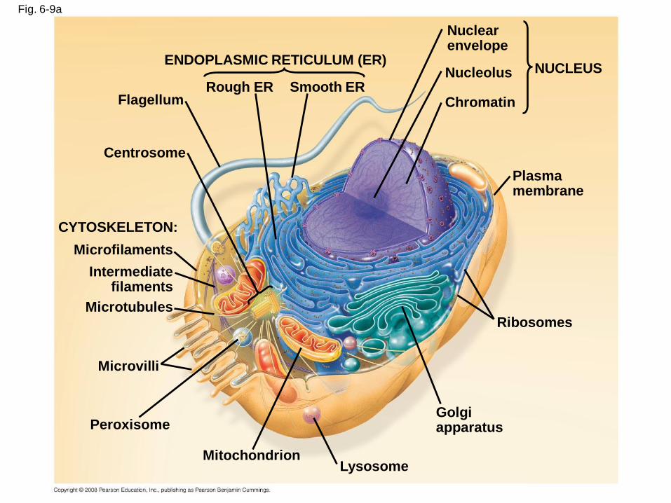
Fig. 6-9a
ENDOPLASMIC RETICULUM (ER)
Smooth ERRough ERFlagellum
Centrosome
CYTOSKELETON:
Microfilaments
Intermediatefilaments
Microtubules
Microvilli
Peroxisome
MitochondrionLysosome
Golgiapparatus
Ribosomes
Plasma membrane
Nuclearenvelope
Nucleolus
Chromatin
NUCLEUS
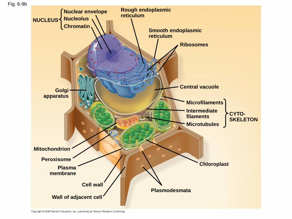
Fig. 6-9b
NUCLEUS
Nuclear envelope
Nucleolus
Chromatin
Rough endoplasmic reticulum
Smooth endoplasmic reticulum
Ribosomes
Central vacuole
Microfilaments
Intermediate filaments
Microtubules
CYTO-SKELETON
Chloroplast
Plasmodesmata
Wall of adjacent cell
Cell wall
Plasma membrane
Peroxisome
Mitochondrion
Golgiapparatus
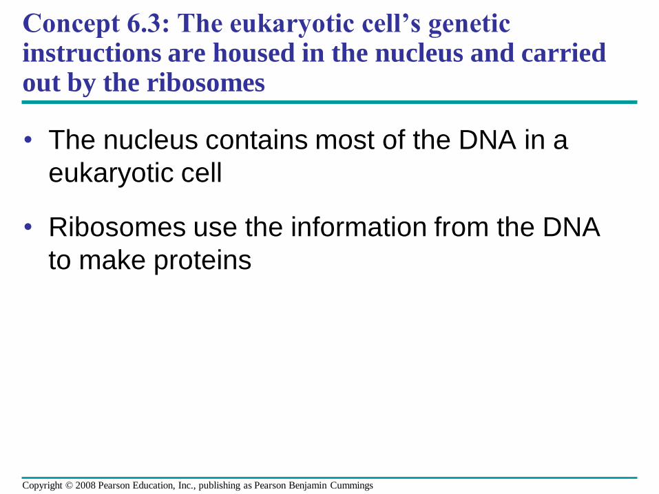
Concept 6.3: The eukaryotic cell’s genetic instructions are housed in the nucleus and carried out by the ribosomes
• The nucleus contains most of the DNA in a
eukaryotic cell
• Ribosomes use the information from the DNA
to make proteins
Copyright © 2008 Pearson Education, Inc., publishing as Pearson Benjamin Cummings
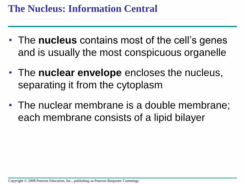
The Nucleus: Information Central
• The nucleus contains most of the cell’s genes
and is usually the most conspicuous organelle
• The nuclear envelope encloses the nucleus,
separating it from the cytoplasm
• The nuclear membrane is a double membrane;
each membrane consists of a lipid bilayer
Copyright © 2008 Pearson Education, Inc., publishing as Pearson Benjamin Cummings
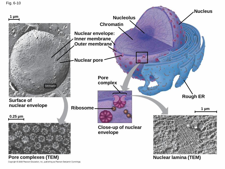
Fig. 6-10
Nucleolus
Nucleus
Rough ER
Nuclear lamina (TEM)
Close-up of nuclear envelope
1 µm
1 µm
0.25 µm
Ribosome
Pore complex
Nuclear pore
Outer membraneInner membraneNuclear envelope:
Chromatin
Surface ofnuclear envelope
Pore complexes (TEM)
• Pores regulate the entry and exit of molecules
from the nucleus
• The shape of the nucleus is maintained by the
nuclear lamina, which is composed of protein
Copyright © 2008 Pearson Education, Inc., publishing as Pearson Benjamin Cummings
• In the nucleus, DNA and proteins form genetic
material called chromatin
• Chromatin condenses to form discrete
chromosomes
• The nucleolus is located within the nucleus
and is the site of ribosomal RNA (rRNA)
synthesis
Copyright © 2008 Pearson Education, Inc., publishing as Pearson Benjamin Cummings
Ribosomes: Protein Factories
• Ribosomes are particles made of ribosomal
RNA and protein
• Ribosomes carry out protein synthesis in two
locations:
– In the cytosol (free ribosomes)
– On the outside of the endoplasmic reticulum or
the nuclear envelope (bound ribosomes)
Copyright © 2008 Pearson Education, Inc., publishing as Pearson Benjamin Cummings
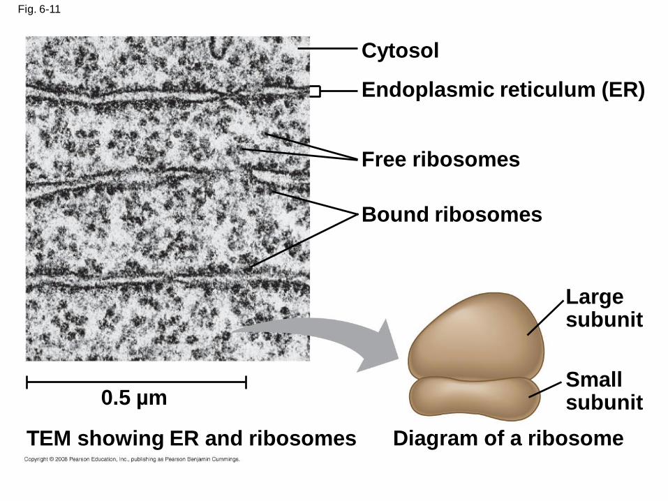
Fig. 6-11
Cytosol
Endoplasmic reticulum (ER)
Free ribosomes
Bound ribosomes
Large subunit
Small subunit
Diagram of a ribosomeTEM showing ER and ribosomes
0.5 µm
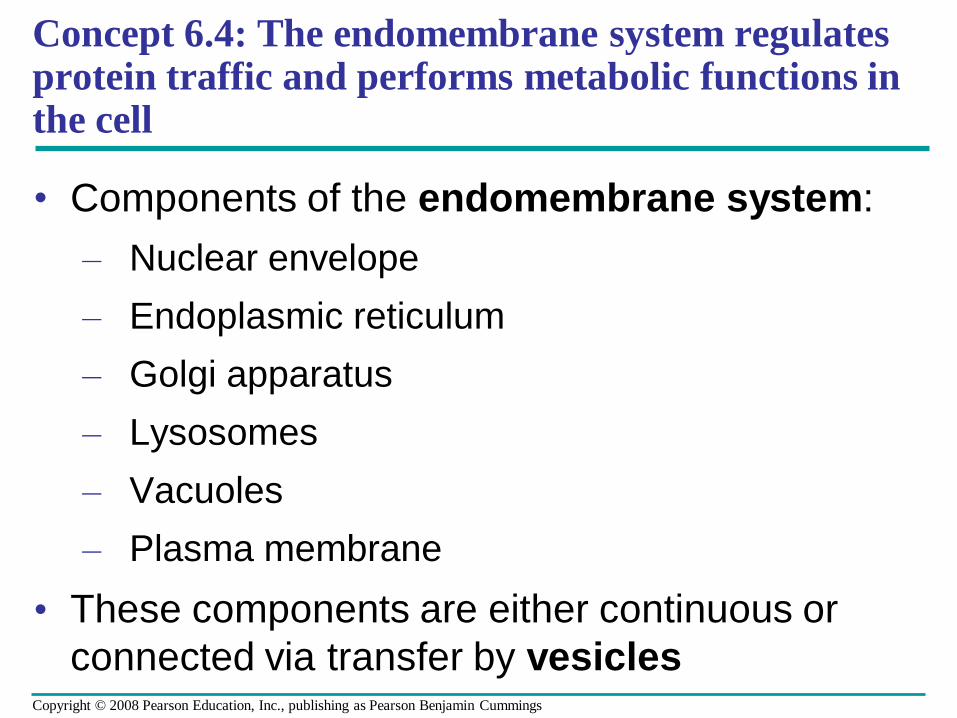
Concept 6.4: The endomembrane system regulates protein traffic and performs metabolic functions in the cell
• Components of the endomembrane system:
– Nuclear envelope
– Endoplasmic reticulum
– Golgi apparatus
– Lysosomes
– Vacuoles
– Plasma membrane
• These components are either continuous or
connected via transfer by vesiclesCopyright © 2008 Pearson Education, Inc., publishing as Pearson Benjamin Cummings
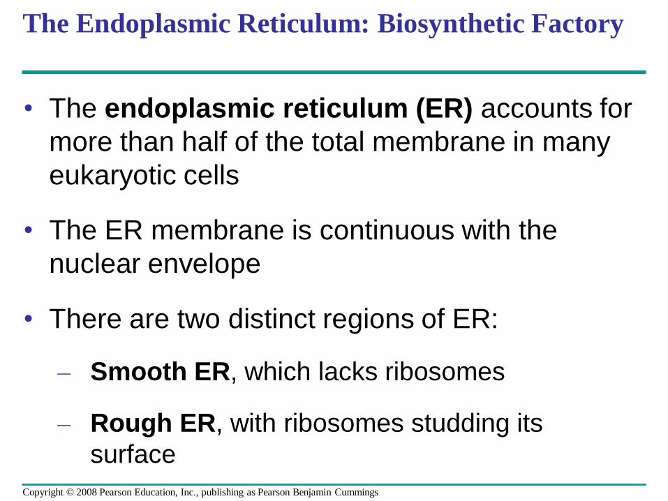
The Endoplasmic Reticulum: Biosynthetic Factory
• The endoplasmic reticulum (ER) accounts for
more than half of the total membrane in many
eukaryotic cells
• The ER membrane is continuous with the
nuclear envelope
• There are two distinct regions of ER:
– Smooth ER, which lacks ribosomes
– Rough ER, with ribosomes studding its
surface
Copyright © 2008 Pearson Education, Inc., publishing as Pearson Benjamin Cummings
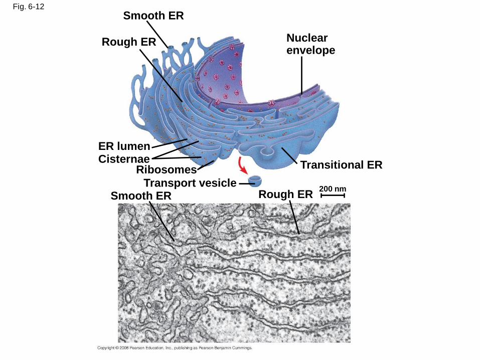
Fig. 6-12
Smooth ER
Rough ER Nuclear envelope
Transitional ER
Rough ERSmooth ERTransport vesicle
RibosomesCisternaeER lumen
200 nm
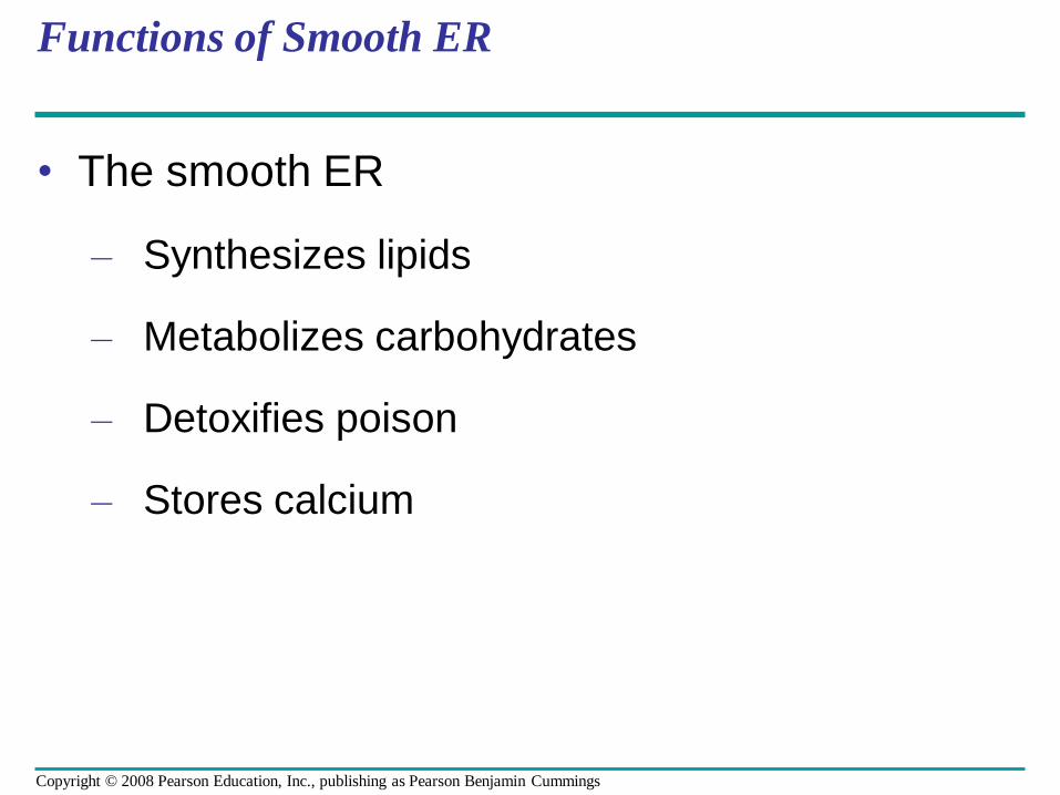
Functions of Smooth ER
• The smooth ER
– Synthesizes lipids
– Metabolizes carbohydrates
– Detoxifies poison
– Stores calcium
Copyright © 2008 Pearson Education, Inc., publishing as Pearson Benjamin Cummings
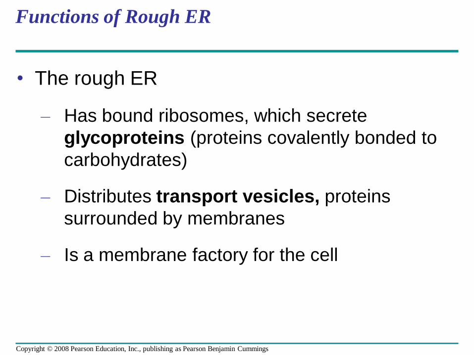
Functions of Rough ER
• The rough ER
– Has bound ribosomes, which secrete
glycoproteins (proteins covalently bonded to
carbohydrates)
– Distributes transport vesicles, proteins
surrounded by membranes
– Is a membrane factory for the cell
Copyright © 2008 Pearson Education, Inc., publishing as Pearson Benjamin Cummings
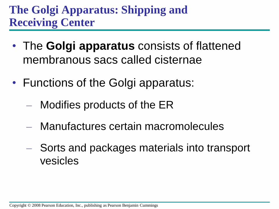
• The Golgi apparatus consists of flattened
membranous sacs called cisternae
• Functions of the Golgi apparatus:
– Modifies products of the ER
– Manufactures certain macromolecules
– Sorts and packages materials into transport
vesicles
The Golgi Apparatus: Shipping and Receiving Center
Copyright © 2008 Pearson Education, Inc., publishing as Pearson Benjamin Cummings
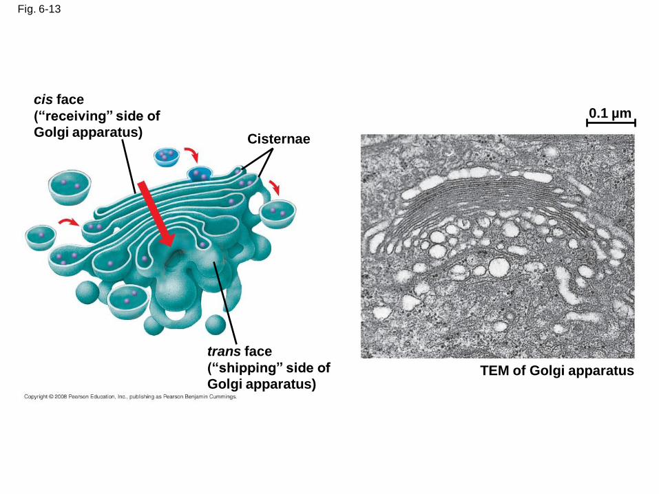
Fig. 6-13
cis face
(“receiving” side of
Golgi apparatus)Cisternae
trans face
(“shipping” side of
Golgi apparatus)TEM of Golgi apparatus
0.1 µm
Lysosomes: Digestive Compartments
• A lysosome is a membranous sac of hydrolytic
enzymes that can digest macromolecules
• Lysosomal enzymes can hydrolyze proteins, fats, polysaccharides, and nucleic acids
Animation: Lysosome Formation
Copyright © 2008 Pearson Education, Inc., publishing as Pearson Benjamin Cummings
• Some types of cell can engulf another cell by phagocytosis; this forms a food vacuole
• A lysosome fuses with the food vacuole and digests the molecules
• Lysosomes also use enzymes to recycle the cell’s own organelles and macromolecules, a process called autophagy
Copyright © 2008 Pearson Education, Inc., publishing as Pearson Benjamin Cummings
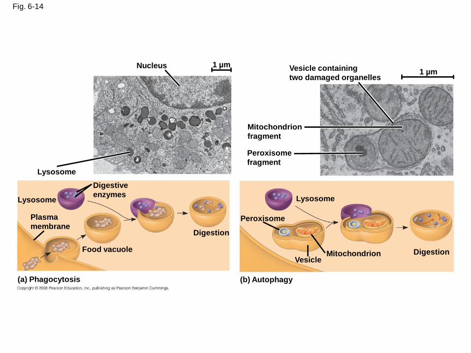
Fig. 6-14
Nucleus 1 µm
Lysosome
Digestive
enzymesLysosome
Plasma
membrane
Food vacuole
(a) Phagocytosis
Digestion
(b) Autophagy
Peroxisome
Vesicle
Lysosome
Mitochondrion
Peroxisome
fragment
Mitochondrion
fragment
Vesicle containing
two damaged organelles1 µm
Digestion
Fig. 6-14a
Nucleus 1 µm
Lysosome
Lysosome
Digestive enzymes
Plasma membrane
Food vacuole
Digestion
(a) Phagocytosis
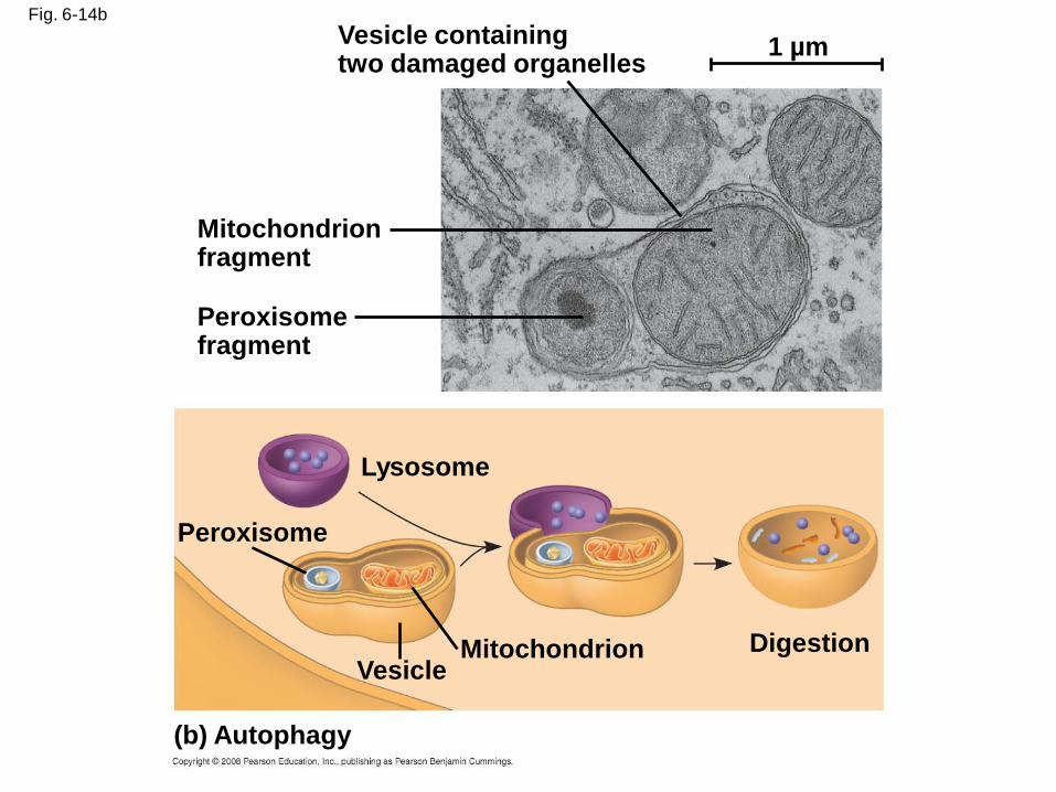
Fig. 6-14b
Vesicle containingtwo damaged organelles
Mitochondrion fragment
Peroxisome fragment
Peroxisome
Lysosome
DigestionMitochondrionVesicle
(b) Autophagy
1 µm
Vacuoles: Diverse Maintenance Compartments
• A plant cell or fungal cell may have one or several vacuoles
Copyright © 2008 Pearson Education, Inc., publishing as Pearson Benjamin Cummings
• Food vacuoles are formed by phagocytosis
• Contractile vacuoles, found in many
freshwater protists, pump excess water out of
cells
• Central vacuoles, found in many mature plant
cells, hold organic compounds and water
Video: Paramecium Vacuole
Copyright © 2008 Pearson Education, Inc., publishing as Pearson Benjamin Cummings
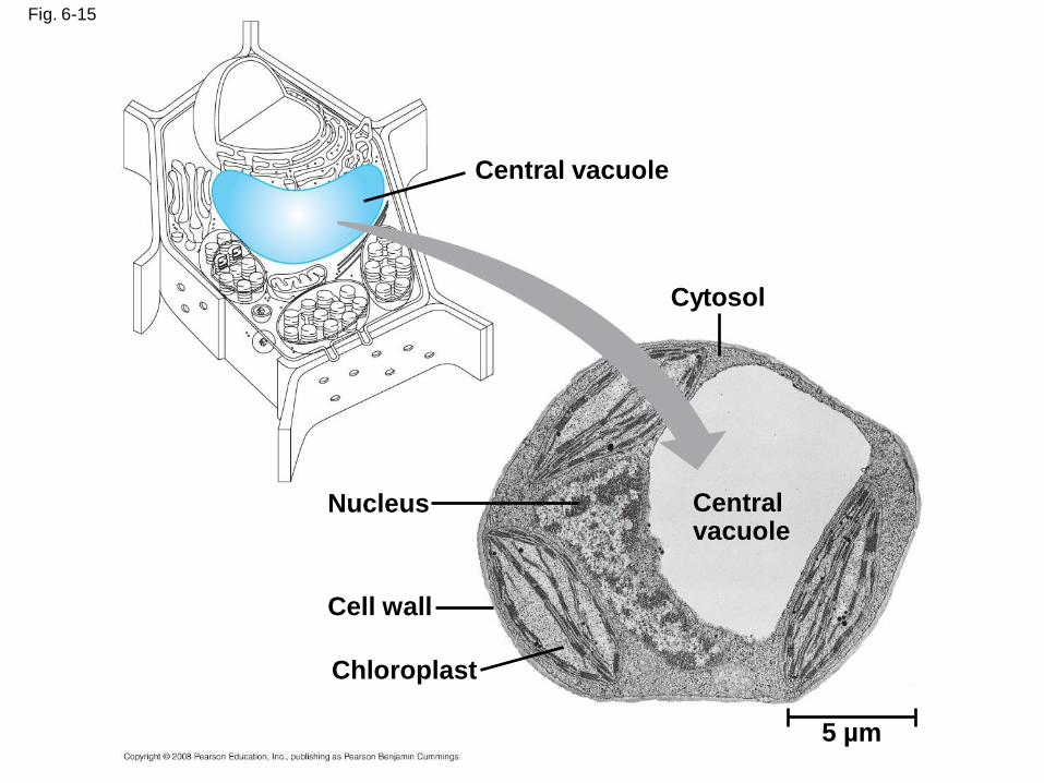
Fig. 6-15
Central vacuole
Cytosol
Central vacuole
Nucleus
Cell wall
Chloroplast
5 µm
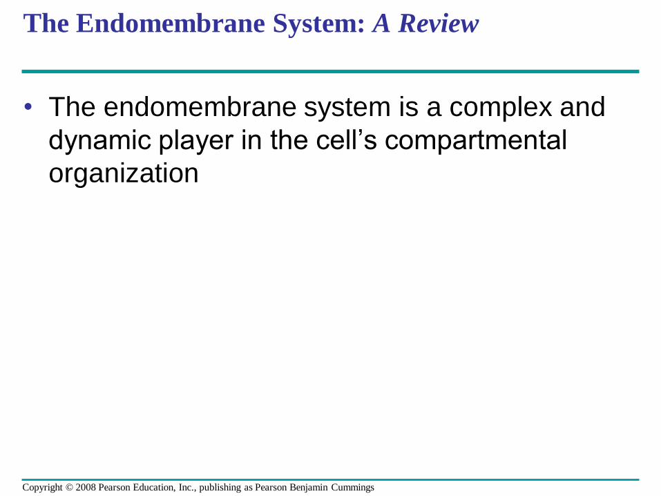
The Endomembrane System: A Review
• The endomembrane system is a complex and
dynamic player in the cell’s compartmental
organization
Copyright © 2008 Pearson Education, Inc., publishing as Pearson Benjamin Cummings
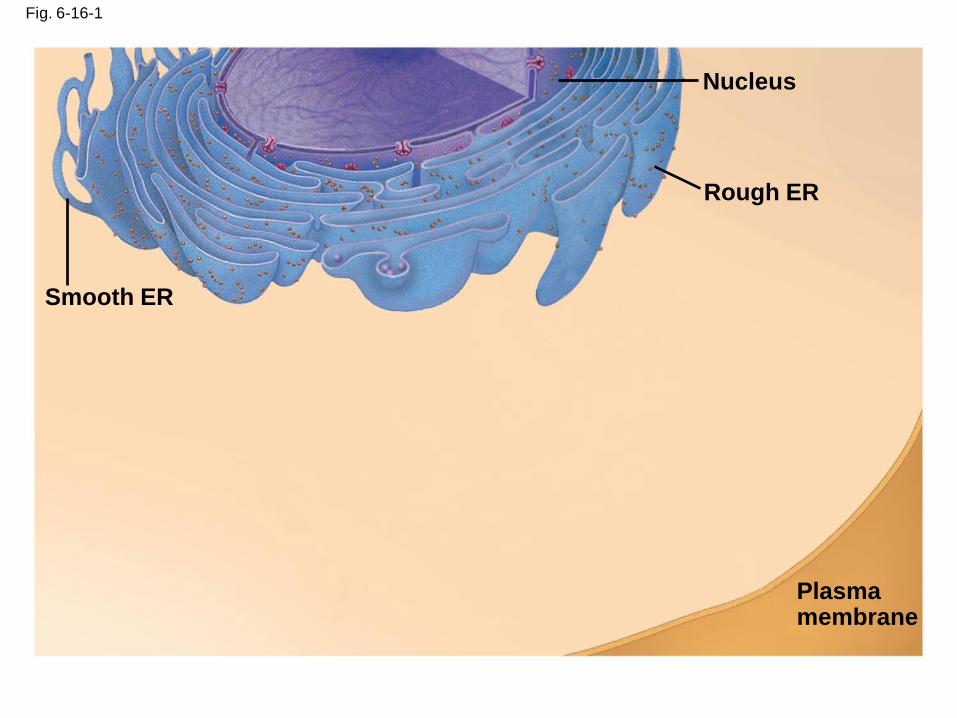
Fig. 6-16-1
Smooth ER
Nucleus
Rough ER
Plasma membrane
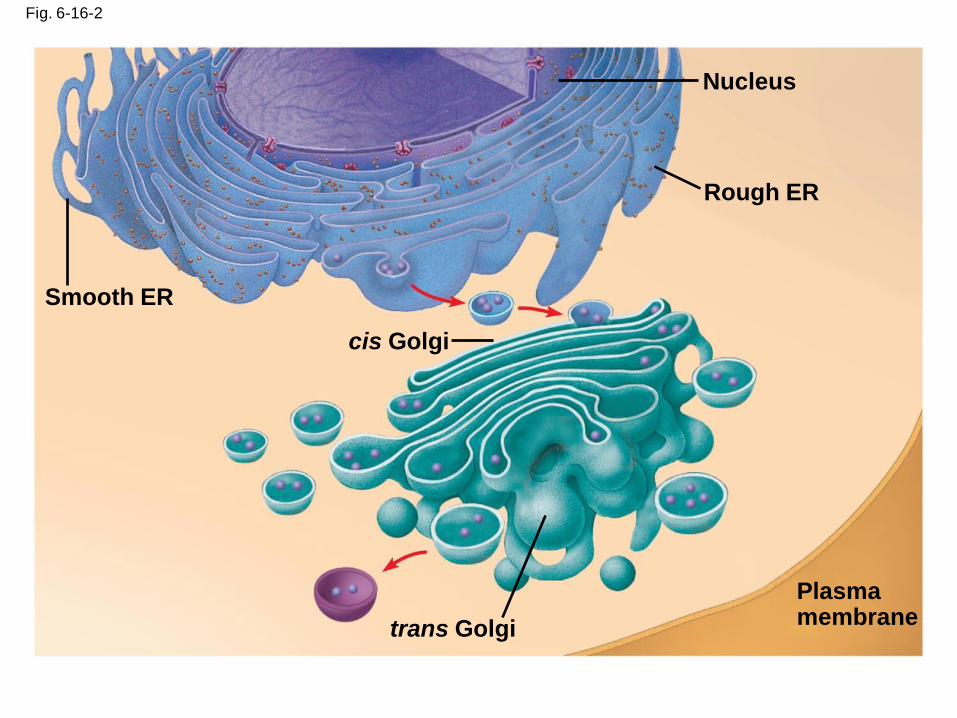
Fig. 6-16-2
Smooth ER
Nucleus
Rough ER
Plasma membrane
cis Golgi
trans Golgi
Fig. 6-16-3
Smooth ER
Nucleus
Rough ER
Plasma membrane
cis Golgi
trans Golgi
Concept 6.5: Mitochondria and chloroplasts change energy from one form to another
• Mitochondria are the sites of cellular
respiration, a metabolic process that generates
ATP
• Chloroplasts, found in plants and algae, are
the sites of photosynthesis
• Peroxisomes are oxidative organelles
Copyright © 2008 Pearson Education, Inc., publishing as Pearson Benjamin Cummings
• Mitochondria and chloroplasts
– Are not part of the endomembrane system
– Have a double membrane
– Have proteins made by free ribosomes
– Contain their own DNA
Copyright © 2008 Pearson Education, Inc., publishing as Pearson Benjamin Cummings
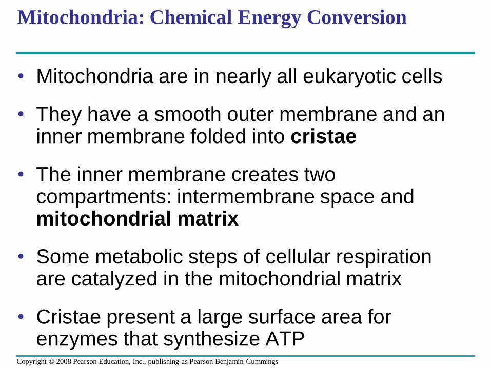
Mitochondria: Chemical Energy Conversion
• Mitochondria are in nearly all eukaryotic cells
• They have a smooth outer membrane and an inner membrane folded into cristae
• The inner membrane creates two compartments: intermembrane space and mitochondrial matrix
• Some metabolic steps of cellular respiration are catalyzed in the mitochondrial matrix
• Cristae present a large surface area for enzymes that synthesize ATP
Copyright © 2008 Pearson Education, Inc., publishing as Pearson Benjamin Cummings
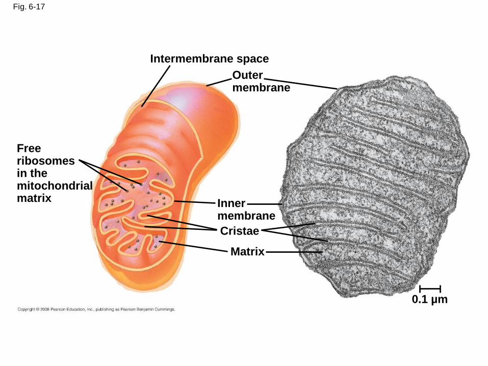
Fig. 6-17
Free ribosomesin the mitochondrial matrix
Intermembrane space
Outer membrane
Inner membrane
Cristae
Matrix
0.1 µm
Chloroplasts: Capture of Light Energy
• The chloroplast is a member of a family of
organelles called plastids
• Chloroplasts contain the green pigment
chlorophyll, as well as enzymes and other
molecules that function in photosynthesis
• Chloroplasts are found in leaves and other
green organs of plants and in algae
Copyright © 2008 Pearson Education, Inc., publishing as Pearson Benjamin Cummings
• Chloroplast structure includes:
– Thylakoids, membranous sacs, stacked to form a granum
– Stroma, the internal fluid
Copyright © 2008 Pearson Education, Inc., publishing as Pearson Benjamin Cummings
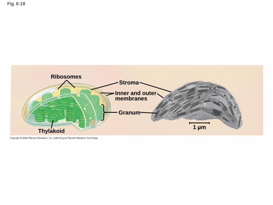
Fig. 6-18
Ribosomes
Thylakoid
Stroma
Granum
Inner and outer membranes
1 µm
Peroxisomes: Oxidation
• Peroxisomes are specialized metabolic
compartments bounded by a single membrane
• Peroxisomes produce hydrogen peroxide and
convert it to water
• Oxygen is used to break down different types
of molecules
Copyright © 2008 Pearson Education, Inc., publishing as Pearson Benjamin Cummings
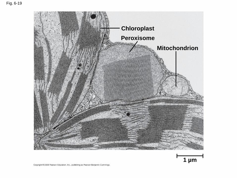
Fig. 6-19
1 µm
Chloroplast
Peroxisome
Mitochondrion
Concept 6.6: The cytoskeleton is a network of fibers that organizes structures and activities in the cell
• The cytoskeleton is a network of fibers
extending throughout the cytoplasm
• It organizes the cell’s structures and activities,
anchoring many organelles
• It is composed of three types of molecular
structures:
– Microtubules
– Microfilaments
– Intermediate filaments
Copyright © 2008 Pearson Education, Inc., publishing as Pearson Benjamin Cummings
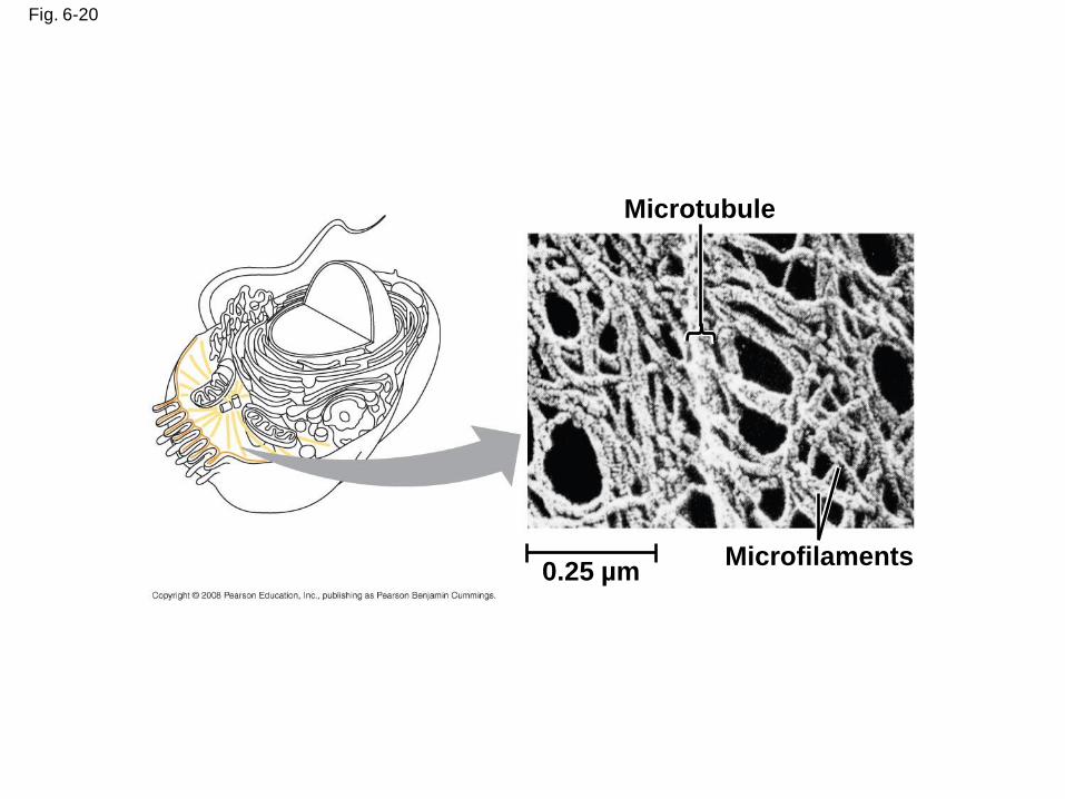
Fig. 6-20
Microtubule
Microfilaments0.25 µm
Roles of the Cytoskeleton: Support, Motility, and Regulation
• The cytoskeleton helps to support the cell and
maintain its shape
• It interacts with motor proteins to produce
motility
• Inside the cell, vesicles can travel along
“monorails” provided by the cytoskeleton
• Recent evidence suggests that the
cytoskeleton may help regulate biochemical
activities
Copyright © 2008 Pearson Education, Inc., publishing as Pearson Benjamin Cummings
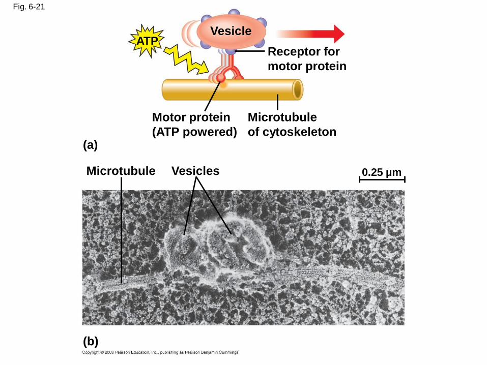
Fig. 6-21
VesicleATP
Receptor for
motor protein
Microtubule
of cytoskeleton
Motor protein
(ATP powered)(a)
Microtubule Vesicles
(b)
0.25 µm
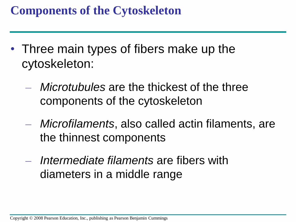
Components of the Cytoskeleton
• Three main types of fibers make up the
cytoskeleton:
– Microtubules are the thickest of the three
components of the cytoskeleton
– Microfilaments, also called actin filaments, are
the thinnest components
– Intermediate filaments are fibers with
diameters in a middle range
Copyright © 2008 Pearson Education, Inc., publishing as Pearson Benjamin Cummings
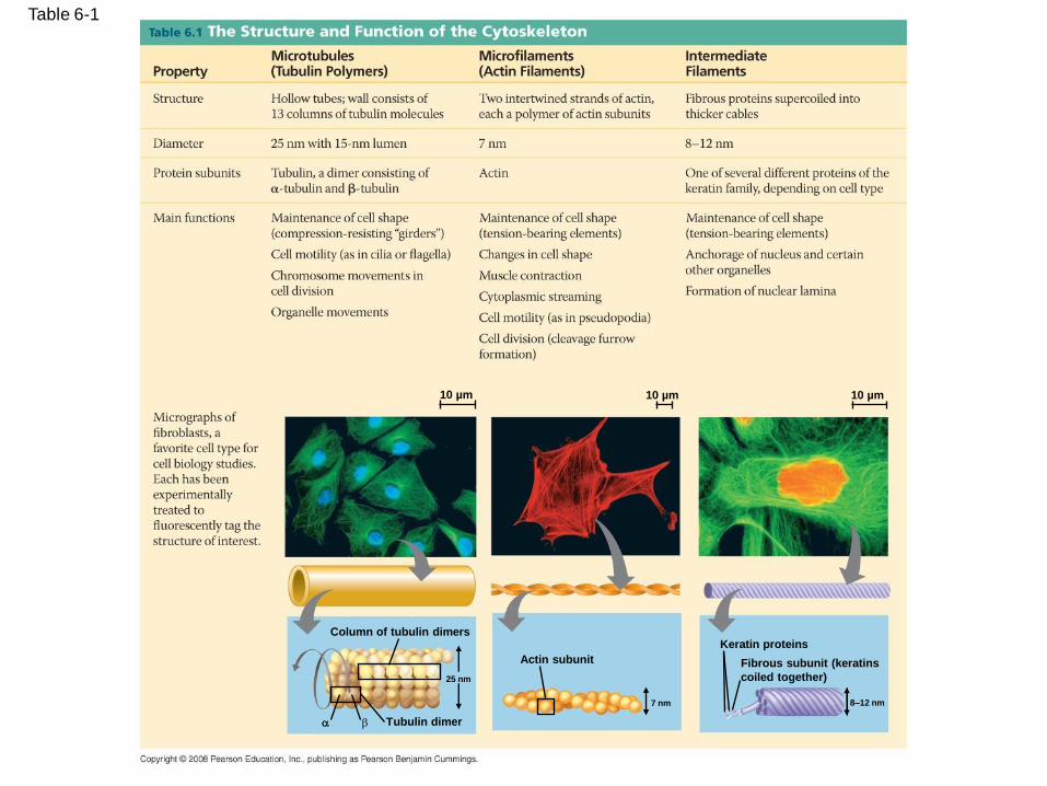
Table 6-1
10 µm 10 µm 10 µm
Column of tubulin dimers
Tubulin dimer
Actin subunit
25 nm
7 nm
Keratin proteins
Fibrous subunit (keratins
coiled together)
8–12 nm
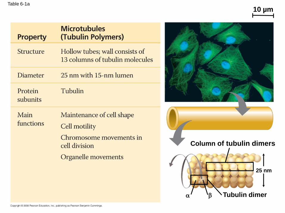
Table 6-1a
10 µm
Column of tubulin dimers
Tubulin dimer
25 nm

Table 6-1b
Actin subunit
10 µm
7 nm
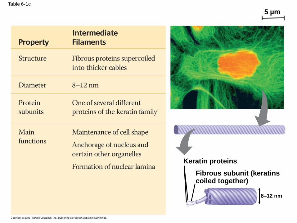
Table 6-1c
5 µm
Keratin proteins
Fibrous subunit (keratinscoiled together)
8–12 nm
Microtubules
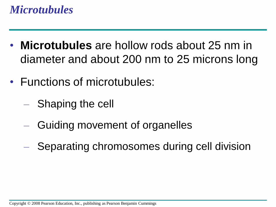
• Microtubules are hollow rods about 25 nm in
diameter and about 200 nm to 25 microns long
• Functions of microtubules:
– Shaping the cell
– Guiding movement of organelles
– Separating chromosomes during cell division
Copyright © 2008 Pearson Education, Inc., publishing as Pearson Benjamin Cummings
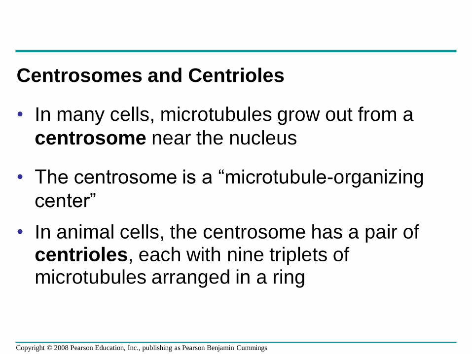
Centrosomes and Centrioles
• In many cells, microtubules grow out from a
centrosome near the nucleus
• The centrosome is a “microtubule-organizing
center”
• In animal cells, the centrosome has a pair of centrioles, each with nine triplets of microtubules arranged in a ring
Copyright © 2008 Pearson Education, Inc., publishing as Pearson Benjamin Cummings
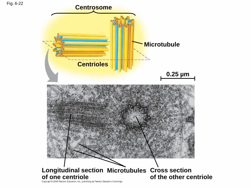
Fig. 6-22Centrosome
Microtubule
Centrioles
0.25 µm
Longitudinal section of one centriole
Microtubules Cross sectionof the other centriole
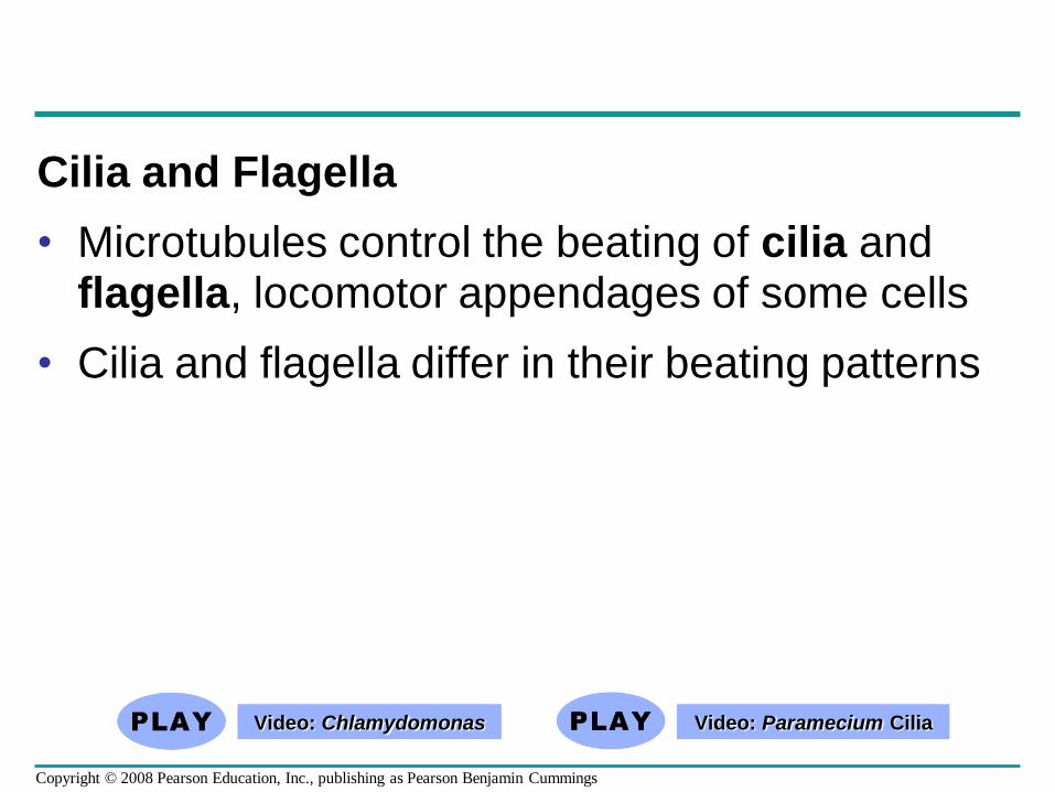
Cilia and Flagella
• Microtubules control the beating of cilia and flagella, locomotor appendages of some cells
• Cilia and flagella differ in their beating patterns
Video: Chlamydomonas Video: Paramecium Cilia
Copyright © 2008 Pearson Education, Inc., publishing as Pearson Benjamin Cummings
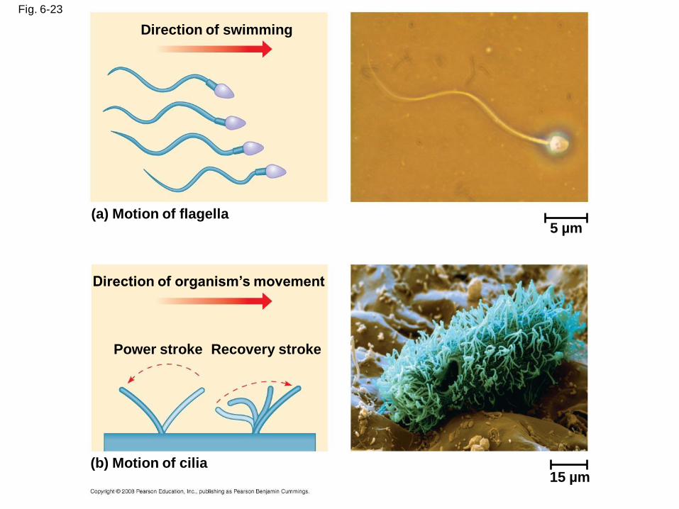
Fig. 6-23
5 µm
Direction of swimming
(a) Motion of flagella
Direction of organism’s movement
Power stroke Recovery stroke
(b) Motion of cilia15 µm
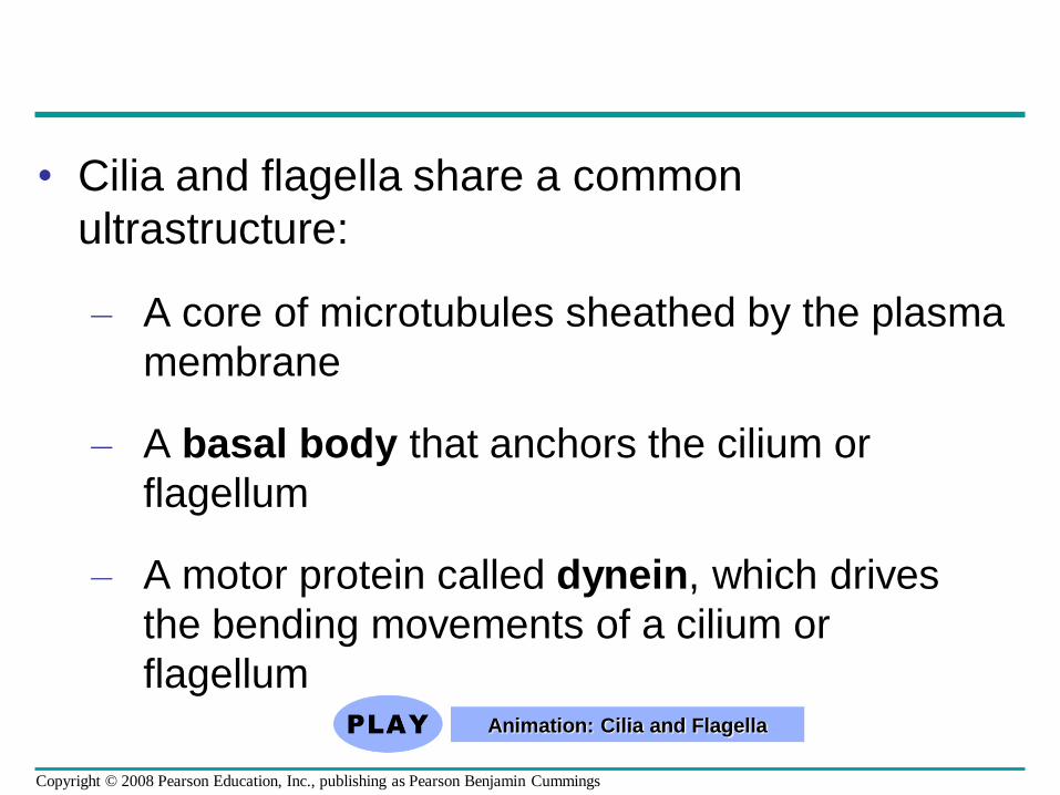
• Cilia and flagella share a common
ultrastructure:
– A core of microtubules sheathed by the plasma
membrane
– A basal body that anchors the cilium or
flagellum
– A motor protein called dynein, which drives
the bending movements of a cilium or
flagellumAnimation: Cilia and Flagella
Copyright © 2008 Pearson Education, Inc., publishing as Pearson Benjamin Cummings
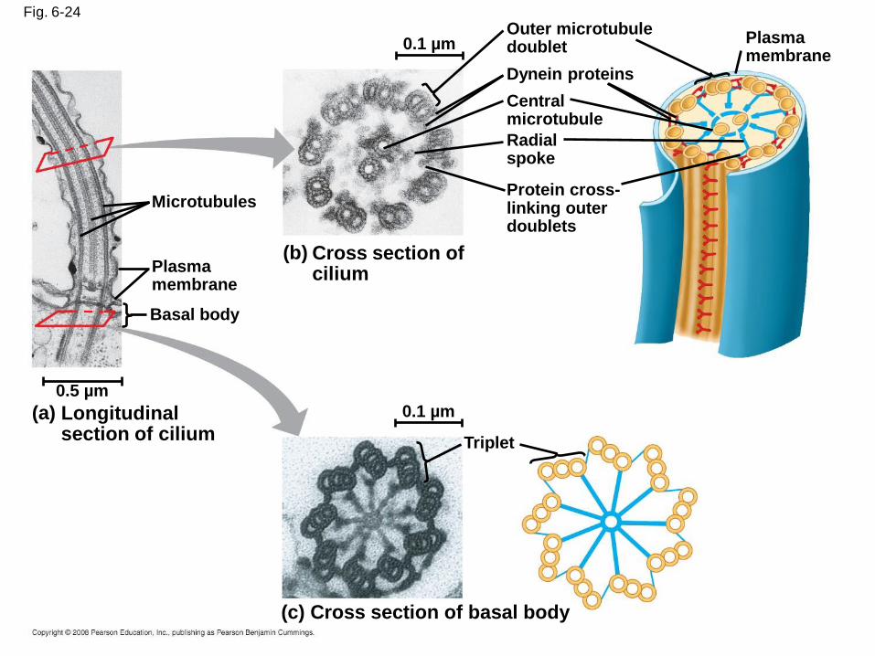
Fig. 6-24
0.1 µm
Triplet
(c) Cross section of basal body
(a) Longitudinal section of cilium
0.5 µm
Plasma membrane
Basal body
Microtubules
(b) Cross section of cilium
Plasma membrane
Outer microtubule doublet
Dynein proteins
Central microtubule
Radial spoke
Protein cross-linking outer doublets
0.1 µm
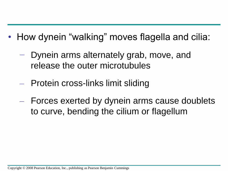
• How dynein “walking” moves flagella and cilia:
− Dynein arms alternately grab, move, and
release the outer microtubules
– Protein cross-links limit sliding
– Forces exerted by dynein arms cause doublets
to curve, bending the cilium or flagellum
Copyright © 2008 Pearson Education, Inc., publishing as Pearson Benjamin Cummings
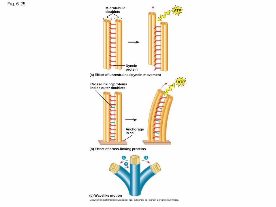
Fig. 6-25Microtubuledoublets
Dyneinprotein
ATP
ATP
(a) Effect of unrestrained dynein movement
Cross-linking proteinsinside outer doublets
Anchoragein cell
(b) Effect of cross-linking proteins
1 3
2
(c) Wavelike motion
Fig. 6-25a
Microtubule doublets
Dynein protein
(a) Effect of unrestrained dynein movement
ATP
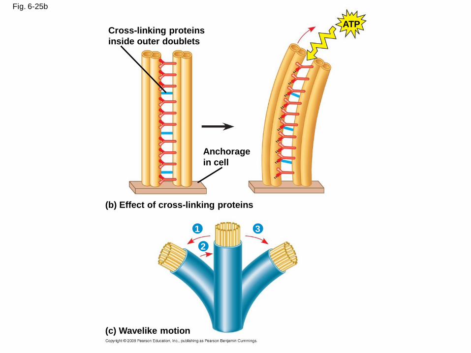
Fig. 6-25b
Cross-linking proteins
inside outer doublets
Anchorage
in cell
ATP
(b) Effect of cross-linking proteins
(c) Wavelike motion
1 3
2
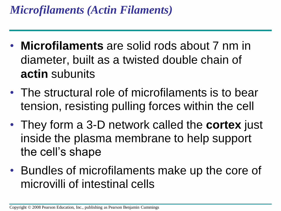
Microfilaments (Actin Filaments)
• Microfilaments are solid rods about 7 nm in
diameter, built as a twisted double chain of
actin subunits
• The structural role of microfilaments is to bear tension, resisting pulling forces within the cell
• They form a 3-D network called the cortex just inside the plasma membrane to help support the cell’s shape
• Bundles of microfilaments make up the core of microvilli of intestinal cells
Copyright © 2008 Pearson Education, Inc., publishing as Pearson Benjamin Cummings
Fig. 6-26
Microvillus
Plasma membrane
Microfilaments (actin
filaments)
Intermediate filaments
0.25 µm
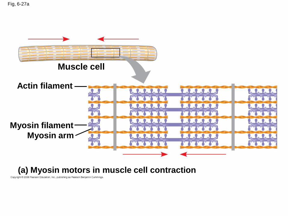
• Microfilaments that function in cellular motility
contain the protein myosin in addition to actin
• In muscle cells, thousands of actin filaments
are arranged parallel to one another
• Thicker filaments composed of myosin
interdigitate with the thinner actin fibers
Copyright © 2008 Pearson Education, Inc., publishing as Pearson Benjamin Cummings
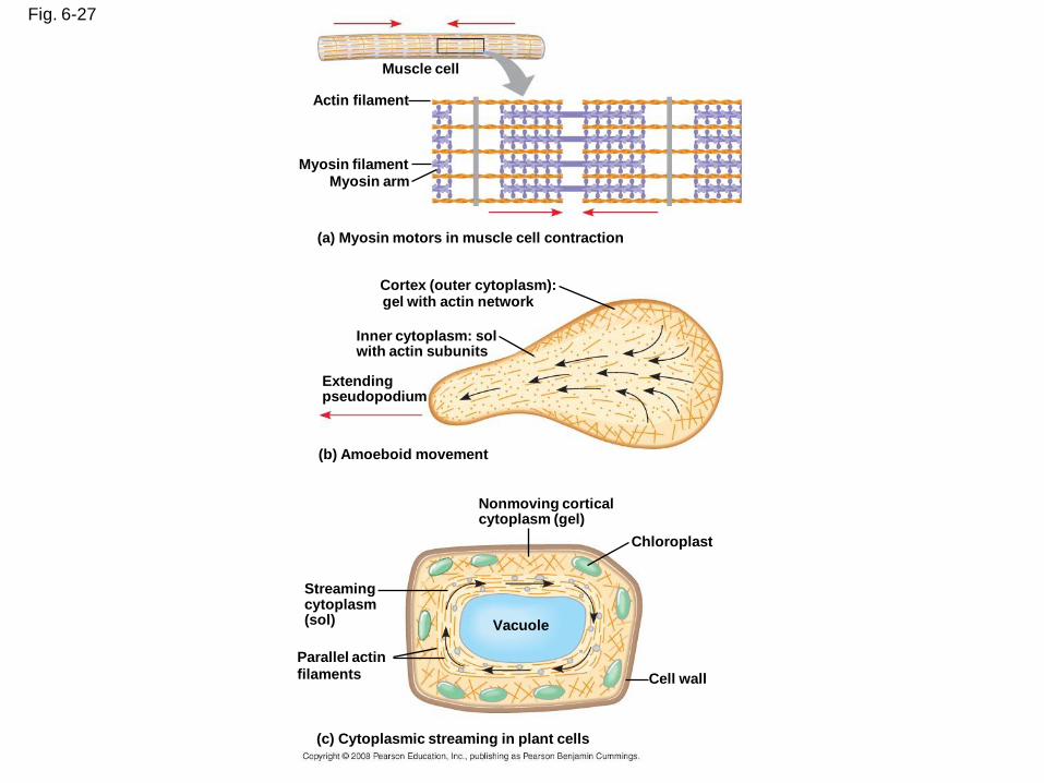
Fig. 6-27
Muscle cell
Actin filament
Myosin filamentMyosin arm
(a) Myosin motors in muscle cell contraction
Cortex (outer cytoplasm):gel with actin network
Inner cytoplasm: solwith actin subunits
Extendingpseudopodium
(b) Amoeboid movement
Nonmoving corticalcytoplasm (gel)
Chloroplast
Streamingcytoplasm(sol) Vacuole
Cell wall
Parallel actinfilaments
(c) Cytoplasmic streaming in plant cells
Fig, 6-27a
Muscle cell
Actin filament
Myosin filament
Myosin arm
(a) Myosin motors in muscle cell contraction
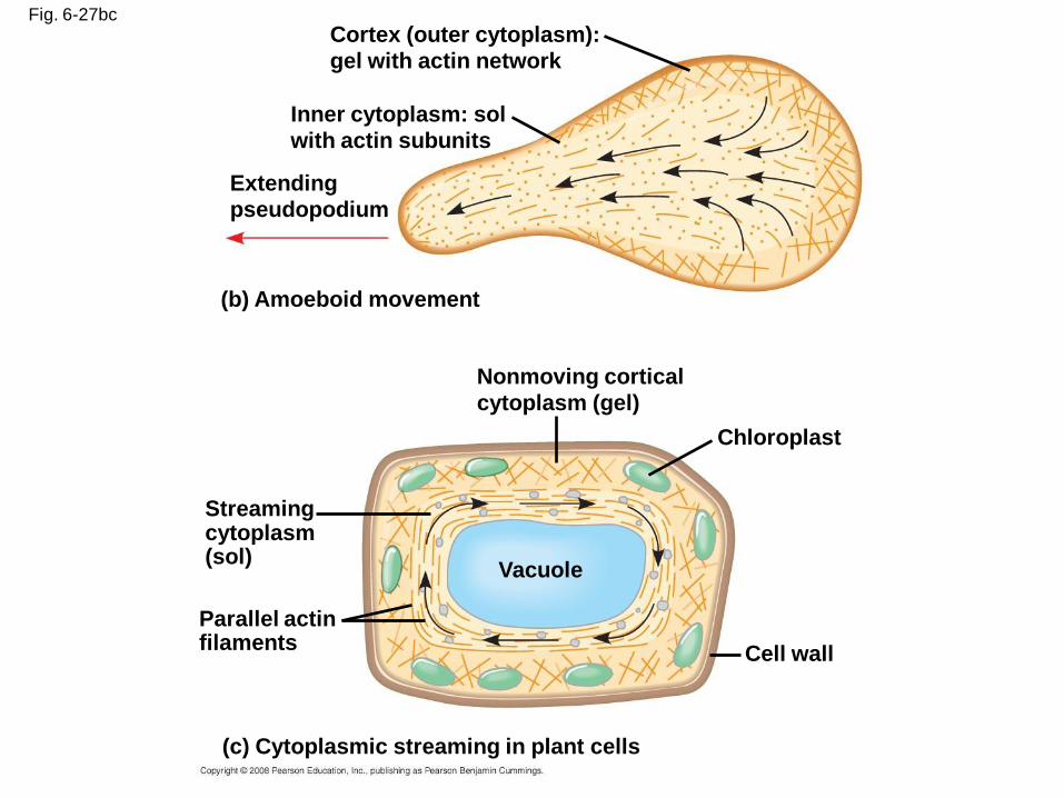
Fig. 6-27bc
Cortex (outer cytoplasm):
gel with actin network
Inner cytoplasm: sol
with actin subunits
Extending
pseudopodium
(b) Amoeboid movement
Nonmoving cortical
cytoplasm (gel)
Chloroplast
Cell wall
Streaming cytoplasm (sol)
Parallel actin filaments
(c) Cytoplasmic streaming in plant cells
Vacuole
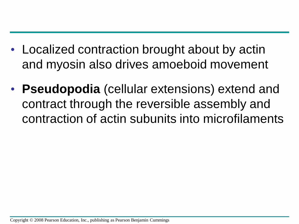
• Localized contraction brought about by actin
and myosin also drives amoeboid movement
• Pseudopodia (cellular extensions) extend and
contract through the reversible assembly and
contraction of actin subunits into microfilaments
Copyright © 2008 Pearson Education, Inc., publishing as Pearson Benjamin Cummings
• Cytoplasmic streaming is a circular flow of
cytoplasm within cells
• This streaming speeds distribution of materials
within the cell
• In plant cells, actin-myosin interactions and sol-
gel transformations drive cytoplasmic
streaming
Video: Cytoplasmic Streaming
Copyright © 2008 Pearson Education, Inc., publishing as Pearson Benjamin Cummings
Intermediate Filaments
• Intermediate filaments range in diameter from
8–12 nanometers, larger than microfilaments
but smaller than microtubules
• They support cell shape and fix organelles in
place
• Intermediate filaments are more permanent
cytoskeleton fixtures than the other two classes
Copyright © 2008 Pearson Education, Inc., publishing as Pearson Benjamin Cummings
Concept 6.7: Extracellular components and connections between cells help coordinate cellular activities
• Most cells synthesize and secrete materials
that are external to the plasma membrane
• These extracellular structures include:
– Cell walls of plants
– The extracellular matrix (ECM) of animal cells
– Intercellular junctions
Copyright © 2008 Pearson Education, Inc., publishing as Pearson Benjamin Cummings
Cell Walls of Plants
• The cell wall is an extracellular structure that
distinguishes plant cells from animal cells
• Prokaryotes, fungi, and some protists also have
cell walls
• The cell wall protects the plant cell, maintains its
shape, and prevents excessive uptake of water
• Plant cell walls are made of cellulose fibers
embedded in other polysaccharides and protein
Copyright © 2008 Pearson Education, Inc., publishing as Pearson Benjamin Cummings
• Plant cell walls may have multiple layers:
– Primary cell wall: relatively thin and flexible
– Middle lamella: thin layer between primary
walls of adjacent cells
– Secondary cell wall (in some cells): added
between the plasma membrane and the
primary cell wall
• Plasmodesmata are channels between
adjacent plant cells
Copyright © 2008 Pearson Education, Inc., publishing as Pearson Benjamin Cummings
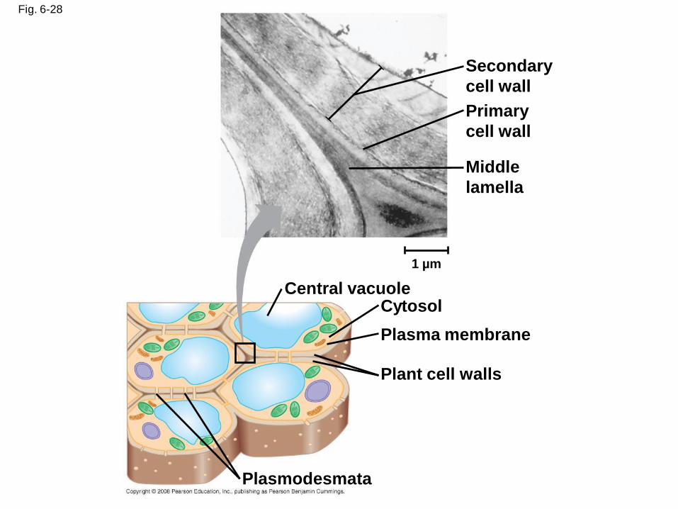
Fig. 6-28
Secondary
cell wall
Primary
cell wall
Middle
lamella
Central vacuoleCytosol
Plasma membrane
Plant cell walls
Plasmodesmata
1 µm
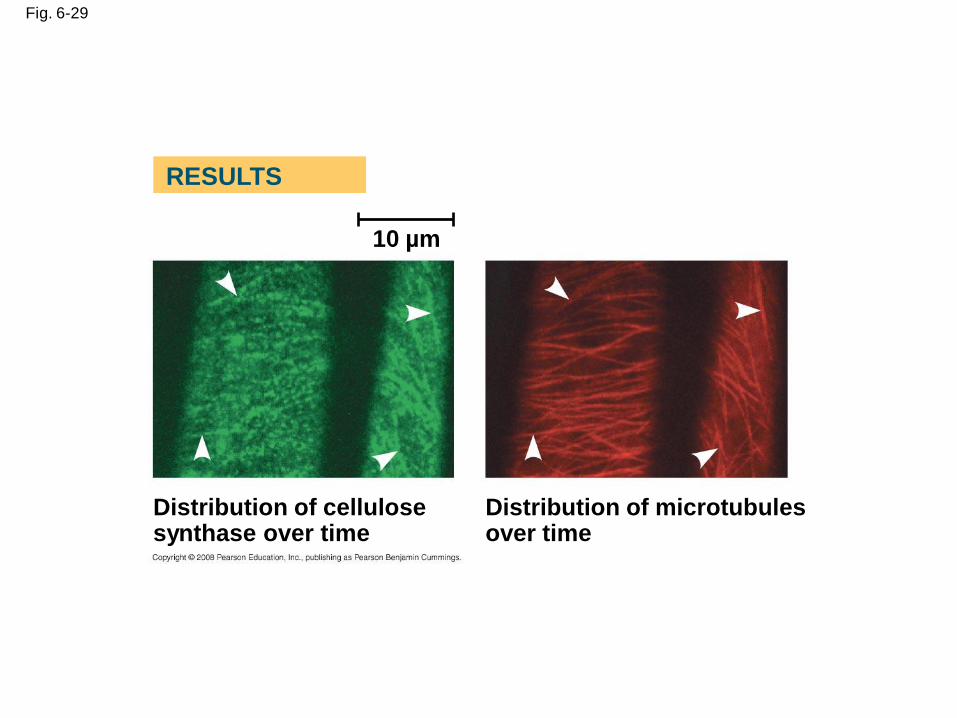
Fig. 6-29
10 µm
Distribution of cellulose synthase over time
Distribution of microtubules over time
RESULTS
The Extracellular Matrix (ECM) of Animal Cells
• Animal cells lack cell walls but are covered by
an elaborate extracellular matrix (ECM)
• The ECM is made up of glycoproteins such as
collagen, proteoglycans, and fibronectin
• ECM proteins bind to receptor proteins in the
plasma membrane called integrins
Copyright © 2008 Pearson Education, Inc., publishing as Pearson Benjamin Cummings
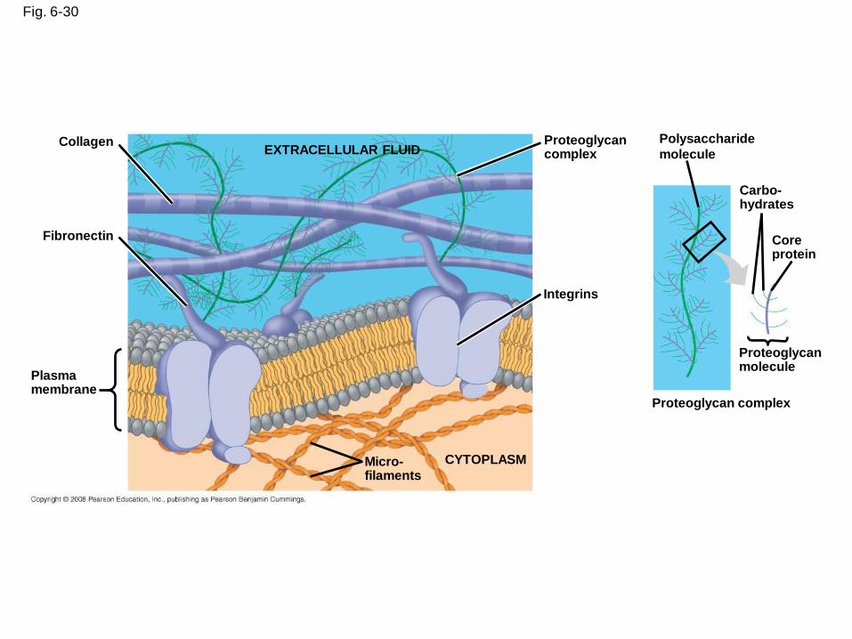
Fig. 6-30
EXTRACELLULAR FLUIDCollagen
Fibronectin
Plasmamembrane
Micro-filaments
CYTOPLASM
Integrins
Proteoglycancomplex
Polysaccharide
molecule
Carbo-hydrates
Coreprotein
Proteoglycanmolecule
Proteoglycan complex
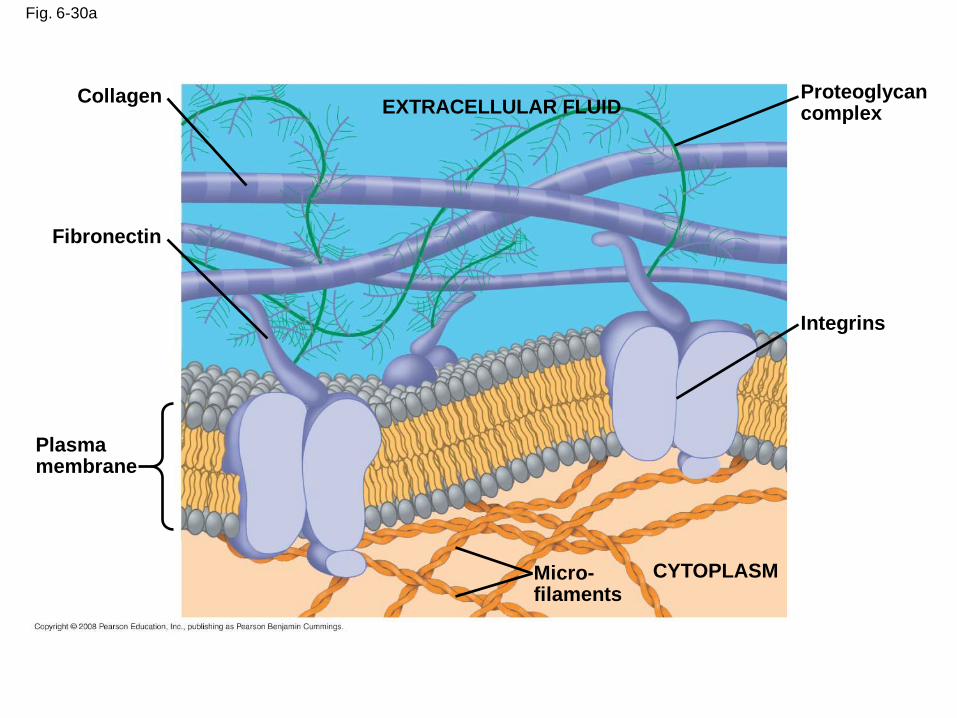
Fig. 6-30a
Collagen
Fibronectin
Plasma membrane
Proteoglycan complex
Integrins
CYTOPLASMMicro-filaments
EXTRACELLULAR FLUID
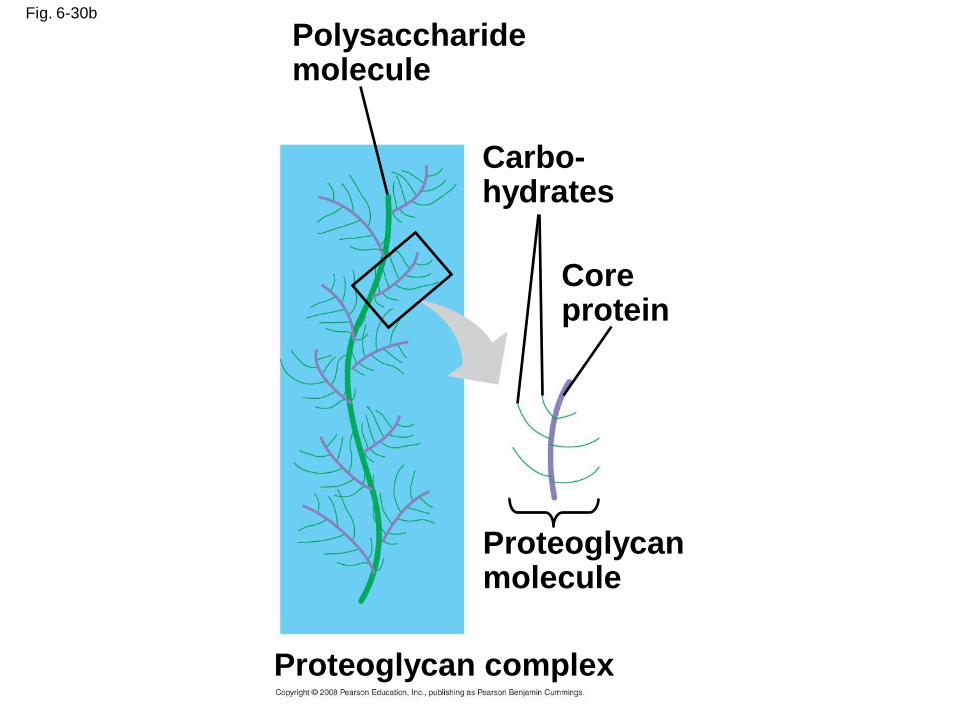
Fig. 6-30b
Polysaccharide molecule
Carbo-hydrates
Core protein
Proteoglycan molecule
Proteoglycan complex
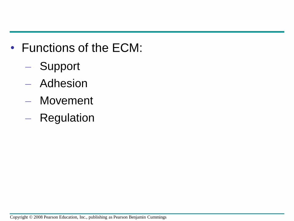
• Functions of the ECM:
– Support
– Adhesion
– Movement
– Regulation
Copyright © 2008 Pearson Education, Inc., publishing as Pearson Benjamin Cummings
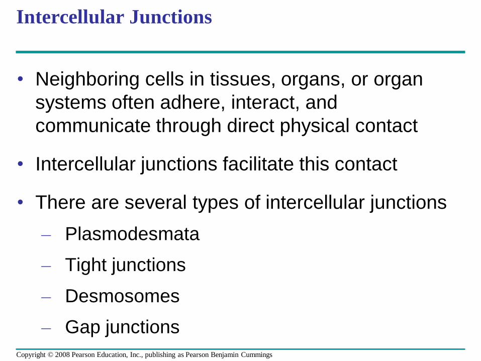
Intercellular Junctions
• Neighboring cells in tissues, organs, or organ
systems often adhere, interact, and
communicate through direct physical contact
• Intercellular junctions facilitate this contact
• There are several types of intercellular junctions
– Plasmodesmata
– Tight junctions
– Desmosomes
– Gap junctions
Copyright © 2008 Pearson Education, Inc., publishing as Pearson Benjamin Cummings
Plasmodesmata in Plant Cells
• Plasmodesmata are channels that perforate
plant cell walls
• Through plasmodesmata, water and small
solutes (and sometimes proteins and RNA) can
pass from cell to cell
Copyright © 2008 Pearson Education, Inc., publishing as Pearson Benjamin Cummings
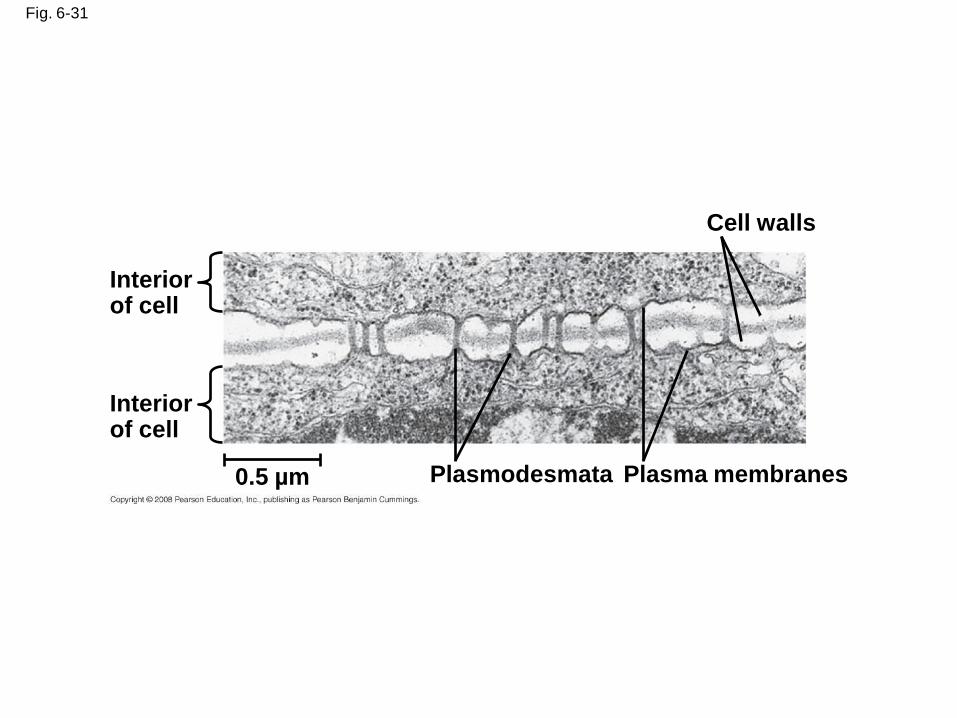
Fig. 6-31
Interior of cell
Interior of cell
0.5 µm Plasmodesmata Plasma membranes
Cell walls
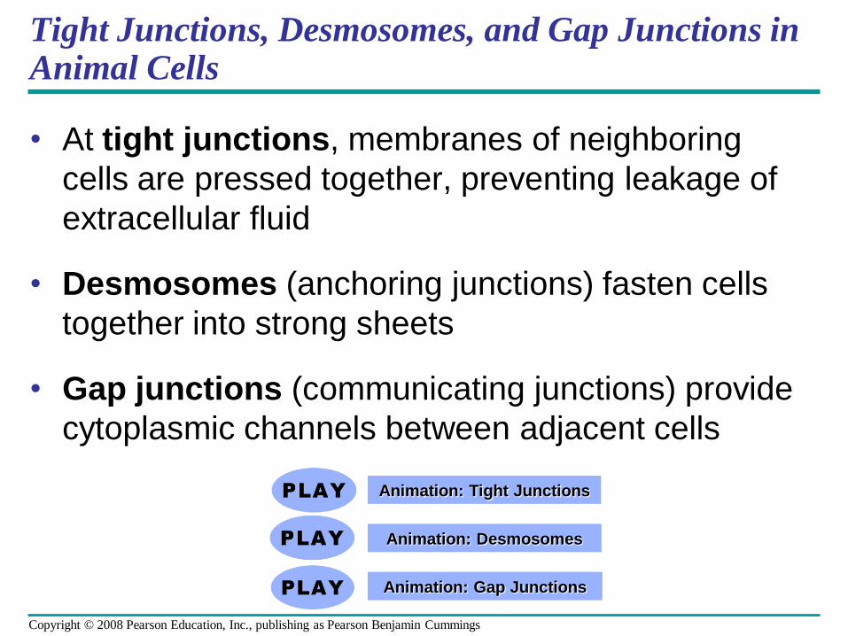
Tight Junctions, Desmosomes, and Gap Junctions in Animal Cells
• At tight junctions, membranes of neighboring
cells are pressed together, preventing leakage of
extracellular fluid
• Desmosomes (anchoring junctions) fasten cells
together into strong sheets
• Gap junctions (communicating junctions) provide
cytoplasmic channels between adjacent cells
Animation: Tight Junctions
Animation: Desmosomes
Animation: Gap Junctions
Copyright © 2008 Pearson Education, Inc., publishing as Pearson Benjamin Cummings
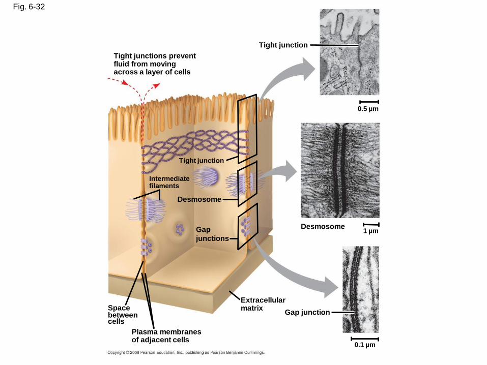
Fig. 6-32
Tight junction
0.5 µm
1 µmDesmosome
Gap junction
Extracellularmatrix
0.1 µm
Plasma membranesof adjacent cells
Spacebetweencells
Gap
junctions
Desmosome
Intermediatefilaments
Tight junction
Tight junctions preventfluid from movingacross a layer of cells
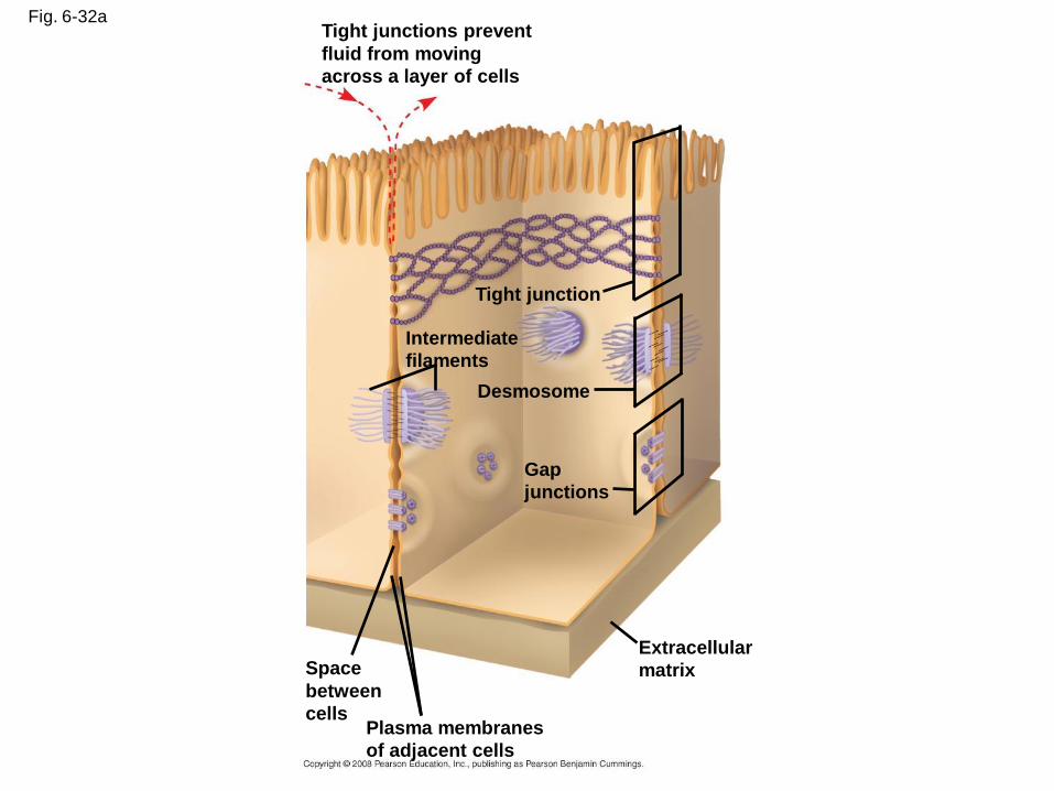
Fig. 6-32aTight junctions prevent
fluid from moving
across a layer of cells
Tight junction
Intermediate
filaments
Desmosome
Gap
junctions
Extracellular
matrixSpace
between
cellsPlasma membranes
of adjacent cells
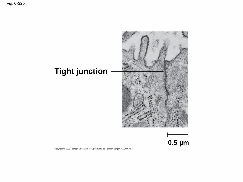
Fig. 6-32b
Tight junction
0.5 µm
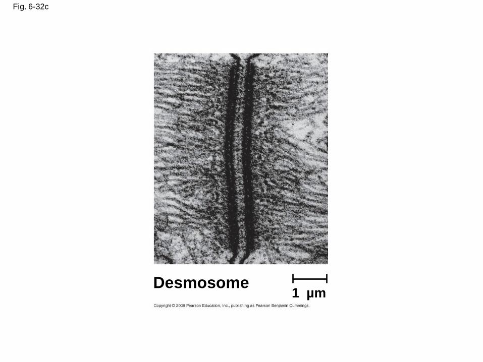
Fig. 6-32c
Desmosome1 µm
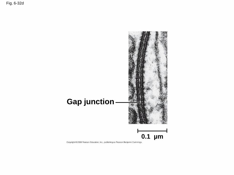
Fig. 6-32d
Gap junction
0.1 µm
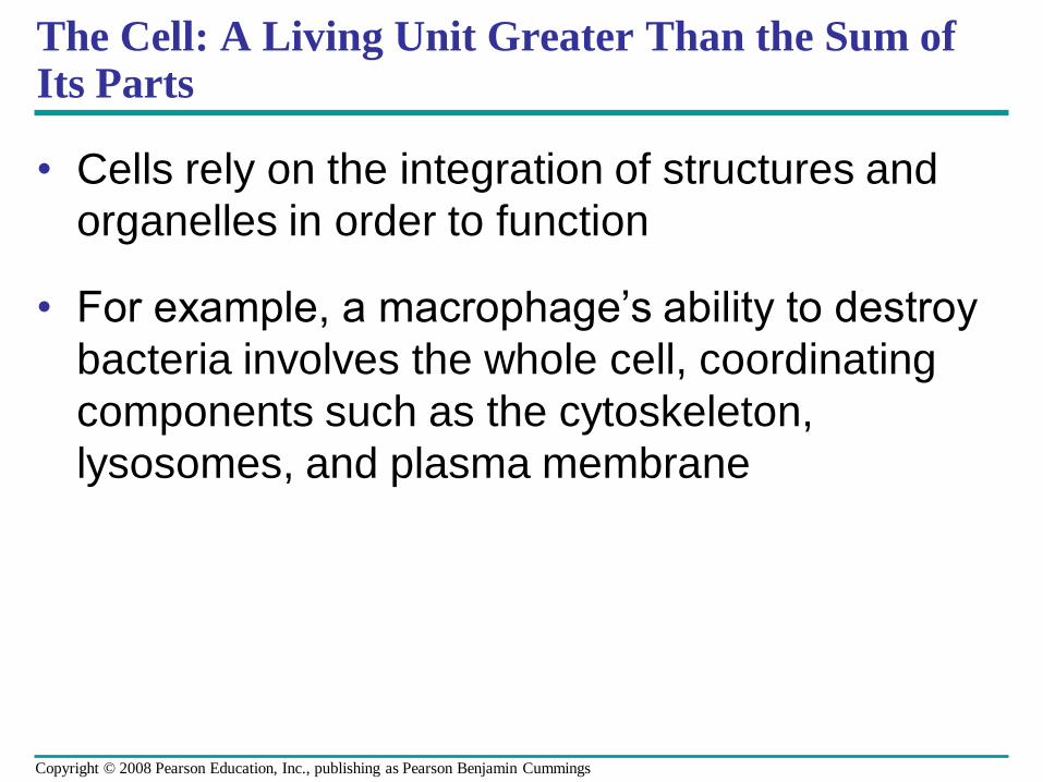
The Cell: A Living Unit Greater Than the Sum of Its Parts
• Cells rely on the integration of structures and
organelles in order to function
• For example, a macrophage’s ability to destroy
bacteria involves the whole cell, coordinating
components such as the cytoskeleton,
lysosomes, and plasma membrane
Copyright © 2008 Pearson Education, Inc., publishing as Pearson Benjamin Cummings
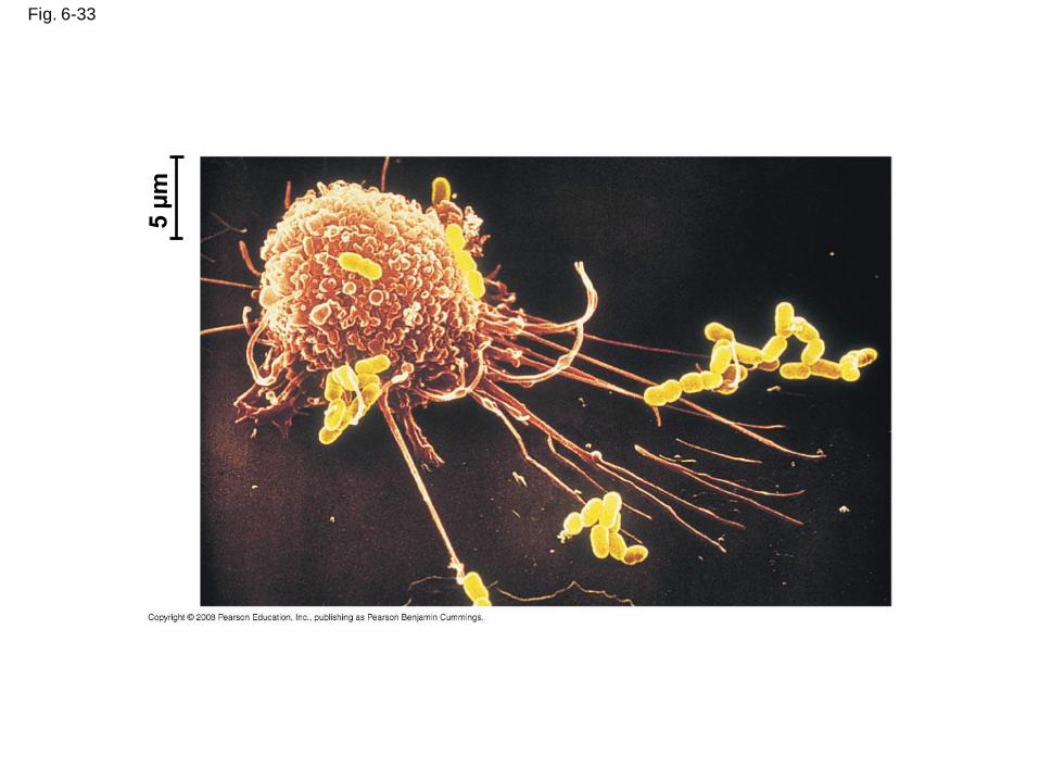
Fig. 6-33
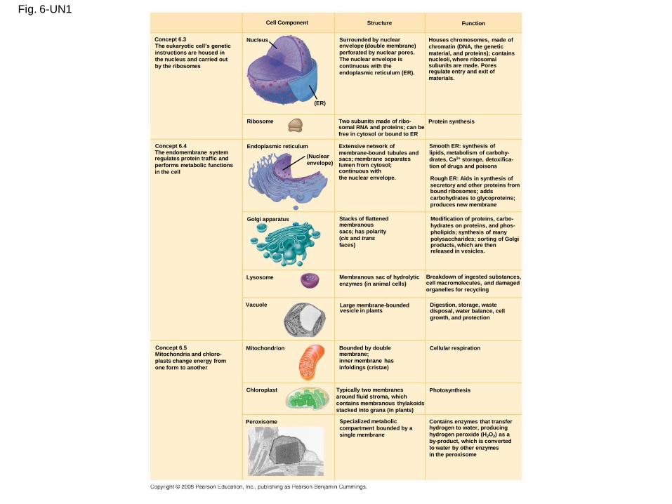
Fig. 6-UN1Cell Component Structure Function
Houses chromosomes, made of
chromatin (DNA, the genetic
material, and proteins); containsnucleoli, where ribosomalsubunits are made. Poresregulate entry and exit of
materials.
Nucleus
(ER)
Concept 6.3 The eukaryotic cell’s genetic
instructions are housed in
the nucleus and carried out
by the ribosomes
Ribosome
Concept 6.4 Endoplasmic reticulum The endomembrane systemregulates protein traffic and
performs metabolic functions
in the cell
(Nuclear
envelope)
Concept 6.5 Mitochondria and chloro-
plasts change energy from
one form to another
Golgi apparatus
Lysosome
Vacuole
Mitochondrion
Chloroplast
Peroxisome
Two subunits made of ribo-somal RNA and proteins; can be
free in cytosol or bound to ER
Extensive network of
membrane-bound tubules andsacs; membrane separateslumen from cytosol;continuous with
the nuclear envelope.
Membranous sac of hydrolytic
enzymes (in animal cells)
Large membrane-boundedvesicle in plants
Bounded by doublemembrane;
inner membrane has
infoldings (cristae)
Typically two membranes
around fluid stroma, which
contains membranous thylakoids
stacked into grana (in plants)
Specialized metabolic
compartment bounded by a
single membrane
Protein synthesis
Smooth ER: synthesis of
lipids, metabolism of carbohy-
drates, Ca2+ storage, detoxifica-
tion of drugs and poisons
Rough ER: Aids in synthesis of
secretory and other proteins frombound ribosomes; adds
carbohydrates to glycoproteins;
produces new membrane
Modification of proteins, carbo-
hydrates on proteins, and phos-
pholipids; synthesis of many
polysaccharides; sorting of Golgiproducts, which are then released in vesicles.
Breakdown of ingested substances,cell macromolecules, and damaged
organelles for recycling
Digestion, storage, wastedisposal, water balance, cell
growth, and protection
Cellular respiration
Photosynthesis
Contains enzymes that transferhydrogen to water, producing
hydrogen peroxide (H2O2) as a
by-product, which is converted
to water by other enzymes
in the peroxisome
Stacks of flattenedmembranous
sacs; has polarity
(cis and trans
faces)
Surrounded by nuclearenvelope (double membrane)
perforated by nuclear pores.
The nuclear envelope is
continuous with the
endoplasmic reticulum (ER).
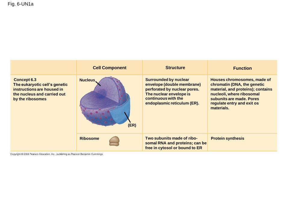
Fig. 6-UN1a
Cell Component Structure Function
Concept 6.3
The eukaryotic cell’s genetic
instructions are housed in
the nucleus and carried out
by the ribosomes
Nucleus Surrounded by nuclear
envelope (double membrane)
perforated by nuclear pores.
The nuclear envelope iscontinuous with the
endoplasmic reticulum (ER).
(ER)
Houses chromosomes, made of
chromatin (DNA, the genetic
material, and proteins); contains
nucleoli, where ribosomal
subunits are made. Poresregulate entry and exit os
materials.
Ribosome Two subunits made of ribo-
somal RNA and proteins; can be
free in cytosol or bound to ER
Protein synthesis
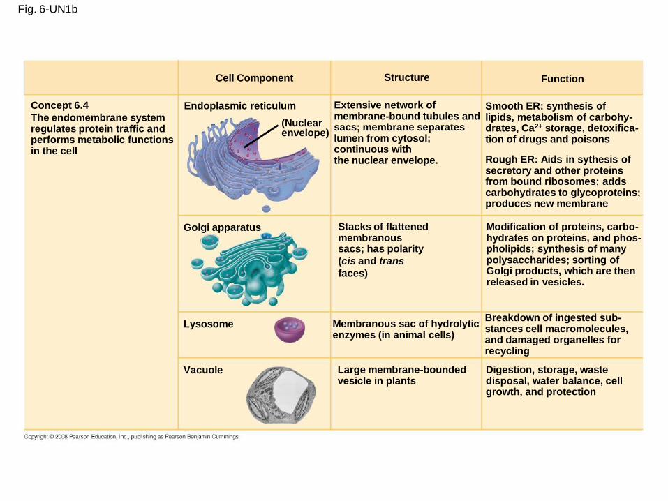
Fig. 6-UN1b
Cell Component Structure Function
Concept 6.4
The endomembrane systemregulates protein traffic andperforms metabolic functionsin the cell
Endoplasmic reticulum
(Nuclearenvelope)
Golgi apparatus
Lysosome
Vacuole Large membrane-boundedvesicle in plants
Membranous sac of hydrolyticenzymes (in animal cells)
Stacks of flattenedmembranoussacs; has polarity
(cis and trans
faces)
Extensive network ofmembrane-bound tubules andsacs; membrane separateslumen from cytosol;continuous withthe nuclear envelope.
Smooth ER: synthesis oflipids, metabolism of carbohy-drates, Ca2+ storage, detoxifica-tion of drugs and poisons
Rough ER: Aids in sythesis ofsecretory and other proteinsfrom bound ribosomes; addscarbohydrates to glycoproteins;produces new membrane
Modification of proteins, carbo-hydrates on proteins, and phos-pholipids; synthesis of manypolysaccharides; sorting ofGolgi products, which are thenreleased in vesicles.
Breakdown of ingested sub-stances cell macromolecules, and damaged organelles for recycling
Digestion, storage, wastedisposal, water balance, cellgrowth, and protection
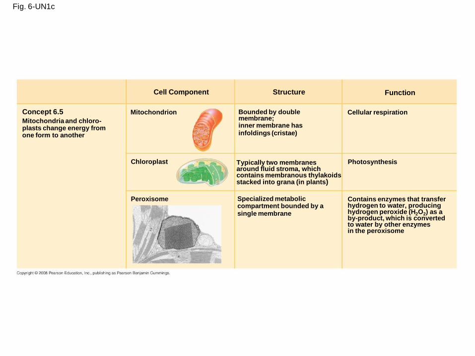
Fig. 6-UN1c
Cell Component
Concept 6.5
Mitochondria and chloro-plasts change energy fromone form to another
Mitochondrion
Chloroplast
Peroxisome
Structure Function
Bounded by doublemembrane;inner membrane hasinfoldings (cristae)
Typically two membranesaround fluid stroma, whichcontains membranous thylakoidsstacked into grana (in plants)
Specialized metaboliccompartment bounded by asingle membrane
Cellular respiration
Photosynthesis
Contains enzymes that transferhydrogen to water, producinghydrogen peroxide (H2O2) as aby-product, which is convertedto water by other enzymesin the peroxisome
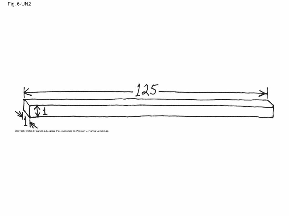
Fig. 6-UN2
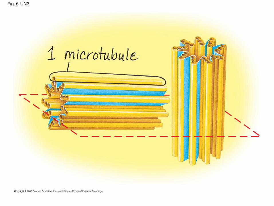
Fig. 6-UN3
You should now be able to:
1. Distinguish between the following pairs of
terms: magnification and resolution;
prokaryotic and eukaryotic cell; free and
bound ribosomes; smooth and rough ER
2. Describe the structure and function of the
components of the endomembrane system
3. Briefly explain the role of mitochondria,
chloroplasts, and peroxisomes
4. Describe the functions of the cytoskeleton
Copyright © 2008 Pearson Education, Inc., publishing as Pearson Benjamin Cummings
5. Compare the structure and functions of
microtubules, microfilaments, and
intermediate filaments
6. Explain how the ultrastructure of cilia and
flagella relate to their functions
7. Describe the structure of a plant cell wall
8. Describe the structure and roles of the
extracellular matrix in animal cells
9. Describe four different intercellular junctionsCopyright © 2008 Pearson Education, Inc., publishing as Pearson Benjamin Cummings
![Peroxisome proliferator activated receptors at the ......Peroxisomes are cellular organelles identified in the late 1960 in rat liver[10,11]; single-membrane bound, they are involved](https://img.pdfslide.us/doc/110x75/5f3ee682dbdf2b618271ecfb/peroxisome-proliferator-activated-receptors-at-the-peroxisomes-are-cellular.jpg)