Embed Size (px)
Citation preview
A Synthetic Superoxide Dismutase/Catalase Mimetic (EUK-134)Inhibits Membrane-Damage-Induced Activation of Mitogen-ActivatedProtein Kinase Pathways and Reduces p53 Accumulation inUltraviolet B-Exposed Primary Human Keratinocytes
David Decraene,� Katrien Smaers,� David Gan,w Tom Mammone,w Mary Matsui,w Daniel Maes,w LieveDeclercq,z and Marjan Garmyn��Department of Dermatology, University of Leuven, Leuven, Belgium; wEstee Lauder Companies, Melville, New York, USA; zEstee Lauder CoordinationCenter, Oevel, Belgium
Salen-manganese complexes exhibit powerful superoxide dismutase and catalase activity, with pharmacologic
efficacy in several oxidative-stress-associated disease models. Ultraviolet (UV) B not only induces direct DNA
damage, but also generates oxidative stress. EUK-134, a salen-manganese complex, might therefore confer a direct
protection against UVB-induced oxidative stress and consequently alleviate UVB-damage-induced signal
transduction. We investigated the effect of EUK-134 on the UVB-induced accumulation and stabilization of the
p53 protein. p53 plays a central role in the UVB response, both as sensor of UVB damage and as a mediator of a
protective response. Cells treated with EUK-134 before UVB irradiation showed a significantly lower accumulation
of the p53 protein in a concentration-dependent fashion. Furthermore, EUK-134 severely reduced N-terminal
phosphorylation of p53. The extracellular signal-regulated kinase ERK and the stress-activated kinases JNK and
p38 have been implicated in the UVB-induced N-terminal phosphorylation and accumulation of p53. Pre-treatment
with EUK-134 inhibited the UVB-induced activation of these mitogen-activated protein kinase (MAPK) pathways. We
hypothesize that EUK-134, by direct protection of the membrane from UVB-induced oxidative damage, reduces
oxidative stress induced MAPK signaling and consequently lowers the level of p53 induction. The protection
conferred by EUK-134 resulted in a significant increase in cell survival following UVB irradiation.
Key words: oxidative stress/skin/p53 phosphorylation/manganese compounds/hydrogen peroxide.J Invest Dermatol 122:484 –491, 2004
Exposure of human skin to ultraviolet (UV) radiation haswell-known acute and chronic effects. The acute effectsinclude erythema, tanning, and immune suppression. Long-term adverse effects of UV exposure are photoaging andphotocarcinogenesis (Gilchrest et al, 1996; Taylor andSober, 1996; Clydesdale et al, 2001). Not only UV-induceddirect DNA damage, but also UV-induced reactive oxygenspecies (ROS) are involved in these processes (de Gruijlet al, 2001; Scharffetter-Kochanek et al, 1997). ROS cancause genetic damage, both to mitochondrial and tonuclear DNA. ROS are also involved in photodamage ofthe dermal connective tissue, characteristic for photoaging,both through direct damage to connective tissue cells andproteins and through the induction of proteolytic pathways(Ma et al, 2001).
In a healthy epidermis, a keratinocyte protects itself fromthe deleterious effects of UVB damage by undergoing a
typical UVB response. The cell undergoes a cell-cycle arrestand repairs its DNA at low UVB doses, whereas high UVBdoses bring about a last escape mechanism: the inductionof programmed cell death or apoptosis. p53 plays a centralrole in this response both as a sensor of UVB damage andas an initiator of a program of expression of genes involvedin growth arrest, repair, or apoptosis. DNA damage elicitedby UVB is thought to an important trigger for p53accumulation and transcriptional activation (Decraene et al,2001).
Nevertheless, a growing amount of data suggest that thecomplex cellular response induced by UVB (290–320 nm)not only involves events triggered by DNA damage, but alsodepends on cytosolic signaling pathways originating at theplasma membrane (Assefa et al, 1997; Kulms et al, 2002).Although UVA is generally regarded as the prime source forROS in sunlight-induced damage, UVB also produces asubstantial amount of ROS through interaction with en-dogenous photosensitizers (Peus et al, 1999a), leading tothe depletion of enzymatic and nonenzymatic antioxidantsystems (Sander et al, 2002). UVB-generated ROS damagethe plasma membrane, resulting in the ligand-independentclustering of membrane receptors. The consequent activa-
Abbreviations: AAPH, 2,20-azobis(2-amidinopropane)dihydrochlor-ide; ERK, extracellular signal-regulated kinase; JNK/SAPK, c-JunN-terminal kinase/stress-activated protein kinase; MAPK, mitogen-activated protein kinase; ROS, reactive oxygen species; SOD,superoxide dismutase; UV, ultraviolet.
Copyright r 2004 by The Society for Investigative Dermatology, Inc.
484
tion of receptors in the absence of ligand binding initiatesspecific signaling cascades, particularly the stress-inducedmitogen-activated protein kinase (MAPK), within minutesof UVB exposure (Rosette and Karin, 1996; Peus et al,1999b).
A reduction in the level of UVB-generated oxidativedamage is therefore bound to have an impact on the majorUVB-responsive pathways. We therefore investigated theimpact of a salen-manganese complex, EUK-134, onthe UVB response of primary human keratinocytes. Thesynthetic salen-manganese complexes, a class of stablelow-molecular-weight compounds that contain tightlybound manganese, exhibit powerful superoxide dismutase(SOD) and catalase activity, targeting and destroying bothsuperoxide and hydrogen peroxide, respectively (Bakeret al, 1998; Baudry et al, 1993; Doctrow et al, 1997). TheseSOD/catalase mimetics have shown efficacy in a variety ofoxidative stress paradigms, including in vivo models forstroke (Baker et al, 1998), Parkinson’s disease (Melov et al,2001), autoimmune disease (Malfroy et al, 1997), andexcitotoxic neuronal death (Rong et al, 1999). Furthermore,an augmentation of the natural antioxidant system withsynthetic salen manganese complexes was shown toincrease life span in certain animal models (Melov et al,2000) and to reverse age-related learning deficits in mice(Melov et al, 2001; Liu et al, 2003). Studies have shown thatactivation of MAPK signal transduction pathways inresponse to UVB is presumably triggered by ROS-mediatedmembrane damage (Peus et al, 1999a; Rosette and Karin,1996).
We show that a pre-treatment with EUK-134 inhibits theUVB-induced activation of MAPK signal transduction path-ways in human keratinocytes. This results in a loweraccumulation of p53, a protein central in the response toUVB damage. In turn, this leads to a significant increase incell survival. Our results indicate that low concentrations ofEUK-134 can effectively protect cells against oxidativedamage by UVB. Our results also show that in addition todirect damage, ROS-dependent events are involved in theaccumulation of p53 upon UVB exposure. These data shednew light on the importance of ROS-mediated signaltransduction in the UVB-response of human skin. Theypoint out a potential use for salen-manganese complexesas protective agents against the ROS-dependent deleter-ious effects of chronic UV exposure: photoaging, andphotocarcinogenesis.
Results
EUK-134 protects primary human keratinocytes againstoxidative stress The structure of the salen-manganesecomplex EUK-134 is shown in Fig 1. EUK-134 is an analogof EUK-8 with increased catalase activity and equivalentSOD activity (Doctrow et al, 2002).
We first investigated whether EUK-134 retains itscatalase- and SOD-like activity under UVB irradiation.EUK-134 solutions were irradiated with doses up to 300mJ per cm2 UVB (Fig 2A) and subsequently subjected toanalysis. The catalase-like activity, the ability to neutralizeH2O2, was assayed by monitoring the rate of conversion of
hydrogen peroxide to oxygen as a decrease in absorbanceat 240 nm, as originally described by Beers and Sober(1952). Catalase activity of sham-irradiated EUK-134,extrapolated from a standard curve of bovine liver catalase,was calculated to be 26 U per mg. The SOD-like activity ofEUK-134, the ability to neutralize O2
–, was determined by itsability to compete with luminol for superoxide anion, leadingto a decrease in the chemiluminescent signal, as modifiedfrom Kimura and Nakano (1988). Superoxide was generatedin situ through the hypoxanthine/xanthine oxidase system.SOD-like activity of sham-irradiated EUK-134, extrapolatedfrom a standard curve of bovine erythrocyte SOD, wasestimated to be 0.33 U per mg. Both the SOD-like and thecatalase-like activity of EUK-134 proved to be stable underincreasing UVB doses (Fig 2A).
Oxidative damage to membranes is thought to be themain cause for the activation of several UVB-inducedsignaling cascades (Rosette and Karin, 1996; Peus et al,1999b). We therefore sought whether the antioxidativeproperties of EUK-134 could confer protection against theoxidation of lipids. The ability of EUK-134 to protect linoleicacid against ROS-induced lipid peroxidation was testedusing the radical initiator AAPH (Fig 2B). Thermal decom-position (371C) of the azo compound AAPH generates freeradicals that rapidly react with oxygen to give peroxylradicals. The peroxyl radicals abstract hydrogen atomsfrom the linoleic acid to give lipid radicals. These lipidradicals then induce a sequence of propagation reactions togive lipid hydroperoxides. The progress of lipid peroxidationwas followed by the rate of appearance of conjugateddienes at 234 nm as a function of time. EUK-134 stronglyreduced the propagation rate of the peroxidation reaction,resulting in a concentration-dependent decrease of theslope (Fig 2B). The concentration causing 50% inhibition(IC50) was estimated to be approximately 0.8 mM EUK-134under the test conditions described.
We next investigated whether EUK-134 could effectivelyprotect cultured human keratinocytes against oxidativestress. To demonstrate a protective effect of EUK-134against oxidative-stress-induced cytotoxicity, we performeda neutral red viability assay (Fautz et al, 1991). Neutral red isreadily incorporated into the lysosomes of living cells. Theuptake of the neutral red dye, monitored by an increase inabsorbance at 540 nm, is therefore a direct indication for
Figure1Structures of the salen-manganese complexes EUK-8 and EUK-134. The ring substituents (R) of EUK-8 and EUK-134 differ as shown(with permission from Eukarion, Inc.).
EUK-134 PROTECTION ALLEVIATES UVB-INDUCED STRESS SIGNALING 485122 : 2 FEBRUARY 2004
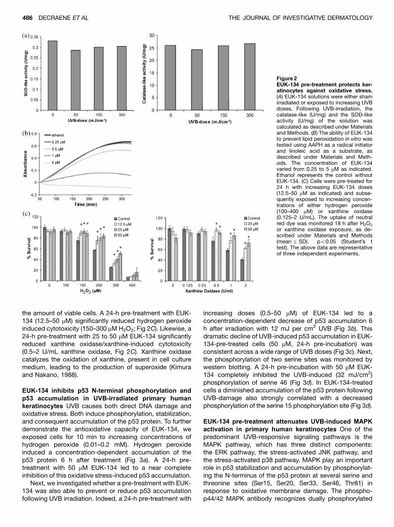
the amount of viable cells. A 24-h pre-treatment with EUK-134 (12.5–50 mM) significantly reduced hydrogen peroxideinduced cytotoxicity (150–300 mM H2O2; Fig 2C). Likewise, a24-h pre-treatment with 25 to 50 mM EUK-134 significantlyreduced xanthine oxidase/xanthine-induced cytotoxicity(0.5–2 U/mL xanthine oxidase, Fig 2C). Xanthine oxidasecatalyzes the oxidation of xanthine, present in cell culturemedium, leading to the production of superoxide (Kimuraand Nakano, 1988).
EUK-134 inhibits p53 N-terminal phosphorylation andp53 accumulation in UVB-irradiated primary humankeratinocytes UVB causes both direct DNA damage andoxidative stress. Both induce phosphorylation, stabilization,and consequent accumulation of the p53 protein. To furtherdemonstrate the antioxidative capacity of EUK-134, weexposed cells for 10 min to increasing concentrations ofhydrogen peroxide (0.01–0.2 mM). Hydrogen peroxideinduced a concentration-dependent accumulation of thep53 protein 6 h after treatment (Fig 3a). A 24-h pre-treatment with 50 mM EUK-134 led to a near completeinhibition of this oxidative stress-induced p53 accumulation.
Next, we investigated whether a pre-treatment with EUK-134 was also able to prevent or reduce p53 accumulationfollowing UVB irradiation. Indeed, a 24-h pre-treatment with
increasing doses (0.5–50 mM) of EUK-134 led to aconcentration-dependent decrease of p53 accumulation 6h after irradiation with 12 mJ per cm2 UVB (Fig 3b). Thisdramatic decline of UVB-induced p53 accumulation in EUK-134-pre-treated cells (50 mM, 24-h pre-incubation) wasconsistent across a wide range of UVB doses (Fig 3c). Next,the phosphorylation of two serine sites was monitored bywestern blotting. A 24-h pre-incubation with 50 mM EUK-134 completely inhibited the UVB-induced (32 mJ/cm2)phosphorylation of serine 46 (Fig 3d). In EUK-134-treatedcells a diminished accumulation of the p53 protein followingUVB-damage also strongly correlated with a decreasedphosphorylation of the serine 15 phosphorylation site (Fig 3d).
EUK-134 pre-treatment attenuates UVB-induced MAPKactivation in primary human keratinocytes One of thepredominant UVB-responsive signaling pathways is theMAPK pathway, which has three distinct components:the ERK pathway, the stress-activated JNK pathway, andthe stress-activated p38 pathway. MAPK play an importantrole in p53 stabilization and accumulation by phosphorylat-ing the N-terminus of the p53 protein at several serine andthreonine sites (Ser15, Ser20, Ser33, Ser46, Thr81) inresponse to oxidative membrane damage. The phospho-p44/42 MAPK antibody recognizes dually phosphorylated
Figure2EUK-134 pre-treatment protects ker-atinocytes against oxidative stress.(A) EUK-134 solutions were either shamirradiated or exposed to increasing UVBdoses. Following UVB-irradiation, thecatalase-like (U/mg) and the SOD-likeactivity (U/mg) of the solution wascalculated as described under Materialsand Methods. (B) The ability of EUK-134to prevent lipid peroxidation in vitro wastested using AAPH as a radical initiatorand linoleic acid as a substrate, asdescribed under Materials and Meth-ods. The concentration of EUK-134varied from 0.25 to 5 mM as indicated.Ethanol represents the control withoutEUK-134. (C) Cells were pre-treated for24 h with increasing EUK-134 doses(12.5–50 mM as indicated) and subse-quently exposed to increasing concen-trations of either hydrogen peroxide(100–400 mM) or xanthine oxidase(0.125–2 U/mL). The uptake of neutralred dye was monitored 18 h after H2O2
or xanthine oxidase exposure, as de-scribed under Materials and Methods(mean � SD). �po0.05 (Student’s ttest). The above data are representativeof three independent experiments.
486 DECRAENE ET AL THE JOURNAL OF INVESTIGATIVE DERMATOLOGY
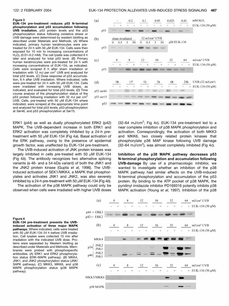
ERK1 (p44) as well as dually phosphorylated ERK2 (p42)MAPK. The UVB-dependent increase in both ERK1 andERK2 activation was completely inhibited by a 24-h pre-treatment with 50 mM EUK-134 (Fig 4a). Basal activation ofthe ERK pathway, owing to the presence of epidermalgrowth factor, was unaffected by EUK-134 pre-treatment.
The UVB-induced activation of JNK protein kinases waslargely inhibited in cells pre-treated with 50 mM EUK-134(Fig 4b). The antibody recognizes two alternative splicingvariants (a 46- and a 54-kDa variant) of both the JNK1 andthe JNK2 protein kinase (Gupta et al, 1996). The UVB-induced activation of SEK1/MKK4, a MAPK that phosphor-ylates and activates JNK1 and JNK2, was also severelyinhibited by a 24-h pre-treatment with 50 mM EUK-134 (Fig 4b).
The activation of the p38 MAPK pathway could only beobserved when cells were irradiated with higher UVB doses
(32–64 mJ/cm2; Fig 4c). EUK-134 pre-treatment led to anear complete inhibition of p38 MAPK phosphorylation andactivation. Correspondingly, the activation of both MKK3and MKK6, two closely related protein kinases thatphosphorylate p38 MAP kinase following UVB damage(32–64 mJ/cm2), was almost completely inhibited (Fig 4c).
Inhibition of the p38 MAPK pathway decreases p53N-terminal phosphorylation and accumulation followingUVB-damage By use of a pharmacologic inhibitor, wewanted to investigate whether an inhibition of the p38MAPK pathway had similar effects on the UVB-inducedN-terminal phosphorylation and accumulation of the p53protein. By binding to the ATP pocket of p38 MAPK, thepyridinyl imidazole inhibitor PD169316 potently inhibits p38MAPK activation (Young et al, 1997). Inhibition of the p38
Figure 3EUK-134 pre-treatment reduces p53 N-terminalphosphorylation and p53 accumulation followingUVB irradiation. p53 protein levels and the p53phosphorylation status following oxidative stress orUVB damage were determined by western blotting asdescribed under Materials and Methods. (A) Whereindicated, primary human keratinocytes were pre-treated for 24 h with 50 mM EUK-134. Cells were thenexposed for 10 min to increasing concentrations ofH2O2 (0.01–0.2 mM). The cell lysate was collected 6 hlater and analyzed for total p53 level. (B) Primaryhuman keratinocytes were pre-treated for 24 h withincreasing concentrations of EUK-134, as indicated.Cells were scraped 6 h after sham irradiation orirradiation with 12 mJ per cm2 UVB and analyzed fortotal p53 levels. (C) Dose response of p53 accumula-tion, 6 h after UVB irradiation. Where indicated, cellswere pre-treated for 24 h with 50 mM EUK-134. Cellswere irradiated with increasing UVB doses, asindicated, and evaluated for total p53 levels. (D) Timecourse analysis of the phosphorylation status of thep53 protein following irradiation with 32 mJ per cm2
UVB. Cells, pre-treated with 50 mM EUK-134 whereindicated, were scraped at the appropriate time pointand analyzed for total p53 levels, p53 phosphorylationat Ser46, and p53 phosphorylation at Ser15.
Figure 4EUK-134 pre-treatment prevents the UVB-induced activation of three major MAPKpathways. Where indicated, cells were treatedwith 50 mM EUK-134 24 h before UVB irradia-tion. Cell lysates were collected 15 min afterirradiation with the indicated UVB dose. Pro-teins were separated by Western blotting asdescribed under Materials and Methods. Mem-branes were probed with phosphospecificantibodies. (A) ERK1 and ERK2 phosphoryla-tion status (ERK-MAPK pathway). (B) MKK4,JNK1, and JNK2 phosphorylation status (JNK/SAPK pathway). (C) MKK3, MKK6, and p38MAPK phosphorylation status (p38 MAPKpathway).
EUK-134 PROTECTION ALLEVIATES UVB-INDUCED STRESS SIGNALING 487122 : 2 FEBRUARY 2004
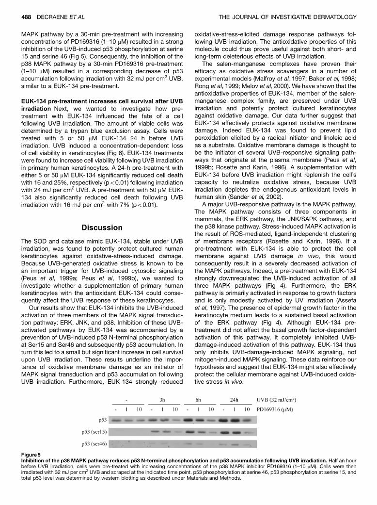
MAPK pathway by a 30-min pre-treatment with increasingconcentrations of PD169316 (1–10 mM) resulted in a stronginhibition of the UVB-induced p53 phosphorylation at serine15 and serine 46 (Fig 5). Consequently, the inhibition of thep38 MAPK pathway by a 30-min PD169316 pre-treatment(1–10 mM) resulted in a corresponding decrease of p53accumulation following irradiation with 32 mJ per cm2 UVB,similar to a EUK-134 pre-treatment.
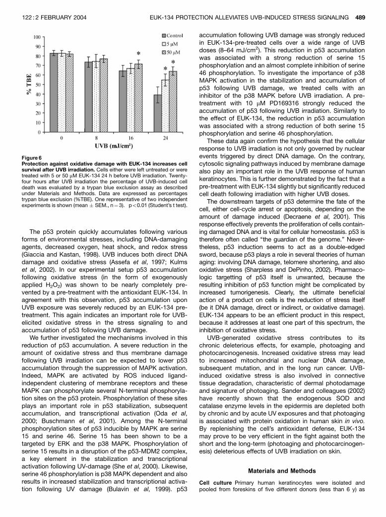
EUK-134 pre-treatment increases cell survival after UVBirradiation Next, we wanted to investigate how pre-treatment with EUK-134 influenced the fate of a cellfollowing UVB irradiation. The amount of viable cells wasdetermined by a trypan blue exclusion assay. Cells weretreated with 5 or 50 mM EUK-134 24 h before UVBirradiation. UVB induced a concentration-dependent lossof cell viability in keratinocytes (Fig 6). EUK-134 treatmentswere found to increase cell viability following UVB irradiationin primary human keratinocytes. A 24-h pre-treatment witheither 5 or 50 mM EUK-134 significantly reduced cell deathwith 16 and 25%, respectively (po0.01) following irradiationwith 24 mJ per cm2 UVB. A pre-treatment with 50 mM EUK-134 also significantly reduced cell death following UVBirradiation with 16 mJ per cm2 with 7% (po0.01).
Discussion
The SOD and catalase mimic EUK-134, stable under UVBirradiation, was found to potently protect cultured humankeratinocytes against oxidative-stress-induced damage.Because UVB-generated oxidative stress is known to bean important trigger for UVB-induced cytosolic signaling(Peus et al, 1999a; Peus et al, 1999b), we wanted toinvestigate whether a supplementation of primary humankeratinocytes with the antioxidant EUK-134 could conse-quently affect the UVB response of these keratinocytes.
Our results show that EUK-134 inhibits the UVB-inducedactivation of three members of the MAPK signal transduc-tion pathway: ERK, JNK, and p38. Inhibition of these UVB-activated pathways by EUK-134 was accompanied by aprevention of UVB-induced p53 N-terminal phosphorylationat Ser15 and Ser46 and subsequently p53 accumulation. Inturn this led to a small but significant increase in cell survivalupon UVB irradiation. These results underline the impor-tance of oxidative membrane damage as an initiator ofMAPK signal transduction and p53 accumulation followingUVB irradiation. Furthermore, EUK-134 strongly reduced
oxidative-stress-elicited damage response pathways fol-lowing UVB-irradiation. The antioxidative properties of thismolecule could thus prove useful against both short- andlong-term deleterious effects of UVB irradiation.
The salen-manganese complexes have proven theirefficacy as oxidative stress scavengers in a number ofexperimental models (Malfroy et al, 1997; Baker et al, 1998;Rong et al, 1999; Melov et al, 2000). We have shown that theantioxidative properties of EUK-134, member of the salen-manganese complex family, are preserved under UVBirradiation and potently protect cultured keratinocytesagainst oxidative damage. Our data further suggest thatEUK-134 effectively protects against oxidative membranedamage. Indeed EUK-134 was found to prevent lipidperoxidation elicited by a radical initiator and linoleic acidas a substrate. Oxidative membrane damage is thought tobe the initiator of several UVB-responsive signaling path-ways that originate at the plasma membrane (Peus et al,1999b; Rosette and Karin, 1996). A supplementation withEUK-134 before UVB irradiation might replenish the cell’scapacity to neutralize oxidative stress, because UVBirradiation depletes the endogenous antioxidant levels inhuman skin (Sander et al, 2002).
A major UVB-responsive pathway is the MAPK pathway.The MAPK pathway consists of three components inmammals, the ERK pathway, the JNK/SAPK pathway, andthe p38 kinase pathway. Stress-induced MAPK activation isthe result of ROS-mediated, ligand-independent clusteringof membrane receptors (Rosette and Karin, 1996). If apre-treatment with EUK-134 is able to protect the cellmembrane against UVB damage in vivo, this wouldconsequently result in a severely decreased activation ofthe MAPK pathways. Indeed, a pre-treatment with EUK-134strongly downregulated the UVB-induced activation of allthree MAPK pathways (Fig 4). Furthermore, the ERKpathway is primarily activated in response to growth factorsand is only modestly activated by UV irradiation (Assefaet al, 1997). The presence of epidermal growth factor in thekeratinocyte medium leads to a sustained basal activationof the ERK pathway (Fig 4). Although EUK-134 pre-treatment did not affect the basal growth factor-dependentactivation of this pathway, it completely inhibited UVB-damage-induced activation of this pathway. EUK-134 thusonly inhibits UVB-damage-induced MAPK signaling, notmitogen-induced MAPK signaling. These data reinforce ourhypothesis and suggest that EUK-134 might also effectivelyprotect the cellular membrane against UVB-induced oxida-tive stress in vivo.
Figure 5Inhibition of the p38 MAPK pathway reduces p53 N-terminal phosphorylation and p53 accumulation following UVB irradiation. Half an hourbefore UVB irradiation, cells were pre-treated with increasing concentrations of the p38 MAPK inhibitor PD169316 (1–10 mM). Cells were thenirradiated with 32 mJ per cm2 UVB and scraped at the indicated time point. p53 phosphorylation at serine 46, p53 phosphorylation at serine 15, andtotal p53 level was determined by western blotting as described under Materials and Methods.
488 DECRAENE ET AL THE JOURNAL OF INVESTIGATIVE DERMATOLOGY
The p53 protein quickly accumulates following variousforms of environmental stresses, including DNA-damagingagents, decreased oxygen, heat shock, and redox stress(Giaccia and Kastan, 1998). UVB induces both direct DNAdamage and oxidative stress (Assefa et al, 1997; Kulmset al, 2002). In our experimental setup p53 accumulationfollowing oxidative stress (in the form of exogenouslyapplied H2O2) was shown to be nearly completely pre-vented by a pre-treatment with the antioxidant EUK-134. Inagreement with this observation, p53 accumulation uponUVB exposure was severely reduced by an EUK-134 pre-treatment. This again indicates an important role for UVB-elicited oxidative stress in the stress signaling to andaccumulation of p53 following UVB damage.
We further investigated the mechanisms involved in thisreduction of p53 accumulation. A severe reduction in theamount of oxidative stress and thus membrane damagefollowing UVB irradiation can be expected to lower p53accumulation through the suppression of MAPK activation.Indeed, MAPK are activated by ROS induced ligand-independent clustering of membrane receptors and theseMAPK can phosphorylate several N-terminal phosphoryla-tion sites on the p53 protein. Phosphorylation of these sitesplays an important role in p53 stabilization, subsequentaccumulation, and transcriptional activation (Oda et al,2000; Buschmann et al, 2001). Among the N-terminalphosphorylation sites of p53 inducible by MAPK are serine15 and serine 46. Serine 15 has been shown to be atargeted by ERK and the p38 MAPK. Phosphorylation ofserine 15 results in a disruption of the p53-MDM2 complex,a key element in the stabilization and transcriptionalactivation following UV-damage (She et al, 2000). Likewise,serine 46 phosphorylation is p38 MAPK dependent and alsoresults in increased stabilization and transcriptional activa-tion following UV damage (Bulavin et al, 1999). p53
accumulation following UVB damage was strongly reducedin EUK-134-pre-treated cells over a wide range of UVBdoses (8–64 mJ/cm2). This reduction in p53 accumulationwas associated with a strong reduction of serine 15phosphorylation and an almost complete inhibition of serine46 phosphorylation. To investigate the importance of p38MAPK activation in the stabilization and accumulation ofp53 following UVB damage, we treated cells with aninhibitor of the p38 MAPK before UVB irradiation. A pre-treatment with 10 mM PD169316 strongly reduced theaccumulation of p53 following UVB irradiation. Similarly tothe effect of EUK-134, the reduction in p53 accumulationwas associated with a strong reduction of both serine 15phosphorylation and serine 46 phosphorylation.
These data again confirm the hypothesis that the cellularresponse to UVB irradiation is not only governed by nuclearevents triggered by direct DNA damage. On the contrary,cytosolic signaling pathways induced by membrane damagealso play an important role in the UVB response of humankeratinocytes. This is further demonstrated by the fact that apre-treatment with EUK-134 slightly but significantly reducedcell death following irradiation with higher UVB doses.
The downstream targets of p53 determine the fate of thecell, either cell-cycle arrest or apoptosis, depending on theamount of damage induced (Decraene et al, 2001). Thisresponse effectively prevents the proliferation of cells contain-ing damaged DNA and is vital for cellular homeostasis. p53 istherefore often called ‘‘the guardian of the genome.’’ Never-theless, p53 induction seems to act as a double-edgedsword, because p53 plays a role in several theories of humanaging: involving DNA damage, telomere shortening, and alsooxidative stress (Sharpless and DePinho, 2002). Pharmaco-logic targetting of p53 itself is unwanted, because theresulting inhibition of p53 function might be complicated byincreased tumorigenesis. Clearly, the ultimate beneficialaction of a product on cells is the reduction of stress itself(be it DNA damage, direct or indirect, or oxidative damage).EUK-134 appears to be an efficient product in this respect,because it addresses at least one part of this spectrum, theinhibition of oxidative stress.
UVB-generated oxidative stress contributes to itschronic deleterious effects, for example, photoaging andphotocarcinogenesis. Increased oxidative stress may leadto increased mitochondrial and nuclear DNA damage,subsequent mutation, and in the long run cancer. UVB-induced oxidative stress is also involved in connectivetissue degradation, characteristic of dermal photodamageand signature of photoaging. Sander and colleagues (2002)have recently shown that the endogenous SOD andcatalase enzyme levels in the epidermis are depleted bothby chronic and by acute UV exposures and that photoagingis associated with protein oxidation in human skin in vivo.By replenishing the cell’s antioxidant defense, EUK-134may prove to be very efficient in the fight against both theshort and the long-term (photoaging and photocarcinogen-esis) deleterious effects of UVB irradiation on skin.
Materials and Methods
Cell culture Primary human keratinocytes were isolated andpooled from foreskins of five different donors (less than 6 y) as
Figure 6Protection against oxidative damage with EUK-134 increases cellsurvival after UVB irradiation. Cells either were left untreated or weretreated with 5 or 50 mM EUK-134 24 h before UVB irradiation. Twenty-four hours after UVB irradiation the percentage of UVB-induced celldeath was evaluated by a trypan blue exclusion assay as describedunder Materials and Methods. Data are expressed as percentagestrypan blue exclusion (%TBE). One representative of two independentexperiments is shown (mean � SEM., n¼ 3). �po0.01 (Student’s t test).
EUK-134 PROTECTION ALLEVIATES UVB-INDUCED STRESS SIGNALING 489122 : 2 FEBRUARY 2004
described (Gilchrest, 1983). The procedure has been approved bythe ethical committee of the University of Leuven. Keratinocyteswere grown in serum-free medium (Keratinocyte-SFM, Invitrogen,Merelbeke, Belgium) supplemented with bovine pituitary extract(50 mg/mL) and human recombinant epidermal growth factor (5 ng/mL). Third- to fifth-passage cells were used in experiments.
Antibodies and reagents Phospho-p44/42 MAP kinase (Thr202/Tyr204, recognizes dually phosphorylated extracellular signal-regulated kinase (ERK) 1 and ERK2), phospho-stress-activatedprotein kinase (SAPK)/c-Jun N-terminal kinase (JNK) (Thr183/Tyr185, recognizes dually phosphorylated JNK1 and JNK2 iso-forms), phospho-MKK3/6 (Ser189/207), phospho-SEK1/MKK4(Thr261), phospho-p53 (Ser46), phospho-p38 MAP kinase(Thr180/Tyr182), and p38 MAP kinase antibodies were purchasedfrom Cell Signaling (Beverly, MA). Anti-human p53 (clone DO-1)antibody was purchased from PharMingen (BD Biosciences,Erembodegem, Belgium). EUK-134 was purchased from EukarionInc. (Bedford, MA). Hydrogen peroxide and xanthine oxidase werepurchased from Sigma-Aldrich (Bornem, Belgium).
UVB irradiation Before UVB irradiation cells were washed twicewith phosphate-buffered saline, irradiated through a thin film ofphosphate-buffered saline, and then refed with their own medium.Cells were exposed through the cover of the dish, which filters outresidual UVC (Brown et al, 2001). The UVB source was a parallelbank of three Philips TL 20W12 tubes with a peak output of 310 nm.Output was measured through the cover of a tissue culture dishwith an IL700 radiometer (International light, Newburyport, MA).
Western blotting At the indicated time points, cells were scrapedin lysis buffer (25 mM HEPES, 0.3 mM NaCl, 1.5 mM MgCl2, 20 mMb-glycerolphosphate, 2 mM ethylenediaminetetraacetic acid, 2 mM
ethylene glycol-bis(b-amino ethyl ether), pH 7.5) containing 1%Triton, 10% glycerol, 1 mM Na3VO4, 0.5 mM dithiothreitol, 10 mgper mL leupeptin, 10 mg per mL aprotinin, and 10 mg per mLantipain. Extracts were incubated on ice and spun down at21,000 g for 20 min. Protein concentration was determined usingthe bicinchoninic acid protein assay reagent (Pierce ChemicalCompany, Rockford, IL). A quantity of 50 to 80 mg of total proteinextract was separated by a 10% SDS-PAGE, followed by wetelectrotransfer onto Hybond-C Super membrane (AmershamPharmacia, Roosendaal, Netherlands). Equal loading of proteinswas verified using Ponceau-S in coloring (Klein et al, 1995).Membranes were blocked for 1 h at room temperature in Tris-buffered saline containing 0.1% Tween and 5% nonfat dry milk.The membrane was incubated overnight at 41C with the primaryantibody, washed, and incubated for 1 h at room temperature withthe peroxidase-conjugated secondary antibody. Protein bandswere visualized using enhanced chemiluminescence as describedby the supplier (Amersham Pharmacia, Roosendaal, Netherlands).
Quantification of the Catalase-like and SOD-like activity ofEUK-134 The catalase-like activity of EUK-134 was assayed bymonitoring the rate of conversion of hydrogen peroxide to oxygenas a decrease in absorbance at 240 nm, as originally described byBeers and Sober (1952). EUK-134 solutions (6 mg/mL in 50 mMphosphate buffer, pH 7.0) were incubated on ice and exposed toUVB irradiation. Following UVB irradiation, an equal volume ofH2O2 (Acros, Geel, Belgium; 34 mM in phosphate buffer) wasadded. The decrease in UV absorbance at 240 nm was monitoredover a period of 2 min. Catalase-like activity (U/mg) wasextrapolated from a standard curve of different concentrations ofcatalase (from bovine liver).
SOD-like activity of EUK-134 was determined as modified fromKimura and Nakano (1988). The protocol is based on the ability ofEUK-134 to compete with luminol for the superoxide anion,produced in situ using a hypoxanthine/xanthine oxidase systemand resulting in a decrease in chemiluminescent signal. Briefly,EUK-134 solutions (120 mg/mL in 50 mM phosphate buffer, pH 7.0)
were incubated on ice and exposed to UVB. Following UVBirradiation, 30 mL of EUK-134 solution was transferred to an equalvolume of xanthine oxidase (0.1 U/mL). Fifteen microliters ofluminol (0.5 mM) and 75 mL of hypoxanthine (0.735 mM) wereadded and the chemiluminescence intensity was measured for 30s. SOD-like activity (U/mg) was extrapolated from a standard curvewith different concentrations of SOD (from bovine erythrocytes). Allreagents were purchased from Sigma, unless stated otherwise.
Linoleic acid peroxidation assay The antioxidant activity ofEUK-134 was expressed as the ability to inhibit linoleic acidperoxidation in vitro according to Liegeois et al (2000). Briefly,oxidation of 16 mM linoleic acid in aqueous dispersions (in boratebuffer, pH 9, 0.5% Tween 20, 25 mM KOH, with or without EUK-134) was initiated by addition of 4 mM of the radical initiator 2,20-azobis(2-amidinopropane)dihydrochloride (AAPH) (Aldrich Chemi-cal Company, Milwaukee, WI). The progress of lipid peroxidation at371C in the presence of EUK-134 was continuously monitored byspectrophotometric reading of the rate of appearance of con-jugated dienes at 234 nm as a function of time.
Neutral red viability assay Primary human keratinocytes weregrown in 96-well plates to approximately 75% confluency. A pre-treatment of EUK-134 at 12.5, 25, and 50 mM was carried out. Cellswere then exposed to increasing concentrations of H2O2 (100–400mM) or xanthine oxidase (0.5–2 U/mL). Following an 18-hincubation, 50 mg per mL neutral red dye (Aldrich ChemicalCompany) was added to the medium. Three hours later, cells werefixed in 1% formaldehyde-1% calcium chloride, rinsed three timeswith phosphate-buffered saline, and lyzed with 1% acetic acid-50% ethanol for 30 min. Absorbance at 540 nm was measured byspectrophotometry.
Trypan blue exclusion assay Primary human keratinocytes wereplated in 60-mm cell culture dishes and grown to 75% confluency.The keratinocytes were pre-incubated with EUK-134 at 0, 5, and50 mM for a 24-h period before UVB irradiation at 8, 16, and 24 mJper cm2. Cells were harvested 24 h after UVB irradiation bytrypsinization and centrifugation. Cell viability was determined bythe trypan blue exclusion assay (Kaltenbach et al, 1958). Briefly,pelleted cells where stained with 0.4% trypan blue (AldrichChemical Company, Milwaukee, WI), followed by examination witha hemacytometer under an inverted microscope. Cells thatexcluded the dye were considered viable and the data areexpressed as the percentage of trypan blue exclusion.
This work was supported in part by Grant OT/00/33 from the Universityof Leuven and Grant 0211.99 from the ‘‘Fonds voor WetenschappelijkOnderzoek-Vlaanderen,’’ Belgium. M.G. is clincal research associate(FWO).
DOI: 10.1046/j.0022-202X.2004.22215.x
Manuscript received February 14, 2003; revised August 4, 2003;accepted for publication October 22, 2003
Address correspondence to: Marjan Garmyn, Department of Derma-tology, UZ St. Rafael, Kapucijnenvoer 33, B-3000 Leuven, Belgium.Email: [email protected]
References
Assefa Z, Garmyn M, Bouillon R, Merlevede W, Vandenheede JR, Agostinis P:
Differential stimulation of ERK and JNK activities by ultraviolet B
irradiation and epidermal growth factor in human keratinocytes. J Invest
Dermatol 108:886–891, 1997
Baker K, Marcus CB, Huffman K, Kruk H, Malfroy B, Doctrow SR: Synthetic
combined superoxide dismutase/catalase mimetics are protective as a
delayed treatment in a rat stroke model: A key role for reactive oxygen
490 DECRAENE ET AL THE JOURNAL OF INVESTIGATIVE DERMATOLOGY
species in ischemic brain injury. J Pharmacol Exp Ther 284:215–221,
1998
Baudry M, Etienne S, Bruce A, Palucki M, Jacobsen E, Malfroy B: Salen-
manganese complexes are superoxide dismutase-mimics. Biochem
Biophys Res Commun 192:964–968, 1993
Beers RF, Sober AJ: A spectrophotometric method for measuring the breakdown
of hydrogen peroxide by catalase. J Biol Chem 195:133–144, 1952
Brown DB, Peritz AE, Uitto J, Gasparro FP: Ultraviolet-filtering properties of
commonly used tissue cell culture plasticware. Photodermatol Photo-
immunol Photomed 17:126–129, 2001
Bulavin DV, Saito S, Hollander MC, Sakaguchi K, Anderson CW, Appella E,
Fornace AJ Jr: Phosphorylation of human p53 by p38 kinase coordinates
N-terminal phosphorylation and apoptosis in response to UV radiation.
EMBO J 18:6845–6854, 1999
Buschmann T, Potapova O, Bar-Shira A, et al: Jun NH2-terminal kinase
phosphorylation of p53 on Thr-81 is important for p53 stabilization and
transcriptional activities in response to stress. Mol Cell Biol 21:2743–
2754, 2001
Clydesdale GJ, Dandie GW, Muller HK: Ultraviolet light induced injury:
Immunological and inflammatory effects. Immunol Cell Biol 79:547–568,
2001
Decraene D, Agostinis P, Pupe A, de Haes P, Garmyn M: Acute response of
human skin to solar radiation: Regulation and function of the p53 protein.
J Photochem Photobiol B 63:78–83, 2001
de Gruijl FR, van Kranen HJ, Mullenders LH: UV-induced DNA damage, repair,
mutations and oncogenic pathways in skin cancer. J Photochem
Photobiol B 63:19–27, 2001
Doctrow SR, Huffman K, Marcus CB, et al: Salen-manganese complexes as
catalytic scavengers of hydrogen peroxide and cytoprotective agents:
Structure–activity relationship studies. J Med Chem 45:4549–4558, 2002
Doctrow SR, Huffman K, Marcus CB, Musleh W, Bruce A, Baudry M, Malfroy B:
Salen-manganese complexes: Combined superoxide dismutase/catalase
mimics with broad pharmacological efficacy. Adv Pharmacol 38:247–269,
1997
Fautz R, Husein B, Hechenberger C: Application of the neutral red assay (NR
assay) to monolayer cultures of primary hepatocytes: Rapid colorimetric
viability determination for the unscheduled DNA synthesis test (UDS).
Mutat Res 253:173–179, 1991
Giaccia AJ, Kastan MB: The complexity of p53 modulation: Emerging patterns
from divergent signals. Genes Dev 12:2973–2983, 1998
Gilchrest BA: In vitro assessment of keratinocyte aging. J Invest Dermatol
81:184s–189s, 1983
Gilchrest BA, Park HY, Eller MS, Yaar M: Mechanisms of ultraviolet light-induced
pigmentation. Photochem Photobiol 63:1–10, 1996
Gupta S, Barrett T, Whitmarsh AJ, Cavanagh J, Sluss HK, Derijard B, Davis RJ:
Selective interaction of JNK protein kinase isoforms with transcription
factors. EMBO J 15:2760–2770, 1996
Kaltenbach JP, Kaltenbach MH, Lyons WB: Nigrosin as a dye for differentiating
live and dead ascites cells. Exp Cell Res 15:112–117, 1958
Kimura H, Nakano M: Highly sensitive and reliable chemiluminescence method
for the assay of superoxide dismutase in human erythrocytes. FEBS Lett
239:347–350, 1988
Klein D, Kern RM, Sokol RZ: A method for quantification and correction of
proteins after transfer to immobilization membranes. Biochem Mol Biol Int
36:59–66, 1995
Kulms D, Zeise E, Poppelmann B, Schwarz T: DNA damage, death receptor
activation and reactive oxygen species contribute to ultraviolet radiation-
induced apoptosis in an essential and independent way. Oncogene
21:5844–5851, 2002
Liegeois C, Lermusieau G, Collin S: Measuring antioxidant efficiency of wort,
malt, and hops against the 2,2-azobis(2-amidinopropane) dihydrochlor-
ide-induced oxidation of an aqueous dispersion of linoleic acid. J Agric
Food Chem 48:1129–1134, 2000
Liu R, Liu IY, Bi X, Thompson RF, Doctrow SR, Malfroy B, Baudry M: Reversal of
age-related learning deficits and brain oxidative stress in mice with
superoxide dismutase/catalase mimetics. Proc Natl Acad Sci USA
100:8526–8531, 2003
Ma W, Wlaschek M, Tantcheva-Poor I, et al: Chronological ageing and
photoageing of the fibroblasts and the dermal connective tissue. Clin
Exp Dermatol 26:592–599, 2001
Malfroy B, Doctrow SR, Orr PL, Tocco G, Fedoseyeva EV, Benichou G:
Prevention and suppression of autoimmune encephalomyelitis by EUK-
8, a synthetic catalytic scavenger of oxygen-reactive metabolites. Cell
Immunol 177:62–68, 1997
Melov S, Doctrow SR, Schneider JA, et al: Lifespan extension and rescue of
spongiform encephalopathy in superoxide dismutase 2 nullizygous mice
treated with superoxide dismutase-catalase mimetics. J Neurosci
21:8348–8353, 2001
Melov S, Ravenscroft J, Malik S, et al: Extension of life-span with superoxide
dismutase/catalase mimetics. Science 289:1567–1569, 2000
Oda K, Arakawa H, Tanaka T, et al: p53AIP1, a potential mediator of p53-
dependent apoptosis, and its regulation by Ser-46-phosphorylated p53.
Cell 102:849–862, 2000
Peus D, Meves A, Vasa RA, Beyerle A, O’Brien T, Pittelkow MR: H2O2 is required
for UVB-induced EGF receptor and downstream signaling pathway
activation. Free Radic Biol Med 27:1197–1202, 1999a
Peus D, Vasa RA, Beyerle A, Meves A, Krautmacher C, Pittelkow MR: UVB
activates ERK1/2 and p38 signaling pathways via reactive oxygen
species in cultured keratinocytes. J Invest Dermatol 112:751–756, 1999b
Rong Y, Doctrow SR, Tocco G, Baudry M: EUK-134, a synthetic superoxide
dismutase and catalase mimetic, prevents oxidative stress and attenu-
ates kainate-induced neuropathology. Proc Natl Acad Sci USA 96:9897–
9902, 1999
Rosette C, Karin M: Ultraviolet light and osmotic stress: Activation of the JNK
cascade through multiple growth factor and cytokine receptors. Science
274:1194–1197, 1996
Sander CS, Chang H, Salzmann S, Muller CS, Ekanayake-Mudiyanselage S,
Elsner P, Thiele JJ: Photoaging is associated with protein oxidation in
human skin in vivo. J Invest Dermatol 118:618–625, 2002
Scharffetter-Kochanek K, Wlaschek M, Brenneisen P, Schauen M, Blaudschun R,
Wenk J: UV-induced reactive oxygen species in photocarcinogenesis and
photoaging. Biol Chem 378:1247–1257, 1997
Sharpless NE, DePinho RA: p53: Good cop/bad cop. Cell 110:9–12, 2002
She QB, Chen N, Dong Z: ERKs and p38 kinase phosphorylate p53 protein at
serine 15 in response to UV radiation. J Biol Chem 275:20444–20449,
2000
Taylor CR, Sober AJ: Sun exposure and skin disease. Annu Rev Med 47:181–191,
1996
Young PR, McLaughlin MM, Kumar S, et al: Pyridinyl imidazole inhibitors of p38
mitogen-activated protein kinase bind in the ATP site. J Biol Chem
272:12116–12121, 1997
EUK-134 PROTECTION ALLEVIATES UVB-INDUCED STRESS SIGNALING 491122 : 2 FEBRUARY 2004