Embed Size (px)
Citation preview
Rice Mitogen Activated Protein Kinase Kinase andMitogen Activated Protein Kinase Interaction NetworkRevealed by In-Silico Docking and Yeast Two-HybridApproachesDhammaprakash Pandhari Wankhede, Mohit Misra, Pallavi Singh, Alok Krishna Sinha*
National Institute of Plant Genome Research, New Delhi, India
Abstract
Protein-protein interaction is one of the crucial ways to decipher the functions of proteins and to understand their role incomplex pathways at cellular level. Such a protein-protein interaction network in many crop plants remains poorly definedowing largely to the involvement of high costs, requirement for state of the art laboratory, time and labour intensivetechniques. Here, we employed computational docking using ZDOCK and RDOCK programmes to identify interactionnetwork between members of Oryza sativamitogen activated protein kinase kinase (MAPKK) and mitogen activated proteinkinase (MAPK). The 3-dimentional (3-D) structures of five MAPKKs and eleven MAPKs were determined by homologymodelling and were further used as input for docking studies. With the help of the results obtained from ZDOCK andRDOCK programmes, top six possible interacting MAPK proteins were predicted for each MAPKK. In order to assess thereliability of the computational prediction, yeast two-hybrid (Y2H) analyses were performed using rice MAPKKs and MAPKs.A direct comparison of Y2H assay and computational prediction of protein interaction was made. With the exception of one,all the other MAPKK-MAPK pairs identified by Y2H screens were among the top predictions by computational dockings.Although, not all the predicted interacting partners could show interaction in Y2H, yet, the harmony between the twoapproaches suggests that the computational predictions in the present work are reliable. Moreover, the present Y2Hanalyses per se provide interaction network among MAPKKs and MAPKs which would shed more light on MAPK signallingnetwork in rice.
Citation: Wankhede DP, Misra M, Singh P, Sinha AK (2013) Rice Mitogen Activated Protein Kinase Kinase and Mitogen Activated Protein Kinase InteractionNetwork Revealed by In-Silico Docking and Yeast Two-Hybrid Approaches. PLoS ONE 8(5): e65011. doi:10.1371/journal.pone.0065011
Editor: Nicholas James Provart, University of Toronto, Canada
Received August 16, 2012; Accepted April 23, 2013; Published May 30, 2013
Copyright: � 2013 Wankhede et al. This is an open-access article distributed under the terms of the Creative Commons Attribution License, which permitsunrestricted use, distribution, and reproduction in any medium, provided the original author and source are credited.
Funding: The work is supported by the core grant of National Institute of Plant Genome Research, New Delhi, India from the Department of Biotechnology, NewDelhi, Government of India. DPW thanks University Grants Commission, India while PS thanks Council of Scientific and Industrial Research, India for fellowship. Thefunders had no role in study design, data collection and analysis, decision to publish, or preparation of the manuscript.
Competing Interests: The authors have declared that no competing interests exist.
* E-mail: [email protected]
Introduction
The genetic potential of improved crop cultivars to perform
better in field conditions is often constrained by a wide spectrum of
biotic and abiotic stresses. These factors directly and indirectly
affect the growth and development of plants and thus ultimately
result in lower production of food grains. In order to develop crop
cultivars resistant/tolerant to an array of stresses, it is a
prerequisite to understand how plants perceive and transduce
the cues and generate responses to cope up with the stress in
question. Protein kinases are crucial players, not only in regulating
various stress responses but also in growth and development.
Among the protein kinases, the highly conserved mitogen-
activated protein kinase (MAPK) pathways in eukaryotes play
pivotal role in basic cellular process, development, hormone
biosynthesis/signalling, senescence, plant immunity as well as in
producing responses to several stress conditions [1–3]. Much of
our knowledge comes from the model plant Arabidopsis thaliana
while in the case of most of the crop plants the knowledge about
these facets is still in its nascent stage.
The MAPK cascades are composed of three main components,
MAPKKK (MKKK/MEKK), MAPKK (MKK/MEK) and
MAPK (MPK) which are activated through consecutive phos-
phorylations. Arabidopsis genome comprises 60–80 MAPKKK,
10 MAPKK and 20 MAPK genes [4,5]. On the basis of rice
genome sequence, the members of rice MAPK cascade have been
identified, which comprise 75 putative MAPKKKs, 8 MAPKKs
and 15 MAPKs [5,6]. Later, an additional MAPK was identified
in our laboratory (OsMPK16-2, Acc No. EU675865) thus making
the number of MAPK to be 16 in indica cultivar of rice. However,
in order to understand MAPK signalling completely, it is
important to understand the interaction networks between the
members of MAPK cascade.
The available literature showing activation of downstream
kinases by its upstream kinase module in response to specific
stimuli seems to be quite complex as well as redundant.
Discrepancy in the number of MAPKK and MAPK genes
suggests that a single MAPKK is likely to activate multiple
MAPKs. Further, an individual MAPK protein may serve as a
target for multiple upstream MAPKK [3,7–10]. Although the
interactions between the components of MAPK cascade have been
PLOS ONE | www.plosone.org 1 May 2013 | Volume 8 | Issue 5 | e65011
well studied in A. thaliana, there is lack of such studies in crop
plants. Surpisingly in rice very few MAPKKs-MAPKs interactions
have been identified either through yeast two-hybrid (Y2H) or
phosphorylation assays [11–14]. Comprehensive Y2H analysis of
several rice protein kinases has also revealed only a few MAPKK-
MAPK interactions [12]. Recently Singh et al. [15] have reported
rice MAPK interactome analysis using directed as well as
proteome wide protein-protein interactions. Although, the study
has presented a comprehensive interaction network between rice
MAPKK, a few MAPKs and transcription factors, yet it throws
limited light on MAPKK-MAPK aspects of interaction network
and showed protein-protein interactions of only four of the fifteen
MAPKs. Therefore, it is important to study the interactions
among the components of the MAPK cascade which would form a
pivotal in signalling and regulatory controls as well as in
machinery of cellular function.
The advancements in the field of bioinformatics have given us
efficient tools to understand several biological processes at
molecular level. In recent times a significant progress has been
made in computational modelling of protein structures and
molecular docking, which holds a great promise in prediction of
protein-protein interactions. Docking is the computational scheme
that attempts to find the best matching between two molecules: a
receptor and ligand [16]. Protein-protein docking is one of the
potential means to study the structure of protein-protein
complexes such as antibody-antigen complexes [17,18,19]. Similar
methodology can be used to study if a given protein has a potential
to interact with itself. However, the availability of the individual
protein structures as either X-ray crystal structures or structures
determined through nuclear magnetic resonance (NMR) is always
a prerequsite for such studies. Homology models designed using
high sequence similarity template can also be used in the docking
studies [20].
In the present work, homology modelling approach was
employed to determine 3D structure of rice MAPKKs and
MAPKs. These 3D structures were further used as an input for
protein-protein docking using ZDOCK and RDOCK pro-
grammes, to predict MAPKK-MAPK interactions. Simultaneous-
ly, Y2H analyses were used to study rice MAPKK-MAPK protein-
protein interaction networks. A direct comparison of computa-
tional prediction and Y2H analyses of MAPKK and MAPK was
made to assess the reliability of computational docking for
prediction of protein-protein interactions.
Materials and Methods
In-silico Homology ModellingHomology modelling was performed as mentioned [21]. For
selection of templates for homology modelling of selected proteins,
PSI BLAST [22] was performed against the PDB database
(http://www.pdb.org/pdb/home/home.do) [23]. Only the hits
with .30% sequence identity were selected. Proteins used as
templates for homology modelling, along with PDB IDs and their
identity with the target proteins are shown in Table S1. Discovery
studio 2.5.5.9350 (http://www.accelrys.com/dstdio) suite was
used, which is a ClustalW hybrid for sequence alignment and
modeler [24], for the homology model building. 3D Models were
refined with the help of loop refinement (MODELER and looper
algorithm based) and side chain refinement protocols. Evaluation
of 3D models was done by drawing Ramachandran plot and
running the verify protein (Profiles 3D) protocol. Prepare protein
protocol was finally run on protein models. The protocol executes
the following steps, (i) cleans the protein, (ii) optimizes side-chain
conformation for residues with inserted atoms, (iii) removes water
molecules (optional), (iv) breaks bonds between metal and protein
atoms (optional), (v) models missing loop regions based on
SEQRES information or by user-definition (optional).
Protein–protein DockingFor protein–protein docking, the ZDOCK and RDOCK
programs were used as mentioned in [21]. ZDOCK is a rigid
body protein docking algorithm that explicitly searches rotational
space and uses a Fast Fourier Transformation (FFT) algorithm, to
significantly speed up searching in translational spaces [25].
ZDOCK score is the shape complementarity score calculated by
the ZDOCK program [26]. ZRank score is the energy of the
docked posed calculated by the ZRank rescoring method. The
Process Poses (ZDOCK) protocol allows the selection of a subset
from a set of docked protein poses, generated by the Dock Proteins
(ZDOCK) protocol, either according to pose rank or by specifying
residues on the docking interface. RDOCK program is an energy
minimization algorithm [27], designed as refinement re-ranking
tool for ZDOCK’s top predictions. In initial stage the protein
receptor (MAPKs) and protein ligand (MAPKKs) were treated as
rigid bodies and all rotational and translational degrees of freedom
were fully explored, with the scoring functions that were tolerant
to conformational changes. An angular step of 15u was used which
resulted in 2600 poses. In the refinement stage, RDOCK top poses
of near native structure obtained in the initial stage were refined
and reranked. RDOCK minimization of the complexes generated
by ZDOCK comprised small clashes removal to allow small
conformational changes, optimization of polar interactions and
charged interactions.
Yeast Two Hybrid Assay for One to One ProteinInteractionIn our laboratory, all the sixteen rice MAPKs and five of the six
functional MAPKKs have already been cloned from the Pusa
Basmati 1 cultivar of indica rice and these sequences are available
in GenBank database [28]. Plasmids from these clones were used
as templates for PCR amplification of OsMPKs and OsMKKs, using
gene specific primers which had specific restriction enzyme
recognition sequence as an adapter. Fourteen rice MAPKs,
OsMPK3 (DQ826422), OsMPK4 (FJ621301), OsMPK6 (FJ621301),
OsMPK7 (DQ826424), OsMPK14 (EU675864), OsMPK16-1
(EU779804), OsMPK16-2 (EU675865), OsMPK17-1 (EU675866),
OsMPK17-2 (DQ826423), OsMPK20-1 (DQ826423), OsMPK20-2
(FJ907414), OsMPK20-3 (EU675869), OsMPK20-4 (DQ826425),
OsMPK20-5 (EU675870), OsMPK21-2 (FJ621303) and five
MAPKKs, OsMKK1 (EF529623.1), OsMKK3 (EF392366), OsMKK4
(JQ886088), OsMKK6 (DQ779790.1) and OsMKK10-2
(EF666056.1) were cloned in pGADT7 and pGBKT7 vectors
(BD Biosciences). Rice MAPKs nomeclature accroding to Hamel
et al. [5] has been followed. These clones were then used for Y2H
screening to study OsMAPKK and OsMAPKs interactions. A
Matchmaker yeast two-hybrid system (BD bioscience, USA) was
used to check protein-protein interactions. For yeast transforma-
tion, yeast competent cells (AH109) were prepared according to
manufacturer’s instructions. OsMAPKs and OsMAPKKs con-
structs were co-transformed in AH109 competent cells. Co
transformants were initially selected on nutrient medium lacking
Leu and Trp (SD/2Leu/2Trp). The resultant co-transformed
cells were then streaked on drop-out medium deficient in Ade, His
Leu and Trp (SD/2Ade/2His/2Leu/2Trp).
Rice MAPKK and MAPK Interaction Network
PLOS ONE | www.plosone.org 2 May 2013 | Volume 8 | Issue 5 | e65011
Results
Homology Modelling of OsMAPKKs and OsMAPKsIn order to understand the molecular interaction properties of a
protein, it is a prerequisite to have the information about its 3D
structure. However, in the absence of crystallographic structures
for rice MAPKs and MAPKK, the homology modelling approach
was employed to determine a reasonable 3D structure of these
proteins based on the known structure of the template proteins. To
select the template for homology modelling Psi Blast [22] was
performed for rice MAPKK and MAPK proteins against the PDB
database (Protein Data Bank) [23] in order to obtain even the
remote homologs of target proteins. Homologs only with .30%
sequence similarity were selected. Unfortunately owing to
unavailability of suitable templates, five MAPKs (OsMPK16-2,
OsMPK17-2, OsMPK20-1, OsMPK20-4 and OsMPK21-1) and
three MAPKKs (OsMKK1, OsMKK10-1 and OsMKK10-3)
were not considered for homology modelling. The proteins used as
templates for homology modelling along with PDB identities (IDs)
are shown in Table S1. In order to have precise 3D model of
target protein, multiple templates were used so that the entire
length of the proteins was covered. The template proteins were
aligned through modeller. To build the homology models Modeler
9v8 [24] was employed. Thus using Discovery studio 2.5.5.9350
(http://www.accelrys.com/dstdio) suite, 3D structure of twelve
rice MAPKs and five MAPKKs were determined.
All the 3D models were refined with the help of loop refinement
(MODELER and looper algorithm based) and side chain
refinement protocols. The modelled 3D structure of each of the
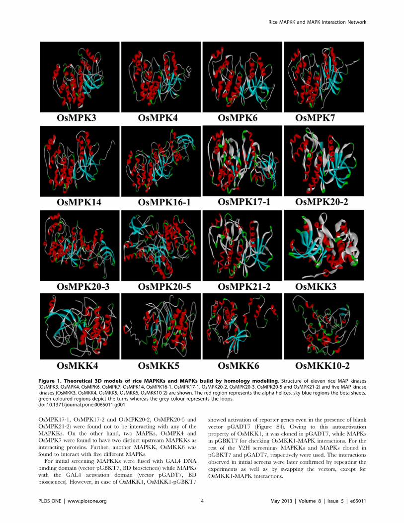
eleven MAPKs and five MAPKKs have been shown (Figure 1).
The DOPE (Discrete Optimized Protein Energy) and PDF
(Probability Density Function) total score for all the 3D structure
are given in Table 1. DOPE score is an atomic based statistical
potential in MODELER package for model evaluation and
structure prediction. The DOPE score of a protein can be viewed
as a conformational energy which measures the relative stability of
a conformation with respect to other conformations of the same
protein. The PDF energy is useful for evaluating the relative
overall condition of each model. As per the protocol, lower values
of DOPE score and PDF total energy represent a better model.
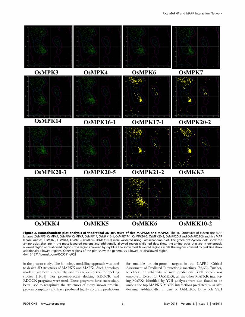
Model AssessmentThe overall stereochemical quality of the modelled 3D structure
of proteins were evaluated by using Ramachandran plot which is
based on psi (Ca-C bond) and phi (N-Ca bond) angles of the
protein and provides information about the number of amino acid
residues present in allowed and disallowed regions. All the
modelled proteins showed maximum residues in the most favoured
region followed by allowed region and least in the generously
allowed regions in the Ramachandran plot (Figure 2). Very few
residues were also found lying in the disallowed regions. In all, the
results suggest the reliability of the modelled proteins.
Additionally reliability of the structures for docking studies was
evaluated by employing the ‘Verify Protein protocol’ (profile 3-D
method) for testing a preliminary protein structure based on
experimental data. It depends on the principle, that a protein’s
structure must be compatible with its own sequence. The ‘Verify
Protein’ was used to calculate local 3D-1D scores in a fixed length
sliding window (typically about 5 to 20 residues long) and plotted
against residue position. This reveals local regions of relatively
high or low 3D-1D compatibility [29]. The program evaluates
fitness of a protein sequence in its current 3D environment. Line
plots for all the 3D structures of proteins were drawn and the
values of the verify score are given in Table 1. The verification
scores of all the sixteen modelled proteins lie between the low and
high expected verify score indicating that the modelled protein
structures are of acceptable quality. After verification, the ‘prepare
protein’ protocol was run on the generated models which ensured
removal of any alternate conformations.
Protein-protein Docking for Identification of Interactionbetween Rice MAPKKs and MAPKsThe docking of rice MAPKKs and MAPKs was performed
following ZDOCK and RDOCK programs. ZDOCK is a docking
program that predicts several protein complexes using Pairwise
Shape Complementarity (PSC) of input protein structure [26].
The output results of RDOCK contain two important scores,
ZRANK and the E_RDOCK (Energy RDOCK). For prediction
of the better docking pose ‘E_RDOCK score’ is often preferred
over ZRANK score [27]. The clashes in the selected poses were
zero thereby suggesting better docking positions. Higher negative
score of E_RDOCK could be used to predict the possible protein-
protein interactions from a set of proteins since it indicates
stronger interaction.
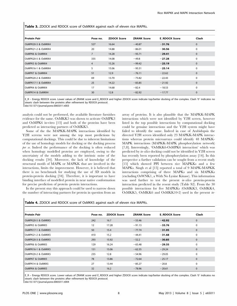
The results obtained from docking studies involving OsMKK3
and eleven MAPK modules have been presented in Table 2. The
representative docking positions of OsMKK3-OsMAPKs com-
plexes have been shown in Figure 3. The lowest ‘E_RDOCK
score’ was recorded for OsMKK3-OsMPK20-3 (227.31) followed
by OsMKK3-OsMPK21-2 (227.20) while OsMKK3-OsMPK6
showed highest E_RDOCK score (212.49). Among the eleven
OsMAPKs top six putative interacting partners of OsMKK3 were
OsMPK20-3, OsMPK21-2, OsMPK20-2 (224.48), OsMPK20-5
(224.20), OsMPK14 (220.9), and OsMPK7 (220.46).
Docking of OsMKK4 with eleven MAPKs was performed and
output values are presented in Table 3. Figure S1 shows the
docking positions of OsMKK4-OsMAPKs complex. Based on
E_RDOCK score top six putative OsMKK4 interacting OsMPKs
were OsMPK20-3 (231.76), OsMPK21-2 (230.56), OsMPK6
(229.41), OsMPK20-5 (227.28), OsMPK3 (225.19) and
OsMPK16-1 (225.14).
For OsMKK5, top six interacting MAPKs were OsMPK20-5,
OsMPK3, OsMPK17-1, OsMPK21-2, OsMPK20-3 and
OsMPK6 (Table 4). The best docking positions of OsMKK5-
OsMAPKs are shown in Figure S2.
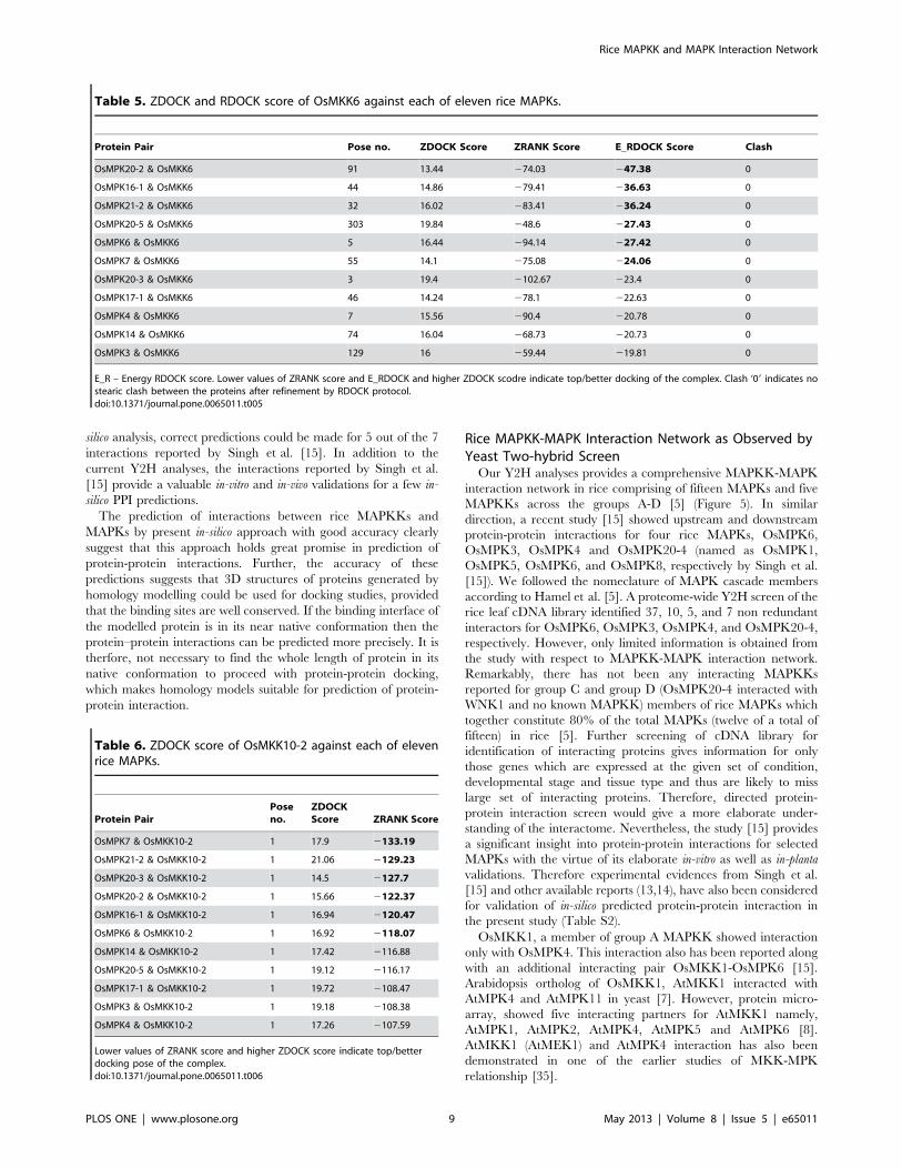
Top six predictions of OsMKK6 interacting OsMAPKs were
OsMPK20-2, OsMPK16-1, OsMPK21-2, OsMPK20-5,
OsMPK6 and OsMPK7 (Table 5). Figure S3 shows the top
docking positions of OsMKK6-OsMPKs complexes.
In case of OsMKK10-2, after the initial docking (ZDOCK),
OsMKK10-2 could not yield successful RDOCK with any of the
OsMAPKs. Nonetheless, in the absence of E_RDOCK score, the
other next important ‘ZRANK score’ [30] obtained from initial
docking studies (ZDOCK) was used to predict the interaction of
OsMKK10-2 with OsMAPKs (Table 6). Similar to E_RDOCK
score, lower values of ZRANK score indicate better docking pair
and thus can be used to predict top interacting pair of proteins.
Following this approach, the top putative OsMKK10-2 interacting
OsMAPKs were OsMPK7, OsMPK21-2, OsMPK20-3,
OsMPK20-2, OsMPK16-1 and OsMPK6.
Study of Interactions between Rice MAPKKs and MAPKsusing Yeast Two-hybrid (Y2H) AssayIn Y2H analysis a total of 11 interactions were identified from
75 combinations of five MAPKKs and fifteen MAPKs. Each
MAPKK was found to have at least one MAPK as interacting
partner whereas, interestingly six MAPKs (OsMPK16-2,
Rice MAPKK and MAPK Interaction Network
PLOS ONE | www.plosone.org 3 May 2013 | Volume 8 | Issue 5 | e65011
OsMPK17-1, OsMPK17-2 and OsMPK20-2, OsMPK20-5 and
OsMPK21-2) were found not to be interacting with any of the
MAPKKs. On the other hand, two MAPKs, OsMPK4 and
OsMPK7 were found to have two distinct upstream MAPKKs as
interacting proteins. Further, another MAPKK, OsMKK6 was
found to interact with five different MAPKs.
For initial screening MAPKKs were fused with GAL4 DNA
binding domain (vector pGBKT7, BD biosciences) while MAPKs
with the GAL4 activation domain (vector pGADT7, BD
biosciences). However, in case of OsMKK1, OsMKK1-pGBKT7
showed activation of reporter genes even in the presence of blank
vector pGADT7 (Figure S4). Owing to this autoactivation
property of OsMKK1, it was cloned in pGADT7, while MAPKs
in pGBKT7 for checking OsMKK1-MAPK interactions. For the
rest of the Y2H screenings MAPKKs and MAPKs cloned in
pGBKT7 and pGADT7, respectively were used. The interactions
observed in initial screens were later confirmed by repeating the
experiments as well as by swapping the vectors, except for
OsMKK1-MAPK interactions.
Figure 1. Theoretical 3D models of rice MAPKKs and MAPKs build by homology modelling. Structure of eleven rice MAP kinases(OsMPK3, OsMPK4, OsMPK6, OsMPK7, OsMPK14, OsMPK16-1, OsMPK17-1, OsMPK20-2, OsMPK20-3, OsMPK20-5 and OsMPK21-2) and five MAP kinasekinases (OsMKK3, OsMKK4, OsMKK5, OsMKK6, OsMKK10-2) are shown. The red region represents the alpha helices, sky blue regions the beta sheets,green coloured regions depict the turns whereas the grey colour represents the loops.doi:10.1371/journal.pone.0065011.g001
Rice MAPKK and MAPK Interaction Network
PLOS ONE | www.plosone.org 4 May 2013 | Volume 8 | Issue 5 | e65011
OsMKK1, a group A MAPKK showed interaction with
OsMPK4, a group B MAPK (Figure 4, panel 1-2). OsMKK6,
the other member of group A MAPKK showed interaction with
five MAPKs namely OsMPK4, OsMPK16-1, OsMPK20-1,
OsMPK20-3 and OsMPK20-4 (Figure 4, panel 7-8).
OsMKK3 which alone constitutes group B rice MAPKK
interacted with OsMPK7 and OsMPK14. Interestingly both
OsMPK7 and OsMPK14 belonged to the same group of MAPKs
(group C) (Figure 4, panel 3-4). However both the interactions
were moderate in nature in comparison to the positive control.
A representative member of group C MAPKKs, OsMKK4 was
found to interact with OsMPK3 and OsMPK6 (Figure 4, panel 5-
6). The OsMKK4-OsMPK6 interaction was found to be stronger
than the OsMKK4-OsMPK3 interaction.
OsMKK10-2, the only member of group D MAPKK which
shows active transcription (data not shown) showed interaction
with OsMPK7, albeit weak in nature (Figure 4, panel 9-10).
Harmony between in-silico Predicted Protein-proteinInteractions by Docking Study and Yeast Two-hybridScreenIn order to substantiate the prediction of protein-protein
interaction by computational docking, a vis-a-vis comparison of
the same was made with Y2H screen (Table 7). In case of
OsMKK3, the interacting MAPKs, OsMPK14 and OsMPK7
identified by Y2H were also present in the top six possible
interactions and were found to be at 5th and 6th positions,
respectively. For OsMKK4, two interactions identified by Y2H
analyses OsMKK4-OsMPK6 and OsMKK4-OsMPK3 were also
found to be at the top six possible interacting pairs and were at 3rd
and 5th positions, respectively. Although, OsMKK5 was not
included in the Y2H screening, a previous study demonstrated that
OsMKK5 could activate OsMPK3 and OsMPK6 [13]. Both
OsMPK3 and OsMPK6 were among the top six predicted
interacting MAPKs of OsMKK5. In Y2H screen, OsMKK6 was
shown to be interacting with five MAPKs namely, OsMPK4,
OsMPK16-1, OsMPK20-1, OsMPK20-3, and OsMPK20-4.
Since, OsMPK20-1 and OsMPK20-4 were not included in the
docking study, only OsMPK16-1 could make to the list of top six
predictions by computational dockings. In case of OsMKK10-2
the RDOCK protocol could not be successfully operated. Hence
the second best preferred score, ZRANK was considered for
prediction of possible interacting partner of OsMKK10-2. In Y2H
screens OsMKK10-2 was shown to interact with OsMPK7, and
interestingly the same was also the top most possible interacting
MAPK in the computational docking studies.
Over all, with the exception of OsMKK6, all the other
MAPKK-MAPK interactions identified by Y2H study as well as
those reported elsewhere were among the topmost interactions
predicted by present computational docking. These results indicate
that the computational docking approach using ZDOCK and
RDOCK programmes are reliable and can be used for
identification of possible protein-protein interactions.
Discussion
The in-silico approach was used to predict protein-protein
interaction between rice MAPKKs and MAPKs. The computa-
tional protein-protein docking, used in the present study can be
utilized to predict large scale protein-protein interactions, espe-
cially for kinases. In the present study, this approach was followed
in order to answer the following questions: 1) if the homology
based 3-D structures of proteins could be used for protein–protein
docking in the absence of experimentally elucidated structure? 2) If
the computational protein-protein docking approach be employed
to predict protein-protein interactions? If yes, 3) how reliable such
an approach could be?
Since there are very limited reports which use the in-silico
approach to study protein-protein interactions, it is considered
necessary to render experimental basis to the outcomes of the
docking studies. To achieve the same, computational protein-
protein docking was further confirmed by directed Y2H analyses
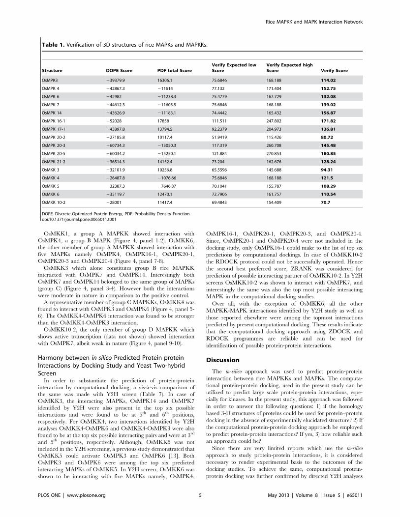
Table 1. Verification of 3D structures of rice MAPKs and MAPKKs.
Structure DOPE Score PDF total ScoreVerify Expected lowScore
Verify Expected highScore Verify Score
OsMPK3 239379.9 16306.1 75.6846 168.188 114.02
OsMPK 4 242867.3 211614 77.132 171.404 152.75
OsMPK 6 242982 211238.3 75.4779 167.729 132.08
OsMPK 7 244612.3 211605.5 75.6846 168.188 139.02
OsMPK 14 243626.9 211183.1 74.4442 165.432 156.87
OsMPK 16-1 252028 17858 111.511 247.802 171.82
OsMPK 17-1 243897.8 13794.5 92.2379 204.973 136.81
OsMPK 20-2 227185.8 10117.4 51.9419 115.426 80.72
OsMPK 20-3 260734.3 215050.3 117.319 260.708 145.48
OsMPK 20-5 260034.2 215250.1 121.884 270.853 180.85
OsMPK 21-2 236514.3 14152.4 73.204 162.676 128.24
OsMKK 3 232101.9 10256.8 65.5596 145.688 94.31
OsMKK 4 226487.8 21076.66 75.6846 168.188 121.5
OsMKK 5 232387.3 27646.87 70.1041 155.787 108.29
OsMKK 6 235119.7 12470.1 72.7906 161.757 110.54
OsMKK 10-2 228001 11417.4 69.4843 154.409 70.7
DOPE–Discrete Optimized Protein Energy, PDF–Probability Density Function.doi:10.1371/journal.pone.0065011.t001
Rice MAPKK and MAPK Interaction Network
PLOS ONE | www.plosone.org 5 May 2013 | Volume 8 | Issue 5 | e65011
in the present study. The homology modelling approach was used
to design 3D structures of MAPKK and MAPKs. Such homology
models have been successfully used by earlier workers for docking
studies [19,31]. For protein-protein docking ZDOCK and
RDOCK programs were used. These programs have successfully
been used to recapitulat the structures of many known protein-
protein complexes and have produced highly accurate predictions
for multiple protein-protein targets in the CAPRI (Critical
Assessment of Predicted Interactions) meetings [32,33]. Further,
to check the reliability of such predictions, Y2H screen was
employed. Except for OsMKK6, all the other MAPKK interact-
ing MAPKs identified by Y2H analyses were also found to be
among the top MAPKK-MAPK interactions predicted by in-silico
docking. Additionally, in case of OsMKK5, for which Y2H
Figure 2. Ramachandran plot analysis of theoretical 3D structure of rice MAPKKs and MAPKs. The 3D Structures of eleven rice MAPkinases (OsMPK3, OsMPK4, OsMPK6, OsMPK7, OsMPK14, OsMPK16-1, OsMPK17-1, OsMPK20-2, OsMPK20-3, OsMPK20-5 and OsMPK21-2) and five MAPkinase kinases (OsMKK3, OsMKK4, OsMKK5, OsMKK6, OsMKK10-2) were validated using Ramachandran plot. The green dots/yellow dots show theamino acids that are in the most favoured regions and additionally allowed region while red dots show the amino acids that are in generouslyallowed region or disallowed regions. The regions covered by sky blue line show most favoured regions, while the regions covered by pink line showadditionally allowed regions. Other regions of the plot show the generously allowed or disallowed region.doi:10.1371/journal.pone.0065011.g002
Rice MAPKK and MAPK Interaction Network
PLOS ONE | www.plosone.org 6 May 2013 | Volume 8 | Issue 5 | e65011
Figure 3. Top docking poses of OsMKK3 with rice MAPKs. The best docking positions of OsMKK3 with each of the eleven rice MAPKs(OsMPK3, OsMPK4, OsMPK6, OsMPK7, OsMPK14, OsMPK16-1, OsMPK17-1, OsMPK20-2, OsMPK20-3, OsMPK20-5 and OsMPK21-2) are shown. OsMKK3is represented as a wireframe pattern whereas all OsMAPKs as a solid ribbon form.doi:10.1371/journal.pone.0065011.g003
Table 2. ZDOCK and RDOCK score of OsMKK3 against each of eleven rice MAPKs.
Protein Pair Pose no. ZDOCK Score ZRANK Score E_RDOCK Score Clash
OsMPK20-3 & OsMKK3 671 14.98 226.10 227.31 0
OsMPK21-2 & OsMKK3 1 14.1 2116.38 227.20 0
OsMPK20-2 & OsMKK3 815 13.30 219.67 224.48 0
OsMPK20-5 & OsMKK3 9 16.48 230.96 224.20 0
OsMPK14 & OsMKK3 162 13.46 255.36 220.9 0
OsMPK7 & OsMKK3 29 13.52 275.1 220.46 0
OsMPK3 & OsMKK3 211 14.86 248.21 219.42 0
OsMPK4 & OsMKK3 11 14.26 288.23 219.26 0
OsMPK16-1 & OsMKK3 110 15.62 263.91 218.72 0
OsMPK17-1 & OsMKK3 143 15.04 257.92 217.3 0
OsMPK6 & OsMKK3 48 18.08 270.21 212.49 0
E_R – Energy RDOCK score. Lower values of ZRANK score and E_RDOCK and higher ZDOCK score indicate top/better docking of the complex. Clash ‘09 indicates nostearic clash between the proteins after refinement by RDOCK protocol.doi:10.1371/journal.pone.0065011.t002
Rice MAPKK and MAPK Interaction Network
PLOS ONE | www.plosone.org 7 May 2013 | Volume 8 | Issue 5 | e65011
analysis could not be performed, the available literature furnishes
evidence for the same. OsMKK5 was shown to activate OsMPK3
and OsMPK6 in-vitro [13] and both of the proteins have been
predicted as interacting partners of OsMKK5.
Some of the the MAPKK-MAPK interactions identified by
Y2H screens were not among the top most predictions by
computational dockings. This could be due to inherent limitations
of the use of homology models for docking or the docking process
per se. Indeed the performance of the docking is often reduced
when homology modelled proteins are employed, owing to the
uncertainty of the models adding to the intrinsic noise of the
docking results [34]. Moreover, the lack of knowledge of the
structural motifs of MAPK or MAPKK that are involved in the
interactions, limits the improvement. However, it is believed that
there is no benchmark for studying the use of 3D models in
protein-protein docking [34]. Therefore, it is important to have
binding interface of modelled protein in near native conformation
for precise prediction of protein–protein interactions.
In the present stuy this approach could be used to narrow down
the number of interacting partners for protein in question from an
array of proteins. It is also plausible that the MAPKK-MAPK
interactions which were not identified by Y2H screen, however
listed in the top possible interactions by computational docking
could be genuine interactions and the Y2H system might have
failed to identify the same. Indeed in case of Arabidopsis the
directed Y2H screen identified only 23 MAPKK-MAPK interac-
tions whereas protein microarrays could identify 48 MAPKK-
MAPK interactions (MAPKK-MAPK phosphorylation network)
[7,8]. Interestingly, ‘OsMKK6-OsMPK6 interaction’ which was
predicted by in-silico docking could not be identified in Y2H screen
has recently been reported by phosphorylation assay [14]. In this
perspective a further validation can be sought from a recent study
[15] which showed PPI between rice MAPKKs and a few
MAPKs. Singh et al [15] reported a total of 9 MAPK-MAPKK
interactions comprising of three MAPKs and six MAPKKs
(excluding OsWNK1, a With No Lysine Kinase). This information
was used further to test the present in-silico protein-protein
interaction predicted in the resent study (Table S2). From the 30
possible interactions for five MAPKKs (OsMKK3, OsMKK4,
OsMKK5, OsMKK6 and OsMKK10-2) used in the present in-
Table 3. ZDOCK and RDOCK score of OsMKK4 against each of eleven rice MAPKs.
Protein Pair Pose no. ZDOCK Score ZRANK Score E_RDOCK Score Clash
OsMPK20-3 & OsMKK4 527 16.64 240.87 231.76 0
OsMPK21-2 & OsMKK4 20 14.88 286.01 230.56 0
OsMPK6 & OsMKK4 7 14.28 293.71 229.41 0
OsMPK20-5 & OsMKK4 335 14.08 249.8 227.28 0
OsMPK3 & OsMKK4 4 15.26 299.42 225.19 0
OsMPK16-1 & OsMKK4 5 15.06 295.51 225.14 0
OsMPK7 & OsMKK4 51 12.9 276.11 222.62 0
OsMPK20-2 & OsMKK4 69 13.70 273.82 222.43 0
OsMPK17-1 & OsMKK4 25 14.22 283.85 221.92 0
OsMPK4 & OsMKK4 17 14.68 282.4 218.53 0
OsMPK14 & OsMKK4 30 12.8 282.92 217.77 0
E_R – Energy RDOCK score. Lower values of ZRANK score and E_RDOCK and higher ZDOCK score indicate top/better docking of the complex. Clash ‘09 indicates nostearic clash between the proteins after refinement by RDOCK protocol.doi:10.1371/journal.pone.0065011.t003
Table 4. ZDOCK and RDOCK score of OsMKK5 against each of eleven rice MAPKs.
Protein Pair Pose no. ZDOCK Score ZRANK Score E_RDOCK Score Clash
OsMPK20-5 & OsMKK5 242 16.7 255.46 242.92 0
OsMPK3 & OsMKK5 12 17.28 291.13 233.76 0
OsMPK17-1 & OsMKK5 50 15.4 277.74 231.95 0
OsMPK21-2 & OsMKK5 410 15.2 244.41 231.68 0
OsMPK20-3 & OsMKK5 293 15.92 252.2 230.85 0
OsMPK6 & OsMKK5 129 16.24 265.48 229.35 0
OsMPK16-1 & OsMKK5 151 15.06 262.35 229.24 0
OsMPK20-2 & OsMKK5 235 12.8 254.96 229.05 0
OsMPK7 & OsMKK5 78 13.86 272.64 225.17 0
OsMPK14 & OsMKK5 27 13.44 285.27 220.8 0
OsMPK4 & OsMKK5 32 16.2 278.96 220.61 0
E_R – Energy RDOCK score. Lower values of ZRANK score and E_RDOCK and higher ZDOCK score indicate top/better docking of the complex. Clash ‘09 indicates nostearic clash between the proteins after refinement by RDOCK protocol.doi:10.1371/journal.pone.0065011.t004
Rice MAPKK and MAPK Interaction Network
PLOS ONE | www.plosone.org 8 May 2013 | Volume 8 | Issue 5 | e65011
silico analysis, correct predictions could be made for 5 out of the 7
interactions reported by Singh et al. [15]. In addition to the
current Y2H analyses, the interactions reported by Singh et al.
[15] provide a valuable in-vitro and in-vivo validations for a few in-
silico PPI predictions.
The prediction of interactions between rice MAPKKs and
MAPKs by present in-silico approach with good accuracy clearly
suggest that this approach holds great promise in prediction of
protein-protein interactions. Further, the accuracy of these
predictions suggests that 3D structures of proteins generated by
homology modelling could be used for docking studies, provided
that the binding sites are well conserved. If the binding interface of
the modelled protein is in its near native conformation then the
protein–protein interactions can be predicted more precisely. It is
therfore, not necessary to find the whole length of protein in its
native conformation to proceed with protein-protein docking,
which makes homology models suitable for prediction of protein-
protein interaction.
Rice MAPKK-MAPK Interaction Network as Observed byYeast Two-hybrid ScreenOur Y2H analyses provides a comprehensive MAPKK-MAPK
interaction network in rice comprising of fifteen MAPKs and five
MAPKKs across the groups A-D [5] (Figure 5). In similar
direction, a recent study [15] showed upstream and downstream
protein-protein interactions for four rice MAPKs, OsMPK6,
OsMPK3, OsMPK4 and OsMPK20-4 (named as OsMPK1,
OsMPK5, OsMPK6, and OsMPK8, respectively by Singh et al.
[15]). We followed the nomeclature of MAPK cascade members
according to Hamel et al. [5]. A proteome-wide Y2H screen of the
rice leaf cDNA library identified 37, 10, 5, and 7 non redundant
interactors for OsMPK6, OsMPK3, OsMPK4, and OsMPK20-4,
respectively. However, only limited information is obtained from
the study with respect to MAPKK-MAPK interaction network.
Remarkably, there has not been any interacting MAPKKs
reported for group C and group D (OsMPK20-4 interacted with
WNK1 and no known MAPKK) members of rice MAPKs which
together constitute 80% of the total MAPKs (twelve of a total of
fifteen) in rice [5]. Further screening of cDNA library for
identification of interacting proteins gives information for only
those genes which are expressed at the given set of condition,
developmental stage and tissue type and thus are likely to miss
large set of interacting proteins. Therefore, directed protein-
protein interaction screen would give a more elaborate under-
standing of the interactome. Nevertheless, the study [15] provides
a significant insight into protein-protein interactions for selected
MAPKs with the virtue of its elaborate in-vitro as well as in-planta
validations. Therefore experimental evidences from Singh et al.
[15] and other available reports (13,14), have also been considered
for validation of in-silico predicted protein-protein interaction in
the present study (Table S2).
OsMKK1, a member of group A MAPKK showed interaction
only with OsMPK4. This interaction also has been reported along
with an additional interacting pair OsMKK1-OsMPK6 [15].
Arabidopsis ortholog of OsMKK1, AtMKK1 interacted with
AtMPK4 and AtMPK11 in yeast [7]. However, protein micro-
array, showed five interacting partners for AtMKK1 namely,
AtMPK1, AtMPK2, AtMPK4, AtMPK5 and AtMPK6 [8].
AtMKK1 (AtMEK1) and AtMPK4 interaction has also been
demonstrated in one of the earlier studies of MKK-MPK
relationship [35].
Table 5. ZDOCK and RDOCK score of OsMKK6 against each of eleven rice MAPKs.
Protein Pair Pose no. ZDOCK Score ZRANK Score E_RDOCK Score Clash
OsMPK20-2 & OsMKK6 91 13.44 274.03 247.38 0
OsMPK16-1 & OsMKK6 44 14.86 279.41 236.63 0
OsMPK21-2 & OsMKK6 32 16.02 283.41 236.24 0
OsMPK20-5 & OsMKK6 303 19.84 248.6 227.43 0
OsMPK6 & OsMKK6 5 16.44 294.14 227.42 0
OsMPK7 & OsMKK6 55 14.1 275.08 224.06 0
OsMPK20-3 & OsMKK6 3 19.4 2102.67 223.4 0
OsMPK17-1 & OsMKK6 46 14.24 278.1 222.63 0
OsMPK4 & OsMKK6 7 15.56 290.4 220.78 0
OsMPK14 & OsMKK6 74 16.04 268.73 220.73 0
OsMPK3 & OsMKK6 129 16 259.44 219.81 0
E_R – Energy RDOCK score. Lower values of ZRANK score and E_RDOCK and higher ZDOCK scodre indicate top/better docking of the complex. Clash ‘09 indicates nostearic clash between the proteins after refinement by RDOCK protocol.doi:10.1371/journal.pone.0065011.t005
Table 6. ZDOCK score of OsMKK10-2 against each of elevenrice MAPKs.
Protein PairPoseno.
ZDOCKScore ZRANK Score
OsMPK7 & OsMKK10-2 1 17.9 2133.19
OsMPK21-2 & OsMKK10-2 1 21.06 2129.23
OsMPK20-3 & OsMKK10-2 1 14.5 2127.7
OsMPK20-2 & OsMKK10-2 1 15.66 2122.37
OsMPK16-1 & OsMKK10-2 1 16.94 2120.47
OsMPK6 & OsMKK10-2 1 16.92 2118.07
OsMPK14 & OsMKK10-2 1 17.42 2116.88
OsMPK20-5 & OsMKK10-2 1 19.12 2116.17
OsMPK17-1 & OsMKK10-2 1 19.72 2108.47
OsMPK3 & OsMKK10-2 1 19.18 2108.38
OsMPK4 & OsMKK10-2 1 17.26 2107.59
Lower values of ZRANK score and higher ZDOCK score indicate top/betterdocking pose of the complex.doi:10.1371/journal.pone.0065011.t006
Rice MAPKK and MAPK Interaction Network
PLOS ONE | www.plosone.org 9 May 2013 | Volume 8 | Issue 5 | e65011
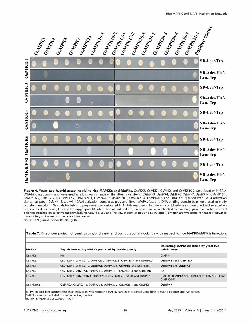
Figure 4. Yeast two-hybrid assay involving rice MAPKKs and MAPKs. OsMKK3, OsMKK4, OsMKK6 and OsMKK10-2 were fused with GAL4DNA-binding domain and were used as a bait against each of the fifteen rice MAPKs (OsMPK3, OsMPK4, OsMPK6, OsMPK7, OsMPK14, OsMPK16-1,OsMPK16-2, OsMPK17-1, OsMPK17-2, OsMPK20-1, OsMPK20-2, OsMPK20-3, OsMPK20-4, OsMPK20-5 and OsMPK21-2) fused with GAL4 activationdomain as preys. OsMKK1 fused with GAL4 activation domain as prey and fifteen MAPKs fused to DNA-binding domain baits were used to studyprotein interactions. Plasmids for bait and prey were co-transformed in AH109 yeast strain in different combinations as mentioned and selected onnutrient medium lacking Leu and Trp (upper panels). Interaction of bait and prey combinations were checked by assessing growth of co-transformedcolonies streaked on selective medium lacking Ade, His, Leu and Trp (lower panels). p53 and SV40 large T-antigen are two proteins that are known tointeract in yeast were used as a positive control.doi:10.1371/journal.pone.0065011.g004
Table 7. Direct comparison of yeast two-hybrid assay and computational dockings with respect to rice MAPKK-MAPK interaction.
MAPKK Top six interacting MAPKs predicted by docking studyInteracting MAPKs identified by yeast two-hybrid screen
OsMKK1 NA OsMPK4
OsMKK3 OsMPK20-3, OsMPK21-2, OsMPK20-2, OsMPK20-5, OsMPK14 and OsMPK7 OsMPK14 and OsMPK7
OsMKK4 OsMPK20-3, OsMPK21-2, OsMPK6, OsMPK20-5, OsMPK3 and OsMPK16-1 OsMPK6 and OsMPK3
OsMKK5 OsMPK20-5, OsMPK3, OsMPK21-2, OsMPK17-1, OsMPK20-3 and OsMPK6 NA
OsMKK6 OsMPK20-2, OsMPK16-1, OsMPK21-2, OsMPK20-5, OsMPK6 and OsMPK7 OsMPK4, OsMPK16-1, OsMPK20-1*, OsMPK20-3 andOsMPK20-4*
OsMKK10-2 OsMPK7, OsMPK21-2, OsMPK20-3, OsMPK20-2, OsMPK16-1 and OsMPK6 OsMPK7
MAPKs in bold font suggests that their interaction with respective MAPKK have been reported using both in-silico prediction and Y2H screen.*MAPKs were not included in in-silico docking studies.doi:10.1371/journal.pone.0065011.t007
Rice MAPKK and MAPK Interaction Network
PLOS ONE | www.plosone.org 10 May 2013 | Volume 8 | Issue 5 | e65011

OsMKK6 the other member of group A MAPKK, was found to
interact with the maximum number of MAPKs (OsMPK4,
OsMPK16-1, OsMPK20-1, OsMPK20-3 and OsMPK20-4).
Among the identified interactions OsMKK6-OsMPK4 interaction
has also been reported earlier [12] using Y2H system as well as the
BiFC system. Other OsMKK6-OsMAPKs interactions have not
been reported earlier. Although, OsMKK6-OsMPK3 and
OsMKK6-OsMPK6 interactions have been reported earlier using
Y2H studies/phosphorylation [11,12,14], in the present Y2H
screen we could not obtain these interactions. Interestingly,
another study using Y2H showed OsMKK6 (OsMEK1) interact-
ing with OsMPK6 (OsMPK1) and OsMPK4 (OsMPK6) [15]. The
inherent limitation of Y2H assay in producing both false positive
and false negatives results may be accounted for such discrepan-
cies. Further, it is also believed that different Y2H formats can also
identify different protein-protein interactions [7]. The differenece
in the Y2H format used in the above mentioned studies in
comparision to that used in the present work could attribute to the
failure to demonstrate a few known interactions. Another reason
could be the transient nature of protein-protein interaction
especially in the case of the signalling molecules. Additionally, it
is interesting to note that AtMPK3, an ortholog of OsMPK3 in
Arabidopsis showed no interaction with AtMKK6, but could
interact with only one MAPKK, AtMKK4 [7]. Moreover, this
interaction was also among the weakest interactions observed in
the study.
OsMKK3, the only member of group B MAPKK, was found to
interact with two rice MAPKs (OsMPK7 and OsMPK14) both
belonging to group C of MAPKs [5]. However, another study has
demonstrated the interaction of OsMKK3 (OsMEK8a) with
OsMPK6 (OsMPK1), though the interaction was found to be
weak in nature [15]. Further, two independent studies showed
interaction of AtMKK3 (OsMKK3 ortholog in Arabidopsis) with
group C members of Arabidopsis MAPKs (AtMPK1, AtMPK2,
AtMPK7 and AtMPK14) [7,36].
OsMKK4, a representative of group C MAPKK showed
interaction with two MAPKs, OsMPK3 and OsMPK6. This is in
agreement with another study where OsMKK4 was shown to
activate OsMPK3 and OsMPK6 in-vivo and involved in phyto-
alexin biosynthesis in rice cell culture [13]. An independent study
also reported similar interacting partner for OsMKK4 [15].
OsMKK10-2, the only member of group D MAPKKs, showed
interaction with OsMPK7 albeit weak in nature. In contrast to
this, in a previous study, OsMKK10-2 (OsMEK3) was found to
interact with OsMPK6 (OsMPK1) [15]. Intriguingly, OsMKK10-
2 showed only partial MAPKK consensus motif. The other
paralogs, OsMKK10-1 and OsMKK10-3 also lack MAPKK
consensus motif and showed no transcripts in any of the libraries of
MPSS database (Data not shown). Further, the Arabidopsis
Figure 5. Protein-protein interaction network among rice MAPKKs and MAPKs based on Y2H analyses and in-silico predictions. Solidlines from upper panel OsMAPKKs to middle panel OsMAPKs indicate findings from yeast two-hybrid screen while dashed line from lower panelOsMAPKKs to middle panel OsMAPKs indicate findings from in-silico protein-protein dockings analyses. Lines originating from specific OsMAPKK arerepresented in same colour.doi:10.1371/journal.pone.0065011.g005
Rice MAPKK and MAPK Interaction Network
PLOS ONE | www.plosone.org 11 May 2013 | Volume 8 | Issue 5 | e65011

AtMKK10 also lacks part of MAPKK consensus motif and is
thought to be biologically non-functional [5,37]. Y2H screen of
AtMKK1 showed interaction only with one MAPK, AtMPK17
[7]. However, protein microarray revealed activation of five
MAPKs including, AtMPK1, AtMPK2, AtMPK4, AtMPK5 and
AtMPK6 [8].
Findings from the Y2H work showed that none of the rice
MAPKKs included in the Y2H screen could interact with
OsMPK16-2, OsMPK17-1, OsMPK17-2, OsMPK20-2,
OsMPK20-5 and OsMPK21-1. Similarly, a Y2H study [7]
comprising seven Arabidopsis MAPKs (AtMPK5, 8, 9, 12, 16,
18, and 19) showed no interactions with any of the selected
AtMAPKKs. In order to obtain a substatial interaction of
MAPKKs with MAPKs, we might need scaffold proteins as
observed in yeast. Scaffold proteins bring MAPK components
together to enhance specificity and accelerate their activation and
reaction rates. For example, yeast Ste5 was found to interact with
an upstream G-protein and along with all three components of the
MAPK cascade [38].
Comparison of MAPKKs and MAPKs orthologs between rice
and Arabidopsis revealed a few new interacting modules. Changes
in docking domain over the period of evolution could be
accounted for these new interactions since docking domains are
major determinants of the specificity of interactions between
MAPKKs and MAPKs [39–41].
The present work apart from improving our understanding
about the MAPK-MAPKK interaction network in rice, also shows
that the computational approach could be employed to explore
large scale protein-protein interactions.
ConclusionsThe MAPK and MAPKK interaction network is crucial to
understand the MAPK signalling pathways at cellular level. The
present work represents an in-silico docking approach to predict
protein-protein interactions between rice MAPKKs and MAPKs.
To achieve the same, 3D structures of eleven MAPKs and five
MAPKKs were predicted by homology modelling. ZDOCK and
RDOCK docking programmes were used for computational
dockings and top possible MAPKK-MAPK interacting pairs were
predicted. Further to confirm the reliability of this approach, Y2H
analysis was performed for conforming MAPKK-MAPK interac-
tions. Except for one MAPKK, all the other interacting partners
identified in Y2H assay were listed in the top possible interactions
by computational dockings. The results suggest that 3D structure
built by homology modelling could be used for docking studies.
Since the top interacting pairs identified by docking in the present
work could not be confirmed by Y2H assay in each case, this
approach may be suitable mainly for narrowing down the
interacting partners from several proteins to a fewer members.
The protein-protein interaction study of MAPKKs and MAPKs
gives a comprehensive interaction network for rice MAPKK and
MAPK.
Supporting Information
Figure S1 Top docking poses of OsMKK4 with riceMAPKs. The best docking positions of OsMKK4 with each of the
eleven rice MAPKs (OsMPK3, OsMPK4, OsMPK6, OsMPK7,
OsMPK14, OsMPK16-1, OsMPK17-1, OsMPK20-2,
OsMPK20-3, OsMPK20-5 and OsMPK21-2) are shown.
OsMKK4 is represented as a wireframe pattern whereas all
OsMAPKs as a solid ribbon form.
(PDF)
Figure S2 Top docking poses of OsMKK5 with riceMAPKs. The best docking positions of OsMKK5 with each of the
eleven rice MAPKs (OsMPK3, OsMPK4, OsMPK6, OsMPK7,
OsMPK14, OsMPK16-1, OsMPK17-1, OsMPK20-2,
OsMPK20-3, OsMPK20-5 and OsMPK21-2) are shown.
OsMKK5 is represented as a wireframe pattern whereas all
OsMAPKs as a solid ribbon form.
(PDF)
Figure S3 Top docking poses of OsMKK6 with riceMAPKs. The best docking positions of OsMKK6 with each of the
eleven rice MAPKs (OsMPK3, OsMPK4, OsMPK6, OsMPK7,
OsMPK14, OsMPK16-1, OsMPK17-1, OsMPK20-2,
OsMPK20-3, OsMPK20-5 and OsMPK21-2) are shown.
OsMKK6 is represented as a wireframe pattern whereas all
OsMAPKs as a solid ribbon form.
(PDF)
Figure S4 Yeast two-hybrid assay control experiment.OsMKK1, OsMKK3, OsMKK4, OsMKK6 and OsMKK10-2 cloned in
pGBKT7 were co-transformed with blank pGADT7 to AH109.
The co-transformants were selected on double drop out medium
and later patched on quadruple drop out medium to check auto-
activation of the reporter genes.
(PDF)
Table S1 Details of the proteins used as templates forhomology modelling of rice MAPKKs and MAPKs.(PDF)
Table S2 Rice MAPK and MAPKKs interactions pre-dicted by in-silico docking and its validation as observedin the experimental evidences from current Y2H anal-yses and available literatures.(PDF)
Acknowledgments
Authors thank Accelrys India for providing a trial version of the Discovery
Studio Software. Dr. Shaveta Kanoria, NIPGR is acknowledged for
critically reading and editing the manuscript.
Author Contributions
Conceived and designed the experiments: AKS DPW MM. Performed the
experiments: DPW MM PS. Analyzed the data: AKS DPW MM.
Contributed reagents/materials/analysis tools: AKS. Wrote the paper:
AKS DPW.
References
1. Suarez-Rodriguez MC, Petersen M, Mundy J (2010) Mitogen-Activated Protein
Kinase signalling in plants. Annu Rev Plant Biol 61: 621–649.
2. Sinha AK, Jaggi M, Raghuram B, Tuteja N (2011) Mitogen-activated protein
kinase signalling in plants under abiotic stress Plant Signal Behav 6: 196–203.
3. Doczi R, Okresz L, Romero AE, Paccanaro A, Bogre L (2012) Exploring the
evolutionary path of plant MAPK networks. Trends Plant Sci (In press).
4. MAPK Group (2002) Mitogen-activated protein kinase cascades in plants: a new
nomenclature. Trends Plant Sci 7: 301–308.
5. Hamel LP, Nicole MC, Sritubtim S, Morency MJ, Ellis M, et al. (2006) Ancient
signals: comparative genomics of plant MAPK and MAPKK gene families.
Trends Plant Sci 11: 192–198.
6. Rao KP, Richa T, Kumar K, Raghuram B, Sinha AK (2010) In-silico analysis
reveals 75 members of mitogen-activated protein kinase kinase kinase gene
family in rice. DNA Res 17: 139–153.
7. Lee JS, Huh KW, Bhargava A, Ellis BE (2008) Comprehensive analysis of
protein-protein interactions between Arabidopsis MAPKs and MAPK kinases
Rice MAPKK and MAPK Interaction Network
PLOS ONE | www.plosone.org 12 May 2013 | Volume 8 | Issue 5 | e65011
helps define potential MAPK signalling modules. Plant Signal Behav 3: 1037–
1041.8. Popescu SC, Popescu GV, Bachan S, Zhang Z, Gerstein M, et al. (2009) MAPK
target networks in Arabidopsis thaliana revealed using functional protein
microarrays. Genes Dev 23: 80–92.9. Andreasson E, Ellis B (2010) Convergence and specificity in the Arabidopsis
MAPK nexus. Trends in Plant Science 15: 106–113.10. Kumar K, Wankhede DP, Sinha AK (2012) Signal convergence through the
lenses of MAP kinases: paradigms of stress and hormone signaling in plants.
Frontiers in Biology DOI 10.1007/s11515–012–1207–1.11. Wen JQ, Oono K, Imai R (2002) Two novel Mitogen-Activated Protein
signalling components, OsMEK1 and OsMAP1, are involved in a moderate lowtemperature signaling pathway in rice. Plant Physiol 129: 1880–1891.
12. Ding X, Richter T, Chen M, Fujii H, Seo YS, et al. (2009) A rice kinase-proteininteraction map. Plant Physiol. 149: 1478–1492.
13. Kishi-Kaboshi M, Okada K, Kurimoto L, Murakami S, Umezawa T, et al.
(2010) A rice fungal MAMP responsive MAPK cascade regulates metabolic flowto antimicrobial metabolite synthesis. Plant J 63: 599–612.
14. Xie G, Kato H, Imai R (2012) Biochemical identification of the OsMKK6-OsMPK3 signalling pathway for chilling stress tolerance in rice. Biochem J 443:
95–102.
15. Singh R, Lee MO, Lee JE, Choi J, Park JH, et al. (2012) Rice mitogen-activatedprotein kinase interactome analysis using the yeast two-hybrid system. Plant
Physiol 160: 477–488.16. Halperin I, Ma B, Wolfson H, Nussinov R (2002) Principles of docking: an
overview of search algorithms and a guide to scoring functions. Proteins 47: 409–443.
17. Gray JJ (2006) High resolution protein-protein docking. Curr Opin Struct Biol
16: 183–193.18. Sivasubramanian A, Chao G, Pressler HM, Wittrup KD, Gray JJ (2006)
Structural model of the mAb 806-EGFR complex using computational dockingfollowed by computational and experimental mutagenesis. Structures 14: 401–
414.
19. Sharma B (2008) Structure and mechanism of a transmission blocking vaccinecandidate protein Pfs25 from P. falciparum: A molecular modeling and docking
study. In Silico Biol 8: 193–206.20. Tovchigrechko A, Wells CA, Vakser IA (2002) Docking of protein models.
Protein Sci 11: 1888–1896.21. Rao KP, Vani G, Kumar K, Wankhede DP, Mishra M, et al. (2011) Arsenic
stress activates MAP kinase in rice roots and leaves. Arch Biochem Biophys 506:
73–82.22. Altschul SF, Madden TL, Schaffer AA, Zhang J, Miller W, et al. (1997) Gapped
BLAST and PSI-BLAST: a new generation of protein database search program.Nucleic Acids Research 25: 3389–3402.
23. Berman HM, Westbrook J, Feng Z, Gilliland G, Bhat TN, et al. (2000) The
Protein Data Bank. Nucleic Acids Res 28: 235–242.24. Sali A, Potterton L, Yuan F, van Vlijmen H, Karplus M (1995) Evaluation of
comparative protein modeling by MODELLER. Proteins 23: 318–326.
25. Chen R, Mintseris J, Janin J, Weng Z (2003) A protein-protein docking
benchmark. Proteins 52: 88–91.
26. Chen R, Weng ZP (2002) Docking unbound proteins using shape complemen-
tarity, desolvation, and electrostatics. Proteins 47: 281–294.
27. Li L, Chen R, Weng Z (2003) RDOCK: refinement of rigid-body protein
docking predictions. Proteins 53: 693–707.
28. Kumar K, Rao KP, Sharma P, Sinha AK (2008) Differential regulation of rice
mitogen activated protein kinase kinase (MKK) by abiotic stress. Plant Physiol
Biochem 46: 891–897.
29. Luthy R, Bowie JU, Eisenberg D (1992) Assessment of protein models with three
dimensional profiles. Nature 356: 83–85.
30. Pierce B, Weng Z (2007) ZRANK: reranking protein docking predictions with
an optimized energy function. Proteins 67: 1078–1086.
31. Schafferhans A, Klebe G (2001) Docking ligands onto binding site representa-
tions derived from proteins built by homology modelling. J Mol Biol 307: 407–
427.
32. Wiehe K, Pierce B, Mintseris J, Tong WW, Anderson R, et al. (2005) ZDOCK
and RDOCK performance in CAPRI rounds 3, 4, and 5. Proteins 60: 207–213.
33. Wiehe K, Pierce B, Tong WW, Hwang H, Mintseris J, et al. (2007) The
performance of ZDOCK and ZRANK in rounds 6–11 of CAPRI. Proteins 69:
719–725.
34. Pons C, Grosdidier S, Solernou A, Perez-Cano L, Fernandez-Recio J (2010)
Present and future challenges and limitations in protein-protein docking.
Proteins 78: 95–108.
35. Mizoguchi T, Ichimura K, Irie K, Morris P, Giraudat J, et al. (1998)
Identification of a possible MAP kinase cascade in Arabidopsis thaliana based on
pairwise yeast two-hybrid analysis and functional complementation tests of yeast
mutants. FEBS Lett 437: 56–60.
36. Doczi R, Brader G, Pettko-Szandtner A, Rajh I, Djamei A, et al. (2007) The
Arabidopsis mitogen-activated protein kinase kinase MKK3 is upstream of
group C mitogen-activated protein kinases and participates in pathogen
signaling. Plant Cell 19: 3266–3279.
37. Colcombet J, Hirt H (2008) Arabidopsis MAPKs: a complex signalling network
involved in multiple biological processes. Biochem J 413, 217–226.
38. Bhattacharyya RP, Remenyi A, Good MC, Bashor CJ, Falick AM, et al. (2006)
The Ste5 scaffold allosterically modulates signaling output of the yeast mating
pathway. Science 311: 822–26Chen R, Weng ZP (2002) Docking unbound
proteins using shape complementarity, desolvation, and electrostatics. Proteins
47: 281–294.
39. Bardwell AJ, Flatauer LJ, Matsukuma K, Thorner J, Bardwell L (2001) A
conserved docking site in MEKs mediates high-affinity binding to MAP kinases
and cooperates with a scaffold protein to enhance signal transmission. J Biol
Chem 276, 10374–10386.
40. Bardwell AJ, Frankson E, Bardwell L (2009) Selectivity of docking sites in
MAPK kinases. J Biol Chem 284: 13165–13173.
41. Mody A, Weiner J, Ramanathan S (2009) Modularity of MAP kinases allows
deformation of their signalling pathways. Nat Cell Biol 11: 484–491.
Rice MAPKK and MAPK Interaction Network
PLOS ONE | www.plosone.org 13 May 2013 | Volume 8 | Issue 5 | e65011

![Human Mitogen-activated Protein Kinase Kinase 4 as a ......(CANCERRESEARCH57. 4177—4182,October 1, 1997] Advances in Brief Human Mitogen-activated Protein Kinase Kinase 4 as](https://img.pdfslide.us/doc/110x75/6082557b7810d746a5071f39/human-mitogen-activated-protein-kinase-kinase-4-as-a-cancerresearch57.jpg)