Embed Size (px)
Citation preview
A Sperm–Plasma b-N-Acetyl-D-HexosaminidaseInteracting with a Chitinolytic b-N-Acetyl-D-Hexosaminidase in Insect Molting FluidMingbo Qu, Tian Liu, Peng Chen, Qing Yang*
School of Life Science and Biotechnology, Dalian University of Technology, Dalian, China
Abstract
Insects require molting fluids to shed the old cuticle during molting. b-N-acetyl-D-hexosaminidase, known as Hex1, togetherwith various chitinases, is responsible for degrading the chitin component of the old cuticle. This study showed that anotherb-N-acetyl-D-hexosaminidase, termed OfHex3, interacted with Hex1 and functioned in the molting fluid, although thehomolog of OfHex3 was known as a sperm–plasma enzyme functioning in egg–sperm recognition. OfHex3 is an enzymecloned from the insect Asian corn borer, Ostrinia furnacalis, which is one of the most destructive pests of maize. Theenzymatic activity analysis indicated that OfHex3 was able to degrade chitooligosaccharides, but at a lower rate than that ofOfHex1. Because OfHex3 did not have substrate inhibition, we deduced that the presence of OfHex3 might help OfHex1relieve substrate inhibition during chitin degradation during molting. The expression patterns of OfHex3 during O. furnacalisdevelopment were studied by real-time PCR as well as western blot. The results showed that both gene transcription andprotein translation levels of OfHex3 were up-regulated during larval–larval molting. The tissue-specific expression patternanalysis indicated that OfHex3 was mostly localized in the fat body and testis. All these data further supported that Hex3was involved in molting as well as in fertilization. This study may help to understand the complexity of cuticle degradationduring insect molting, and may provide a possible target for pest control.
Citation: Qu M, Liu T, Chen P, Yang Q (2013) A Sperm–Plasma b-N-Acetyl-D-Hexosaminidase Interacting with a Chitinolytic b-N-Acetyl-D-Hexosaminidase inInsect Molting Fluid. PLoS ONE 8(8): e71738. doi:10.1371/journal.pone.0071738
Editor: Wolfgang Blenau, Goethe University Frankfurt, Germany
Received March 27, 2013; Accepted July 3, 2013; Published August 12, 2013
Copyright: � 2013 Qu et al. This is an open-access article distributed under the terms of the Creative Commons Attribution License, which permits unrestricteduse, distribution, and reproduction in any medium, provided the original author and source are credited.
Funding: The authors acknowledge the financial support provided by the National Key Project for Basic Research (2010CB126100), the National Natural ScienceFoundation (31070715, 31101671), the National High Technology Research and Development Program of China (2011AA10A204), the National Key TechnologyR&D Program (2011BAE06B05), the Fundamental Research Funds for the Central Universities (DUT11RC(3)73) of China, and China Postdoctoral ScienceFoundation (2013M530122). The funders had no role in study design, data collection and analysis, decision to publish, or preparation of the manuscript.
Competing Interests: The authors have declared that no competing interests exist.
* E-mail: [email protected]
Introduction
Molting is an important process in insect growth and
development [1]. To molt, an insect synthesizes and secrets
molting fluids into a lumen located between the old and new
cuticle [2]. Because chitin is the primary component of insect
cuticle, one of the main functions of molting fluid is to hydrolyze
this highly polymerized saccharide. The chitinolytic activity of
molting fluids was first reported in the exuvia fluid of Bombyx mori
(Lepidoptera) [2,3], then two kinds of chitinolytic enzymes,
previously termed endo-chitinase and exo-chitinase but now
named chitinase (EC3.2.1.14) and b-N-acetyl-D-hexosaminidase
(Hex; EC 3.2.1.52), respectively, were isolated and characterized.
Chitinases degrade chitin into oligosaccharides, and Hexes
degrade oligosaccharides to N-acetyl-b-D-glucosamine (GlcNAc)
[4].
Insect genomes contain as many as seven genes encoding Hex
that function during different life processes. These genes fall into
four phylogenetic classes [5]. The class I Hexes (Hex1s) possess
chitinolytic activities and have been found in several insects
[6,7,8]. It occurs in dimeric and glycosylated form [6]. Studies of
the enzymatic properties of Hex1 from both Manduca sexta
(Lepidoptera) and B. mori suggested that it could degrade
(GlcNAc)6 to GlcNAc [9] and was more active toward the
synthesized substrate pNP-b-GlcNAc (p-nitrophenyl-N-acetyl-b-D-
glucosaminide) than toward pNP-b-(GlcNAc)2 (p-nitrophenyl -b-
D-chitobiose) [6]. The first crystal structure of an insect Hex1,
OfHex1 from Ostrinia furnacalis (Lepidoptera), provided an elegant
structural explanation for why chitinolytic Hex1s possess highly
catalytic activity towards chitooligosaccharide substrates [10]. The
expression pattern of HEX1 was also studied. In M. sexta, MsHEX1
was mainly expressed in the epidermis on the sixth and seventh
days of the fifth instar [11]. In Tribolium castaneum (Coleoptera),
RNA interference against TcHEX1 interrupted larval–larval,
larval–pupal, and pupal–adult development [5]. In Choristoneura
fumiferana (Lepidoptera), immunohistochemistry staining indicated
that CfHex1 was localized in molting fluid, epidermis and trachea
[12]. Taken together, these data demonstrated that Hex1 is an
enzyme involved in chitin degradation.
Here, we cloned another Hex, named OfHEX3, from the Asian
corn borer, O. furnacalis, an important lepidopteran pest of maize
and cotton. OfHex3 belongs to class III Hexes. Although
previously reported class III enzymes were involved in sperm–
egg interaction [13,14,15,16], this study found that OfHex3
occurred with OfHex1 in molting fluids. Enzymatic activity and
expression pattern both suggested that besides functioning in
fertilization, OfHex3 worked with OfHex1 in cuticle degradation.
PLOS ONE | www.plosone.org 1 August 2013 | Volume 8 | Issue 8 | e71738
Materials and Methods
Molecular Cloning and Sequence Analysis of OfHEX3Total RNA was isolated from fifth-instar larvae of O. furnacalis
using RNAiso Reagent (TaKaRa, Dalian, China). cDNA was
synthesized using 39-Full RACE Core Set Ver.2.0 (TaKaRa) from
2 mg total RNA using random hexamer primers and oligo-(dT) as
reverse transcript primers. Gene-specific primers were designed
based on conserved sequences within insect Hexes (Table S1) and
used for PCRs (Figure S1). The 59- and 39- ends were obtained
using 39-Full RACE Core Set Ver.2.0 and 59-Full RACE Kit
(TaKaRa), respectively.
The signal peptide of OfHex3 was predicted by Signal P 4.0
program [17]. Structure-based multiple sequence alignments of
OfHex3 and other insect Hexes were performed with PRO-
MALS3D [18] using the crystal structure of OfHex1 (PDB code:
3NSM) as structure input. Sequence alignment was performed by
using the software ESpript 2.2 [19]. Phylogenetic trees of insect
Hexes were constructed with MEGA 5 [20] using the neighbor-
joining method with a 5,000 bootstrap replicates.
The structure model of the catalytic domain of OfHex3 was
generated by Modeller 9.10 [21] using the catalytic domain of
OfHex1 as template [10]. The best of 20 generated models was
selected and refined using the loop refinement module in Modeller
9.10 according to the reports by PROCHECK [22] and
Verify_3D [23]. The structure figure was prepared using PyMOL.
Recombinant Expression and Purification of OfHex3OfHEX3 cDNA lacking the first 54 nucleotides encoding the
signal peptide was cloned into pPIC9 vector (Invitrogen, Carlsbad,
CA, USA) between the EcoR I and Not I sites with a-factor at the
N-terminus and 66His-tag at the C-terminus in frame. The
generated plasmid, named pPIC9-OfHex3, was linearized using
Pme I and transformed into Pichia pastoris strain GS115 by
electroporation. Positive clones were selected as previously
described [24].
The selected recombinant P. pastoris was precultured in BMGY
medium and then transferred to BMMY medium for inducible
expression by 1% methanol according to the manufacture’s
instruction. After 144 h of induction, the recombinant protein was
obtained first by 75% saturation of ammonium sulfate precipita-
tion and then by affinity chromatography on IMAC Sepharose
High Performance column (5 mL, GE Healthcare) [24]. The
purity of recombinant OfHex3 was analyzed by 10% (w/v) SDS–
PAGE. For determination of deglycosylation, 20 mg of the purified
OfHex3 were denatured and reacted with either 2 mL glycopepti-
dase F (TaKaRa) or 2 mL water according to the manufacturer’s
instruction, and then analyzed by 10% SDS–PAGE.
Enzymatic AssayFor the substrates pNP-b-GlcNAc and pNP-b-GalNAc (p-
nitrophenyl-N-acetyl-b-D-galactosaminide) (Sigma-Aldrich, St.
Louis, MO, USA), the reaction mixture contained substrate
(0.05–5 mM) and 2 mL enzyme in 60 mL of Britton–Robinson’s
wide range buffer (pH 2–12). After incubation at 25uC for 30 min,
the reaction was stopped by adding 60 mL of 0.5 M Na2CO3 and
absorbance was measured at 405 nm using a Sunrise microplate
reader (Tecan, Mannedorf, Switzerland). The reaction velocity
was quantified by comparing the absorbance of the product pNP
(p-nitrophenol) with a standard curve of pNP with known
concentrations.
For the substrates (GlcNAc)n (n = 2–4), GlcNAcb1,2Man, and
GlcNAcb1,3GalNAc (Toronto Research Chemicals, Toronto,
Canada), the enzymatic reaction mixture consisted of 30 mL
substrate (0.1–1 mM) in 5 mM sodium phosphate buffer, 25 mL of
5 mM sodium phosphate buffer (pH 6.0), and 5 mL of enzyme.
The reaction mixtures were incubated at 25uC for an appropriate
time, and then the reaction was immediately stopped by
incubation on ice. The hydrolysis products were analyzed by
HPLC using a TSKgel Amide-80 column [25]. The hydrolytic
products were quantified by converting the peak area to
concentration values according to a standard curve of GlcNAc
made with known concentrations. Enzymatic reactions were
terminated before 15% of substrate was consumed. The Km and
kcat values were also calculated by linear regression of the data
using Lineweaver–Burk plots.
For the pyridylaminated (PA) substrates GnGn [GlcNAcb1,2-
Mana1,6(GlcNAcb1,2Mana1,3)Manb1,4GlcNAcb1,4GlcNAc],
GA2 [GalNAcb1,4Galb1,4Glc] and Gb4 [GalNAcb1,3Gala1,4-
Galb1,4Glc], the enzymatic reaction mixture consisted of 25 mL of
10 mM substrate in 5 mM sodium phosphate buffer, 20 mL of
5 mM sodium phosphate buffer (pH 6.0), and 5 mL of enzyme.
The reaction mixtures were incubated at 25uC for 24 h and the
hydrolysis products were analyzed by HPLC as previously
described [25].
Expression Pattern of OfHEX3 During O. furnacalisDevelopment
To evaluate the temporal expression pattern of OfHEX3, insects
ranging from the third-instar day-2 to pupa day-2 were sampled.
For tissue–specific expression, samples of the integument, midgut,
fat body, trachea, Malpighian tubule, silk gland, and testis of the
fifth-instar day-3 larvae were isolated.
Gene–specific primers for real-time PCR were designed
according to the most unique sequence of OfHEX3 (Table S2).
The housekeeping gene OfRpS3 (GenBank ID: EU275206) was
chosen as the endogenous control [26]. The standard curves for
each gene were generated by serial (56) dilutions of PCR
products. RNAs were isolated from the samples collected and
cDNAs were synthesized. Real-time PCR was performed and the
expression levels of OfHEX3 were calculated as described
previously [27].
A unique peptide (‘‘KDPTPIVYEPSWVFKC’’) of OfHex3 was
synthesized and used as an antigen to produce an antibody. This
peptide was highly conserved among lepidopteran Hex3s. The
OfHex3 antiserum was further purified by affinity purification
using peptide-conjugated agarose gel. Proteins were extracted
from the samples and separated by 10% SDS-PAGE. Western
blotting was performed as described previously [25]. The
antibodies used for western blot were 1:400 dilution for OfHex3
and 1:2000 dilution for tubulin.
To determine the specificity of Hex3 antibody for B. mori Hex3
(BmHex3), the gene encoding BmHex3 (GenBank ID:
NM_001085364) was cloned into the vector pET22b and
expressed in E. coli. After induction by 1 mM of IPTG for 4 h,
cells were collected. Western blot analysis was applied to
determine if the antibody for Hex3 could recognize BmHex3.
The effect of tissue–specific expression of Hex3 in B. mori on
protein level was detected using the Hex3 specific antibody.
Samples from different tissues of day-5 fifth-instar larvae were
collected, including the integument, midgut, fat body, trachea,
Malpighian tubule, silk gland, ovary, and testis. Proteins were
extracted and western blotting was performed as described above.
His-tag Pull-down Assay of OfHex1Molting fluid was collected from pharate pupae of O. furnacalis
and B. mori (unpublished data, Mingbo Qu). Hex3-specific
antibody was used to detect protein in the molting fluid. A His-
Insect Molting Fluid Finds a Sperm-Plasma Hex
PLOS ONE | www.plosone.org 2 August 2013 | Volume 8 | Issue 8 | e71738
tag pull-down assay was used to detect the interaction between
Hex1 and Hex3. The purified OfHex1-His protein was prepared
as previously described [24]. In the experimental group, OfHex1-
His protein dissolved in binding buffer (20 mM sodium phosphate,
0.5 M NaCl, pH 7.4) was incubated with molting fluid from B.
mori. One negative control was purified OfHex1-His incubated
with phosphate buffered saline (PBS). Another negative control
was PBS incubated with molting fluid, so as to exclude the
possibility of any proteins bind to the NTA beads. After incubation
for 4 h, the samples were loaded on to Ni-NTA beads (GE
Healthcare) and incubated for 1 h. After five washes with binding
buffer, the beads were washed five times with binding buffer plus
75 mM imidazole. Finally, the protein was eluted with binding
buffer containing 250 mM imidazole and separated on 10% SDS-
PAGE. Hex3 antibody was used to detect Hex3 in each sample as
described above.
Subcellular Localization of OfHex3 in Insect CellsOfHex3 was expressed in the insect cell line Sf9 using insect
expression vector pIB-V5/His (Invitrogen). Full-length OfHex3
was inserted in pIB-V5/His with a C-terminal V5 tag and 66His-
tag in frame. Following the same strategy as with full-length
OfHex3, several N-terminal truncations (D19–55, D19–104, and
D19–155) of OfHex3 were also constructed to investigate whether
the subcellular localization was influenced by its N-terminal
sequence. All the recombinant expression vectors were transfected
into Sf9 using Cellfectin II (Invitrogen) according to the
manufacturer’s instructions. The subcellular localization of the
recombinant proteins were determined by immunofluorescence as
described previously [28]. Mouse anti-V5 antibody (Invitrogen)
was used to detect the recombinant protein expressed and Cy3
conjugated goat anti-mouse antibody was used for visualization.
Results
Sequence Analysis of OfHex3The cDNA of OfHEX3 was cloned from total RNA of fifth-
instar larvae of O. furnacalis by RT-PCR and RACEs. Sequence
analysis indicated that OfHEX3 contains an open reading frame of
1827 nucleotides encoding 609 amino acid residues. OfHex3 was
predicted by Signal P 4.0 to be a secreted protein with a signal
peptide (residues 1–18) [17].
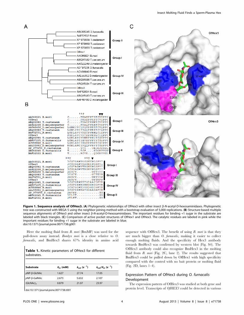
The phylogenetic analysis of insect glycoside hydrolase family
20 (GH20) Hexes from four insect species, including B. mori, O.
furnacalis, Drosophila melanogaster (Diptera) and T. castaneum, indicat-
ed these Hexes fell into four groups (Fig. 1A). Group I included
chitinolytic Hexes, e.g. OfHex1 (GenBank ID: ABI81756) [29].
Group II comprised Hexes with a broad substrate spectrum, e.g.
OfHex2 (GenBank ID: ABO65045) [25]. Group IV consisted of
N-glycan modifying Hexes, e.g. DmFDL (GenBank ID:
AAL55992) [30]. OfHex3 belonged to Group III, which also
included BmHex3 (GenBank ID: BAF52531) [31], DmHexo2
(GenBank ID: AAF46406) [32] and TcNag2 (GenBank ID:
ABQ95983) [5].
The structure-based multiple sequence alignment showed that
OfHex3 contained all the critical residues that are important for
the activity of GH20 Hexes, including Asp267, His321, Asp391 and
Glu392 for catalysis, Arg238, Tyr503, Asp505 and Glu541 for
substrate binding and Trp452, Trp476 and Trp539 for substrate
binding via stacking interactions (Fig. S2). The most obvious
differences among the four groups of insect Hexes were the lengths
and amino acid compositions of two loops, L314–335 and L478–496,
located at the entrance of the active pocket of OfHex1 (Fig. 1B).
Group III and Group IV Hexes possessed the longest L314–335.
The residues Val and Glu (black triangles in Fig. 1B), which
interacted with the +1 sugar of the substrate in Group I enzymes,
were changed to Ile and Gln in the Group III enzymes (Ile351 and
Gln352 in OfHex3), respectively.
The structure model of the catalytic domain of OfHex3 was
built using the catalytic domain of OfHex1 (PDB ID: 3NSN) as a
template and was validated using PROCHECK (89.8% of the
residues were in the most favored regions) and Verify_3D (93.4%
of the residues had an average three- to one-dimensional score
.0.2). The coordinates of (GlcNAc)2 were built based on the
structure of the (GlcNAc)2 complexed chitobiase from Serratia
marcescens (PDB ID: 1QBB) [33]. The structures of the active
pockets of OfHex1 and OfHex3 were then compared (Fig. 1C).
Although catalytic residues (Asp367 and Glu368 in OfHex1, Asp391
and Glu392 in OfHex3) were conserved, the key residues
comprising the +1 subsites were very different. OfHex3 did not
have the aromatic residue (Trp490 in OfHex1) to stack with the +1
sugar of the substrate. Unlike in OfHex1, the residues Ile351 and
Gln352 in OfHex3 were set in a position outward of the active
pocket such that they could not interact with the +1 sugar of the
substrate.
Recombinant Expression, Purification, andCharacterization of OfHex3
The gene encoding mature OfHex3 was successfully expressed
in the yeast strain P. pastoris GS115 and the recombinant OfHex3
was purified from the culture supernatant with a yield of about
1 mg/L. The purified OfHex3 was resolved as a single band with
a molecular weight of 75 kDa by SDS-PAGE analysis and was
verified by western blotting using anti-His-tag antibody (Fig. S3).
The molecular weight of the recombinant OfHex3 was reduced by
15 kDa after glycopeptidase F treatment (Fig. S3).
The kinetic parameters of OfHex3 for pNP-b-GlcNAc, pNP-b-
GalNAc, and (GlcNAc)2 were determined. As indicated by kcat/Km
values, OfHex3 hydrolyzed the putative physiological substrate,
(GlcNAc)2, 1.4 times faster than it did pNP-b-GlcNAc (Table 1).
Moreover, OfHex3 hydrolyzed pNP-b-GlcNAc at an 8.1-fold
higher efficiency than it did pNP-b-GalNAc (Table 1). No
substrate inhibition was observed using physiological substrate
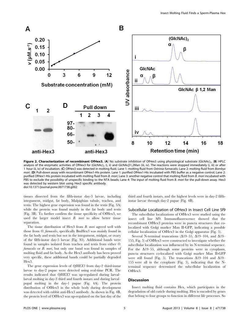
(GlcNAc)2 even with a concentration as high as 10 mM (Fig. 2A).
The glycosidic bond preference of OfHex3 was tested using the
substrates GlcNAcb1,4GlcNAc, GlcNAcb1,3GalNAc and
GlcNAcb1,2Man. OfHex3 could hydrolyze GlcNAcb1,4GlcNAc
at a rate of 20.6 mM min21, which was 10-fold higher than that of
GlcNAcb1,2Man (Fig. 2B), but it could not hydrolyze
GlcNAcb1,3GalNAc (data not shown).
To determine whether OfHex3 could hydrolyze GlcNAc-
containing glycans from glycoproteins and glycolipids, several
commercial available PA substrates including GnGn-PA(Glc-
NAcb1,2Mana1,6(GlcNAcb1,2Mana1,3)Manb1,4Glc-
NAcb1,4GlcNAc-PA), GA2-PA (GalNAcb1,4Galb1,4Glc-PA) and
Gb4-PA (GalNAcb1,3Gala1,4Galb1,4Glc-PA) were tested. The
results indicated that there were no hydrolytic products after 24 h
co-incubation of OfHex3 with these substrates (data not shown).
OfHex3 Occurred with OfHex1 in Molting FluidsSince OfHex3 exhibited the activity of hydrolyzing chitooligo-
saccharides, we tested the molting fluid to see if it contained
OfHex3. The molting fluid was collected during larval–pupal
molting of O. furnacalis and the specific antibody against OfHex3
was applied for western blot analysis. The results showed that
OfHex3 was present in the molting fluid (Fig. 2C, lane 1). As Hex1
also occurs in molting fluid, the interaction between Hex3 and
Hex1 in molting fluid was tested by pull-down assay.
Insect Molting Fluid Finds a Sperm-Plasma Hex
PLOS ONE | www.plosone.org 3 August 2013 | Volume 8 | Issue 8 | e71738
Here the molting fluid from B. mori (BmMF) was used for the
pull-down assay instead. Bombyx mori is a close relative to O.
furnacalis, and BmHex3 shares 67% identity in amino acid
sequence with OfHex3. The benefit of using B. mori is that they
are much bigger than O. furnacalis, making it easier to collect
enough molting fluids. And the specificity of Hex3 antibody
towards BmHex3 was confirmed by western blot (Fig. S4). The
OfHex3 antibody could also recognize BmHex3 in the molting
fluid from B. mori (Fig. 2C, lane 2). The results suggested that
BmHex3 could be pulled down by OfHex1 with high specificity
compared with the control with no bait protein or molting fluid
(Fig. 2D, lanes 1–4).
Expression Pattern of OfHex3 during O. furnacalisDevelopment
The expression pattern of OfHex3 was studied at both gene and
protein level. Transcripts of OfHEX3 could be detected in various
Figure 1. Sequence analysis of OfHex3. (A) Phylogenetic relationships of OfHex3 with other insect b-N-acetyl-D-hexosaminidases. Phylogenetictree was constructed with MEGA 5 using the neighbor-joining method with a bootstrap evaluation of 5,000 replications. (B) Structure-based multiplesequence alignments of OfHex3 and other insect b-N-acetyl-D-hexosaminidases. The important residues for binding +1 sugar in the substrate arelabeled with black triangles. (C) Comparison of active pocket structures of OfHex1 and OfHex3. The catalytic residues are labeled in pink while theimportant residues for binding +1 sugar in the substrate are labeled in blue.doi:10.1371/journal.pone.0071738.g001
Table 1. Kinetic parameters of OfHex3 for differentsubstrates.
Substrate Km (mM) kcat (s21) kcat/Km (s21)
pNP-b-GlcNAc 1.627 27.74 17.05
pNP-b-GalNAc 2.673 5.632 2.107
(GlcNAc)2 0.879 21.07 23.97
doi:10.1371/journal.pone.0071738.t001
Insect Molting Fluid Finds a Sperm-Plasma Hex
PLOS ONE | www.plosone.org 4 August 2013 | Volume 8 | Issue 8 | e71738
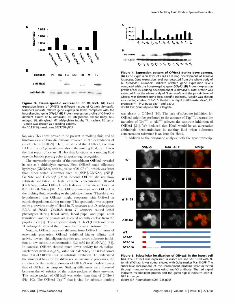
tissues dissected from the fifth-instar day-3 larvae, including
integument, midgut, fat body, Malpighian tubule, trachea, and
testis. The highest gene expression was found in the testis (Fig. 3A)
while the protein was found mainly in the fat body and testis
(Fig. 3B). To further confirm the tissue specificity of OfHex3, we
used the larger model insect B. mori to allow better tissue
separation.
The tissue distribution of Hex3 from B. mori agreed well with
those from O. furnacalis, specifically BmHex3 was mainly found in
the fat body and testis but not in the integument, midgut, or ovary
of the fifth-instar day-5 larvae (Fig. S5). Additional bands were
found in samples isolated from trachea and testis from either O.
furnacalis or B. mori, but only one band was found in samples of
molting fluid and fat body. As the Hex3 antibody has been proved
very specific, these additional bands could be partially degraded
Hex3.
The gene expression levels of OfHEX3 from day-2 third-instar
larvae to day-2 pupae were detected using real-time PCR. The
results indicated that OfHEX3 was up-regulated during larval–
larval molting in day-3 third and fourth instars and during larval–
pupal molting in the day-1 pupae (Fig. 4A). The protein
distribution of OfHex3 in the whole body during development
was detected with rabbit anti-Hex3 antibody. As shown in Fig. 4B,
the protein level of OfHex3 was up-regulated on the last day of the
third and fourth instars, and the highest levels were in day-2 fifth-
instar larvae through day-2 pupae (Fig. 4B).
Subcellular Localization of OfHex3 in Insect Cell Line Sf9The subcellular localizations of OfHex3 were studied using the
insect cell line Sf9. Immunofluorescence showed that the
recombinant OfHex3 proteins were in puncta structures that co-
localized with Golgi marker Man II-GFP, indicating a possible
cellular localization of OfHex3 in the Golgi apparatus (Fig. 5).
Several N-terminal truncations (D19–55, D19–104, and D19–
155, Fig. 5) of OfHex3 were constructed to investigate whether the
subcellular localization was influenced by its N-terminal sequence.
For the D19–55, although some proteins were in cytoplasm,
puncta structures co-localized with Golgi marker Man II-GFP
were still found (Fig. 5). The truncations D19–104 and D19–
155 were all in the cytoplasm (Fig. 5), indicating that the N-
terminal sequence determined the subcellular localization of
OfHex3.
Discussion
Insect molting fluid contains Hex, which participates in the
degradation of old cuticle during molting. Hex is encoded by genes
that belong to four groups to function in different life processes. So
Figure 2. Characterization of recombinant OfHex3. (A) No substrate inhibition of OfHex3 using physiological substrate (GlcNAc)2. (B) HPLCanalysis of the enzymatic activities of OfHex3 for (GlcNAc)2 (i, ii) and GlcNAcb1,2Man (iii, iv). The reactions were stopped immediately (i, iii) or after1 hour (ii, iv) of incubation. (C) OfHex3 was detected in molting fluid. Lane 1: molting fluid from Ostrinia furnacalis; Lane 2: molting fluid from Bombyxmori. (D) Pull-down assay with recombinant OfHex1-His protein. Lane 1: purified OfHex1-His incubated with PBS buffer as a negative control; Lane 2:purified OfHex1-His protein incubated with molting fluid from B. mori; Lane 3: another negative control that molting fluid from B. mori incubated withPBS to exclude the possibility of unspecific binding to the NTA beads; Lane 4: The input of molting fluid from B. mori for the pull-down assay. Hex3was detected by western blot using Hex3 specific antibody.doi:10.1371/journal.pone.0071738.g002
Insect Molting Fluid Finds a Sperm-Plasma Hex
PLOS ONE | www.plosone.org 5 August 2013 | Volume 8 | Issue 8 | e71738
far, only Hex1 was proved to be present in molting fluid and to
function as a chitinolytic enzyme involved in the degradation of
cuticle chitin [9,10,29]. Here, we showed that OfHex3, the class
III Hex from O. furnacalis, was also in the molting fluid, too. This is
the first report of a class III Hex that functions as a molting fluid
enzyme besides playing roles in sperm–egg recognition.
The enzymatic properties of the recombinant OfHex3 revealed
its role as a chitinolytic enzyme. First, OfHex3 could efficiently
hydrolyze (GlcNAc)2 with kcat value of 21.07 s21, which was faster
than other tested substrates such as pNP-b-GlcNAc, pNP-b-
GalNAc, and GlcNAcb1,2Man. Second, OfHex3 did not show
substrate inhibition at high substrate concentration (10 mM
(GlcNAc)2), unlike OfHex1, which showed substrate inhibition at
0.2 mM (GlcNAc)2 [34]. Also, OfHex3 interacted with OfHex1 in
the molting fluid according to the pull-down assay. Therefore, we
hypothesized that OfHex3 might cooperate with OfHex1 in
cuticle degradation during molting. This speculation was support-
ed by a previous study of Hex3 in T. castaneum and D. melanogaster.
RNAi of HEX3 (TcNAG2) from T. castaneum caused lethal
phenotypes during larval–larval, larval–pupal and pupal–adult
transitions, and the pharate adults could not fully enclose from the
pupal cuticle [5]. The enzymatic study of Hex3 (DmHexo2) from
D. melanogaster showed that it could hydrolyze chitotriose [30].
Notably, OfHex3 was very different from OfHex1 in terms of
enzymatic properties. OfHex1 exhibited higher affinity and
activity toward chitooligosaccharides and severe substrate inhibi-
tion at low substrate concentration (0.2 mM for (GlcNAc)2) [34].
In contrast, OfHex3 showed much lower activity for chitooligo-
saccharides (with a kcat/Km value for (GlcNAc)2 142.8-fold lower
than that of OfHex1) but no substrate inhibition. To understand
the structural basis for the difference in enzymatic properties, the
structure of the catalytic domain of OfHex3 was modeled using
that of OfHex1 as template. Striking differences were observed
between the +1 subsites of the active pockets of these enzymes.
The active pocket of OfHex3 was wider than that of OfHex1
(Fig. 1C). The OfHex1 Trp490 that is vital for substrate binding
was absent in OfHex3 [10]. The lack of substrate inhibition for
OfHex3 might be attributed to the absence of Trp490, because the
mutation of Trp490 to Ala490 relieved the substrate inhibition of
OfHex1 [34]. We deduced that Hex3 would be an alternative
chitinolytic hexosaminidase in molting fluid when substrates
concentration tolerance is an issue for Hex1.
In addition to the enzymatic analysis, both the gene transcrip-
Figure 3. Tissue-specific expression of OfHex3. (A) Geneexpression levels of OfHEX3 in different tissues of Ostrinia furnacalis.Numbers indicate relative gene expression levels compared with thehousekeeping gene OfRpS3. (B) Protein expression profile of OfHex3 indifferent tissues of O. furnacalis. IN: integument, FB: fat body, MG:midgut, SG: silk gland, MT: Malpighian tubule, TR: trachea, TE: testis.Tubulin was chosen as a loading control.doi:10.1371/journal.pone.0071738.g003
Figure 4. Expression pattern of OfHex3 during development.(A) Gene expression level of OfHEX3 during development of Ostriniafurnacalis. Gene expression level was detected from the whole body ofO. furnacalis. Numbers indicate relative gene expression levelscompared with the housekeeping gene OfRpS3. (B) Protein expressionprofile of OfHex3 during development of O. furnacalis. Total protein wasextracted from the whole body of O. furnacalis and the protein level ofOfHex3 was detected using Hex3 specific antibody. Tubulin was chosenas a loading control. 3L2–5L5: third-instar day-2 to fifth-instar day-5; PP:prepupa; P-1, P-2: pupa day-1 and day-2.doi:10.1371/journal.pone.0071738.g004
Figure 5. Subcellular localization of OfHex3 in the insect cellline Sf9. OfHex3 was expressed in insect cell line Sf9 fused with N-terminal V5 tag. It was co-transfected with Golgi marker Man II-GFP. Thesubcellular localizations of the recombinant proteins were detectedthrough immunofluorescence using anti-V5 antibody. The red signalindicates recombinant protein and the green signal indicates Man II-GFP in merge.doi:10.1371/journal.pone.0071738.g005
Insect Molting Fluid Finds a Sperm-Plasma Hex
PLOS ONE | www.plosone.org 6 August 2013 | Volume 8 | Issue 8 | e71738
tion and translation levels indicated OfHex3’s involvement in
molting. During the development of O. furnacalis, both gene and
protein levels of OfHex3 were highly up-regulated during larval–
larval molting and larval–pupal metamorphosis, suggesting that
OfHex3 was involved in chitin degradation during molting.
In summary, OfHex3, a class III Hex, cooperated with OfHex1,
a class I Hex, to function as chitinolytic enzymes in the molting
fluids of insect.
Supporting Information
Figure S1 The cloning strategy of OfHEX3 gene. The full
length cDNA of OfHEX3 was determined by 4 fragments.
Fragment 1was the PCR products. Fragment 2 was obtained by
39-RACE and fragments 3 and 4 were the products of 59-RACE.
(TIF)
Figure S2 Multiple sequence alignment of OfHex3 withother insect b-N-acetyl-D-hexosaminidases. Structure-
based multiple sequence alignments of OfHex3 and other insect
Hexes were performed with PROMALS3D using the crystal
structure of OfHex1 (PDB code: 3NSM) as structure input.
Sequence alignment was performed by using the software ESpript
2.2.
(TIF)
Figure S3 SDS-PAGE and western blot analysis of therecombinant OfHex3. Proteins were separated by 10% SDS-
PAGE. The molecular weight of the recombinant OfHex3 was
reduced by 15 kDa after glycopeptidase F treatment. His-tag
antibody was used for western blot to detect the recombinant
OfHex3.
(TIF)
Figure S4 Hex3 antibody recognizing BmHex3 fromBombyx mori. The gene encoding Hex3 (Genbank ID:
NM_001085364) from B. mori was cloned into the vector pET22b
and expressed in E. coli. Protein expression was induced by 1 mM
of IPTG for 4 hours at 37uC. Both SDS-PAGE and western blot
were applied to determine the expression of the recombinant
BmHex3 and antibody specificity. Lane 1, 3: cell lysates of E. coli
harboring the expression vector pET22b-BmHex3; 2, 4: cell
lysates of E. coli harboring the vector pET22b alone. Arrow
indicates the recombinant BmHex3.
(TIF)
Figure S5 Protein expression profile of Hex3 protein inBombyx mori. Proteins were extracted from different tissues of
fifth-instar day-5 B. mori. The protein expression profile of Hex3
was detected through western blot using Hex3 specific antibody.
IN: integument, FB: fat body, MG: midgut, SG: silk gland, MT:
Malpighian tubule, TR: trachea, OV: ovary, TE: testis. Tubulin
was chosen as a loading control.
(TIF)
Table S1 Primers used in cloning of OfHEX3.
(DOCX)
Table S2 Primers used for Real-Time PCR.
(DOCX)
Author Contributions
Conceived and designed the experiments: MQ TL QY. Performed the
experiments: MQ TL PC. Analyzed the data: MQ QY TL. Wrote the
paper: QY MQ TL.
References
1. Merzendorfer H, Zimoch L (2003) Chitin metabolism in insects: structure,function and regulation of chitin synthases and chitinases. J Exp Biol 206: 4393–
4412.
2. Reynolds SE, Samuels RI (1996) Physiology and biochemistry of insect moulting
fluid. Adv Insect Physiol 26: 157–232.
3. Hamamura Y, Kanehara Y (1940) Enzymatic studies of exuvial fluid of Bombyx
mori. II Chitinase. J Agric Chem Soc Jpn 16: 907–909.
4. Kramer KJ, Koga D (1986) Insect chitin: physical state, synthesis, degradation
and metabolic regulation. Insect Biochem 16: 851–877.
5. Hogenkamp DG, Arakane Y, Kramer KJ, Muthukrishnan S, Beeman RW
(2008) Characterization and expression of the b-N-acetylhexosaminidase gene
family of Tribolium castaneum. Insect Biochem Mol Biol 38: 478–489.
6. Dziadik-Turner C, Koga D, Mai MS, Kramer KJ (1981) Purification and
characterization of two b-N-acetylhexosaminidases from the tobacco hornworm,Manduca sexta (L.)(Lepidoptera: Sphingidae). Arch Biochem Biophys 212: 546–
560.
7. Koga D, Mai MS, Kramer KJ (1983) Comparative biochemistry of insect exo-b-
N-acetylglucosaminidases: Characterization of a third enzyme from pupalhemolymph of the tobacco hornworm, Manduca sexta L. Comp Biochem Physiol
B: Biochem Mol Biol 74: 515–520.
8. Koga D, Nakashima M, Matsukura T, Kimura S, Ide A (1986) Purification andproperties of b-N-acetyl-D-glucosaminidase from alimentary canal of the
silkworm, Bombyx mori. Agric Biol Chem 50: 2357–2368.
9. Nagamatsu Y, Yanagisawa I, Kimoto M, Okamoto E, Koga D (1995)
Purification of a chitooligosaccharidolytic b-N-Acetylglucosaminidase fromBombyx Mori larvae during metamorphosis and the nucleotide-sequence of its
cDNA. Biosci Biotechnol Biochem 59: 219–225.
10. Liu T, Zhang H, Liu F, Wu Q, Shen X, et al. (2011) Structural determinants of
an insect b-N-acetyl-D-hexosaminidase specialized as a chitinolytic enzyme.
J Biol Chem 286: 4049–4058.
11. Zen KC, Choi HK, Krishnamachary N, Muthukrishnan S, Kramer KJ (1996)
Cloning, expression, and hormonal regulation of an insect b-N-acetylglucosa-minidase gene. Insect Biochem Mol Biol 26: 435–444.
12. Zheng Y, Krell PJ, Doucet D, Arif BM, Feng Q (2008) Cloning, expression, andlocalization of a molt-related b-N-acetylglucosaminidase in the spruce budworm,
Choristoneura fumiferana. Arch Insect Biochem Physiol 68: 49–59.
13. Perotti ME, Cattaneo F, Pasini ME, Intra J, Matsumoto M, et al. (2006)
Identification and expression analysis of Drosophila melanogaster genes encoding b-hexosaminidases of the sperm plasma membrane. Glycobiology 16: 786–800.
14. Perotti ME, Intra J, Cenni F, Pavesi G, Pasini M (2009) Interspecific Analysis of
the Glycosidases of the Sperm Plasma Membrane in Drosophila. Mol Reprod Dev
76: 85–100.
15. Pasini ME, Intra J, Gomulski LM, Calvenzani V, Petroni K, et al. (2011)
Identification and expression profiling of Ceratitis capitata genes coding for b-
hexosaminidases. Gene 473: 44–56.
16. Pasini ME, Intra J, De Caro D, Perotti ME (2011) Glycosidases in the plasma
membrane of Ceratitis capitata spermatozoa. Insect Biochem Mol Biol 41: 90–100.
17. Petersen TN, Brunak S, von Heijne G, Nielsen H (2011) SignalP 4.0:
discriminating signal peptides from transmembrane regions. Nature Methods
8: 785–786.
18. Pei J, Kim BH, Grishin NV (2008) PROMALS3D: a tool for multiple protein
sequence and structure alignments. Nucleic Acids Res 36: 2295–2300.
19. Gouet P, Courcelle E (2002) ENDscript: a workflow to display sequence and
structure information. Bioinformatics 18: 767–768.
20. Tamura K, Peterson D, Peterson N, Stecher G, Nei M, et al. (2011) MEGA5:
molecular evolutionary genetics analysis using maximum likelihood, evolution-
ary distance, and maximum parsimony methods. Mol Biol Evol 28: 2731–2739.
21. Sali A, Blundell TL (1993) Comparative protein modelling by satisfaction of
spatial restraints. J Mol Biol 234: 779–815.
22. Laskowski RA, MacArthur MW, Moss DS, Thornton JM (1993) PROCHECK:
a program to check the stereochemical quality of protein structures. J Appl
Crystallogr 26: 283–291.
23. Bowie JU, Luthy R, Eisenberg D (1991) A method to identify protein sequences
that fold into a known three-dimensional structure. Science 253: 164–170.
24. Liu T, Liu F, Yang Q, Yang J (2009) Expression, purification and
characterization of the chitinolytic b-N-acetyl-D-hexosaminidase from the insect
Ostrinia furnacalis. Protein Expr Purif 68: 99–103.
25. Liu F, Liu T, Qu M, Yang Q (2012) Molecular and biochemical characterization
of a novel b-N-acetyl-D-hexosaminidase with broad substrate-spectrum from the
Aisan corn borer, Ostrinia furnacalis. Int J Biol Sci 8: 1085–1096.
26. Qu M, Yang Q (2012) Physiological significance of alternatively spliced exon
combinations of the single-copy gene class A chitin synthase in the insect Ostrinia
furnacalis (Lepidoptera). Insect Mol Biol 21: 395–404.
27. Qu M, Yang Q (2011) A novel alternative splicing site of class A chitin synthase
from the insect Ostrinia furnacalis - gene organization, expression pattern and
physiological significance. Insect Biochem Mol Biol 41: 923–931.
Insect Molting Fluid Finds a Sperm-Plasma Hex
PLOS ONE | www.plosone.org 7 August 2013 | Volume 8 | Issue 8 | e71738
28. Qu M, Liu T, Yang J, Yang Q (2011) The gene, expression pattern and
subcellular localization of chitin synthase B from the insect Ostrinia furnacalis.
Biochem Biophys Res Commun 404: 302–307.
29. Yang Q, Liu T, Liu F, Qu M, Qian X (2008) A novel b-N-acetyl-D-
hexosaminidase from the insect Ostrinia furnacalis (Guenee). FEBS J 275: 5690–
5702.
30. Leonard R, Rendic D, Rabouille C, Wilson IBH, Preat T, et al. (2006) The
Drosophila fused lobes gene encodes an N-acetylglucosaminidase involved in N-
glycan processing. J Biol Chem 281: 4867–4875.
31. Okada T, Ishiyama S, Sezutsu H, Usami A, Tamura T, et al. (2007) Molecular
cloning and expression of two novel b-N-acetylglucosaminidases from silkwormBombyx mori. Biosci Biotechnol Biochem 71: 1626–1635.
32. Cattaneo F, Ogiso M, Hoshi M, Perotti ME, Pasini ME (2002) Purification and
characterization of the plasma membrane glycosidases of Drosophila melanogaster
spermatozoa. Insect Biochem Mol Biol 32: 929–941.
33. Tews I, Perrakis A, Oppenheim A, Dauter Z, Wilson KS, et al. (1996) Bacterialchitobiase structure provides insight into catalytic mechanism and the basis of
Tay-Sachs disease. Nat Struct Biol 3: 638–648.
34. Liu T, Wu Q, Liu L, Yang Q (2012) Elimination of substrate inhibition of a b-N-acetyl-D-hexosaminidase by single site mutation. Process Biochem 48: 103–108.
Insect Molting Fluid Finds a Sperm-Plasma Hex
PLOS ONE | www.plosone.org 8 August 2013 | Volume 8 | Issue 8 | e71738