Embed Size (px)
Citation preview

JOURNAL OF VIROLOGY, June 2010, p. 5868–5879 Vol. 84, No. 120022-538X/10/$12.00 doi:10.1128/JVI.02383-09Copyright © 2010, American Society for Microbiology. All Rights Reserved.
A Single Coxsackievirus B2 Capsid Residue Controls Cytolysis andApoptosis in Rhabdomyosarcoma Cells�
Maria Gullberg,1 Conny Tolf,1 Nina Jonsson,1 Charlotta Polacek,2 Jana Precechtelova,3Miriam Badurova,3 Martin Sojka,3 Camilla Mohlin,1 Stina Israelsson,1 Kjell Johansson,1
Shubhada Bopegamage,3 Susan Hafenstein,4 and A. Michael Lindberg1*School of Natural Sciences, Linnaeus University, SE-391 82 Kalmar, Sweden1; National Veterinary Institute, Technical University of
Denmark, Lindholm, DK-4771, Kalvehave, Denmark2; Department of Virology, Slovak Medical University, Limbova 12,83303 Bratislava, Slovak Republic3; and Department of Microbiology and Immunology, The Pennsylvania State University College of
Medicine, 500 University Drive, Hershey, Pennsylvania 170334
Received 12 November 2009/Accepted 26 March 2010
Coxsackievirus B2 (CVB2), one of six human pathogens of the group B coxsackieviruses within theenterovirus genus of Picornaviridae, causes a wide spectrum of human diseases ranging from mild upperrespiratory illnesses to myocarditis and meningitis. The CVB2 prototype strain Ohio-1 (CVB2O) wasoriginally isolated from a patient with summer grippe in the 1950s. Later on, CVB2O was adapted tocytolytic replication in rhabdomyosarcoma (RD) cells. Here, we present analyses of the correlationbetween the adaptive mutations of this RD variant and the cytolytic infection in RD cells. Using reversegenetics, we identified a single amino acid change within the exposed region of the VP1 protein (glutamineto lysine at position 164) as the determinant for the acquired cytolytic trait. Moreover, this cytolytic virusinduced apoptosis, including caspase activation and DNA degradation, in RD cells. These findingscontribute to our understanding of the host cell adaptation process of CVB2O and provide a valuable toolfor further studies of virus-host interactions.
Virus infections depend on complex interactions betweenviral and cellular proteins. Consequently, the nature of theseinteractions has important implications for viral cell type spec-ificity, tissue tropism, and pathogenesis. Group B coxsackievi-ruses (CVB1 to CVB6), members of the genus Enteroviruswithin the family of Picornaviridae, are human pathogens thatcause a broad spectrum of diseases, ranging from mild upperrespiratory illnesses to more severe infections of the centralnervous system, heart, and pancreas (61). These viruses havealso been associated with certain chronic muscle diseases andmyocardial infarction (2, 3, 12, 13, 22).
The positive single-stranded RNA genome (approximately7,500 nucleotides in length) of CVBs is encapsidated within asmall T�1, icosahedral shell (30 nm in diameter) comprised ofrepeating identical subunits made up of four structural pro-teins (VP1 to VP4). Parts of VP1, VP2, and VP3 are exposedon the outer surface of the capsid, whereas VP4 is positionedon the interior. The virion morphology is characterized by astar-shaped mesa at each 5-fold icosahedral symmetry axis,surrounded by a narrow depression referred to as the “canyon”(69). All six serotypes of CVB can use the coxsackie and ade-novirus receptor (CAR) for cell attachment and entry (9, 55,82). Some strains of CVB1, -3, and -5 also use decay acceler-ating factor ([DAF] CD55) for initial attachment to the hostcell; however, binding to DAF alone is insufficient to permitentry into the cell (10, 54, 76).
Picornaviruses are generally characterized by their cytolytic
nature in cell culture. However, several in vivo and in vitrostudies have shown that some picornaviruses, e.g., poliovirus,Theiler’s murine encephalomyelitis virus, foot-and-mouth dis-ease virus, CVB3, CVB4, and CVB5, may also establish per-sistent, noncytolytic infections (4, 29, 35, 39, 62, 74). Recently,it has been shown that the diverse outcomes of picornaviralinfections may depend on interactions between the virus andthe apoptotic machinery of the infected cell (14, 30, 71). Sev-eral picornaviral proteins have been identified as inducers ofan apoptotic response, including viral capsid proteins VP1,VP2, and VP3, as well as nonstructural proteins 2A and 3C (7,20, 32, 33, 42, 50, 63). In addition, antiapoptotic activity hasbeen assigned to the nonstructural proteins 2B and 3A (16, 59).
Picornaviruses have the potential to adapt rapidly to newhost environments. Virus features affecting adaptability in-clude high mutation rates, short replication times, large pop-ulations, and frequent incidences of recombination (25–27,53). Consequently, picornaviruses exist as genetically heterog-enous populations, referred to as viral quasispecies (25, 26).
Previously, the CVB2 prototype strain Ohio-1 (CVB2O) wasadapted to cytolytic replication in rhabdomyosarcoma (RD)cells (66). Two amino acid changes were identified in the cap-sid-coding region, and one was identified in the 2C-codingregion of the adapted virus. Further characterization of thevirus-host interaction showed that the infection was not af-fected by anti-DAF antibodies, indicating the use of an alter-native receptor.
In this study, the amino acid substitutions associated withthe adaptation of CVB2O to cytolytic infection of RD cellswere evaluated. Site-directed mutagenesis studies showed thata single amino acid change in the VP1 capsid protein wasresponsible for the cytolytic RD phenotype. In addition, as
* Corresponding author. Mailing address: School of Natural Sci-ences, Linnaeus University, SE-391 82 Kalmar, Sweden. Phone: 46 480446240. Fax: 46 480 446262. E-mail: [email protected].
� Published ahead of print on 7 April 2010.
5868

indicated by caspase activation and DNA degradation, theapoptotic pathway was activated in RD cells infected by thecytolytic virus.
MATERIALS AND METHODS
Cells and viruses. Cultures of a local variant of green monkey kidney (GMK)cells, human epithelial (HEp-2) cells, and human RD cells, obtained from theAmerican Tissue Culture Collection (ATCC), were maintained in Dulbecco’smodified Eagles medium (DMEM) supplemented with 2 mM L-glutamine, 100U/ml penicillin, 0.1 mg/ml streptomycin, and 10% newborn calf serum (NCS) at37°C in 7.5% CO2. The prototype strain CVB2 Ohio-1 (VR-29; ATCC) (56, 67)and the CVB2ORD variant (66) were propagated in GMK and RD cells, re-spectively, as previously described (66).
Flow cytometry. The flow cytometry procedure has been described previously(66). RD cells were stained with an anti-CAR (RmcB) antibody (37) (hybridomakindly provided by L. Philipson and R. Pettersson, Karolinska Institute, Sweden;also available as ATCC CRL-2379) or an anti-DAF (BRIC110) antibody (Cym-bus Biotechnologies). A monoclonal mouse IgG1 antibody (X0931; Dako) wasused as a negative control. After 1 h of incubation at 4°C, the cells were stainedwith a secondary R-phycoerythrin-conjugated polyclonal rabbit anti-mouse an-tibody (R0439; Dako). Data were acquired using a FACSCalibur (Becton Dick-inson) and analyzed with CellQuest, version 3.3, software (Becton Dickinson).
Infectious viral cDNA clones. The complete CVB2O genome was amplifiedand cloned into the pCR-Script Direct SK(�) vector (Stratagene) by using theAscI and NotI restriction enzyme cleavage sites, as previously described (Fig.1A) (49, 67). In this pCVB2Owt (where wt is wild type) construct, the individualCVB2ORD substitutions in the protein VP1 (I to F and Q to K) and 2C (K toR) were introduced by site-specific mutagenesis (Fig. 1). The PflFI and SexAIsites were used to generate the single-amino acid mutant pCVB2OVP1I118Fclone (vVP1I118F) encoding the VP1 I118F substitution (names of viruses de-rived from the infectious cDNA clones are given in parenthesis). The SexAI andEcoRI restriction sites were used to construct the pCVB2OVP1Q164K clone(vVP1Q164K), while EcoRI and BamHI sites were used to produce thepCVB2O2CK185R clone (v2CK185R). The CVB2O constructs were propagatedin Escherichia coli, isolated, and verified by sequencing.
Generation of viruses from infectious viral cDNA clones. Viruses were gen-erated by transfection (Lipofectamine 2000; Invitrogen) of 2.5 �g of the proto-type (pCVB2Owt) or mutant (pCVB2OVP1I118F, pCVB2OVP1Q164K, andpCVB2O2CK185R) DNA into RD cells according to manufacturer’s protocol.At 5 days posttransfection, the virus from transfected cells was released by threefreeze-thaw cycles and further propagated by one subsequent passage in RD cellsas described previously (57). Viral RNA was extracted from infected cell cultures(QIAamp viral RNA mini kit; Qiagen), reverse transcribed (Superscript III;Invitrogen), and PCR amplified (PicoMaxx high fidelity PCR system; Stratagene)using virus-specific primers. PCR amplicons were visualized on an agarose gel,purified (QIAquick gel extraction kit; Qiagen), and sequenced. Virus titers weredetermined by 50% tissue culture infectious dose (TCID50) assays in GMK cells,according to standard procedures (34).
Virus infection. Cells were infected with the different CVB2O variants accord-ing to standard procedures (57). Briefly, subconfluent monolayers of RD cellsgrown in 25-cm2 flasks were inoculated with clone-derived viruses at a multiplic-ity of infection (MOI) ranging from 1 to 100 TCID50s/cell. After a 1-h adsorptionat room temperature, the inoculum was removed, and cells were washed threetimes to remove unbound virus before addition of DMEM supplemented with 2mM L-glutamine, 100 U/ml penicillin, and 0.1 mg/ml streptomycin. Incubationwas continued for 7 days or until cytopathic effect (CPE) was observed.
In order to determine the number of mutations that were introduced in thevVP1Q164K genome during serial passages in RD cells, infected cells wereincubated until complete CPE was observed. These samples were subjected tothree freeze-thaw cycles and further passaged five times in RD cells at anapparently high MOI (in two parallel experiments). After the fifth passage, theviral progeny was sequenced.
For studies of virus production during repeated noncytolytic infections, RDcells infected with CVB2Owt were detached by EDTA treatment and passagedevery fourth day at a 1/3 dilution. The titer of virus released into the medium ofeach passage was measured by the TCID50 method in GMK cells. After the 10thpassage, the viral progeny was sequenced.
Release of CVB2Owt from RD cells. Confluent RD cells cultured in 24-wellplates were infected with CVB2Owt at an MOI of 1 TCID50/cell. In order toassess both the intra- and extracellular virus production, samples (only mediumor medium together with cells) were frozen at different time points postinfection
FIG. 1. Schematic overview of the CVB2O genome organization and the construction of CVB2O variants containing adaptive mutations. (A) Arepresentation of the CVB2O genome is depicted at the top, with boxes delineating viral genes as well as approximate positions of the relevantrestriction enzyme sites used to construct infectious viral cDNA clones. Below, the location of amino acid substitutions in the CVB2ORD genomeare indicated next to the VP1- and 2C-encoding genes; the first letter refers to the amino acid residue of the CVB2O protein and the numberindicates the amino acid position in the individual protein. Infectious CVB2O cDNA clone variants, as inserted in the pCR-Script Direct SK�vector, are shown in the lower part of the figure. The designation of constructed cDNA clones and simplified names of viruses generated from theseclones (given within parentheses) are used throughout the article. (B) A surface-rendered view of a pentameric subunit of the viral capsid showingthe location of the exposed CVB2O substitution that evolved during propagation in RD cells. The entire capsid of the closely related CVB3 (PDB1COV) (58) is shown at the top with five protomers related by an icosahedral 5-fold symmetry axis colored to denote proteins VP1 (blue), VP2(green), and VP3 (red), whereas the remaining virus surface is shown in shades of gray. Below is an enlarged view of the pentamer showing thelocation of the surface-exposed amino acid change of CVB2O VP1 (Q164K; cyan and encircled) given as the equivalent CVB3 VP1 residue 160,based on multiple sequence alignment (ClustalW) (79). The icosahedral asymmetric unit of the virus is indicated by the triangular boundary.
VOL. 84, 2010 CVB2 CYTOLYSIS AND APOPTOSIS IN RD CELLS 5869

(p.i.), and virus titers were determined after three cycles of freezing and thawing(to release intracellular viruses) by a TCID50 assay in GMK cells.
Quantitation of viral infection. The replication of viral plus-strand RNA inRD cells infected with the CVB2O variants was determined by real-time PCR,according to established procedures (43). Confluent RD cells in 24-well plateswere infected with the different CVB2O variants at an MOI of 1 TCID50/cell asdescribed above. Infected cell samples were harvested at 0 h and 96 h p.i. andsubjected to three freeze-thaw cycles. Viral RNA was extracted as describedabove, followed by reverse transcription of extracted RNA using an AppliedBiosystems TaqMan reverse transcriptase kit and random hexamers. Reversetranscribed viral cDNA was quantified using real-time PCR and a Power SYBRgreen Master Mix in accordance with the manufacturer’s instructions (AppliedBiosystems). The CVB2-specific primers were designed to amplify a 147-bpproduct from nucleotide positions 454 to 601 in the genomic 5� untranslatedregion (UTR) region. The final PCR mixture contained 100 nM (each) forward(5�-GCCCCTGAATGCGGCTAAT-3�) and reverse (5�-ATTGTCACCATAAGCAGCCA-3�) primers, 10 �l of Power SYBR green PCR Master Mix (AppliedBiosystems), and 1 �l of cDNA. Samples were analyzed in triplicates in a finalvolume of 20 �l. Thermal cycling was performed with an ABI Prism 7500 SDSmachine (Applied Biosystems) and universal cycling conditions (2 min at 50°C,10 min at 95°C, followed by 40 cycles of 15 s at 95°C and 1 min at 60°C).Nuclease-free water was used as a negative control. Cycle threshold (CT) valueswere determined by automated threshold analysis included in the ABI sequencedetection software, version 1.3.1. Dissociation curves were recorded after eachrun. The difference between the copy numbers of cDNA (i.e., viral RNA) de-tected at 0 h and 96 h p.i. was presented as the fold change (2CT,0h�CT,96h) underthe assumption of 100% amplification efficiency according to the 2���CT method(52). Furthermore, virus production in these samples (at 0 h and 96 h p.i.) wasmeasured by the TCID50 method in GMK cells.
Plaque formation assay. The plaque phenotype of the CVB2O variants wasdetermined by utilizing a gum tragacanth overlay as previously described (24).Briefly, the CVB2O variants were titrated by 10-fold dilutions onto confluentmonolayers of RD cells in six-well plates. Following adsorption for 1 h at 37°C,the inoculum was aspirated and replaced with an overlay medium, which con-sisted of 0.8% (wt/vol) gum tragacanth (Sigma), DMEM, 1% NCS, 2 mML-glutamine, 100 U/ml penicillin, and 0.1 mg/ml streptomycin. RD cells werestained at 96 h p.i. with a crystal violet-ethanol solution for visualization ofplaques.
Single-step growth kinetics. Confluent monolayers of RD cells cultured in24-well plates were infected with virus at an MOI of 1 TCID50/cell as describedabove. For assessing virus growth, samples were frozen at different time pointsp.i., and virus titers were determined by a TCID50 assay in GMK cells.
Immunofluorescence staining of virus-infected cells. For immunofluorescenceassays, monolayers of RD cells cultured on Lab-TEK II chamber glass slides(Nalge Nunc International) were infected with the CVB2O variants at an MOIof 1 TCID50/cell. Alternatively, to analyze expression of CVB2 antigens duringrepeated noncytolytic infection, a fraction of CVB2Owt-infected RD cells frompassages 1, 5, and 10 were transferred and cultured on glass slides. At indicatedtime points (see Fig. 3C and 4B), the cells were fixed with 4% formaldehyde for30 min at 4°C, washed twice, and stained with a monoclonal mouse anti-CVB2antibody (MAB946; Chemicon International, Inc.) for 1 h at room temperature.For analyses of apoptosis, the fixed cells were incubated with a polyclonal anti-body against cleaved/activated caspase-3 ([CC-3] 9661; Cell Signaling Technol-ogy). After the slides were washed, a secondary goat anti-mouse or goat anti-rabbit antibody labeled with Alexa Fluor 488 (A11001 or A11034; Molecularprobes Inc.) was added for 1 h at room temperature. Finally, the slides weremounted with Vectashield (Immunkemi, Sweden) containing 4�,6-diamidino-2-phenylindole (DAPI). Images were captured with an epifluorescence micro-scope.
DNA fragmentation assay. DNA fragmentation in CVB2O-infected and un-infected RD cells was analyzed as described previously (80). Briefly, EDTA-treated RD cells were lysed with 0.2% Triton X-100 and 1 mM EDTA in 10 mMTris-HCl, pH 7.4. The nucleic acid components were separated by centrifugation(20,000 � g for 10 min at 4°C) and collected from the supernatant fraction byprecipitation. The purified nucleic acids were dissolved in 1 mM EDTA–10 mMTris-HCl (pH 7.4) and treated with RNase A (10 �g/ml) for 30 min at 37°C. DNAfragmentation was analyzed by electrophoresis in 1% agarose gels. RD cellstreated with 0.5 �M staurosporine ([STS] S4400; Sigma-Aldrich), a broad-spec-trum protein kinase inhibitor, were used as a positive control.
Western blot analysis. Protein extraction was performed as described previ-ously (8). Virus-infected, uninfected, and STS-treated RD cells were detached byEDTA treatment and collected together with the floating cell material by cen-trifugation. The pellet was washed with phosphate-buffered saline (PBS) and
resuspended in lysis buffer (2% SDS, 35 mM �-mercaptoethanol, 50 mM Tris-HCl, pH 6.8) supplemented, immediately before use, with 1 mM phenylmethyl-sulfonyl fluoride and a Complete Mini protease inhibitor cocktail (Roche). Theobtained lysate was incubated in boiling water for 10 min and sonicated. Thetotal protein concentration was determined with a spectrophotometer (Nano-Drop ND-1000; Saveen Werner). The protein samples (5 to 40 �g) were frac-tionated by SDS-PAGE (12 or 14% polyacrylamide) and electroblotted onto a0.45-�m-pore-size polyvinylidene difluoride membrane (Amersham). Nonspe-cific sites were blocked with 5% nonfat dry milk and 0.1% Tween-20 in Tris-buffered saline (TBS) for 1 h at room temperature and incubated overnight withprimary antibodies diluted in TBS with 5% bovine serum albumin (BSA) and0.1% Tween-20. The blocking solution was also used for the dilution of second-ary antibodies. For Western blot analyses, the following primary antibodies wereused: anti-caspase-3 (9665; Cell Signaling Technology), anti-Bid (2002; CellSignaling Technology), anti-caspase-8 (551242; BD Bioscience), anti-caspase-9(ab28131; Abcam), and anti-actin (ab8227; Abcam). Immunoreactive proteinswere visualized by using secondary antibodies conjugated with horseradish per-oxidase (P0448 and P0260; Dako) and a chemiluminescence detection system(ECL; Amersham).
In vivo infection assay. Male A/JOlaHsd mice with the H-2a locus (Harlan,United Kingdom) and weighing 15 to 17 g were housed three per cage in sterilepathogen-free conditions and supplied with sterile water and commercial foodpellets (Topdovo, Trnava, Slovak Republic). Groups of mice (4 weeks of age)were injected intraperitoneally with CVB2O (2 � 106 TCID50s/mouse), a cor-responding amount of CVB2ORD, or PBS (control) as described previously(11). Mice were sacrificed at day 5 postinoculation, a time point selected basedon previous in vivo studies of coxsackieviruses (87, 88). Pancreas and heart tissuespecimens were fixed in 4% formalin, embedded in paraffin, and sectioned (5- to7-�m thickness) for subsequent staining with hematoxylin and eosin. Pancreatictissues were subjected to three freeze-thaw cycles and homogenized in PBS to an10% organ suspension. After removal of cell debris by centrifugation andaddition of antibiotics (200 U/ml penicillin and 0.2 mg/ml streptomycin), thevirus titer was determined by the TCID50 assay in HEp-2 cells. The animal studywas conducted according to directives of the European Commission and ap-proved by the State Veterinary and Food Administration of the Slovak Republic.
Prediction of amino acid changes in the VP1 protein. The locations ofCVB2ORD-specific amino acid substitutions were mapped based on multiplesequence alignment (ClustalW) (79) with the closely related CVB3 (ProteinData Bank [PDB] accession number 1COV) (58) that identified the equivalentCVB3 residues. The X-ray crystal structure of the CVB3 capsid was then mod-eled and visualized using Chimera (64, 73).
Statistical analysis. Data are presented as means standard errors of themean (SEM) of triplicate observations. One-way analysis of variance (ANOVA)followed by Bonferroni’s test was used for multiple comparisons, and a P valueof �0.05 was considered statistically significant.
RESULTS
A surface-exposed VP1 substitution in CVB2O controls cy-tolysis. CVB2 is an enterovirus that causes CPE in susceptibleCAR-expressing cells such as GMK and HeLa (55). A previousstudy has shown that by serial passages in RD cells, CVB2Ocan be transformed from a noncytolytic virus to a cytolyticvariant, CVB2ORD (66). Sequence analysis revealed that thegenome of the RD-adapted virus contained three nonsynony-mous mutations, one in the 2C-encoding gene (K to R atamino acid position 185) and two in the gene that codes for thecapsid protein VP1 (I to F at position 118 and Q to K atposition 164) (Fig. 1A). The structural proteins of CVB2and CVB3 share a high amino acid sequence identity (77%).Therefore, the published tertiary structure of CVB3 (58) wasused to model the capsid locations of amino acid substitutionsunique to the CVB2ORD variant. The highly conserved VP1residue (Q) (67) was mapped to be exposed (�B helix) on thesurface of the capsid, whereas the amino acid at position 118(I) was positioned in proximity to the hydrophobic pocketbelow the canyon floor surrounding the 5-fold axis of the virion(�D strand) (Fig. 1B) (66).
5870 GULLBERG ET AL. J. VIROL.

To characterize how individual mutations were associatedwith the cytolytic RD phenotype, three different CVB2O vari-ants were constructed by employing a reverse genetics ap-proach (Fig. 1A). The substitutions in the VP1 protein (I118Fand Q164K) and 2C protein (K185R) where introduced indi-vidually into a full-length CVB2O cDNA clone. Viruses werederived from cDNA clones in RD cells that expressed DAF butwithout evident expression of CAR (Fig. 2A). Sequencing ofprogeny virus populations showed that the mutations wereindividually tolerated and also that no other substitutions orsilent mutations were introduced during viral replication. Inorder to determine the yield of infectious viral particles pro-duced in RD cells after transfection, the different CVB2Ovariants were titrated on GMK cells (Fig. 2B), a cell line whereboth the parental and the RD variant of CVB2O cause cytol-ysis. Similar virus titers (105.0 to 105.9 TCID50s/ml) were ob-tained for CVB2Owt and the three CVB2O variants (96 h p.i.).Hence, both the wild-type CVB2O virus and the CVB2O singlemutants were able to replicate with similar efficiency in RDcells.
To investigate the cellular effects caused by viruses derivedfrom viral cDNA clones, RD cells were infected at an MOI of1 TCID50/cell. The mutant virus expressing the surface-ex-posed amino acid substitution (vVP1Q164K) induced partialcell lysis at 72 h p.i. and complete CPE at 96 h p.i. (Fig. 3A).
In addition, CVB2ORD encoding a combination of all threesubstitutions induced cytolysis by 48 h p.i. (data not shown). Incontrast, CVB2Owt, vVP1I118F, or v2CK185R virus did notinduce CPE in RD cells (Fig. 3A). No differences in CPE wereobserved when cells were infected with vVP1Q164K orCVB2Owt at a higher MOI (100 TCID50s/cell) (data notshown). The association between the VP1 Q164K substitutionand cytolytic infection was verified by a plaque assay, whereonly the Q164K-expressing variant formed visible plaques inthe RD cell monolayers at 96 h p.i. (Fig. 3B). In order toanalyze the accumulation of mutations in the vVP1Q164K ge-nome during replication in RD cells, the virus was passagedfive times in these cells and then sequenced. Sequence analysesof two independent passages revealed that new mutations hadbeen introduced in the 5� UTR (C to A at nucleotide position655) or in the VP3 protein (Y to F at amino acid position 107).However, it is worth noting that the Q164K substitution in theVP1 protein was retained, indicating that this mutation is con-served during replication in RD cells.
An immunofluorescence assay was employed to furthercharacterize the CVB2O infection. Uninfected and CVB2O-infected RD cells were stained with a CVB2-specific antibody(24 h p.i.). Surprisingly, the production of viral antigens wasequally efficient in cells infected with each one of the CVB2Ovariants, regardless of their respective propensities to causecytolysis (Fig. 3C). At a later stage of the infection (72 h p.i.),a majority of RD cells subjected to virus infection showedvarious degrees of staining (data not shown).
The viral positive-sense RNAs of viruses adsorbed to RDcells (0 h) as well as those of synthesized progeny viruses (96 hp.i.) were quantified by real-time PCR (Fig. 3D). This quanti-fication showed that the amount of viral RNA increased ap-proximately 800-fold for the cytolytic vVP1Q164K virus. Asignificant increase of viral plus-strand RNA (100- to 400-fold)was also detected at 96 h p.i. for the noncytolytic viruses(CVB2Owt, vVP1I118F, and v2CK185R). Further, the produc-tion of infectious particles was determined by titration in GMKcells. All CVB2O variants showed similar binding characteris-tics to RD cells (approximately 104 TCID50s/ml) and producedsimilar amounts of viral progeny (approximately 106 to 107
TCID50s/ml) during the following 4 days (Fig. 3E).Previously, it has been shown that several types of CVB can
establish a persistent infection in RD cells (4, 29). To furthercharacterize the noncytolytic CVB2O infection, RD cells in-fected with CVB2Owt were repeatedly passaged. After 10 pas-sages, no signs of CPE could be observed (Fig. 4A) althoughinfection was verified by detection of CVB2 antigens and bya continuous production of infectious progeny (105 to 107
TCID50s/ml at 96 h p.i. of each passage) (Fig. 4B and C).Sequence analysis of this progeny virus showed that none ofthe RD-adaptive mutations were introduced during RD cellpassages. However, due to the canonical propensity of the viralRNA-dependent RNA polymerase, other mutations were ob-served in the 5� UTR (C to T at nucleotide position 391), theVP2 protein (R to C at amino acid position 40), the VP1protein (T to M at amino acid position 93), and the 3D protein(a silent mutation from a T to a C at amino acid position 199).In addition, detailed analysis of the CVB2Owt infection in RDcells showed that the virus progeny was first released at 12 to16 h p.i. (Fig. 4D). These analyses demonstrated that the
FIG. 2. Generation of virus from cDNA clones in RD cells.(A) Flow cytometric analysis of CAR and DAF expression on RDcells. For detection of CAR and DAF, anti-CAR (RmcB) and anti-DAF (BRIC110) monoclonal antibodies were used (black histograms).A mouse IgG1 antibody was used as a negative control (gray histo-grams). (B) Total virus yield (extracellular and intracellular) deter-mined for indicated viral variants by a TCID50 assay in GMK cells.Viruses were generated from infectious cDNA clones by transfectionand one passage in RD cells (96 h p.i.). Error bars represent mean SEM of triplicate samples.
VOL. 84, 2010 CVB2 CYTOLYSIS AND APOPTOSIS IN RD CELLS 5871

CVB2 infection was maintained during serial passages of RDcells and that the virus was released from the RD cells withoutsigns of CPE.
Cytolytic and noncytolytic CVB2O viruses replicate withequal efficiencies. In order to compare the replication kineticsof CVB2Owt and vVP1Q164K in RD cells, one-step growthcurves were established. In this analysis, the wild-type virus andthe vVP1Q164K variant showed comparable replication kinet-ics (Fig. 5). Only minor differences at the initial phase of theinfection (2 h p.i.) and at the plateau of the growth curve (96h p.i.) were detected. Conclusively, the growth kinetics analysis
did not reveal any major differences between the cytolytic andnoncytolytic virus.
The VP1 Q164K substitution induces an apoptotic response.Picornaviruses have been shown to manipulate the apoptoticresponse of their host cells, a characteristic that is most likelyassociated with the pathology of these viruses (14, 30, 71). Toinvestigate whether apoptosis plays a role during cytolytic andnoncytolytic CVB2O infection in RD cells, a number of apop-totic hallmarks were analyzed. As a first step, we evaluated thelevel of DNA fragmentation in cells infected with virus. Nu-clear DNA extracted from virus-infected cells was compared
FIG. 3. Characterization of CVB2O infection in RD cells. (A) Uninfected RD cells (control) and RD cells infected with CVB2Owt, vVP1I118F,vVP1Q164K, and v2CK185R at an MOI of 1 TCID50/cell. Cells were visualized by light microscopy (96 h p.i.). Bar, 100 �m. (B) Plaque morphologyof the CVB2O variants (96 h p.i.). Plaques were visualized by crystal violet staining of RD cells. (C) Immunostaining of RD cells infected with thedifferent CVB2O viruses (MOI of 1). Viruses were detected (24 h p.i.) with an anti-CVB2 monoclonal antibody and a secondary antibody labeledwith Alexa Fluor 488 (green). The cellular nuclei were visualized with DAPI (blue). Bar, 50 �m. (D) Quantitation of viral genome replication. RDcells were infected with CVB2O virus variants (MOI of 1) and the amount of positive-sense RNA was quantified by real-time PCR (0 h and 96 hp.i.). The amount of viral RNA was determined by the cycle threshold (CT) value and is presented as the fold change (mean SEM) of viral RNAfor triplicate samples (from 0 h to 96 h p.i.). (E) CVB2O progeny production in RD cells measured by titration. RD cells were infected (MOI of1), and the number of infectious viral particles was determined by the TCID50 method for bound viruses (0 h p.i.) and virus progeny (96 h p.i.).Results are presented as the mean SEM (n � 3).
5872 GULLBERG ET AL. J. VIROL.
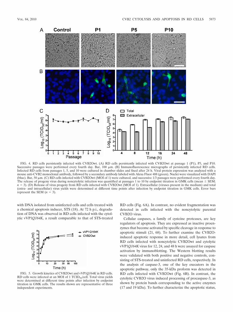
with DNA isolated from uninfected cells and cells treated witha chemical apoptosis inducer, STS (18). At 72 h p.i., degrada-tion of DNA was observed in RD cells infected with the cytol-ytic vVP1Q164K, a result comparable to that of STS-treated
RD cells (Fig. 6A). In contrast, no evident fragmentation wasdetected in cells infected with the noncytolytic parentalCVB2O virus.
Cellular caspases, a family of cysteine proteases, are keyregulators of apoptosis. They are expressed as inactive proen-zymes that become activated by specific cleavage in response toapoptotic stimuli (21, 68). To further examine the CVB2O-induced apoptotic response in more detail, cell lysates fromRD cells infected with noncytolytic CVB2Owt and cytolyticvVP1Q164K virus for 12, 24, and 48 h were assayed for caspaseactivation by immunoblotting. The Western blotting resultswere validated with both positive and negative controls, con-sisting of STS-treated and uninfected RD cells, respectively. Inthe analysis of caspase-3, one of the key executors in theapoptotic pathway, only the 35-kDa proform was detected inRD cells infected with CVB2Owt (Fig. 6B). In contrast, thecytolytic CVB2O virus induced processing of procaspase-3, asshown by protein bands corresponding to the active enzymes(17 and 19 kDa). To further characterize the apoptotic status,
FIG. 4. RD cells persistently infected with CVB2Owt. (A) RD cells persistently infected with CVB2Owt at passage 1 (P1), P5, and P10.Successive passages were performed every fourth day. Bar, 100 �m. (B) Immunofluorescence micrographs of persistently infected RD cells.Infected RD cells from passages 1, 5, and 10 were cultured in chamber slides and fixed after 24 h. Viral protein expression was analyzed with amouse anti-CVB2 monoclonal antibody, followed by a secondary antibody labeled with Alexa Fluor 488 (green). Nuclei were visualized with DAPI(blue). Bar, 50 �m. (C) RD cells infected with CVB2Owt (MOI of 1) were cultured, and successive 1/3 passages were performed every fourth day.The release of progeny virus during noncytolytic infection was quantified at passages 1 to 10 by endpoint titration in GMK cells (mean SEM;n � 3). (D) Release of virus progeny from RD cells infected with CVB2Owt (MOI of 1). Extracellular (viruses present in the medium) and total(extra- and intracellular) virus yields were determined at different time points after infection by endpoint titration in GMK cells. Error barsrepresent the SEM (n � 3).
FIG. 5. Growth kinetics of CVB2Owt and vVP1Q164K in RD cells.RD cells were infected at an MOI of 1 TCID50/cell. Total virus yieldswere determined at different time points after infection by endpointtitration in GMK cells. The results shown are representative of threeindependent experiments.
VOL. 84, 2010 CVB2 CYTOLYSIS AND APOPTOSIS IN RD CELLS 5873

two upstream caspases (caspase-8 and caspase-9) were ana-lyzed. The active forms of caspase-8 (36 and 40 kDa) andcaspase-9 (35 and 37 kDa) were detected in RD cells infectedwith vVP1Q164K. However, only the precursors of caspase-8and caspase-9 were detected in cells infected with the noncy-tolytic CVB2O virus. Surprisingly, at 48 h p.i., a downregula-tion of procaspase-8 was observed in these cells.
The Bid protein, a proapoptotic protein of the Bcl-2 family,is an important regulator of apoptosis. Upon activation by theinitiator caspase-8, the active form of Bid translocates to themitochondria where it triggers Bax activation, which, in turn, isfollowed by cytochrome c efflux into the cytosol (46). To fur-ther assess the CVB2O-triggered apoptosis in RD cells, theprocessing of Bid was determined. The proform of Bid (22kDa) was clearly processed into its active form (15 kDa) in RD
cells infected with the cytolytic vVP1Q164K while no activationwas observed for CVB2Owt-infected cells (Fig. 6B).
The association between apoptosis and the cytolytic infec-tion (vVP1Q164K) in RD cells was also quantified by immu-nofluorescence based on caspase-3 activation. Only a smallfraction of uninfected (1%) and CVB2Owt-infected(3.6%) RD cells showed staining of active caspase-3 (Fig. 6Cand D). However, a significantly higher percentage (12%) ofcells infected with the vVP1Q164K mutant stained positivelyfor cleaved caspase-3 (24 h p.i.).
Taken together, these results suggested that the VP1 substi-tution Q164K in CVB2O was associated with apoptosis in RDcells via a number of key players of the apoptotic cascade.
CVB2ORD causes pancreatic inflammation in mice. CVBsare known to have pancreatic and myocardial tropism in
FIG. 6. Apoptotic status of RD cells infected with CVB2Owt and vVP1Q164K. RD cells were infected with the CVB2O viruses at an MOI of1 and incubated at 37°C. (A) At the indicated times postinfection, cells were harvested, and DNA was extracted and resolved by electrophoresisin a 1% agarose gel as described in Materials and Methods. Uninfected RD cells (�) and cells treated with STS for 12 h (�) were used as negativeand positive controls, respectively. M, molecular weight marker. (B) At the indicated times postinfection, RD cell lysates were prepared, and equalamounts of proteins (5 to 40 �g) were assayed for caspase-3, caspase-9, caspase-8, and Bid by Western blotting. Uninfected RD cells and cellstreated with STS for 8 h were used as negative and positive controls, respectively, while expression of actin was used as a control of equal proteinloading. Activated forms of Bid and caspase-3 are shown after longer durations of exposure than the unprocessed fragment. The molecular massesof the active fragments are indicated in the right margin. The results shown are representative of three independent experiments. (C) RD cellsinfected with CVB2Owt and vVP1Q164K were fixed at 24 h postinfection and analyzed with a polyclonal antibody against cleaved/activatedcaspase-3 (CC-3). CC-3 was visualized with a secondary antibody conjugated with Alexa Fluor 488 (green), and nuclei of RD cells were visualizedwith DAPI (blue). (D) Induction of apoptosis determined by caspase-3 activation. The percentage of CC-3-labeled RD cells from each sample wasestimated from 300 randomly counted nuclei. Error bars represent the SEM (n � 3). A P value of �0.001 (���) was statistically significant forvVP1Q164K versus CVB2Owt or control cells. NS, not significant (P 0.05).
5874 GULLBERG ET AL. J. VIROL.

murine models (31, 36, 38, 83, 86). In order to investigatethe relevance of the cytolytic RD phenotype (CVB2ORD)in a murine model, A/J mice susceptible to CVB infection(83, 87, 88) were inoculated intraperitoneally with CVB2Oor CVB2ORD. At day 5 p.i., CVB2ORD-infected miceshowed mild to intense perivascular lymphocyte infiltrationwith focal vasculopathy (i.e., inflammation observed as blebformation of endothelial cells in blood vessels) in the exo-crine pancreas (Fig. 7A). In contrast, no visible signs ofinfection were observed in mice infected with CVB2O.Moreover, neither CVB2O nor CVB2ORD induced his-topathological changes in the endocrine pancreas or in theheart of infected mice. Titrations of the pancreatic tissueshowed that the viral titer was higher in the pancreas frommice subjected to the CVB2ORD virus variant (Fig. 7B).
DISCUSSION
In the present study, we examined specific amino acidchanges associated with adaptation of CVB2O to cytolytic in-fection in RD cells. Our results showed that a single amino acidchange on the capsid surface of CVB2O transforms the virusfrom a noncytolytic variant to a virus causing cytolysis. Thecharacterization of the viral infection suggested that the
CVB2O-induced cytolysis was associated with an apoptoticresponse (Fig. 8).
Previously published results have shown that CVB2O hasthe capacity to adapt to cytolytic replication in RD cells andthat this novel property was associated with three nonsynony-mous mutations (66). The results from reverse genetics studieswith the cloned single mutants presented here demonstratedthat a single surface-exposed amino acid change (Q164K) inthe VP1 capsid protein of this virus is sufficient for the trans-formation to a cytolytic phenotype. This observation supportsthe view that specific capsid residues influence picornaviral celltype specificity and tissue tropism (1, 6, 15, 39, 44, 45, 62).
In detailed studies of the virus replication in RD cells byreal-time PCR and titration on permissive GMK cells, thedifferent cDNA clone-derived CVB2O variants showed similarproperties regarding genome replication and virus progenyproduction. The productive infection of the cytolytic virus andnoncytolytic CVB2O variants was further confirmed by immu-nofluorescence studies. This study also showed, together withthe single-step growth curve and the observed onset of CPE,that there is a lag phase between the initial phase of viralreplication and the CPE. The same phenomenon has previ-ously been observed for another picornavirus, the Ljunganvirus of the Parechovirus genus (28, 80). Possibly, this delay
FIG. 7. Histological analysis of pancreatic tissue from mice infected with CVB2O or CVB2ORD. Male A/J mice were intraperitoneally injectedwith CVB2O (dose of 2 � 106 TCID50s), CVB2ORD (dose of 2 � 106 TCID50s), or PBS (control). At day 5 postinoculation, the pancreata wereformalin fixed and paraffin embedded, and tissue sections were stained with hematoxylin and eosin. Arrows indicate pancreatic tissue regions withmild to intense exocrine and perivascular lymphocyte infiltration with focal vasculopathy. Representative histopathological sections are shown.Original magnification, �20. (B) CVB2O and CVB2ORD titers in murine pancreas at 5 days after inoculation. No viral titer was measurable inpancreas of control mice. Titers were determined by endpoint titration in HEp-2 cells. Error bars represent the SEM (n � 3).
VOL. 84, 2010 CVB2 CYTOLYSIS AND APOPTOSIS IN RD CELLS 5875

of CPE is a consequence of a virus that is not yet completelyadapted to its host cell. However, the mechanism(s) in-volved in this delay of the CPE in RD cells infected with thecytolytic CVB2 variant remains to be elucidated. Takentogether, the CVB2Owt and all CVB2O mutants were ableto replicate in RD cells, a property that was not linked tocytolysis.
The nonstructural 2C protein of picornaviruses is a multi-functional protein with reported activities, including guidanceof viral replication complexes to cytoplasmic membranes, en-zymatic nucleotide triphosphatase activity, and involvement invirion assembly (48, 65, 78, 84). Results from analyses of theCVB2O mutant expressing a single 2C substitution (K185R)presented here suggested that this genetic change was notan essential determinant for the cytolytic phenotype ofCVB2ORD. However, infection of RD cells with CVB2ORD,expressing all three adaptive substitutions, resulted in an ear-lier onset of cytolysis than infection with vVP1Q164K. Thus,the additional substitution in VP1 (I118F) together with theamino acid change of 2C contributed to the cytolytic pheno-type in RD cells by a mechanism that remains to be elucidated.
Previously, it has been shown that CVB can establish per-sistent infections in RD cells (4, 29). Monitoring of repeatedpassages of the wild-type CVB2O in RD cells revealed a con-
tinuous release of infectious progeny although no signs of CPEwere observed. The ability of the CVB2O wild-type strain toreplicate in RD cells without evident signs of CPE has, to ourknowledge, never been characterized before. Studies of polio-virus have shown that persistent infections may be establishedwhen HEp-2 cells are subjected to virus at a very low MOI(62). In contrast, the CVB2O infection of RD cells seems to beindependent of virus dosage since these cells remained persis-tently infected even when they were exposed to a very highviral dose (MOI of 100). The release of noncytolytic CVB2Owtis possibly facilitated by viral proteins such as nonstructuralprotein 2B, which has been shown to modify membrane per-meability (23, 60, 85). Conclusively, these results suggest thatCVB2O established a persistent infection in cultured RD cells.This may have implications for CVB2 infections in vivo wheremuscle cells possibly serve as virus reservoirs. Indeed, CVB2RNA has been detected in muscle tissue of patients withchronic muscle diseases (3, 12, 13, 22).
The virus-host cell system represented by the differentCVB2O variants and the RD cells provided a well-definedmodel system for studies of persistent and cytolytic CVB2Oinfection. This model system also made it possible to examinewhether apoptosis played a role during CVB2O infection inthese cells. Theiler’s murine encephalomyelitis virus is highlycytolytic in permissive BHK-21 cells, causing rapid cell destruc-tion without signs of apoptosis; however, in less permissivecells, virus growth is markedly reduced, and viral replication isaccompanied by induction of apoptosis (40, 41). Other picorna-viruses, including CVB3, enterovirus 71, foot-and-mouth dis-ease virus, poliovirus and avian encephalomyelitis virus, havealso been shown to interact with the cellular apoptotic pathway(17, 19, 47, 50, 51, 63, 81). Although one-step growth analysisdemonstrated that RD cells were equally susceptible to repli-cation of the cytolytic and persistent CVB2O variants, majordisparities were revealed when the apoptotic status of infectedcells was examined. Prior to cell cytolysis, distinctive apoptotichallmarks, i.e., extensive DNA degradation and activation ofcaspase-8, caspase-9, and caspase-3, were observed in RD cellsinfected with the cytolytic CVB2O (vVP1Q164K) variant. Inaddition, the infection was accompanied by an activation ofBid, an activation previously described for RD cells infectedwith enterovirus 71 (19). Conversely, RD cells persistentlyinfected with the parental CVB2O virus showed no signs ofDNA degradation and caspase activation. In conclusion, thesedata add to the increasing knowledge of the interplay betweenpicornaviruses and the cellular apoptotic pathway during in-fection.
Several reports have suggested that the structural proteins ofpicornaviruses are involved in the induction of apoptosis (33,50, 63). For example, the VP1 protein of foot-and-mouth dis-ease virus has been shown to activate a proapototic response bybinding to integrins and by deactivation of the Akt signalingpathway (63). In other experiments, the VP3 protein of avianencephalomyelitis virus colocalized with mitochondria (50),whereas the VP2 protein of CVB3 interacted with a proapop-totic factor, called Siva (33). Hence, picornaviral structuralproteins are associated with induction of apoptosis by differentmechanisms. Whether any of these mechanisms are involved inthe apoptotic response associated with the VP1 protein ofCVB2O remains to be elucidated.
FIG. 8. Schematic illustration of the CVB2O-induced apoptotic re-sponse in RD cells. Cytolytic CVB2O infection (vVP1Q164K) of RDcells results in activation of both the extrinsic (procaspase-8) andintrinsic (procaspase-9) pathways. These pathways are interconnectedby Bid. When Bid is proteolytically activated by caspase-8, it translo-cates to the mitochondria and induces cytochrome c release. This, inturn, causes a successive activation of caspase-9 and caspase-3. Theactivated apoptotic signal eventually results in apoptotic cell death. Incontrast, CVB2Owt causes a persistent infection in RD cells withoutdetectable signs of apoptosis.
5876 GULLBERG ET AL. J. VIROL.

Although induction of apoptosis in RD cells was linked tocaspase activation, the involvement of other apoptotic mecha-nisms cannot be excluded. For example, the large quantities ofviral products that accompany a poliovirus infection result indrastic rearrangement of the endoplasmic reticulum (ER)(72). ER-mediated stress resulting in activation of apoptosishas previously been reported for cells infected with a virus ofthe Flaviviridae family, the Japanese encephalitis virus (77). Inaddition, a previous study has shown that caspase inhibitiondoes not prevent cell death of CVB3-infected HeLa cells (17).Thus, apoptotic activity is certainly not the only mechanismthat can induce cell death and cytolysis during picornavirusinfections. For example, the multifunctional 2A and 3C pro-teins are involved in the breakdown process of the cytoskeletonat the time of cell lysis (5, 42, 75). Consequently, cell lysisinduced by a virus infection is probably a result of severaldifferent intracellular processes.
CVB infections cause myocarditis and pancreatitis (31, 38).As suggested by serological studies, these viruses also appearto be associated with insulin-dependent diabetes mellitus (38).Several experimental murine models have been developed tostudy CVB infections. In this report, a comparative study in amouse model indicated that the cytolytic CVB2ORD variantinduced inflammation in the exocrine pancreas while no signsof inflammation were observed in pancreatic tissue of miceinfected with the parental CVB2O strain. This pancreatic vir-ulence is consistent with other studies of CVB infections inmice, which have shown that virus-induced inflammation waslocated to the exocrine pancreas and not the endocrine tissue(70). However, these initial observations, including the possi-ble association between the cytolytic RD phenotype and theincreased propensity to induce pancreatic inflammation inmice, need to be explored further.
Knowledge about virus-host cell interactions is important inorder to elucidate mechanisms by which the virus causes dam-age and to enable development of new antiviral treatments.The present study illustrates the adaptive potential of picorna-viruses, where a single capsid substitution in CVB2O played apivotal role for both cytolytic infection and induction of anapoptotic response in RD cells. In the absence of this aminoacid change, CVB2O established a persistent, noncytolytic in-fection without signs of apoptosis. Thus, this study shows thatthe fate of the infected cell depends on a complex balancebetween the host and the virus and that this balance can bedisrupted by a single substitution.
ACKNOWLEDGMENTS
We thank Marlene Norrby for outstanding technical assistance andKjell Edman for valuable discussions.
Figure 1B was produced using the UCSF Chimera package from theComputer Graphics Laboratory, University of California, San Fran-cisco (supported by NIH P41 RR-01081). This work was also sup-ported by grants from the Swedish Knowledge Foundation, the Uni-versity of Kalmar, the Helge Ax:son Johnsons Foundation, theSparbanken Kronan Foundation, and the Slovak Ministry of Health(MZSR code.2005/23-SZU-01).
REFERENCES
1. Al-Hello, H., P. Ylipaasto, T. Smura, E. Rieder, T. Hovi, and M. Roivainen.2009. Amino acids of Coxsackie B5 virus are critical for infection of themurine insulinoma cell line, MIN-6. J. Med. Virol. 81:296–304.
2. Andreoletti, L., L. Venteo, F. Douche-Aourik, F. Canas, G. Lorin de la
Grandmaison, J. Jacques, H. Moret, N. Jovenin, J. F. Mosnier, M. Matta, S.Duband, M. Pluot, B. Pozzetto, and T. Bourlet. 2007. Active coxsackieviral Binfection is associated with disruption of dystrophin in endomyocardial tissueof patients who died suddenly of acute myocardial infarction. J. Am. Coll.Cardiol. 50:2207–2214.
3. Archard, L. C., N. E. Bowles, P. O. Behan, E. J. Bell, and D. Doyle. 1988.Postviral fatigue syndrome: persistence of enterovirus RNA in muscle andelevated creatine kinase. J. R Soc Med. 81:326–329.
4. Argo, E., B. Gimenez, and P. Cash. 1992. Non-cytopathic infection of rhabdo-myosarcoma cells by coxsackie B5 virus. Arch. Virol. 126:215–229.
5. Badorff, C., G. H. Lee, B. J. Lamphear, M. E. Martone, K. P. Campbell, R. E.Rhoads, and K. U. Knowlton. 1999. Enteroviral protease 2A cleaves dystro-phin: evidence of cytoskeletal disruption in an acquired cardiomyopathy.Nat. Med. 5:320–326.
6. Bae, Y. S., and J. W. Yoon. 1993. Determination of diabetogenicity attrib-utable to a single amino acid, Ala776, on the polyprotein of encephalomyo-carditis virus. Diabetes 42:435–443.
7. Barco, A., E. Feduchi, and L. Carrasco. 2000. Poliovirus protease 3C(pro)kills cells by apoptosis. Virology 266:352–360.
8. Belov, G. A., L. I. Romanova, E. A. Tolskaya, M. S. Kolesnikova, Y. A.Lazebnik, and V. I. Agol. 2003. The major apoptotic pathway activated andsuppressed by poliovirus. J. Virol. 77:45–56.
9. Bergelson, J. M., J. A. Cunningham, G. Droguett, E. A. Kurt-Jones, A.Krithivas, J. S. Hong, M. S. Horwitz, R. L. Crowell, and R. W. Finberg. 1997.Isolation of a common receptor for coxsackie B viruses and adenoviruses 2and 5. Science 275:1320–1323.
10. Bergelson, J. M., J. G. Mohanty, R. L. Crowell, N. F. St John, D. M. Lublin,and R. W. Finberg. 1995. Coxsackievirus B3 adapted to growth in RD cellsbinds to decay-accelerating factor (CD55). J. Virol. 69:1903–1906.
11. Bopegamage, S., M. Borsanyiova, A. Vargova, A. Petrovicova, M. Benkovi-cova, and P. Gomolcak. 2003. Coxsackievirus infection of mice. I. Viralkinetics and histopathological changes in mice experimentally infected withcoxsackieviruses B3 and B4 by oral route. Acta Virol. 47:245–251.
12. Bowles, N. E., V. Dubowitz, C. A. Sewry, and L. C. Archard. 1987. Dermato-myositis, polymyositis, and Coxsackie-B-virus infection. Lancet 1:1004–1007.
13. Bowles, N. E., P. J. Richardson, E. G. Olsen, and L. C. Archard. 1986.Detection of Coxsackie-B-virus-specific RNA sequences in myocardialbiopsy samples from patients with myocarditis and dilated cardiomyo-pathy. Lancet 1:1120–1123.
14. Buenz, E. J., and C. L. Howe. 2006. Picornaviruses and cell death. TrendsMicrobiol. 14:28–36.
15. Caggana, M., P. Chan, and A. Ramsingh. 1993. Identification of a singleamino acid residue in the capsid protein VP1 of coxsackievirus B4 thatdetermines the virulent phenotype. J. Virol. 67:4797–4803.
16. Campanella, M., A. S. de Jong, K. W. Lanke, W. J. Melchers, P. H. Willems,P. Pinton, R. Rizzuto, and F. J. van Kuppeveld. 2004. The coxsackievirus 2Bprotein suppresses apoptotic host cell responses by manipulating intracellu-lar Ca2� homeostasis. J. Biol. Chem. 279:18440–18450.
17. Carthy, C. M., D. J. Granville, K. A. Watson, D. R. Anderson, J. E. Wilson,D. Yang, D. W. Hunt, and B. M. McManus. 1998. Caspase activation andspecific cleavage of substrates after coxsackievirus B3-induced cytopathiceffect in HeLa cells. J. Virol. 72:7669–7675.
18. Chae, H. J., J. S. Kang, J. O. Byun, K. S. Han, D. U. Kim, S. M. Oh, H. M.Kim, S. W. Chae, and H. R. Kim. 2000. Molecular mechanism of stauro-sporine-induced apoptosis in osteoblasts. Pharmacol. Res. 42:373–381.
19. Chang, S. C., J. Y. Lin, L. Y. Lo, M. L. Li, and S. R. Shih. 2004. Diverseapoptotic pathways in enterovirus 71-infected cells. J. Neurovirol. 10:338–349.
20. Chau, D. H., J. Yuan, H. Zhang, P. Cheung, T. Lim, Z. Liu, A. Sall, and D.Yang. 2007. Coxsackievirus B3 proteases 2A and 3C induce apoptotic celldeath through mitochondrial injury and cleavage of eIF4GI but not DAP5/p97/NAT1. Apoptosis 12:513–524.
21. Cohen, G. M. 1997. Caspases: the executioners of apoptosis. Biochem. J.326:1–16.
22. Cunningham, L., N. E. Bowles, R. J. Lane, V. Dubowitz, and L. C. Archard.1990. Persistence of enteroviral RNA in chronic fatigue syndrome is associ-ated with the abnormal production of equal amounts of positive and negativestrands of enteroviral RNA. J. Gen. Virol. 71:1399–1402.
23. de Jong, A. S., W. J. Melchers, D. H. Glaudemans, P. H. Willems, and F. J.van Kuppeveld. 2004. Mutational analysis of different regions in the coxsackie-virus 2B protein: requirements for homo-multimerization, membrane per-meabilization, subcellular localization, and virus replication. J. Biol. Chem.279:19924–19935.
24. Dobos, P. 1976. Use of gum tragacanth overlay, applied at room tempera-ture, in the plaque assay of fish and other animal viruses. J. Clin. Microbiol.3:373–375.
25. Domingo, E., C. K. Biebricher, M. Eigen, and J. J. Holland. 2001. Quasi-species and RNA virus evolution: principles and consequences. Landes Bio-science, Austin, TX.
26. Domingo, E., E. Martinez-Salas, F. Sobrino, J. C. de la Torre, A. Portela,J. Ortin, C. Lopez-Galindez, P. Perez-Brena, N. Villanueva, R. Najera, et al.
VOL. 84, 2010 CVB2 CYTOLYSIS AND APOPTOSIS IN RD CELLS 5877
1985. The quasispecies (extremely heterogeneous) nature of viral RNA ge-nome populations: biological relevance—a review. Gene 40:1–8.
27. Drake, J. W., and J. J. Holland. 1999. Mutation rates among RNA viruses.Proc. Natl. Acad. Sci. U. S. A. 96:13910–13913.
28. Ekstrom, J. O., C. Tolf, C. Fahlgren, E. S. Johansson, G. Arbrandt, B.Niklasson, K. A. Edman, and A. M. Lindberg. 2007. Replication of Ljunganvirus in cell culture: the genomic 5�-end, infectious cDNA clones and hostcell response to viral infections. Virus Res. 130:129–139.
29. Frisk, G., M. A. Lindberg, and H. Diderholm. 1999. Persistence of coxsackie-virus B4 infection in rhabdomyosarcoma cells for 30 months. Brief report.Arch. Virol. 144:2239–2245.
30. Galluzzi, L., C. Brenner, E. Morselli, Z. Touat, and G. Kroemer. 2008. Viralcontrol of mitochondrial apoptosis. PLoS Pathog. 4:e1000018.
31. Gauntt, C., and S. Huber. 2003. Coxsackievirus experimental heart diseases.Front Biosci. 8:e23–35.
32. Goldstaub, D., A. Gradi, Z. Bercovitch, Z. Grosmann, Y. Nophar, S. Luria,N. Sonenberg, and C. Kahana. 2000. Poliovirus 2A protease induces apop-totic cell death. Mol. Cell. Biol. 20:1271–1277.
33. Henke, A., H. Launhardt, K. Klement, A. Stelzner, R. Zell, and T. Munder.2000. Apoptosis in coxsackievirus B3-caused diseases: interaction betweenthe capsid protein VP2 and the proapoptotic protein Siva. J. Virol. 74:4284–4290.
34. Hierholzer, J. C., and R. A. Killington. 1996. Quantitation of virus, p. 35–37.In B. W. J. Mahy and H. O. Kangro (ed.), Virology methods manual.Academic Press, London, United Kingdom.
35. Horsington, J., and Z. Zhang. 2007. Consistent change in the B-C loop ofVP2 observed in foot-and-mouth disease virus from persistently infectedcattle: implications for association with persistence. Virus Res. 125:114–118.
36. Horwitz, M. S., A. Ilic, C. Fine, E. Rodriguez, and N. Sarvetnick. 2003.Coxsackievirus-mediated hyperglycemia is enhanced by reinfection and thisoccurs independent of T cells. Virology 314:510–520.
37. Hsu, K. H., K. Lonberg-Holm, B. Alstein, and R. L. Crowell. 1988. A mono-clonal antibody specific for the cellular receptor for the group B coxsackie-viruses. J. Virol. 62:1647–1652.
38. Huber, S., and A. I. Ramsingh. 2004. Coxsackievirus-induced pancreatitis.Viral Immunol. 17:358–369.
39. Jarousse, N., R. A. Grant, J. M. Hogle, L. Zhang, A. Senkowski, R. P. Roos,T. Michiels, M. Brahic, and A. McAllister. 1994. A single amino acid changedetermines persistence of a chimeric Theiler’s virus. J. Virol. 68:3364–3368.
40. Jelachich, M. L., and H. L. Lipton. 1999. Restricted Theiler’s murine en-cephalomyelitis virus infection in murine macrophages induces apoptosis.J. Gen. Virol. 80:1701–1705.
41. Jelachich, M. L., and H. L. Lipton. 1996. Theiler’s murine encephalomyelitisvirus kills restrictive but not permissive cells by apoptosis. J. Virol. 70:6856–6861.
42. Joachims, M., K. S. Harris, and D. Etchison. 1995. Poliovirus protease 3Cmediates cleavage of microtubule-associated protein 4. Virology 211:451–461.
43. Jonsson, N., M. Gullberg, and A. M. Lindberg. 2009. Real-time polymerasechain reaction as a rapid and efficient alternative to estimation of picorna-virus titers by tissue culture infectious dose 50% or plaque forming units.Microbiol. Immunol. 53:149–154.
44. Jun, H. S., Y. Kang, A. L. Notkins, and J. W. Yoon. 1997. Gain or loss ofdiabetogenicity resulting from a single point mutation in recombinant en-cephalomyocarditis virus. J. Virol. 71:9782–9785.
45. Knowlton, K. U., E. S. Jeon, N. Berkley, R. Wessely, and S. Huber. 1996. Amutation in the puff region of VP2 attenuates the myocarditic phenotype ofan infectious cDNA of the Woodruff variant of coxsackievirus B3. J. Virol.70:7811–7818.
46. Kruidering, M., and G. I. Evan. 2000. Caspase-8 in apoptosis: the beginningof “the end”? IUBMB Life 50:85–90.
47. Kuo, R. L., S. H. Kung, Y. Y. Hsu, and W. T. Liu. 2002. Infection withenterovirus 71 or expression of its 2A protease induces apoptotic cell death.J. Gen. Virol. 83:1367–1376.
48. Li, J. P., and D. Baltimore. 1990. An intragenic revertant of a poliovirus 2Cmutant has an uncoating defect. J. Virol. 64:1102–1107.
49. Lindberg, A. M., C. Polacek, and S. Johansson. 1997. Amplification andcloning of complete enterovirus genomes by long distance PCR. J. Virol.Methods 65:191–199.
50. Liu, J., T. Wei, and J. Kwang. 2002. Avian encephalomyelitis virus inducesapoptosis via major structural protein VP3. Virology 300:39–49.
51. Liu, J., T. Wei, and J. Kwang. 2004. Avian encephalomyelitis virus nonstruc-tural protein 2C induces apoptosis by activating cytochrome c/caspase-9pathway. Virology 318:169–182.
52. Livak, K. J., and T. D. Schmittgen. 2001. Analysis of relative gene expressiondata using real-time quantitative PCR and the 2(-��CT) method. Methods25:402–408.
53. Lukashev, A. N. 2005. Role of recombination in evolution of enteroviruses.Rev. Med. Virol. 15:157–167.
54. Martino, T. A., M. Petric, M. Brown, K. Aitken, C. J. Gauntt, C. D. Rich-ardson, L. H. Chow, and P. P. Liu. 1998. Cardiovirulent coxsackieviruses andthe decay-accelerating factor (CD55) receptor. Virology 244:302–314.
55. Martino, T. A., M. Petric, H. Weingartl, J. M. Bergelson, M. A. Opavsky,C. D. Richardson, J. F. Modlin, R. W. Finberg, K. C. Kain, N. Willis, C. J.Gauntt, and P. P. Liu. 2000. The coxsackie-adenovirus receptor (CAR) isused by reference strains and clinical isolates representing all six serotypes ofcoxsackievirus group B and by swine vesicular disease virus. Virology 271:99–108.
56. Melnick, J. L., N. Ledinko, et al. 1950. Ohio strains of a virus pathogenic forinfant mice, Coxsackie group; simultaneous occurrence with poliomyelitisvirus in patients with summer grippe. J. Exp. Med. 91:185–195.
57. Minor, P. 1985. Growth, assay and purification of picornaviruses, p. 25–41. InB. Mahy (ed.), Virology, a practical approach. IRL Press, Oxford, UnitedKingdom.
58. Muckelbauer, J. K., M. Kremer, I. Minor, G. Diana, F. J. Dutko, J. Groarke,D. C. Pevear, and M. G. Rossmann. 1995. The structure of coxsackievirus B3at 3.5 Å resolution. Structure 3:653–667.
59. Neznanov, N., A. Kondratova, K. M. Chumakov, B. Angres, B. Zhum-abayeva, V. I. Agol, and A. V. Gudkov. 2001. Poliovirus protein 3A inhibitstumor necrosis factor (TNF)-induced apoptosis by eliminating the TNFreceptor from the cell surface. J. Virol. 75:10409–10420.
60. Nieva, J. L., A. Agirre, S. Nir, and L. Carrasco. 2003. Mechanisms ofmembrane permeabilization by picornavirus 2B viroporin. FEBS Lett. 552:68–73.
61. Pallansch, M. A., and R. P. Roos. 2001. Enteroviruses: polioviruses, coxsackie-viruses, echoviruses and newer enteroviruses, p. 723–775. In D. M. Knipe,P. M. Howley, D. E. Griffin, R. A. Lamb, M. A. Martin, B. Roizman, andS. E. Straus (ed.), Fields virology, 4th ed. Lippincott Williams & Wilkins,Philadelphia, PA.
62. Pelletier, I., G. Duncan, and F. Colbere-Garapin. 1998. One amino acidchange on the capsid surface of poliovirus Sabin 1 allows the establishmentof persistent infections in HEp-2c cell cultures. Virology 241:1–13.
63. Peng, J. M., S. M. Liang, and C. M. Liang. 2004. VP1 of foot-and-mouthdisease virus induces apoptosis via the Akt signaling pathway. J. Biol. Chem.279:52168–52174.
64. Pettersen, E. F., T. D. Goddard, C. C. Huang, G. S. Couch, D. M. Greenblatt,E. C. Meng, and T. E. Ferrin. 2004. UCSF Chimera—a visualization systemfor exploratory research and analysis. J. Comput. Chem. 25:1605–1612.
65. Pfister, T., and E. Wimmer. 1999. Characterization of the nucleoside triphos-phatase activity of poliovirus protein 2C reveals a mechanism by whichguanidine inhibits poliovirus replication. J. Biol. Chem. 274:6992–7001.
66. Polacek, C., J. O. Ekstrom, A. Lundgren, and A. M. Lindberg. 2005. Cytolyticreplication of coxsackievirus B2 in CAR-deficient rhabdomyosarcoma cells.Virus Res. 113:107–115.
67. Polacek, C., A. Lundgren, A. Andersson, and A. M. Lindberg. 1999. Genomicand phylogenetic characterization of coxsackievirus B2 prototype strainOhio-1. Virus Res. 59:229–238.
68. Pop, C., and G. S. Salvesen. 2009. Human caspases: activation, specificity,and regulation. J. Biol. Chem. 284:21777–21781.
69. Racaniello, V. R. 2001. Picornaviridae: the viruses and their replication, p.685–722. In D. M. Knipe, P. M. Howley, D. E. Griffin, R. A. Lamb, M. A.Martin, B. Roizman, and S. E. Straus (ed.), Fields virology, 4th ed. Lippin-cott Williams & Wilkins, Philadelphia, PA.
70. Ramsingh, A. I. 1997. Coxsackieviruses and pancreatitis. Front Biosci.2:e53–62.
71. Roulston, A., R. C. Marcellus, and P. E. Branton. 1999. Viruses and apop-tosis. Annu. Rev. Microbiol. 53:577–628.
72. Rust, R. C., L. Landmann, R. Gosert, B. L. Tang, W. Hong, H. P. Hauri, D.Egger, and K. Bienz. 2001. Cellular COPII proteins are involved in produc-tion of the vesicles that form the poliovirus replication complex. J. Virol.75:9808–9818.
73. Sanner, M. F., A. J. Olson, and J. C. Spehner. 1996. Reduced surface: anefficient way to compute molecular surfaces. Biopolymers 38:305–320.
74. Schmidtke, M., H. C. Selinka, A. Heim, B. Jahn, M. Tonew, R. Kandolf, A.Stelzner, and R. Zell. 2000. Attachment of coxsackievirus B3 variants tovarious cell lines: mapping of phenotypic differences to capsid protein VP1.Virology 275:77–88.
75. Seipelt, J., H. D. Liebig, W. Sommergruber, C. Gerner, and E. Kuechler.2000. 2A proteinase of human rhinovirus cleaves cytokeratin 8 in infectedHeLa cells. J. Biol. Chem. 275:20084–20089.
76. Shafren, D. R., R. C. Bates, M. V. Agrez, R. L. Herd, G. F. Burns, and R. D.Barry. 1995. Coxsackieviruses B1, B3, and B5 use decay accelerating factoras a receptor for cell attachment. J. Virol. 69:3873–3877.
77. Su, H. L., C. L. Liao, and Y. L. Lin. 2002. Japanese encephalitis virusinfection initiates endoplasmic reticulum stress and an unfolded proteinresponse. J. Virol. 76:4162–4171.
78. Teterina, N. L., D. Egger, K. Bienz, D. M. Brown, B. L. Semler, and E.Ehrenfeld. 2001. Requirements for assembly of poliovirus replication com-plexes and negative-strand RNA synthesis. J. Virol. 75:3841–3850.
79. Thompson, J. D., D. G. Higgins, and T. J. Gibson. 1994. CLUSTAL W:improving the sensitivity of progressive multiple sequence alignment throughsequence weighting, position-specific gap penalties and weight matrix choice.Nucleic Acids Res. 22:4673–4680.
80. Tolf, C., M. Gullberg, J. O. Ekstrom, N. Jonsson, and A. Michael Lindberg.
5878 GULLBERG ET AL. J. VIROL.

2009. Identification of amino acid residues of Ljungan virus VP0 and VP1associated with cytolytic replication in cultured cells. Arch. Virol. 154:1271–1284.
81. Tolskaya, E. A., L. I. Romanova, M. S. Kolesnikova, T. A. Ivannikova, E. A.Smirnova, N. T. Raikhlin, and V. I. Agol. 1995. Apoptosis-inducing andapoptosis-preventing functions of poliovirus. J. Virol. 69:1181–1189.
82. Tomko, R. P., R. Xu, and L. Philipson. 1997. HCAR and MCAR: the humanand mouse cellular receptors for subgroup C adenoviruses and group Bcoxsackieviruses. Proc. Natl. Acad. Sci. U. S. A. 94:3352–3356.
83. Tracy, S., K. Hofling, S. Pirruccello, P. H. Lane, S. M. Reyna, and C. J.Gauntt. 2000. Group B coxsackievirus myocarditis and pancreatitis: connec-tion between viral virulence phenotypes in mice. J. Med. Virol. 62:70–81.
84. Vance, L. M., N. Moscufo, M. Chow, and B. A. Heinz. 1997. Poliovirus 2Cregion functions during encapsidation of viral RNA. J. Virol. 71:8759–8765.
85. van Kuppeveld, F. J., J. G. Hoenderop, R. L. Smeets, P. H. Willems, H. B.Dijkman, J. M. Galama, and W. J. Melchers. 1997. Coxsackievirus protein2B modifies endoplasmic reticulum membrane and plasma membrane per-meability and facilitates virus release. EMBO J. 16:3519–3532.
86. Woodruff, J. F. 1980. Viral myocarditis. A review. Am. J. Pathol. 101:425–484.87. Yang, D., J. Yu, Z. Luo, C. M. Carthy, J. E. Wilson, Z. Liu, and B. M.
McManus. 1999. Viral myocarditis: identification of five differentially ex-pressed genes in coxsackievirus B3-infected mouse heart. Circ Res. 84:704–712.
88. Yu, J. Z., J. E. Wilson, S. M. Wood, R. Kandolf, K. Klingel, D. Yang, andB. M. McManus. 1999. Secondary heterotypic versus homotypic infection byCoxsackie B group viruses: impact on early and late histopathological lesionsand virus genome prominence. Cardiovasc. Pathol. 8:93–102.
VOL. 84, 2010 CVB2 CYTOLYSIS AND APOPTOSIS IN RD CELLS 5879

JOURNAL OF VIROLOGY, Aug. 2010, p. 7922 Vol. 84, No. 150022-538X/10/$12.00 doi:10.1128/JVI.01116-10Copyright © 2010, American Society for Microbiology. All Rights Reserved.
ERRATUM
A Single Coxsackievirus B2 Capsid Residue Controls Cytolysis andApoptosis in Rhabdomyosarcoma Cells
Maria Gullberg, Conny Tolf, Nina Jonsson, Charlotta Polacek, Jana Precechtelova, Miriam Badurova,Martin Sojka, Camilla Mohlin, Stina Israelsson, Kjell Johansson, Shubhada Bopegamage,
Susan Hafenstein, and A. Michael LindbergSchool of Natural Sciences, Linnaeus University, SE-391 82 Kalmar, Sweden; National Veterinary Institute, Technical University of
Denmark, Lindholm, DK-4771, Kalvehave, Denmark; Department of Virology, Slovak Medical University, Limbova 12, 83303 Bratislava,Slovak Republic; and Department of Microbiology and Immunology, The Pennsylvania State University College of
Medicine, 500 University Drive, Hershey, Pennsylvania 17033
Volume 84, no. 12, p. 5868–5879, 2010. Page 5875, legend to Fig. 7, line 1: “Histological analysis” should read “(A) Histologicalanalysis.”
Page 5877. Acknowledgements, paragraph 2: Add “, the EEA Financial Mechanism, the Norwegian Financial Mechanism, andthe State Budget of the Slovak Republic (SK 0082)” to the last sentence.
7922


![Asthma Pharmacology [Read-Only] · Mechanism: IL-5 receptor alpha-directed cytolytic monoclonal antibody Requirements Serum eos ≥ 300 cells/µL ≥2 exacerbations Dosing: SQ injection,](https://img.pdfslide.us/doc/110x75/5f934d223c98053b747f2187/asthma-pharmacology-read-only-mechanism-il-5-receptor-alpha-directed-cytolytic.jpg)