Embed Size (px)
Citation preview

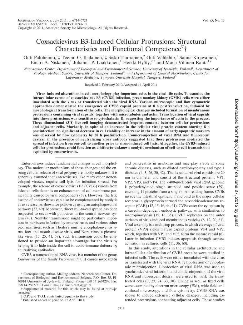
Published Ahead of Print 27 April 2011. 2011, 85(13):6714. DOI: 10.1128/JVI.00247-10. J. Virol.
P. Laakkonen, Heikki Hyöty and Maija Vihinen-RantaVälilehto, Sanna Kirjavainen, Einari A. Niskanen, Johanna Outi Paloheimo, Teemu O. Ihalainen, Sisko Tauriainen, Outi Functional CompetenceProtrusions: Structural Characteristics and Coxsackievirus B3-Induced Cellular
http://jvi.asm.org/content/85/13/6714Updated information and services can be found at:
These include:SUPPLEMENTAL MATERIAL
lhttp://jvi.asm.org/content/suppl/2011/06/02/85.13.6714.DC1.htm
REFERENCEShttp://jvi.asm.org/content/85/13/6714#ref-list-1at:
This article cites 61 articles, 26 of which can be accessed free
CONTENT ALERTS more»articles cite this article),
Receive: RSS Feeds, eTOCs, free email alerts (when new
http://journals.asm.org/site/misc/reprints.xhtmlInformation about commercial reprint orders: http://journals.asm.org/site/subscriptions/To subscribe to to another ASM Journal go to:
on June 27, 2012 by guesthttp://jvi.asm
.org/D
ownloaded from
JOURNAL OF VIROLOGY, July 2011, p. 6714–6724 Vol. 85, No. 130022-538X/11/$12.00 doi:10.1128/JVI.00247-10Copyright © 2011, American Society for Microbiology. All Rights Reserved.
Coxsackievirus B3-Induced Cellular Protrusions: StructuralCharacteristics and Functional Competence!†
Outi Paloheimo,1‡ Teemu O. Ihalainen,1‡ Sisko Tauriainen,2 Outi Valilehto,1 Sanna Kirjavainen,1Einari A. Niskanen,1 Johanna P. Laakkonen,1 Heikki Hyoty,2,3 and Maija Vihinen-Ranta1*
Nanoscience Center, Department of Biological and Environmental Science, University of Jyvaskyla, Finland1; Department ofVirology, Medical School, University of Tampere, Finland2; and Department of Clinical Microbiology, Center for
Laboratory Medicine, Tampere University Hospital, Tampere, Finland3
Received 3 February 2010/Accepted 14 April 2011
Virus-induced alterations in cell morphology play important roles in the viral life cycle. To examine theintracellular events of coxsackievirus B3 (CVB3) infection, green monkey kidney (GMK) cells were eitherinoculated with the virus or transfected with the viral RNA. Various microscopic and flow cytometricapproaches demonstrated the emergence of CVB3 capsid proteins at 8 h posttransfection, followed bymorphological transformation of the cells. The morphological changes included formation of membranousprotrusions containing viral capsids, together with microtubules and actin. Translocation of viral capsidsinto these protrusions was sensitive to cytochalasin D, suggesting the importance of actin in the process.Three-dimensional (3D) live-cell imaging demonstrated frequent contacts between cellular protrusionsand adjacent cells. Markedly, in spite of an increase in the cellular viral protein content starting 8 hpostinfection, no significant decrease in cell viability or increase in the amount of early apoptotic markerswas observed by flow cytometry by 28 h postinfection. Comicroinjection of viral RNA and fluorescentdextran in the presence of neutralizing virus antibody suggested that these protrusions mediated thespread of infection from one cell to another prior to virus-induced cell lysis. Altogether, the CVB3-inducedcellular protrusions could function as a hitherto-unknown nonlytic mechanism of cell-to-cell transmissionexploited by enteroviruses.
Enteroviruses induce fundamental changes in cell morphol-ogy. The molecular mechanisms of these changes and the en-suing cellular release of viral progeny are mostly unknown. It isgenerally assumed that enteroviruses, like many other nonen-veloped viruses, require cell lysis for infection spread. Forexample, the release of coxsackievirus B3 (CVB3) virions frominfected cells depends on enhancement of cell membrane per-meability caused by viral components (53). However, the lyticescape of enteroviruses can also be complemented by nonlyticvirus release, as shown for poliovirus using an autophagosomalpathway (27, 49). Moreover, direct cell-to-cell spread has beensuspected to occur with poliovirus in the central nervous sys-tem (40). Nonlytic transmission might be particularly impor-tant in persistent infection by enteroviruses and various otherpicornaviruses, such as Theiler’s murine encephalomyelitis vi-rus, foot-and-mouth disease virus, and Nora virus, a picorna-like virus (17, 25, 41, 58). Such transmission could be envi-sioned to provide an important advantage for the virus byhelping it to hide inside the cell to avoid immune defense byneutralizing antibodies.
CVB3, a nonenveloped RNA virus, is a member of the genusEnterovirus of the family Picornaviridae. It causes myocarditis
and pancreatitis in newborns and may play a role in somechronic diseases, such as dilated cardiomyopathy and type 1diabetes (4, 5, 26, 30, 42). The icosahedral viral capsids are 29nm in diameter and consist of the structural proteins VP1,VP2, VP3, and VP4. The 7,400-nucleotide viral RNA genomeis polyadenylated, single stranded, and positive sense (39),encoding 11 proteins from a single open reading frame. CVBsinvade the intestinal epithelium and bind to the major cellularreceptor, a glycoprotein termed the coxsackie-adenovirus re-ceptor (CAR) (12, 15, 16, 44, 61). CVBs enter the cytoplasm bya caveolin-dependent endocytic pathway, with similarities tomacropinocytosis (15, 16, 35). CVB3 replicates on the outersurfaces of virus-induced membranous vesicles (8, 12, 20, 61).Viral assembly is a multistep process, in which cleavage of viralprotein (VP0) yields mature capsid proteins VP4 and VP2,which, together with VP1 and VP3, form the mature capsid (6).Later in infection CVB3 induces apoptosis through caspaseactivation in cultured cells (11, 36, 60).
In this study, alterations in the cellular architecture andintracellular distribution of CVB3 proteins were studied ininfected cells. The cells were either inoculated with the virusor transfected with the viral RNA by lipofection or cytoplas-mic microinjection. Lipofection of viral RNA was used tosynchronize viral infection, and comicroinjection of the viralRNA and fluorescent dextran were used to mark the trans-fected cells (7, 23, 24, 33, 38). Living as well as fixed cellswere examined by electron microscopy (EM), wide-field andconfocal microscopy, and flow cytometry. CVB3 RNA wasshown to induce extensive cellular changes, including ex-tended protrusions connecting adjacent cells. These studies
* Corresponding author. Mailing address: Nanoscience Center, De-partment of Biological and Environmental Science, P.O. Box 35, FI-40014 University of Jyvaskyla, Finland. Phone: 358 14 2604209. Fax:358 14 2602221. E-mail: [email protected].
† Supplemental material for this article may be found at http://jvi.asm.org/.
‡ O.P. and T.O.I. contributed equally to this study.! Published ahead of print on 27 April 2011.
6714
on June 27, 2012 by guesthttp://jvi.asm
.org/D
ownloaded from
demonstrated for the first time nonlytic intercellular trans-mission of CVB3.
MATERIALS AND METHODS
Viruses and cells. CVB3 strain Nancy was obtained from the ATCC (Manas-sas, VA). Green monkey kidney (GMK) cells were grown in Earle’s minimumessential medium (MEM) supplemented with 10% heat-inactivated fetal calfserum (FCS), 0.05 mg/ml penicillin, 0.05 mg/ml streptomycin (PAA laboratoriesGmbH, Linz, Austria), 1 mM L-glutamine (Invitrogen, Carlsbad, CA), and 1%glucose at 37°C in 5% CO2. For infection, the cells were inoculated with CVB3and kept at 37°C until live-cell studies or fixation was performed (1).
CVB3, coxsackievirus A9 (CAV9), echovirus 11, echovirus 30, parechovirus,rhinovirus, and rotavirus RNAs were isolated from cell culture supernatants witha QIAamp Viral RNA Mini Kit (Qiagen, Hilden, Germany) according to themanufacturer’s protocol. The CVB3 RNA concentration in experiments was 120ng/!l, and for other RNAs it was 100 ng/!l.
Antibodies and chemicals. CVB3 polyclonal antibody (Ab) was obtained fromPharmacia Fine Chemicals (Uppsala, Sweden). Virus proteins were stained withAb, followed by Alexa 488-conjugated anti-rabbit IgG. Flow cytometry of cell-associated viral proteins was done with anti-CVB3 monoclonal antibody (MAb)(Millipore, Billerica, MA) conjugated to Atto 488 fluorophore according to themanufacturer’s protocol (Innova Biosciences, Cambridge, United Kingdom).Virus neutralization was performed in the presence of virus MAb (2 !g/ml).Microtubules (MTs) were visualized using "-tubulin MAb (Amersham, Buck-inghamshire, United Kingdom), followed by Alexa 633-conjugated anti-mouseIgG (Molecular Probes) and actin with tetramethylrhodamine isothiocyanate(TRITC)-phalloidin (Molecular Probes). CAV9, echovirus 11, echovirus 30,parechovirus, rhinovirus, and rotavirus RNA proteins were stained with entero-virus MAb (Dako, Glostrup, Denmark), followed by Alexa 555-conjugated anti-mouse IgG (Molecular Probes). MT- and actin-disintegrating agents, nocodazoleand cytochalasin D (Sigma Aldrich, St. Louis, MO), were used to detect CVB3capsid protein signal. For immuno-EM studies, nanogold-conjugated goat anti-rabbit Ab and HQ-silver enhancement reagents were purchased from Nano-probes (Yaphank, NY). Epon LX-112 was obtained from Ladd Research Indus-tries (Williston, VT). For microinjection studies, fluorescein isothiocyanate(FITC)-labeled dextrans (40,500 kDa) were obtained from Molecular Probes andTRITC- and FITC-labeled dextrans (150 kDa) from Sigma Aldrich. Cell viabilitywas judged by staining the cells with propidium iodide (PI) (Sigma) and annexinV-Alexa 488 (Molecular Probes). In flow cytometry control studies, apoptosiswas induced by staurosporine (STS) or cycloheximide (CHX) obtained fromSigma Aldrich.
Infection. Viral infection was initiated by either inoculation with virus ormicroinjection or by lipofection with the viral RNA using the DMRIE-C lipo-fection reagent (Invitrogen) according to the manufacturer’s protocol.
Microinjection into cells was carried out using a semiautomatic system com-prising the Transjector 5246 and Micromanipulator 5171 (Eppendorf, Hamburg,Germany) on an inverted microscope. Needles were pulled from glass capillaries(Clark Electromedical Instruments, Reading, United Kingdom) using a P-97needle puller (Sutter Instruments, Novato, CA). For studies with fixed cells,cultures were grown to 80% confluence on glass coverslips and for live imagingon 21.5-cm2 glass bottom culture dishes (MatTek Cultureware, Ashland, MA).Purified CVB3 RNA was microinjected into the cytoplasm in a microinjectionbuffer (10 mM Tris-HCl–120 mM KCl, pH 7.4) at a concentration of 120 ng/ml.At various times after microinjection, cells were fixed with 4% paraformaldehyde(PFA) (20 min at room temperature [RT]), followed by labeling with CVB3 Aband Alexa 488-conjugated anti-rabbit IgG. Three-dimensional (3D) images wereobtained with inverted laser scanning confocal microscopes (LSM 510 Axiovert100 M [Carl Zeiss AG, Jena, Germany] or FV-1000 IX-81 [Olympus, Tokyo,Japan]) using a 63# Plan-Neofluor oil immersion objective with a numericalaperture (NA) of 1.25 or a 60# APO oil immersion objective (NA $ 1.35).Stacks of 25 to 35 images with an image size of 800 by 800 pixels were collected.The pixel resolution was adjusted to 110 nm/pixel. Fluorescent dextran (2 mg/ml)was used to identify the microinjected cells. For cell-to-cell spread studies, singlecells of confluent monolayers were comicroinjected with RNA and 500 kDaFITC-dextran and incubated in the presence of neutralizing CVB3 Ab. The cellswere fixed at 24 h postmicroinjection (p.m.) with 4% PFA and permeabilizedwith 0.1% Triton X-100. Control studies were performed in the absence of Ab.In microinjection experiments, the cells were stained for virus proteins withCVB3 Ab, followed by Alexa 555-conjugated anti-rabbit IgG.
Flow cytometry. Double staining with annexin V/propidium iodide (PI) wasused to assay cell viability. The cells were inoculated with the virus, and after 8to 28 h, they were detached by scraping, washed with phosphate-buffered saline
(PBS), stained with annexin V-Alexa Fluor 488 conjugate (5 !l) and PI at 2!g/ml for 15 min, and analyzed with FACSCalibur and CellQuest software(Becton Dickinson, Heidelberg, Germany). The effects of drugs (1 !g/ml STS or1 !g/ml CHX for 24 h) on cell viability were monitored. Virus proteins weredetected with CVB3 monoclonal antibody (1 !g/1 # 106 cells) conjugated toAtto 488. Shortly after, the detached cells were washed with PBS, fixed in fixationbuffer (R&D Systems Inc., Minneapolis, MN), and incubated at room temper-ature for 10 min prior to treatment with permeabilization/wash buffer (R&DSystems Inc.). MAb was added to the cells and incubated for 60 min at 4°C. Thecells were washed with permeabilization/wash buffer and resuspended in stainingbuffer (R&D Systems Inc.) for flow cytometry analysis. The means of one rep-resentative experiment were counted from quadruplicate measurements with atotal of 4 # 104 cells. Experiments were repeated three times with similarobservations.
Live-cell imaging. Cells were grown to 80% confluence on 21.5-cm2 glassbottom culture dishes (0.16 to 0.19 mm thickness; MatTek Cultureware, Ash-land, MA). The images were captured either with a CellObserver HS wide-fieldmicroscope (Zeiss) or with a laser scanning confocal microscope (LSM510;Zeiss). Time lapse imaging of virus-induced alterations of cell morphology wasperformed with a Zeiss CellObserver HS wide-field microscope. The microscopeincubator was maintained at 37°C, and the CO2 concentration was adjusted to5%. The LD Plan-Neofluar 40# (NA $ 0.6) objective was used. Images weretaken at 5-min intervals. The LSM 510 objective and the sample holder werewarmed to 37°C prior to imaging. The 3D stacks of 15 to 30 cross-sectionalimages were acquired with a Plan-Neofluar 63# oil immersion objective (NA $1.25). The image size was adjusted to 512 by 512 pixels with an xy resolution of100 nm/voxel and z of 350 to 500 nm/voxel. A 488-nm argon laser line was usedfor FITC-dextran excitation, and fluorescence was monitored with a 520- to560-nm band-pass filter. TRITC-dextran was excited with a 543-nm HeNe laserand monitored with a long-pass filter of 560 nm.
For morphological characterization of uninfected cells, two cell populationsindividually transfected either with enhanced yellow fluorescent protein (EYFP)or enhanced cyan fluorescent protein (ECFP) were plated onto the same cellculture dish. The cells were imaged with a laser scanning confocal microsope(FV1000; Olympus) using the UPLSAPO 60# (NA $ 1.2) water immersionobjective. A 514-nm argon laser line was used for EYFP excitation, and fluores-cence was monitored using a 530- to 630-nm band-pass filter. ECFP was excitedby a 405-nm diode laser and was monitored with a band-pass filter of 460 to 500nm. The pixel size was 210 nm in x and y and 460 nm in z. Line averaging was setat 2.
Electron microscopy. For EM, cells on 13-mm glass coverslips were trans-fected with CVB3 RNA, incubated for 24 h, and fixed with 2.5% glutaraldehyde,0.1 M Na-cacodylate buffer, pH 7.4. Postfixation with 1% osmium tetroxide in 0.1M sodium cacodylate buffer (pH 7.4) was followed by dehydration and embed-ding in epoxy resin. Ultrathin sections were cut parallel to the coverslip, post-stained with uranyl acetate and lead citrate, and examined with a JEM-100S(JEOL, Tokyo, Japan). For preembedding immunolabeling, the cells were grownon 8.8-cm2 plastic dishes, transfected prior to fixation, and processed as previ-ously described (48). Briefly, the cells were fixed with periodate-lysine-PFAfixative for 2 h at RT and permeabilized with buffer A (0.01% saponin and 0.1%bovine serum albumin [BSA] in 0.1 M phosphate buffer, pH 7.4). Immunolabel-ing was performed using CVB3 Ab and 1.4-nm gold particle-conjugated goatrabbit IgG. Nanogold was silver enhanced for 9 min with the HQ-silver kit andgold toned with 0.05% gold chloride. The cells were postfixed with reduced 1%osmium tetroxide, dehydrated, and embedded in epoxy resin. Sections parallel tothe bottom were cut, stained with uranyl acetate and lead citrate, and examinedusing a JEM-1200EX electron microscope (JEOL). The center-to-center dis-tances between the virus particles in the protrusions were estimated by 1DFourier transformation of the line profiles (ImageJ) (2). The most prominentfrequency was converted to distance.
Microscopy of fixed cells. For laser scanning confocal microscopy, cells onglass coverslips were transfected or microinjected with viral RNA (CVB3, CAV9,echovirus 11, echovirus 30, parechovirus, rhinovirus, and rotavirus) or virions orinoculated with virions. In some experiments, the transfected cells were firstincubated for 8 h and then for another 8 h with either 60 !M nocodazole or 10!M cytochalasin D. The cells were fixed at set time intervals after transfection ormicroinjection with 4% PFA, permeabilized with 0.1% Triton X-100, and stainedfor virus proteins, MTs, and actin. The cells were embedded with Mowiol-DABCO or ProLong Gold antifade reagent with or without DAPI (4%,6-di-amidino-2-phenylindole) (Molecular Probes). In confocal microscopy (OlympusFV-1000), Alexa 488 was excited with a 488-nm argon laser, and fluorescence wascollected with a 510- to 540-nm band-pass filter; Alexa 555 was excited with a543-nm He-Ne laser, and the fluorescence was collected with a 570- to 620-nm
VOL. 85, 2011 CV RNA TRANSFECTION-INDUCED PROTRUSIONS CONNECT CELLS 6715
on June 27, 2012 by guesthttp://jvi.asm
.org/D
ownloaded from
band-pass filter; and Alexa 633 was excited with a 633-nm He-Ne laser, and thefluorescence was collected with a 647-nm long-pass filter.
Deconvolution. To visualize the geometry of the protrusions, deconvolutionconfocal microscopy was performed with cells comicroinjected with RNA and500-kDa FITC-dextran. A confocal microsope (Zeiss LSM 510) with a Plan-Neofluar 63# (NA $ 1.25; oil) objective was used to acquire 3D stacks. TheFITC was excited with a 488-nm laser line, and the fluorescence was collectedwith a 505- to 550 nm band-pass filter. The image size was adjusted to 1,024 by1,024 pixels, and 55 slices were imaged with averaging of 4. The voxel size was 95nm in xy and 400 nm in z. The pinhole was set to 1 Airy unit. The experimentalpoint spread function was collected by imaging with green PS-Speck nanopar-ticles (Invitrogen). The particles were introduced to cells to simulate the dextranimaging conditions. The cells were washed with PBS prior to the addition of 70!l of 1:2-diluted green PS-Speck particles. The cells were incubated for 15 minat RT and then fixed with 4% PFA for 20 min at RT. Before imaging of the cellsin Dulbecco’s modified Eagle’s medium (DMEM), the dish was washed withPBS. The deconvolution was performed with Huygens Essential software (SVI,Netherlands). The point spread functions were averaged, and the iterative de-convolution was performed with signal-to-noise set to 10 and the quality thresh-old set to 0.l.
RESULTS
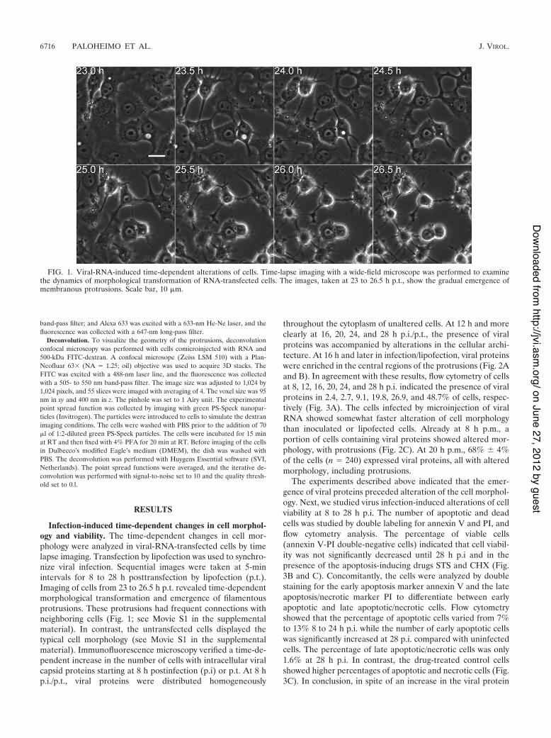
Infection-induced time-dependent changes in cell morphol-ogy and viability. The time-dependent changes in cell mor-phology were analyzed in viral-RNA-transfected cells by timelapse imaging. Transfection by lipofection was used to synchro-nize viral infection. Sequential images were taken at 5-minintervals for 8 to 28 h posttransfection by lipofection (p.t.).Imaging of cells from 23 to 26.5 h p.t. revealed time-dependentmorphological transformation and emergence of filamentousprotrusions. These protrusions had frequent connections withneighboring cells (Fig. 1; see Movie S1 in the supplementalmaterial). In contrast, the untransfected cells displayed thetypical cell morphology (see Movie S1 in the supplementalmaterial). Immunofluorescence microscopy verified a time-de-pendent increase in the number of cells with intracellular viralcapsid proteins starting at 8 h postinfection (p.i) or p.t. At 8 hp.i./p.t., viral proteins were distributed homogeneously
throughout the cytoplasm of unaltered cells. At 12 h and moreclearly at 16, 20, 24, and 28 h p.i./p.t., the presence of viralproteins was accompanied by alterations in the cellular archi-tecture. At 16 h and later in infection/lipofection, viral proteinswere enriched in the central regions of the protrusions (Fig. 2Aand B). In agreement with these results, flow cytometry of cellsat 8, 12, 16, 20, 24, and 28 h p.i. indicated the presence of viralproteins in 2.4, 2.7, 9.1, 19.8, 26.9, and 48.7% of cells, respec-tively (Fig. 3A). The cells infected by microinjection of viralRNA showed somewhat faster alteration of cell morphologythan inoculated or lipofected cells. Already at 8 h p.m., aportion of cells containing viral proteins showed altered mor-phology, with protrusions (Fig. 2C). At 20 h p.m., 68% & 4%of the cells (n $ 240) expressed viral proteins, all with alteredmorphology, including protrusions.
The experiments described above indicated that the emer-gence of viral proteins preceded alteration of the cell morphol-ogy. Next, we studied virus infection-induced alterations of cellviability at 8 to 28 h p.i. The number of apoptotic and deadcells was studied by double labeling for annexin V and PI, andflow cytometry analysis. The percentage of viable cells(annexin V-PI double-negative cells) indicated that cell viabil-ity was not significantly decreased until 28 h p.i and in thepresence of the apoptosis-inducing drugs STS and CHX (Fig.3B and C). Concomitantly, the cells were analyzed by doublestaining for the early apoptosis marker annexin V and the lateapoptosis/necrotic marker PI to differentiate between earlyapoptotic and late apoptotic/necrotic cells. Flow cytometryshowed that the percentage of apoptotic cells varied from 7%to 13% 8 to 24 h p.i. while the number of early apoptotic cellswas significantly increased at 28 p.i. compared with uninfectedcells. The percentage of late apoptotic/necrotic cells was only1.6% at 28 h p.i. In contrast, the drug-treated control cellsshowed higher percentages of apoptotic and necrotic cells (Fig.3C). In conclusion, in spite of an increase in the viral protein
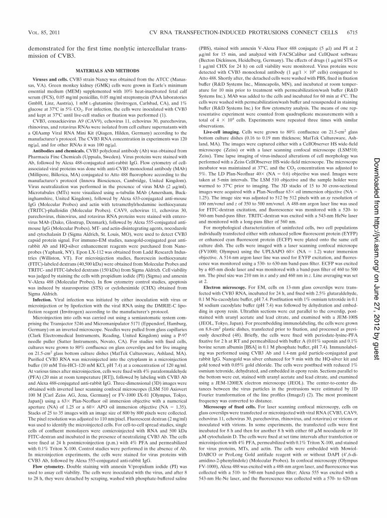
FIG. 1. Viral-RNA-induced time-dependent alterations of cells. Time-lapse imaging with a wide-field microscope was performed to examinethe dynamics of morphological transformation of RNA-transfected cells. The images, taken at 23 to 26.5 h p.t., show the gradual emergence ofmembranous protrusions. Scale bar, 10 !m.
6716 PALOHEIMO ET AL. J. VIROL.
on June 27, 2012 by guesthttp://jvi.asm
.org/D
ownloaded from

content of the cells, only a minor decrease in viability or in-crease in the number of apoptotic cells was observed in in-fected compared to uninfected cells prior to 28 h p.i.
To examine whether it is the viral RNA that explains thealteration of cell morphology, we microinjected positive-sense,single-stranded RNAs ((')ssRNAs) from CAV9, echovirus11, echovirus 30, parechovirus, and rhinovirus and double-stranded RNA (dsRNA) from rotavirus into cells. We utilizeddextran comicroinjection to mark the RNA-containing cellseven in the absence of structural modifications. EnterovirusMAb immunolabeling studies with CAV9, echovirus 11, andechovirus 30 RNAs showed that at 24 h p.m., 20% & 4% (n $300), 11% & 2% (n $ 300), and 15% & 4% (n $ 300) of cells
FIG. 2. Time-related alteration of infected cells. Cells infectedby virus inoculation, lipofection, or microinjection of viral RNAwere incubated for 8, 12, 16, 20, 24, or 28 h prior to fixation,immunolabeling, and imaging. A general image (inset) and aclose-up image of inoculated (A), lipofected (B), and microinjected(C) cells at each time point are shown. Capsid proteins were visu-alized with CVB3 Ab, followed by Alexa 488-conjugated anti-rabbitIgG (green), actin with TRITC-phalloidin (red), and nuclei withDAPI (blue). Scale bars, 20 !m.
FIG. 3. Emergence of viral proteins and infection-induced changesin cell viability. Flow cytometry was performed at 8, 12, 16, 20, 24, and28 h p.i. (A) Percentages of virus protein-expressing cells. The cellswere fixed and labeled with CVB3 monoclonal antibody conjugated toAtto 488. Untreated cells (C() and uninfected immunolabeled cells(C') were used as controls. (B) The percentage of viable cells wasanalyzed with annexin V-Alexa 488/propidium iodide double staining.The viable cells were annexin V and PI negative. (C) Percentages ofapoptotic cells. Cells in early apoptosis were annexin V positive and PInegative, and cells in late apoptosis or necrosis were both annexin Vand PI positive. STS- and CHX-treated cells were included as positivecontrols. The error bars indicate standard deviations.
VOL. 85, 2011 CV RNA TRANSFECTION-INDUCED PROTRUSIONS CONNECT CELLS 6717
on June 27, 2012 by guesthttp://jvi.asm
.org/D
ownloaded from
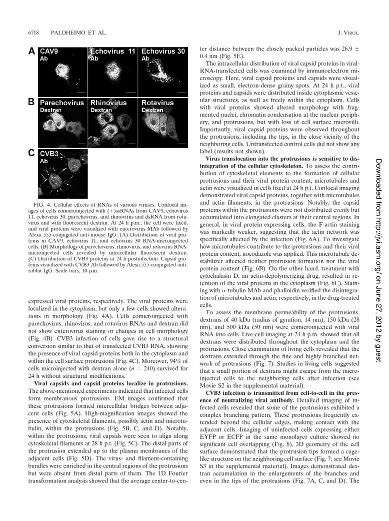
expressed viral proteins, respectively. The viral proteins werelocalized in the cytoplasm, but only a few cells showed altera-tions in morphology (Fig. 4A). Cells comicroinjected withparechovirus, rhinovirus, and rotavirus RNAs and dextran didnot show enterovirus staining or changes in cell morphology(Fig. 4B). CVB3 infection of cells gave rise to a structuralconversion similar to that of transfected CVB3 RNA, showingthe presence of viral capsid proteins both in the cytoplasm andwithin the cell surface protrusions (Fig. 4C). Moreover, 94% ofcells microinjected with dextran alone (n $ 240) survived for24 h without structural modifications.
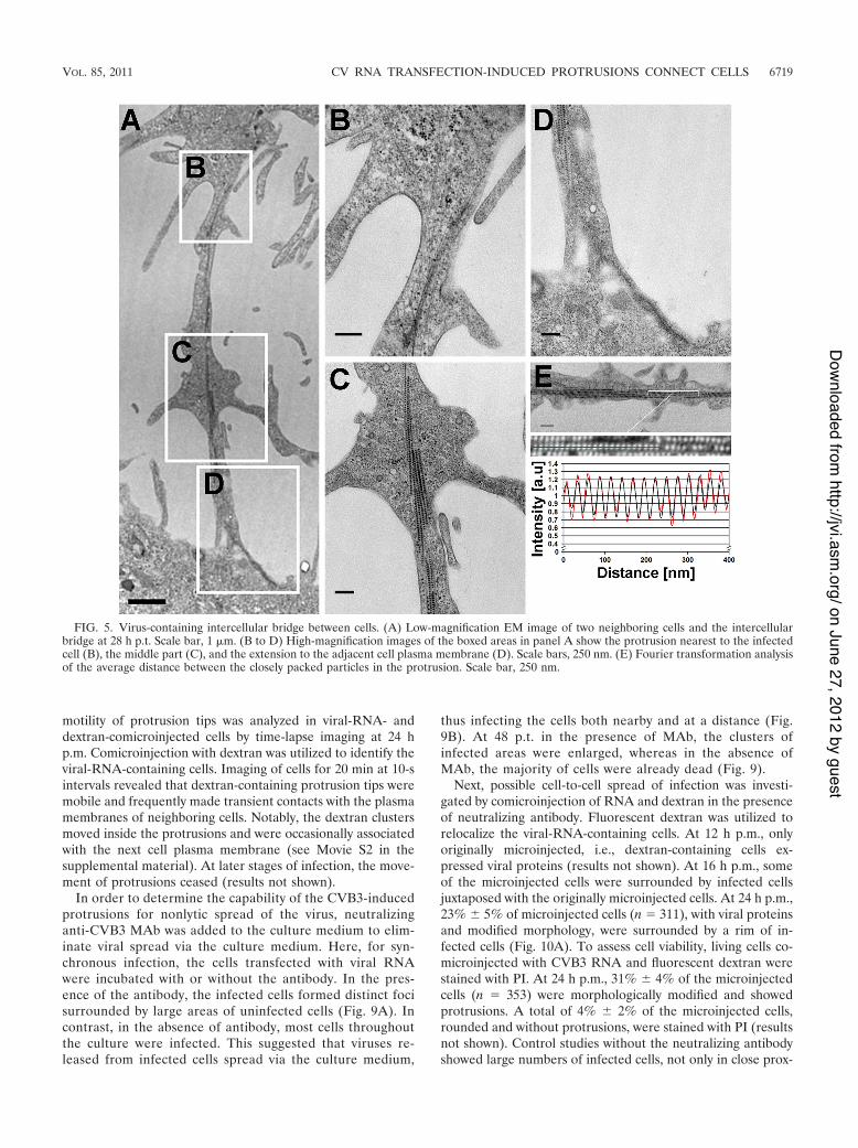
Viral capsids and capsid proteins localize in protrusions.The above-mentioned experiments indicated that infected cellsform membranous protrusions. EM images confirmed thatthese protrusions formed intercellular bridges between adja-cent cells (Fig. 5A). High-magnification images showed thepresence of cytoskeletal filaments, possibly actin and microtu-bulin, within the protrusions (Fig. 5B, C, and D). Notably,within the protrusions, viral capsids were seen to align alongcytoskeletal filaments at 28 h p.t. (Fig. 5C). The distal parts ofthe protrusion extended up to the plasma membranes of theadjacent cells (Fig. 5D). The virus- and filament-containingbundles were enriched in the central regions of the protrusionsbut were absent from distal parts of them. The 1D Fouriertransformation analysis showed that the average center-to-cen-
ter distance between the closely packed particles was 26.9 &0.4 nm (Fig. 5E).
The intracellular distribution of viral capsid proteins in viral-RNA-transfected cells was examined by immunoelectron mi-croscopy. Here, viral capsid proteins and capsids were visual-ized as small, electron-dense grainy spots. At 24 h p.t., viralproteins and capsids were distributed inside cytoplasmic vesic-ular structures, as well as freely within the cytoplasm. Cellswith viral proteins showed altered morphology with frag-mented nuclei, chromatin condensation at the nuclear periph-ery, and protrusions, but with loss of cell surface microvilli.Importantly, viral capsid proteins were observed throughoutthe protrusions, including the tips, in the close vicinity of theneighboring cells. Untransfected control cells did not show anylabel (results not shown).
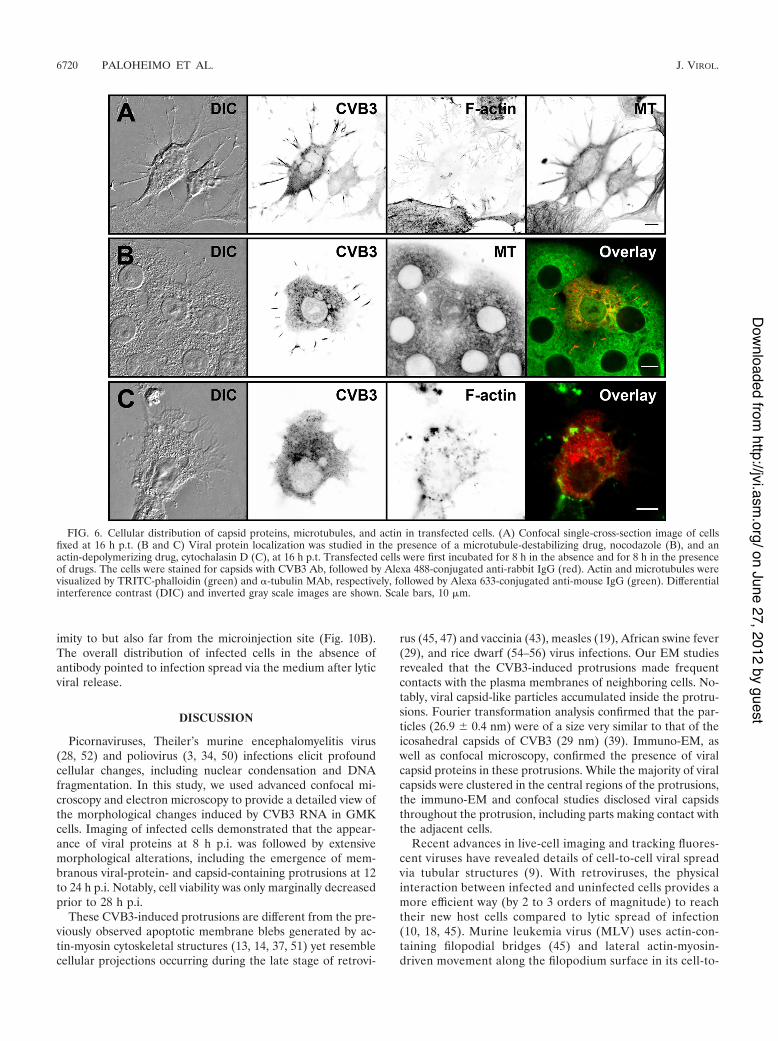
Virus translocation into the protrusions is sensitive to dis-integration of the cellular cytoskeleton. To assess the contri-bution of cytoskeletal elements to the formation of cellularprotrusions and their viral protein content, microtubules andactin were visualized in cells fixed at 24 h p.t. Confocal imagingdemonstrated viral capsid proteins, together with microtubulesand actin filaments, in the protrusions. Notably, the capsidproteins within the protrusions were not distributed evenly butaccumulated into elongated clusters at their central regions. Ingeneral, in viral-protein-expressing cells, the F-actin stainingwas markedly weaker, suggesting that the actin network wasspecifically affected by the infection (Fig. 6A). To investigatehow microtubules contribute to the protrusions and their viralprotein content, nocodazole was applied. This microtubule de-stabilizer affected neither protrusion formation nor the viralprotein content (Fig. 6B). On the other hand, treatment withcytochalasin D, an actin-depolymerizing drug, resulted in re-tention of the viral proteins in the cytoplasm (Fig. 6C). Stain-ing with "-tubulin MAb and phalloidin verified the disintegra-tion of microtubules and actin, respectively, in the drug-treatedcells.
To assess the membrane permeability of the protrusions,dextrans of 40 kDa (radius of gyration, 14 nm), 150 kDa (28nm), and 500 kDa (50 nm) were comicroinjected with viralRNA into cells. Live-cell imaging at 24 h p.m. showed that alldextrans were distributed throughout the cytoplasm and theprotrusions. Close examination of living cells revealed that thedextrans extended through the fine and highly branched net-work of protrusions (Fig. 7). Studies in living cells suggestedthat a small portion of dextrans might escape from the micro-injected cells to the neighboring cells after infection (seeMovie S2 in the supplemental material).
CVB3 infection is transmitted from cell-to-cell in the pres-ence of neutralizing viral antibody. Detailed imaging of in-fected cells revealed that some of the protrusions exhibited acomplex branching pattern. These protrusions frequently ex-tended beyond the cellular edges, making contact with theadjacent cells. Imaging of uninfected cells expressing eitherEYFP or ECFP in the same monolayer culture showed nosignificant cell overlapping (Fig. 8). 3D geometry of the cellsurface demonstrated that the protrusion tips formed a cage-like structure on the neighboring cell surface (Fig. 7; see MovieS3 in the supplemental material). Images demonstrated dex-tran accumulation in the enlargements of the branches andeven in the tips of the protrusions (Fig. 7A, C, and D). The
FIG. 4. Cellular effects of RNAs of various viruses. Confocal im-ages of cells comicroinjected with (')ssRNAs from CAV9, echovirus11, echovirus 30, parechovirus, and rhinovirus and dsRNA from rota-virus and with fluorescent dextran. At 24 h p.m., the cell were fixed,and viral proteins were visualized with enterovirus MAb followed byAlexa 555-conjugated anti-mouse IgG. (A) Distribution of viral pro-teins in CAV9, echovirus 11, and echovirus 30 RNA-microinjectedcells. (B) Morphology of parechovirus, rhinovirus, and rotavirus RNA-microinjected cells revealed by intracellular fluorescent dextran.(C) Distribution of CVB3 proteins at 24 h postinfection. Capsid pro-teins visualized with CVB3 Ab followed by Alexa 555-conjugated anti-rabbit IgG. Scale bars, 10 !m.
6718 PALOHEIMO ET AL. J. VIROL.
on June 27, 2012 by guesthttp://jvi.asm
.org/D
ownloaded from
motility of protrusion tips was analyzed in viral-RNA- anddextran-comicroinjected cells by time-lapse imaging at 24 hp.m. Comicroinjection with dextran was utilized to identify theviral-RNA-containing cells. Imaging of cells for 20 min at 10-sintervals revealed that dextran-containing protrusion tips weremobile and frequently made transient contacts with the plasmamembranes of neighboring cells. Notably, the dextran clustersmoved inside the protrusions and were occasionally associatedwith the next cell plasma membrane (see Movie S2 in thesupplemental material). At later stages of infection, the move-ment of protrusions ceased (results not shown).
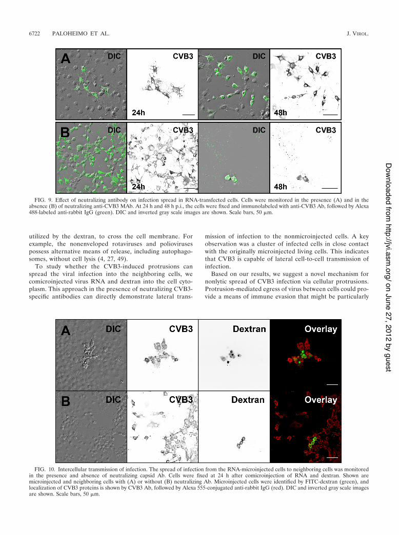
In order to determine the capability of the CVB3-inducedprotrusions for nonlytic spread of the virus, neutralizinganti-CVB3 MAb was added to the culture medium to elim-inate viral spread via the culture medium. Here, for syn-chronous infection, the cells transfected with viral RNAwere incubated with or without the antibody. In the pres-ence of the antibody, the infected cells formed distinct focisurrounded by large areas of uninfected cells (Fig. 9A). Incontrast, in the absence of antibody, most cells throughoutthe culture were infected. This suggested that viruses re-leased from infected cells spread via the culture medium,
thus infecting the cells both nearby and at a distance (Fig.9B). At 48 p.t. in the presence of MAb, the clusters ofinfected areas were enlarged, whereas in the absence ofMAb, the majority of cells were already dead (Fig. 9).
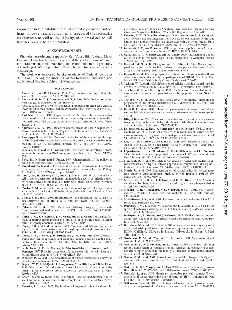
Next, possible cell-to-cell spread of infection was investi-gated by comicroinjection of RNA and dextran in the presenceof neutralizing antibody. Fluorescent dextran was utilized torelocalize the viral-RNA-containing cells. At 12 h p.m., onlyoriginally microinjected, i.e., dextran-containing cells ex-pressed viral proteins (results not shown). At 16 h p.m., someof the microinjected cells were surrounded by infected cellsjuxtaposed with the originally microinjected cells. At 24 h p.m.,23% & 5% of microinjected cells (n $ 311), with viral proteinsand modified morphology, were surrounded by a rim of in-fected cells (Fig. 10A). To assess cell viability, living cells co-microinjected with CVB3 RNA and fluorescent dextran werestained with PI. At 24 h p.m., 31% & 4% of the microinjectedcells (n $ 353) were morphologically modified and showedprotrusions. A total of 4% & 2% of the microinjected cells,rounded and without protrusions, were stained with PI (resultsnot shown). Control studies without the neutralizing antibodyshowed large numbers of infected cells, not only in close prox-
FIG. 5. Virus-containing intercellular bridge between cells. (A) Low-magnification EM image of two neighboring cells and the intercellularbridge at 28 h p.t. Scale bar, 1 !m. (B to D) High-magnification images of the boxed areas in panel A show the protrusion nearest to the infectedcell (B), the middle part (C), and the extension to the adjacent cell plasma membrane (D). Scale bars, 250 nm. (E) Fourier transformation analysisof the average distance between the closely packed particles in the protrusion. Scale bar, 250 nm.
VOL. 85, 2011 CV RNA TRANSFECTION-INDUCED PROTRUSIONS CONNECT CELLS 6719
on June 27, 2012 by guesthttp://jvi.asm
.org/D
ownloaded from
imity to but also far from the microinjection site (Fig. 10B).The overall distribution of infected cells in the absence ofantibody pointed to infection spread via the medium after lyticviral release.
DISCUSSION
Picornaviruses, Theiler’s murine encephalomyelitis virus(28, 52) and poliovirus (3, 34, 50) infections elicit profoundcellular changes, including nuclear condensation and DNAfragmentation. In this study, we used advanced confocal mi-croscopy and electron microscopy to provide a detailed view ofthe morphological changes induced by CVB3 RNA in GMKcells. Imaging of infected cells demonstrated that the appear-ance of viral proteins at 8 h p.i. was followed by extensivemorphological alterations, including the emergence of mem-branous viral-protein- and capsid-containing protrusions at 12to 24 h p.i. Notably, cell viability was only marginally decreasedprior to 28 h p.i.
These CVB3-induced protrusions are different from the pre-viously observed apoptotic membrane blebs generated by ac-tin-myosin cytoskeletal structures (13, 14, 37, 51) yet resemblecellular projections occurring during the late stage of retrovi-
rus (45, 47) and vaccinia (43), measles (19), African swine fever(29), and rice dwarf (54–56) virus infections. Our EM studiesrevealed that the CVB3-induced protrusions made frequentcontacts with the plasma membranes of neighboring cells. No-tably, viral capsid-like particles accumulated inside the protru-sions. Fourier transformation analysis confirmed that the par-ticles (26.9 & 0.4 nm) were of a size very similar to that of theicosahedral capsids of CVB3 (29 nm) (39). Immuno-EM, aswell as confocal microscopy, confirmed the presence of viralcapsid proteins in these protrusions. While the majority of viralcapsids were clustered in the central regions of the protrusions,the immuno-EM and confocal studies disclosed viral capsidsthroughout the protrusion, including parts making contact withthe adjacent cells.
Recent advances in live-cell imaging and tracking fluores-cent viruses have revealed details of cell-to-cell viral spreadvia tubular structures (9). With retroviruses, the physicalinteraction between infected and uninfected cells provides amore efficient way (by 2 to 3 orders of magnitude) to reachtheir new host cells compared to lytic spread of infection(10, 18, 45). Murine leukemia virus (MLV) uses actin-con-taining filopodial bridges (45) and lateral actin-myosin-driven movement along the filopodium surface in its cell-to-
FIG. 6. Cellular distribution of capsid proteins, microtubules, and actin in transfected cells. (A) Confocal single-cross-section image of cellsfixed at 16 h p.t. (B and C) Viral protein localization was studied in the presence of a microtubule-destabilizing drug, nocodazole (B), and anactin-depolymerizing drug, cytochalasin D (C), at 16 h p.t. Transfected cells were first incubated for 8 h in the absence and for 8 h in the presenceof drugs. The cells were stained for capsids with CVB3 Ab, followed by Alexa 488-conjugated anti-rabbit IgG (red). Actin and microtubules werevisualized by TRITC-phalloidin (green) and "-tubulin MAb, respectively, followed by Alexa 633-conjugated anti-mouse IgG (green). Differentialinterference contrast (DIC) and inverted gray scale images are shown. Scale bars, 10 !m.
6720 PALOHEIMO ET AL. J. VIROL.
on June 27, 2012 by guesthttp://jvi.asm
.org/D
ownloaded from

cell transport. Moreover, human immunodeficiency virustype 1 (HIV-1) exploits membrane nanotubes, in which virusmovement is mediated by the underlying actin cytoskeleton,for intercellular spread between T cells (47). Other exam-ples of enveloped viruses utilizing actin-rich cellular projec-tions for intercellular dispersal are herpesviruses (22, 32),African swine fever virus (29), measles virus (19), and vac-cinia virus (46, 59). Nonenveloped viruses are usually re-leased into the extracellular space via virus-induced celllysis. However, some of them exploit other means of cell-to-cell transmission. Prelytic transmission could be of par-ticular use in persistent infection. Direct cell-to-cell spread
has been suspected in the transmission of poliovirus fromperipheral tissues to the central nervous system (40) and hasalso been described in CVB-infected myocytes (31), as wellas in cells infected by another picornavirus, Ljungan virus(21). Rice dwarf virus, a member of the Reoviridae, providesyet another example of a nonenveloped virus exploitingvirus-induced actin-based protrusions for cell-to-cell trans-fer (54–56). Moreover, this virus requires microtubules forrelease from insect cells (57). Our confocal microscopy ex-amination showed that the CVB3-induced protrusions con-tained viral proteins, together with microtubules and actinfilaments. Disintegration of actin resulted in cytoplasmicaccumulation of viral capsid proteins, whereas depolymer-ization of microtubules did not inhibit viral translocationinto the protrusions. This strongly suggests an importantrole for actin in the transport of viral particles into theprotrusions. Furthermore, EM studies demonstrated thealignment of capsid-like particles along cytoskeletal fila-ments in the protrusions. These findings indicated that, inaddition to the capsids freely diffusing into the protrusions,a significant number of capsids interact actively with the cellcytoskeleton.
To estimate the potential of the protrusions in cell-to-celltransmission, it is crucial to show that they make contactsbetween adjacent cells. 3D geometry demonstrated that thetips of the protrusions formed a cage-like structure on theneighboring cell surface. Live-cell studies indicated thatthe branched tips of intercellular protrusions were mobileand frequently located around the periphery of neighboringcells. Although a majority of the dextrans remained in theoriginally microinjected infected cells, a small portion ofdextrans escaped from the microinjected cells after infec-tion. This suggests that viruses might induce nonlytic means,
FIG. 7. 3D structure of protrusion. Cell imaging, deconvolution, and 3D space-filling modeling were used to examine protrusion morphologyand interactions with neighboring cells. (A) Living cells comicroinjected with RNA and FITC-dextran (green) were imaged at 24 h p.m. (B) DICimage of cells. (C) Magnification of the white box in panel A, showing the protrusion area. (D) The xy and xz renderings of the image reveal the3D details of the geometry of protrusions located around the periphery of the neighboring cell. Pseudocolor images with intensity increasing fromblack to white was used to visualize the intracellular distribution of dextran. Scale bars, 10 !m.
FIG. 8. Analysis of cell overlapping in monolayer culture. Shown isconfocal live-cell imaging of mixed populations of uninfected cellsexpressing either EYFP (yellow) or ECFP (cyan). The xz projectionreveals the profiles of cells at a location indicated by the red line. Scalebar, 20 !m.
VOL. 85, 2011 CV RNA TRANSFECTION-INDUCED PROTRUSIONS CONNECT CELLS 6721
on June 27, 2012 by guesthttp://jvi.asm
.org/D
ownloaded from
utilized by the dextran, to cross the cell membrane. Forexample, the nonenveloped rotaviruses and poliovirusespossess alternative means of release, including autophago-somes, without cell lysis (4, 27, 49).
To study whether the CVB3-induced protrusions canspread the viral infection into the neighboring cells, wecomicroinjected virus RNA and dextran into the cell cyto-plasm. This approach in the presence of neutralizing CVB3-specific antibodies can directly demonstrate lateral trans-
mission of infection to the nonmicroinjected cells. A keyobservation was a cluster of infected cells in close contactwith the originally microinjected living cells. This indicatesthat CVB3 is capable of lateral cell-to-cell transmission ofinfection.
Based on our results, we suggest a novel mechanism fornonlytic spread of CVB3 infection via cellular protrusions.Protrusion-mediated egress of virus between cells could pro-vide a means of immune evasion that might be particularly
FIG. 9. Effect of neutralizing antibody on infection spread in RNA-transfected cells. Cells were monitored in the presence (A) and in theabsence (B) of neutralizing anti-CVB3 MAb. At 24 h and 48 h p.i., the cells were fixed and immunolabeled with anti-CVB3 Ab, followed by Alexa488-labeled anti-rabbit IgG (green). DIC and inverted gray scale images are shown. Scale bars, 50 !m.
FIG. 10. Intercellular transmission of infection. The spread of infection from the RNA-microinjected cells to neighboring cells was monitoredin the presence and absence of neutralizing capsid Ab. Cells were fixed at 24 h after comicroinjection of RNA and dextran. Shown aremicroinjected and neighboring cells with (A) or without (B) neutralizing Ab. Microinjected cells were identified by FITC-dextran (green), andlocalization of CVB3 proteins is shown by CVB3 Ab, followed by Alexa 555-conjugated anti-rabbit IgG (red). DIC and inverted gray scale imagesare shown. Scale bars, 50 !m.
6722 PALOHEIMO ET AL. J. VIROL.
on June 27, 2012 by guesthttp://jvi.asm
.org/D
ownloaded from
important in the establishment of nonlytic persistent infec-tions. However, many fundamental aspects of the molecularmechanisms, as well as the ubiquity, of this viral cell-to-celltransfer remain to be elucidated.
ACKNOWLEDGMENTS
First-class experimental support by Kati Vuori, Eija Jokitalo, MerviLindman, Eeva Jokela, Eeva Tolvanen, Milla Vuokko, Sami Willman,Pirjo Kauppinen, Raija Vassinen, and Paavo Niutanen is gratefullyacknowledged. We are grateful to Klaus Hedman for comments on themanuscript.
The work was supported by the Academy of Finland (contracts107311 and 129774), the Juvenile Diabetes Research Foundation, andthe National Graduate School of Nanoscience.
REFERENCES
1. Abraham, G., and R. J. Colonno. 1984. Many rhinovirus serotypes share thesame cellular receptor. J. Virol. 51:340–345.
2. Abramoff, M. D., P. J. Magelhaes, and S. J. Ram. 2004. Image processingwith ImageJ. J. Biophotonics Int. 11:36–42.
3. Agol, V. I., et al. 1998. Two types of death of poliovirus-infected cells: caspaseinvolvement in the apoptosis but not cytopathic effect. Virology 252:343–353.doi:10.1006/viro.1998.9438.
4. Andreoletti, L., et al. 1997. Experimental CVB3-induced chronic myocarditisin two murine strains: evidence of interrelationships between virus replica-tion and myocardial damage in persistent cardiac infection. J. Med. Virol.52:206–214.
5. Andreoletti, L., et al. 1997. Detection of coxsackie B virus RNA sequences inwhole blood samples from adult patients at the onset of type I diabetesmellitus. J. Med. Virol. 52:121–127.
6. Basavappa, R., et al. 1994. Role and mechanism of the maturation cleavageof VP0 in poliovirus assembly: structure of the empty capsid assembly inter-mediate at 2.9 A resolution. Protein Sci. 3:1651–1669. doi:10.1002/pro.5560031005.
7. Belsham, G. J., and C. J. Bostock. 1988. Studies on the infectivity of foot-and-mouth disease virus RNA using microinjection. J. Gen. Virol. 69:265–274.
8. Bienz, K., D. Egger, and T. Pfister. 1994. Characteristics of the poliovirusreplication complex. Arch. Virol. Suppl. 9:147–157.
9. Burckhardt, C. J., and U. F. Greber. 2009. Virus movements on the plasmamembrane support infection and transmission between cells. PLoS Pathog.5:e1000621. doi:10.1371/journal.ppat.1000621.
10. Carr, J. M., H. Hocking, P. Li, and C. J. Burrell. 1999. Rapid and efficientcell-to-cell transmission of human immunodeficiency virus infection frommonocyte-derived macrophages to peripheral blood lymphocytes. Virology265:319–329. doi:10.1006/viro.1999.0047.
11. Carthy, C. M., et al. 1998. Caspase activation and specific cleavage of sub-strates after coxsackievirus B3-induced cytopathic effect in HeLa cells. J. Vi-rol. 72:7669–7675.
12. Chung, S. K., et al. 2005. Internalization and trafficking mechanisms ofcoxsackievirus B3 in HeLa cells. Virology 333:31–40. doi:10.1016/j.virol.2004.12.010.
13. Coleman, M. L., et al. 2001. Membrane blebbing during apoptosis resultsfrom caspase-mediated activation of ROCK I. Nat. Cell Biol. 3:339–345.doi:10.1038/35070009.
14. Cotter, T. G., S. V. Lennon, J. M. Glynn, and D. R. Green. 1992. Microfila-ment-disrupting agents prevent the formation of apoptotic bodies in tumorcells undergoing apoptosis. Cancer Res. 52:997–1005.
15. Coyne, C. B., and J. M. Bergelson. 2006. Virus-induced Abl and Fyn kinasesignals permit coxsackievirus entry through epithelial tight junctions. Cell124:119–131. doi:10.1016/j.cell.2005.10.035.
16. Coyne, C. B., L. Shen, J. R. Turner, and J. M. Bergelson. 2007. Coxsacki-evirus entry across epithelial tight junctions requires occludin and the smallGTPases Rab34 and Rab5. Cell. Host Microbe 2:181–192. doi:10.1016/j.chom.2007.07.003.
17. de la Torre, J. C., B. Alarcon, E. Martinez-Salas, L. Carrasco, and E.Domingo. 1987. Ribavirin cures cells of a persistent infection with foot-and-mouth disease virus in vitro. J. Virol. 61:233–235.
18. Dimitrov, D. S., et al. 1993. Quantitation of human immunodeficiency virustype 1 infection kinetics. J. Virol. 67:2182–2190.
19. Duprex, W. P., S. McQuaid, L. Hangartner, M. A. Billeter, and B. K. Rima.1999. Observation of measles virus cell-to-cell spread in astrocytoma cells byusing a green fluorescent protein-expressing recombinant virus. J. Virol.73:9568–9575.
20. Egger, D., and K. Bienz. 2005. Intracellular location and translocation ofsilent and active poliovirus replication complexes. J. Gen. Virol. 86:707–718.doi:10.1099/vir.0.80442-0.
21. Ekstrom, J. O., et al. 2007. Replication of Ljungan virus in cell culture: the
genomic 5%-end, infectious cDNA clones and host cell response to viralinfections. Virus Res. 130:129–139. doi:10.1016/j.virusres.2007.06.004.
22. Favoreel, H. W., G. Van Minnebruggen, D. Adriaensen, and H. J. Nauwynck.2005. Cytoskeletal rearrangements and cell extensions induced by the US3kinase of an alphaherpesvirus are associated with enhanced spread. Proc.Natl. Acad. Sci. U. S. A. 102:8990–8995. doi:10.1073/pnas.0409099102.
23. Gamarnik, A. V., and R. Andino. 1996. Replication of poliovirus in Xenopusoocytes requires two human factors. EMBO J. 15:5988–5998.
24. Gamarnik, A. V., N. Boddeker, and R. Andino. 2000. Translation and repli-cation of human rhinovirus type 14 and mengovirus in Xenopus oocytes.J. Virol. 74:11983–11987.
25. Habayeb, M. S., S. K. Ekengren, and D. Hultmark. 2006. Nora virus, apersistent virus in Drosophila, defines a new picorna-like virus family.J. Gen. Virol. 87:3045–3051. doi:10.1099/vir.0.81997-0.
26. Hyoty, H., et al. 1995. A prospective study of the role of coxsackie B andother enterovirus infections in the pathogenesis of IDDM. Childhood Dia-betes in Finland (DiMe) Study Group. Diabetes 44:652–657.
27. Jackson, W. T., et al. 2005. Subversion of cellular autophagosomal machin-ery by RNA viruses. PLoS Biol. 3:e156. doi:10.1371/journal.pbio.0030156.
28. Jelachich, M. L., and H. L. Lipton. 1996. Theiler’s murine encephalomyelitisvirus kills restrictive but not permissive cells by apoptosis. J. Virol. 70:6856–6861.
29. Jouvenet, N., et al. 2006. African swine fever virus induces filopodia-likeprojections at the plasma membrane. Cell. Microbiol. 8:1803–1811. doi:10.1111/j.1462-5822.2006.00750.x.
30. Kandolf, R., et al. 1993. Molecular pathogenesis of enterovirus-inducedmyocarditis: virus persistence and chronic inflammation. Intervirology 35:140–151.
31. Klingel, K., et al. 1998. Visualization of enteroviral replication in myocardialtissue by ultrastructural in situ hybridization: identification of target cells andcytopathic effects. Lab. Invest. 78:1227–1237.
32. La Boissiere, S., A. Izeta, S. Malcomber, and P. O’Hare. 2004. Compart-mentalization of VP16 in cells infected with recombinant herpes simplexvirus expressing VP16-green fluorescent protein fusion proteins. J. Virol.78:8002–8014. doi:10.1128/JVI.78.15.8002-8014.2004.
33. Lai, C. J., B. T. Zhao, H. Hori, and M. Bray. 1991. Infectious RNA tran-scribed from stably cloned full-length cDNA of dengue type 4 virus. Proc.Natl. Acad. Sci. U. S. A. 88:5139–5143.
34. Lopez-Guerrero, J. A., M. Alonso, F. Martin-Belmonte, and L. Carrasco.2000. Poliovirus induces apoptosis in the human U937 promonocytic cellline. Virology 272:250–256. doi:10.1006/viro.2000.0405.
35. Marchant, D., et al. 2009. ERK MAP kinase-activated Arf6 trafficking di-rects coxsackievirus type B3 into an unproductive compartment during virushost-cell entry. J. Gen. Virol. 90:854–862. doi:10.1099/vir.0.005868-0.
36. Martin, U., et al. 2004. Characterization of coxsackievirus B3-caused apop-tosis under in vitro conditions. Med. Microbiol. Immunol. 193:133–139.doi:10.1007/s00430-003-0197-7.
37. Mills, J. C., N. L. Stone, J. Erhardt, and R. N. Pittman. 1998. Apoptoticmembrane blebbing is regulated by myosin light chain phosphorylation.J. Cell Biol. 140:627–636.
38. Modalsli, K. R., G. Bukholm, S. O. Mikalsen, and M. Degre. 1991. Micro-injected Coxsackie B1 virus does not replicate in HEp-2 cells. Virology185:888–890.
39. Muckelbauer, J. K., et al. 1995. The structure of coxsackievirus B3 at 3.5 Aresolution. Structure 3:653–667.
40. Ponnuraj, E. M., T. J. John, M. J. Levin, and E. A. Simoes. 1998. Cell-to-cellspread of poliovirus in the spinal cord of bonnet monkeys (Macaca radiata).J. Gen. Virol. 79:2393–2403.
41. Rodriguez, M., E. Oleszak, and J. Leibowitz. 1987. Theiler’s murine enceph-alomyelitis: a model of demyelination and persistence of virus. Crit. Rev.Immunol. 7:325–365.
42. Roivainen, M., et al. 1998. Several different enterovirus serotypes can beassociated with prediabetic autoimmune episodes and onset of overtIDDM. Childhood Diabetes in Finland (DiMe) Study Group. J. Med.Virol. 56:74–78.
43. Sanderson, C. M., M. Way, and G. L. Smith. 1998. Virus-induced cellmotility. J. Virol. 72:1235–1243.
44. Shafren, D. R., D. T. Williams, and R. D. Barry. 1997. A decay-acceleratingfactor-binding strain of coxsackievirus B3 requires the coxsackievirus-ade-novirus receptor protein to mediate lytic infection of rhabdomyosarcomacells. J. Virol. 71:9844–9848.
45. Sherer, N. M., et al. 2007. Retroviruses can establish filopodial bridges forefficient cell-to-cell transmission. Nat. Cell Biol. 9:310–315. doi:10.1038/ncb1544.
46. Smith, G. L., B. J. Murphy, and M. Law. 2003. Vaccinia virus motility. Annu.Rev. Microbiol. 57:323–342. doi:10.1146/annurev.micro.57.030502.091037.
47. Sowinski, S., et al. 2008. Membrane nanotubes physically connect T cellsover long distances presenting a novel route for HIV-1 transmission. Nat.Cell Biol. 10:211–219. doi:10.1038/ncb1682.
48. Suikkanen, S., et al. 2003. Exploitation of microtubule cytoskeleton anddynein during parvoviral traffic toward the nucleus. J. Virol. 77:10270–10279.
VOL. 85, 2011 CV RNA TRANSFECTION-INDUCED PROTRUSIONS CONNECT CELLS 6723
on June 27, 2012 by guesthttp://jvi.asm
.org/D
ownloaded from
49. Taylor, M. P., and K. Kirkegaard. 2008. Potential subversion of autophago-somal pathway by picornaviruses. Autophagy 4:286–289.
50. Tolskaya, E. A., et al. 1995. Apoptosis-inducing and apoptosis-preventingfunctions of poliovirus. J. Virol. 69:1181–1189.
51. Torgerson, R. R., and M. A. McNiven. 1998. The actin-myosin cytoskeletonmediates reversible agonist-induced membrane blebbing. J. Cell Sci. 111:2911–2922.
52. Tsunoda, I., C. I. Kurtz, and R. S. Fujinami. 1997. Apoptosis in acute andchronic central nervous system disease induced by Theiler’s murine enceph-alomyelitis virus. Virology 228:388–393. doi:10.1006/viro.1996.8382.
53. van Kuppeveld, F. J., et al. 1997. Coxsackievirus protein 2B modifies endo-plasmic reticulum membrane and plasma membrane permeability and facil-itates virus release. EMBO J. 16:3519–3532. doi:10.1093/emboj/16.12.3519.
54. Wei, T., H. Chen, T. Ichiki-Uehara, H. Hibino, and T. Omura. 2007. Entry ofRice dwarf virus into cultured cells of its insect vector involves clathrin-mediated endocytosis. J. Virol. 81:7811–7815. doi:10.1128/JVI.00050-07.
55. Wei, T., et al. 2006. The spread of Rice dwarf virus among cells of its insectvector exploits virus-induced tubular structures. J. Virol. 80:8593–8602. doi:10.1128/JVI.00537-06.
56. Wei, T., T. Shimizu, and T. Omura. 2008. Endomembranes and myosinmediate assembly into tubules of Pns10 of Rice dwarf virus and intercellularspreading of the virus in cultured insect vector cells. Virology 372:349–356.doi:10.1016/j.virol.2007.10.034.
57. Wei, T., et al. 2009. Association of Rice gall dwarf virus with microtubules isnecessary for viral release from cultured insect vector cells. J. Virol. 83:10830–10835. doi:10.1128/JVI.01067-09.
58. Whitton, J. L., C. T. Cornell, and R. Feuer. 2005. Host and virus determi-nants of picornavirus pathogenesis and tropism. Nat. Rev. Microbiol. 3:765–776. doi:10.1038/nrmicro1284.
59. Wolffe, E. J., A. S. Weisberg, and B. Moss. 1998. Role for the vaccinia virusA36R outer envelope protein in the formation of virus-tipped actin-contain-ing microvilli and cell-to-cell virus spread. Virology 244:20–26. doi:10.1006/viro.1998.9103.
60. Yuan, J. P., et al. 2003. Coxsackievirus B3-induced apoptosis and caspase-3.Cell Res. 13:203–209. doi:10.1038/sj.cr.7290165.
61. Zhang, Y., and J. M. Bergelson. 2005. Adenovirus receptors. J. Virol. 79:12125–12131. doi:10.1128/JVI.79.19.12125-12131.2005.
6724 PALOHEIMO ET AL. J. VIROL.
on June 27, 2012 by guesthttp://jvi.asm
.org/D
ownloaded from