Embed Size (px)
Citation preview

RESEARCH COMMUNICATION
A shared molecular mechanismunderlies the humanrasopathies Legius syndromeand Neurofibromatosis-1Irma B. Stowe,1 Ellen L. Mercado,1
Timothy R. Stowe,2 Erika L. Bell,1
Juan A. Oses-Prieto,3 Hilda Hernandez,3
Alma L. Burlingame,3 and Frank McCormick1,4
1Helen Diller Family Comprehensive Cancer Center, Universityof California at San Francisco, San Francisco, California 94158,USA; 2Department of Neurological Surgery, University ofCalifornia at San Francisco, San Francisco, California 94143,USA; 3Department of Pharmaceutical Chemistry, University ofCalifornia at San Francisco, San Francisco, California 94143, USA
The Ras/mitogen-activated protein kinase (MAPK) path-way plays a critical role in transducing mitogenic signalsfrom receptor tyrosine kinases. Loss-of-function muta-tions in one feedback regulator of Ras/MAPK signaling,SPRED1 (Sprouty-related protein with an EVH1 domain),cause Legius syndrome, an autosomal dominant humandisorder that resembles Neurofibromatosis-1 (NF1). Spred1functions as a negative regulator of the Ras/MAPK path-way; however, the underlying molecular mechanism ispoorly understood. Here we show that neurofibromin, theNF1 gene product, is a Spred1-interacting protein that isnecessary for Spred1’s inhibitory function. We show thatSpred1 binding induces the plasma membrane localiza-tion of NF1, which subsequently down-regulates Ras-GTPlevels. This novel mechanism for the regulation of neuro-fibromin provides a molecular bridge for understanding theoverlapping pathophysiology of NF1 and Legius syndrome.
Supplemental material is available for this article.
Received February 28, 2012; revised version accepted May25, 2012.
Spred1 (Sprouty-related protein with an EVH1 domain)functions as a negative regulator of growth factor, cyto-kine, and chemokine-induced ERK activation by specifi-cally inhibiting the Ras/Raf/MEK/ERK pathway (Wakiokaet al. 2001; Miyoshi et al. 2004; Nonami et al. 2004).Spred1 is comprised of an N-terminal Ena/Vasp homology1 (EVH1) domain, a central c-Kit-binding domain (KBD),and a cysteine rich C-terminal Sprouty (SPR) domain.Mammals contain three homologs, Spred 1, Spred 2, andSpred 3, each of which can negatively regulate the Ras/Raf/mitogen-activated protein kinase (MAPK) pathway(Kato et al. 2003; King et al. 2006). However, the precisemechanism by which the Spreds act remains unclear, and
it has been reported to function both upstream of anddownstream from Ras (Wakioka et al. 2001; King et al.2005). Overexpression of Spred1 can increase Raf’s re-cruitment to the plasma membrane, where it associateswith Ras without stimulating Raf activation (Wakiokaet al. 2001). Yet, this mechanism has never been fullyrefined and fails to explain how Spred1 prevents Raf ac-tivation. A separate report suggests that Spred1 may pre-vent Ras activation, as evidenced by decreased Ras-GTPlevels when Spred1 is overexpressed (King et al. 2005).
Heterozygous germline loss-of-function mutations havebeen identified in SPRED1 in Legius syndrome, a develop-mental disorder that shares a number of phenotypes withNeurofibromatosis-1 (NF1) (Brems et al. 2007). Both syn-dromes are characterized as rasopathies, congenital devel-opmental syndromes caused by germline mutations thataffect the Ras/MAPK pathway. Legius syndrome has beencharacterized as a milder form of NF1, with individualsdisplaying multiple cafe-au-lait spots, axillary freckling,and macrocephaly, but lacking other common NF1 man-ifestations such as Lisch nodules, neurofibromas, osseouslesions, or optic pathway gliomas. Initial experiments re-vealed that Spred1 mutations were loss-of-function muta-tions, incapable of inhibiting the Ras/MAPK pathway(Brems et al. 2007). SPRED1 mutations account for atleast 2% of the pathogenic mutations associated withpatients clinically diagnosed with NF1 (Brems et al. 2007;Messiaen et al. 2009; Pasmant et al. 2009; Spurlock et al.2009; Muram-Zborovski et al. 2010; Denayer et al. 2011;Laycock-van Spyk et al. 2011; Spencer et al. 2011).
NF1, an autosomal dominant, multisystem disorderthat affects approximately one in 3500 individuals, wasthe first disorder found to originate from a component ofthe Ras/MAPK pathway. The NF1 gene product neuro-fibromin negatively regulates Ras signaling by functioningas a Ras GTPase-activating protein (RasGAP) to acceleratethe hydrolysis of active Ras-GTP to inactive Ras-GDP(Martin et al. 1990; Xu et al. 1990). Loss of neurofibrominleads to hyperactive Ras signaling, as observed by elevatedRas pathway activity in cells (Basu et al. 1992; DeClue et al.1992; Bollag et al. 1996). Despite the identification ofneurofibromin as a RasGAP 20 years ago, the regulationof neurofibromin activity remains poorly understood.Neurofibromin has been reported to be positively andnegatively regulated by various phosphorylation events,but the context of this regulation is largely unknown (Fenget al. 2004; Mangoura et al. 2006; Leondaritis et al. 2009). Inaddition, neurofibromin protein levels are negatively regu-lated by the ubiquitin proteasome system following growthfactor stimulation (Cichowski et al. 2003; McGillicuddyet al. 2009). However, the exact regulatory mechanismthat couples receptor growth factor activation to neuro-fibromin’s suppression of Ras activation remains unclear.
Here we report that Spred1 down-regulates the Ras/MAPK pathway through an interaction with the NF1protein neurofibromin. Importantly, this interaction func-tions to recruit neurofibromin to the plasma membrane.Furthermore, loss-of-function Spred1 mutants observed inLegius syndrome are either unable to bind neurofibrominor incapable of recruiting it to the membrane. Our dataprovide evidence for a molecular link between the phe-notypically overlapping developmental disorders Legiussyndrome and NF1.
[Keywords: Legius syndrome; NF1; signal transduction; Ras/MAPK;Sprouty]4Corresponding authorE-mail [email protected] is online at http://www.genesdev.org/cgi/doi/10.1101/gad.190876.112.
GENES & DEVELOPMENT 26:1421–1426 � 2012 by Cold Spring Harbor Laboratory Press ISSN 0890-9369/12; www.genesdev.org 1421
Cold Spring Harbor Laboratory Press on January 29, 2019 - Published by genesdev.cshlp.orgDownloaded from

Results and Discussion
Ectopic expression of Spred1 results in suppression of ERKactivation following acute agonist stimulation by variousgrowth factors, including EGF and FGF (SupplementalFig. S1; Wakioka et al. 2001). Spred1 has been described asfunctioning both upstream of and downstream from Ras(Wakioka et al. 2001; King et al. 2005). To address thesediscrepancies, we tested the hypothesis that Spred1 actsupstream of Ras by examining whether Spred1 could affectcellular Ras-GTP levels following growth factor stimula-tion (Fig. 1A). Inducible expression of Spred1 in T-REx-293cells significantly reduced Ras-GTP levels following EGFstimulation, suggesting that Spred1 might interfere withRas regulation. Furthermore, ectopic expression of Spred1suppressed downstream MAPK signaling, as evidenced bydecreases in phospho-Raf and phospho-ERK. To furtherinvestigate the hypothesis that Spred1 affects Ras regula-tion, we depleted Spred1 from a cancer cell line withreportedly high Spred1 protein levels (Fig. 1B; Li et al.2010). Depletion of Spred1 from the PC3 prostate cell lineresulted in elevated levels of Ras-GTP following EGFstimulation. Taken together, these data support the modelthat Spred1 can function upstream of Ras to negativelyregulate its activation, although we cannot exclude thepossibility that Spred1 may also independently functiondownstream from Ras activation as well.
We next asked whether SPRED1 loss-of-function mu-tations found in Legius syndrome (Supplemental Table
S1) might similarly affect Ras activity (Fig. 1C). Interest-ingly, we found that ectopic expression of Spred1-bearingpathogenic missense mutations in the N-terminal EVH1domain and C-terminal SPR domain are defective in sup-pressing Ras-GTP levels and inhibiting ERK activationfollowing growth factor treatment. The truncation mu-tant M266fsX4, which retains the N-terminal EVH1 domainbut lacks the SPR and internal Kit-binding domains, was alsodefective in regulating Ras-GTP and phospho-ERK levels.
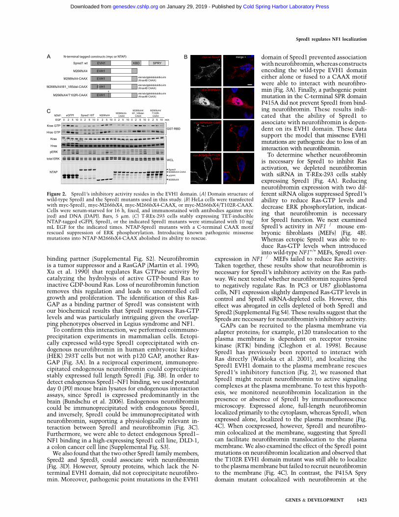
The majority of reported Spred1 mutations produce apremature stop codon, resulting in C-terminally truncatedprotein products (Brems et al. 2007; Messiaen et al. 2009;Pasmant et al. 2009; Spurlock et al. 2009; Muram-Zborovskiet al. 2010; Denayer et al. 2011; Laycock-van Spyk et al.2011; Spencer et al. 2011). As the SPR domain has previouslybeen reported to target Spred1 to the plasma membrane viainteractions with phospholipids and caveolin-1 (Lim et al.2002; Nonami et al. 2005), we surmised that the truncatedmutants were unable to function as a result of mislocaliza-tion. To test this hypothesis, we artificially localized theSpred1 C-terminal truncation mutant (M266fsX4) to themembrane by fusing it to the membrane targeting CAAXmotif of Kras4B (Fig. 2A). Immunofluorescence microscopyrevealed that the mutant Spred1 protein localized primarilyto the cytoplasm, whereas the mutant CAAX fusion pro-tein was localized to the plasma membrane (Fig. 2B). Themutant CAAX protein also showed a restored ability toreduce EGF-induced Ras activation and inhibit ERK activa-tion (Fig. 2C), indicating that the inhibitory activity ofSpred1 resides at the N terminus. Intriguingly, this alsosuggests that defective membrane targeting of Spred1 isresponsible for loss-of-function of Spred1 in Legius syn-drome-associated C-terminal truncation mutants.
Thus far, ;12 missense mutations in SPRED1 have beencharacterized as pathogenic in Legius syndrome (11 pointmutations and one four-amino-acid in-frame deletion), five ofwhich are used in this study (Supplemental Table S1; Bremset al. 2007; Messiaen et al. 2009; Pasmant et al. 2009; Spurlocket al. 2009; Denayer et al. 2011; Spencer et al. 2011). Of these12 missense mutations, nine localize to the EVH1 domain.When we introduced two of these mutations (T102R andI81_V85del) into the truncated CAAX fusion protein, bothprevented the ability of the CAAX motif to rescue Spred1function (Fig. 2C). Together, these results underscore theimportance of the EVH1 domain of Spred1 in inhibitingERK activation, and the importance of the SPR domain indirecting the plasma membrane localization of Spred1.
EVH1 domains are protein–protein interaction modulesthat recognize and bind proline-rich sequences (Gertler et al.1996). The crystal structure of the Xenopus tropicalis Spred1EVH1 domain suggests a distinct peptide-binding mecha-nism compared with other identified EVH1 domains(Harmer et al. 2005). Although an interacting protein forthe Spred2 EVH1 domain has recently been reported (NBR1)(Mardakheh et al. 2009), proteins that specifically interactwith the Spred1 EVH1 domain have yet to be determined.Since the EVH1 domain appears to be essential forSpred’s function, we hypothesized that an unidentifiedbinding partner of the EVH1 domain might be requiredfor Spred1’s inhibitory activity. To identify novel Spred1EVH1-interacting proteins, we performed tandem affinitypurification (TAP) (Burckstummer et al. 2006) using thetruncation mutant-CAAX fusion protein (Fig. 2A) as bait.Following mass spectrometry analysis, we identified theNF1 gene product neurofibromin as a novel Spred1 EVH1-
Figure 1. Spred1 suppresses Ras activity. (A) TET-inducible 6X-HISeGFPf or Spred1-expressing T-REx-293 cells were stimulated withEGF (10 ng/mL) for the indicated times, and whole-cell lysates wereaffinity-purified using GST-Raf1 RBD to precipitate active Ras.Immunoblot analysis was performed with the antibodies indicatedon the left. (B) PC3 cells were transfected with the indicated siRNAoligos, serum-starved for 12 h, and stimulated with EGF for 2 min.Cell lysates were subjected to GST-Raf1 RBD assays to detect Ras-GTP levels. (C) T-REx-293 cells stably expressing Flag-tagged eGFPf,Spred1, or the indicated Spred1 mutants were stimulated with 10ng/mL EGF for the indicated times.
Stowe et al.
1422 GENES & DEVELOPMENT
Cold Spring Harbor Laboratory Press on January 29, 2019 - Published by genesdev.cshlp.orgDownloaded from
binding partner (Supplemental Fig. S2). Neurofibrominis a tumor suppressor and a RasGAP (Martin et al. 1990;Xu et al. 1990) that regulates Ras GTPase activity bycatalyzing the hydrolysis of active GTP-bound Ras toinactive GDP-bound Ras. Loss of neurofibromin functionremoves this regulation and leads to uncontrolled cellgrowth and proliferation. The identification of this Ras-GAP as a binding partner of Spred1 was consistent withour biochemical results that Spred1 suppresses Ras-GTPlevels and was particularly intriguing given the overlap-ping phenotypes observed in Legius syndrome and NF1.
To confirm this interaction, we performed coimmuno-precipitation experiments in mammalian cells. Ectopi-cally expressed wild-type Spred1 coprecipitated with en-dogenous neurofibromin in human embryonic kidney(HEK) 293T cells but not with p120 GAP, another Ras-GAP (Fig. 3A). In a reciprocal experiment, immunopre-cipitated endogenous neurofibromin could coprecipitatestably expressed full length Spred1 (Fig. 3B). In order todetect endogenous Spred1–NF1 binding, we used postnatalday 0 (P0) mouse brain lysates for endogenous interactionassays, since Spred1 is expressed predominantly in thebrain (Bundschu et al. 2006). Endogenous neurofibromincould be immunoprecipitated with endogenous Spred1,and inversely, Spred1 could be immunoprecipitated withneurofibromin, supporting a physiologically relevant in-teraction between Spred1 and neurofibromin (Fig. 3C).Furthermore, we were able to detect endogenous Spred1–NF1 binding in a high-expressing Spred1 cell line, DLD-1,a colon cancer cell line (Supplemental Fig. S3).
We also found that the two other Spred1 family members,Spred2 and Spred3, could associate with neurofibromin(Fig. 3D). However, Sprouty proteins, which lack the N-terminal EVH1 domain, did not coprecipitate neurofibro-min. Moreover, pathogenic point mutations in the EVH1
domain of Spred1 prevented associationwith neurofibromin, whereas constructsencoding the wild-type EVH1 domaineither alone or fused to a CAAX motifwere able to interact with neurofibro-min (Fig. 3A). Finally, a pathogenic pointmutation in the C-terminal SPR domainP415A did not prevent Spred1 from bind-ing neurofibromin. These results indi-cated that the ability of Spred1 toassociate with neurofibromin is depen-dent on its EVH1 domain. These datasupport the model that missense EVH1mutations are pathogenic due to loss of aninteraction with neurofibromin.
To determine whether neurofibrominis necessary for Spred1 to inhibit Rasactivation, we depleted neurofibrominwith siRNA in T-REx-293 cells stablyexpressing Spred1 (Fig. 4A). Reducingneurofibromin expression with two dif-ferent siRNA oligos suppressed Spred1’sability to reduce Ras-GTP levels anddecrease ERK phosphorylation, indicat-ing that neurofibromin is necessaryfor Spred1 function. We next examinedSpred1’s activity in NF1�/� mouse em-bryonic fibroblasts (MEFs) (Fig. 4B).Whereas ectopic Spred1 was able to re-duce Ras-GTP levels when introducedinto wild-type NF1+/+ MEFs, Spred1 over-
expression in NF1�/� MEFs failed to reduce Ras activity.Taken together, these results show that neurofibromin isnecessary for Spred1’s inhibitory activity on the Ras path-way. We next tested whether neurofibromin requires Spredto negatively regulate Ras. In PC3 or U87 glioblastomacells, NF1 expression slightly dampened Ras-GTP levels incontrol and Spred1 siRNA-depleted cells. However, thiseffect was abrogated in cells depleted of both Spred1 andSpred2 (Supplemental Fig S4). These results suggest that theSpreds are necessary for neurofibromin’s inhibitory activity.
GAPs can be recruited to the plasma membrane viaadapter proteins; for example, p120 translocation to theplasma membrane is dependent on receptor tyrosinekinase (RTK) binding (Cleghon et al. 1998). BecauseSpred1 has previously been reported to interact withRas directly (Wakioka et al. 2001), and localizing theSpred1 EVH1 domain to the plasma membrane rescuesSpred1’s inhibitory function (Fig. 2), we reasoned thatSpred1 might recruit neurofibromin to active signalingcomplexes at the plasma membrane. To test this hypoth-esis, we monitored neurofibromin localization in thepresence or absence of Spred1 by immunofluorescencemicroscopy. Expressed alone, full-length neurofibrominlocalized primarily to the cytoplasm, whereas Spred1, whenexpressed alone, localized to the plasma membrane (Fig.4C). When coexpressed, however, Spred1 and neurofibro-min colocalized at the membrane, suggesting that Spred1can facilitate neurofibromin translocation to the plasmamembrane. We also examined the effect of the Spred1 pointmutations on neurofibromin localization and observed thatthe T102R EVH1 domain mutant was still able to localizeto the plasma membrane but failed to recruit neurofibrominto the membrane (Fig. 4C). In contrast, the P415A Sprydomain mutant colocalized with neurofibromin at the
Figure 2. Spred1’s inhibitory activity resides in the EVH1 domain. (A) Domain structure ofwild-type Spred1 and the Spred1 mutants used in this study. (B) HeLa cells were transfectedwith myc-Spred1, myc-M266fsX4, myc-M266fsX4-CAAX, or myc-M266fsX4/T102R-CAAX.Cells were serum-starved for 16 h, fixed, and immunostained with antibodies against myc(red) and DNA (DAPI). Bars, 5 mm. (C) T-REx-293 cells stably expressing TET-inducibleNTAP-tagged eGFPf, Spred1, or the indicated Spred1 mutants were stimulated with 10 ng/mL EGF for the indicated times. NTAP-Spred1 mutants with a C-terminal CAAX motifrescued suppression of ERK phosphorylation. Introducing known pathogenic missensemutations into NTAP-M266fsX4-CAAX abolished its ability to rescue.
Spred1 regulates NF1 localization
GENES & DEVELOPMENT 1423
Cold Spring Harbor Laboratory Press on January 29, 2019 - Published by genesdev.cshlp.orgDownloaded from

cytoplasm, rather than at the plasma membrane. Thesedata were consistent with our observations that the T102Rmutant failed to interact with neurofibromin by coimmu-noprecipitation, whereas the pathogenic P415A mutantcould still bind neurofibromin.
To confirm that neurofibromin recruitment was depen-dent on the EVH1 domain, we also examined neurofibro-min localization after coexpression of the M266fsX4-CAAXmutant (Fig. 4C; Supplemental Fig. S5). Consistent with ourbiochemical interaction data, we observed that neurofibro-min translocated to the membrane in the presence of themembrane targeted (CAAX) Spred1 mutant. However,when the T102R point mutation was introduced intothe M266fsX4-CAAX construct, mutant Spred1 still local-ized to the membrane but failed to recruit neurofibromin,which remained cytoplasmic (Fig. 4C, bottom panel).
To biochemically confirm that Spred1 was capable oflocalizing neurofibromin to the plasma membrane, Spred1-expressing 293 cells were subjected to biochemical fraction-ation (Fig. 4D). Endogenous neurofibromin was distributedprimarily to the cytoplasmic fraction. However, followingexogenous expression of Spred1, there was a robust changein subcellular distribution of neurofibromin in that cyto-plasmic neurofibromin significantly decreased and therewas a large increase in neurofibromin in the membranefraction. Consistent with our immunofluorescence data,the Spred1 EVH1 T102R mutant was found in the mem-brane fraction but did not alter neurofibromin’s cytoplas-mic localization. The ectopic expression of Spred2 also ledto a significant redistribution of neurofibromin from thecytoplasm to the membrane fraction (Fig. 4E).
Additionally, the siRNA knockdown of both Spred1 andSpred2 in PC3 cells led to a significant reduction ofendogenous neurofibromin in the membrane fraction (Sup-
plemental Fig. S6). In summary, Spred’s inhibi-tory activity on the Ras/MAPK pathway dependson the recruitment of neurofibromin to theplasma membrane.
This work identifies a new biochemicallink between Legius syndrome and NF1 andprovides the first evidence for the molecularmechanism of Spred1 function, invoking anovel and critical regulatory mechanism forneurofibromin. On the basis of the data de-scribed here, we propose a new model forSpred1 inhibition of the Ras/MAPK pathway.We show that the N-terminal EVH1 domainof Spred1 interacts with neurofibromin andmediates its translocation to the plasmamembrane, where neurofibromin can performits function as a RasGAP. This model is con-sistent with the ability of Spred1 to reducecellular Ras-GTP levels in the absence of anyknown enzymatic activity. Previously, Spred1and Spred2 have been reported to translocateto the plasma membrane following growthfactor stimulation (Wakioka et al. 2001) andinhibit mitogenic signaling induced by a rangeof growth factors and cytokines (Nonami et al.2004; Bundschu et al. 2006); future studiesshould aim to identify specific receptors thatmay engage Spred1 to recruit neurofibrominand modulate Ras-GTP signaling.
Given the overlapping clinical phenotypesof Legius syndrome and NF1, our findings pro-
vide a molecular basis for these similarities and supporta role for Spred1 as a novel regulator of neurofibrominfunction. Furthermore, since Spred2 and Spred3 can alsointeract with neurofibromin, these two isoforms maycompensate for loss of Spred1 and thus may help explainthe milder phenotype associated with Legius syndrome incomparison with NF1. Thus, these results not only iden-tify a novel regulatory module for neurofibromin andRas/MAPK signaling, but importantly, also provide thefirst molecular link between NF1 and Legius syndrome.
Materials and methods
Plasmid constructs
N-terminal-tagged full-length Spred1 and M266fsX4 were made by Gate-
way cloning (Invitrogen) according to the manufacturer’s instructions.
Spred1 truncation mutants were made by PCR cloning. SPRED1 mis-
sense mutants were made by PCR-directed mutagenesis and verified by
sequencing. Full-length NF1 was synthesized by GeneArt and transferred
to various Gateway Destination vectors (Invitrogen). The N terminus the
GS TAP cassette was obtained from the Euroscarf collection (http://web.
uni-frankfurt.de/fb15/mikro/euroscarf/index.html).
Immunochemical analysis
Cell culture, transfections, and cell lysis were performed as described
(Rodriguez-Viciana et al. 2006). T-REx-293 was used to generate stable
HEK-293 cell lines with tetracycline-inducible expression of His-tagged
or NTAP-tagged eGFPf, Spred1, or Spred1 mutants. NF1+/+ and NF1�/�
MEFs were a kind gift from J. Nakamura (University of California at San
Francisco). The following antibodies were used for immunoblotting: Kras
(F234), Nras (F155), Hras (C20), Myc (A-14), neurofibromin (D), GFP (B-2),
SBP (SB19-C4), and GST (B-14) were from Santa Cruz Biotechnology; total
ERK, phospho-ERK (Thr 202/Tyr 204), and phospho-S338 Raf were from
Cell Signaling; Spred1 (rabbit) was from Abcam; Spred1 (sheep) was from
Figure 3. Neurofibromin is a novel Spred1-binding partner. (A) Flag-tagged eGFPf,RKIP, wild-type Spred1, or the indicated Spred1 mutants were transfected into HEK-293T cells, serum-starved for 16 h, and lysed 24 h post-transfection. Anti-Flag im-munoprecipitates were blotted for endogenous neurofibromin and p120 GAP. (B)Endogenous neurofibromin was immunoprecipitated from T-REx-293 cell lysates stablyexpressing TET-inducible 6X-HIS Spred1. (C) Endogenous neurofibromin coimmuno-precipitated with endogenous Spred1 from P0 whole mouse brain lysates using an anti-Spred1 antibody. Endogenous Spred1 also coimmunoprecipitated with endogenousneurofibromin with two different NF1 antibodies. (D) Ectopic Spred2 and Spred3 alsocoprecipitated endogenous neurofibromin in T-REx-293 cells, whereas Sprouty2 andSprouty4 were unable to do so.
Stowe et al.
1424 GENES & DEVELOPMENT
Cold Spring Harbor Laboratory Press on January 29, 2019 - Published by genesdev.cshlp.orgDownloaded from

R&D Systems; Flag M2 was from Sigma; 6X-His was from Clontech; and
Raf-1 (610151) was from BD Transduction Laboratories.
siRNA transfections
Transfection of siRNA oligonucleotides into cells was performed with
RNAiMAX (Invitrogen) according to the manufacturer’s protocol. siRNA
transfections were performed twice. Forty-eight hours after the first
transfection, cells were serum-starved. Seventy-two hours after the initial
transfection, the appropriate growth factor was added, and cells were
harvested for further analysis.
The following target sequences (Qiagen) were used: Spred1-1, TTC
ACGTATCATTCTGCTAAA; Spred1-2, TAGGGTCCCTTTGAAATCA
AT; Spred2-1, AAGGACTTGGTCTACACCAAA; NF1-1, CAGGTGGCTT
GGGATCAATAA; NF1-2, TACAGTAATAGCACTAACCAA.
Nonsilencing control siRNAs were from Dharmacon.
TAP and mass spectrometry (MS)
T-REx-293 cells (Invitrogen) stably expressing tetracycline-inducible
TAP-tagged wild-type Spred1 or M266fsX4-CAAX were plated in five
15-cm dishes. Twenty-four hours following induction with doxycycline
(1 mg/mL), cells were stimulated with EGF (10 ng/mL). Pooled cell
lysates were purified and analyzed as described (Burckstummer et al.
2006; Rodriguez-Viciana et al. 2006) with the following differences for
the MS analysis: Digests were analyzed using a QSTAR Elite mass
spectrometer (Applied Biosystems/MDS Sciex). Data were searched
against the UniProtKB.2010.08.10 as of August 10, 2010, using in-house
ProteinProspector version 5.2.2 (a public version is available online).
Immunofluorescence
Following transfection, HeLa cells were grown in serum-free medium over-
night, washed with PBS, and fixed in 4% paraformaldehyde or stimulated
with EGF (10 ng/mL) prior to fixation. Cells were washed with PBS, followed
by extraction and blocking with PBS containing 3% BSA (Sigma-Aldrich),
0.1% Triton X-100, and 0.02% sodium azide (PBS-BT). Coverslips were
incubated sequentially with primary antibodies diluted in PBS-BT for 1 h at
room temperature or overnight at 4°C. Alexa Fluor dye-conjugated secondary
antibodies (Invitrogen) were diluted in PBS-BT 1:250 and incubated sequen-
tially for 1 h at room temperature. Coverslips were mounted using anti-fade
mounting medium containing PBS, glycerol, and P-phenylenediamine. For
standard immunofluorescence, images for Figure 2B were acquired with a
Leica SP6; for Figure 4C and Supplemental Figure S5, images were acquired
with Openlab 4.0.4 (PerkinElmer) using an Axiovert 200M (Carl Zeiss, Inc).
Images were processed using Photoshop (Adobe Systems, Inc).
Biochemical fractionation
Twenty-four hours to 48 h after transfection with GFP or Spred constructs,
293Tcells were resuspended in hypotonic lysis buffer (10 mM Tris at pH 7.5,
1 mM EDTA, 1 mM DTT, protease and phosphatase inhibitor cocktails),
incubated for 20 min on ice, and then passed through a 25-gauge needle 15
times to lyse. Lysates were replenished with NaCl to a final concentration
Figure 4. NF1 is necessary for Spred1’s inhibitory function. (A) Neurofibromin was siRNA-depleted from TET-inducible 6X-HIS eGFPf andSpred1 cell lines, serum-starved, and stimulated with EGF for the times indicated. Active Ras was precipitated from whole-cell lysates usingGST-Raf RBD beads. (B) MEFs were transduced with eGFPf- or Spred1-expressing retrovirus. Following serum starvation, cells were treated withEGF for the indicated times, lysed, and analyzed by immunoblot. (C) HeLa cells were transfected with either GFP-NF1 alone, myc-Spred1 alone,GFP-NF1 and myc-Spred1, or GFP-NF1 and the indicated myc-Spred1 mutant and serum-starved and for 16 h. Fixed cells were immunostainedfor myc (red), GFP (green), and DNA (DAPI). (D,E) HEK-293T cells ectopically expressing either wild-type Spred1 or the indicated Spred1 mutant(D) or Spred2 (E) were subjected to biochemical fractionation, and the resulting fractions were resolved by immunoblot analysis.
Spred1 regulates NF1 localization
GENES & DEVELOPMENT 1425
Cold Spring Harbor Laboratory Press on January 29, 2019 - Published by genesdev.cshlp.orgDownloaded from

of 150 mM, spun at 100g for 3 min to pellet nuclei and unbroken cells, and
then spun at 10,000g for 15 min to pellet the membrane fraction. The pellet
was washed twice with lysis buffer, and membrane proteins were solubi-
lized in TNE buffer (20 mM Tris at pH 7.5, 150 mM NaCl, 1 mM EDTA, 1%
Triton X-100, 1 mM DTT, protease and phosphatase inhibitor cocktails); the
membrane fraction was then cleared at 15,000g for 10 min. The supernatant
following the 10,000g spin was spun at 100,000g for 30 min, and the final
supernatant was isolated as the cytoplasmic fraction.
Acknowledgments
We thank A. Young, T. Yuan, and K. Shannon for discussions and critical
reading of the manuscript, and A. Balmain and members of the McCormick
laboratory for support and discussions. This work was supported by a
contribution from Sandra Lloyd and a Young Investigator Award from the
Children’s Tumor Foundation. The University of California at San
Francisco Mass Spectrometry Facility (A. L. Burlingame, director) is
supported by the NIH NCRR (grant no. P41RR001614).
References
Basu TN, Gutmann DH, Fletcher JA, Glover TW, Collins FS, Downward
J. 1992. Aberrant regulation of ras proteins in malignant tumour cells
from type 1 neurofibromatosis patients. Nature 356: 713–715.
Bollag G, Clapp DW, Shih S, Adler F, Zhang YY, Thompson P, Lange BJ,
Freedman MH, McCormick F, Jacks T, et al. 1996. Loss of NF1 results
in activation of the Ras signaling pathway and leads to aberrant
growth in haematopoietic cells. Nat Genet 12: 144–148.
Brems H, Chmara M, Sahbatou M, Denayer E, Taniguchi K, Kato R,
Somers R, Messiaen L, De Schepper S, Fryns JP, et al. 2007. Germline
loss-of-function mutations in SPRED1 cause a neurofibromatosis
1-like phenotype. Nat Genet 39: 1120–1126.
Bundschu K, Walter U, Schuh K. 2006. The VASP–Spred–Sprouty domain
puzzle. J Biol Chem 281: 36477–36481.
Burckstummer T, Bennett KL, Preradovic A, Schutze G, Hantschel O, Superti-
Furga G, Bauch A. 2006. An efficient tandem affinity purification procedure
for interaction proteomics in mammalian cells. Nat Methods 3: 1013–1019.
Cichowski K, Santiago S, Jardim M, Johnson BW, Jacks T. 2003. Dynamic
regulation of the Ras pathway via proteolysis of the NF1 tumor
suppressor. Genes Dev 17: 449–454.
Cleghon V, Feldmann P, Ghiglione C, Copeland TD, Perrimon N, Hughes
DA, Morrison DK. 1998. Opposing actions of CSW and RasGAP
modulate the strength of Torso RTK signaling in the Drosophila
terminal pathway. Mol Cell 2: 719–727.
DeClue JE, Papageorge AG, Fletcher JA, Diehl SR, Ratner N, Vass WC,
Lowy DR. 1992. Abnormal regulation of mammalian p21ras contrib-
utes to malignant tumor growth in von Recklinghausen (type 1)
neurofibromatosis. Cell 69: 265–273.
Denayer E, Chmara M, Brems H, Kievit AM, van Bever Y, Van den
Ouweland AM, Van Minkelen R, de Goede-Bolder A, Oostenbrink R,
Lakeman P, et al. 2011. Legius syndrome in fourteen families. Hum
Mutat 32: E1985–E1998. doi: 10.1002/humu.21404.
Feng L, Yunoue S, Tokuo H, Ozawa T, Zhang D, Patrakitkomjorn S,
Ichimura T, Saya H, Araki N. 2004. PKA phosphorylation and 14-3-3
interaction regulate the function of neurofibromatosis type I tumor
suppressor, neurofibromin. FEBS Lett 557: 275–282.
Gertler FB, Niebuhr K, Reinhard M, Wehland J, Soriano P. 1996. Mena,
a relative of VASP and Drosophila Enabled, is implicated in the
control of microfilament dynamics. Cell 87: 227–239.
Harmer NJ, Sivak JM, Amaya E, Blundell TL. 2005. 1.15 A crystal structure of
the X. tropicalis Spred1 EVH1 domain suggests a fourth distinct peptide-
binding mechanism within the EVH1 family. FEBS Lett 579: 1161–1166.
Kato R, Nonami A, Taketomi T, Wakioka T, Kuroiwa A, Matsuda Y,
Yoshimura A. 2003. Molecular cloning of mammalian Spred-3 which
suppresses tyrosine kinase-mediated Erk activation. Biochem Bio-
phys Res Commun 302: 767–772.
King JAJ, Straffon AFL, D’Abaco GM, Poon CLC, I STT, Smith CM,
Buchert M, Corcoran NM, Hall NE, Callus BA, et al. 2005. Distinct
requirements for the Sprouty domain for functional activity of Spred
proteins. Biochem J 388: 445–454.
King JAJ, Corcoran NM, D’Abaco GM, Straffon AF, Smith CT, Poon CLC,
Buchert M, I S, Hall NE, Lock P, et al. 2006. Eve-3: A liver enriched
suppressor of Ras/MAPK signaling. J Hepatol 44: 758–767.
Laycock-van Spyk S, Jim HP, Thomas L, Spurlock G, Fares L, Palmer-
Smith S, Kini U, Saggar A, Patton M, Mautner V, et al. 2011.
Identification of five novel SPRED1 germline mutations in Legius
syndrome. Clin Genet 80: 93–96.
Leondaritis G, Petrikkos L, Mangoura D. 2009. Regulation of the Ras-GTPase
activating protein neurofibromin by C-tail phosphorylation: Implications
for protein kinase C/Ras/extracellular signal-regulated kinase 1/2 path-
way signaling and neuronal differentiation. J Neurochem 109: 573–583.
Li D, Jackson RA, Yusoff P, Guy GR. 2010. Direct association of Sprouty-related
protein with an EVH1 domain (SPRED) 1 or SPRED2 with DYRK1A
modifies substrate/kinase interactions. J Biol Chem 285: 35374–35385.
Lim J, Yusoff P, Wong ES, Chandramouli S, Lao DH, Fong CW, Guy GR.
2002. The cysteine-rich sprouty translocation domain targets mitogen-
activated protein kinase inhibitory proteins to phosphatidylinositol
4,5-bisphosphate in plasma membranes. Mol Cell Biol 22: 7953–7966.
Mangoura D, Sun Y, Li C, Singh D, Gutmann DH, Flores A, Ahmed M,
Vallianatos G. 2006. Phosphorylation of neurofibromin by PKC is
a possible molecular switch in EGF receptor signaling in neural cells.
Oncogene 25: 735–745.
Mardakheh FK, Yekezare M, Machesky LM, Heath JK. 2009. Spred2
interaction with the late endosomal protein NBR1 down-regulates
fibroblast growth factor receptor signaling. J Cell Biol 187: 265–277.
Martin GA, Viskochil D, Bollag G, McCabe PC, Crosier WJ, Haubruck H,
Conroy L, Clark R, O’Connell P, Cawthon RM, et al. 1990. The GAP-
related domain of the neurofibromatosis type 1 gene product interacts
with ras p21. Cell 63: 843–849.
McGillicuddy LT, Fromm JA, Hollstein PE, Kubek S, Beroukhim R, De
Raedt T, Johnson BW, Williams SM, Nghiemphu P, Liau LM, et al.
2009. Proteasomal and genetic inactivation of the NF1 tumor
suppressor in gliomagenesis. Cancer Cell 16: 44–54.
Messiaen L, Yao S, Brems H, Callens T, Sathienkijkanchai A, Denayer E,
Spencer E, Arn P, Babovic-Vuksanovic D, Bay C, et al. 2009. Clinical
and mutational spectrum of neurofibromatosis type 1-like syndrome.
JAMA 302: 2111–2118.
Miyoshi K, Wakioka T, Nishinakamura H, Kamio M, Yang L, Inoue M,
Hasegawa M, Yonemitsu Y, Komiya S, Yoshimura A. 2004. The
Sprouty-related protein, Spred, inhibits cell motility, metastasis,
and Rho-mediated actin reorganization. Oncogene 23: 5567–5576.
Muram-Zborovski TM, Stevenson DA, Viskochil DH, Dries DC, Wilson
AR, Rong M. 2010. SPRED 1 mutations in a neurofibromatosis clinic.
J Child Neurol 25: 1203–1209.
Nonami A, Kato R, Taniguchi K, Yoshiga D, Taketomi T, Fukuyama S,
Harada M, Sasaki A, Yoshimura A. 2004. Spred-1 negatively regulates
interleukin-3-mediated ERK/mitogen-activated protein (MAP) kinase
activation in hematopoietic cells. J Biol Chem 279: 52543–52551.
Nonami A, Taketomi T, Kimura A, Saeki K, Takaki H, Sanada T, Taniguchi
K, Harada M, Kato R, Yoshimura A. 2005. The Sprouty-related protein,
Spred-1, localizes in a lipid raft/caveola and inhibits ERK activation in
collaboration with caveolin-1. Genes Cells 10: 887–895.
Pasmant E, Sabbagh A, Hanna N, Masliah-Planchon J, Jolly E, Goussard
P, Ballerini P, Cartault F, Barbarot S, Landman-Parker J, et al. 2009.
SPRED1 germline mutations caused a neurofibromatosis type 1
overlapping phenotype. J Med Genet 46: 425–430.
Rodriguez-Viciana P, Oses-Prieto J, Burlingame A, Fried M, McCormick F.
2006. A phosphatase holoenzyme comprised of Shoc2/Sur8 and the
catalytic subunit of PP1 functions as an M-Ras effector to modulate
Raf activity. Mol Cell 22: 217–230.
Spencer E, Davis J, Mikhail F, Fu C, Vijzelaar R, Zackai EH, Feret H,
Meyn MS, Shugar A, Bellus G, et al. 2011. Identification of SPRED1
deletions using RT–PCR, multiplex ligation-dependent probe ampli-
fication and quantitative PCR. Am J Med Genet A 155A: 1352–1359.
Spurlock G, Bennett E, Chuzhanova N, Thomas N, Jim HP, Side L, Davies
S, Haan E, Kerr B, Huson SM, et al. 2009. SPRED1 mutations (Legius
syndrome): Another clinically useful genotype for dissecting the
neurofibromatosis type 1 phenotype. J Med Genet 46: 431–437.
Wakioka T, Sasaki A, Kato R, Shouda T, Matsumoto A, Miyoshi K,
Tsuneoka M, Komiya S, Baron R, Yoshimura A. 2001. Spred is
a Sprouty-related suppressor of Ras signalling. Nature 412: 647–651.
Xu GF, O’Connell P, Viskochil D, Cawthon R, Robertson M, Culver M,
Dunn D, Stevens J, Gesteland R, White R, et al. 1990. The neurofibro-
matosis type 1 gene encodes a protein related to GAP. Cell 62: 599–608.
Stowe et al.
1426 GENES & DEVELOPMENT
Cold Spring Harbor Laboratory Press on January 29, 2019 - Published by genesdev.cshlp.orgDownloaded from

10.1101/gad.190876.112Access the most recent version at doi: 26:2012, Genes Dev.
Irma B. Stowe, Ellen L. Mercado, Timothy R. Stowe, et al. Legius syndrome and Neurofibromatosis-1A shared molecular mechanism underlies the human rasopathies
Material
Supplemental
http://genesdev.cshlp.org/content/suppl/2012/07/02/26.13.1421.DC1
Related Content
Genes Dev. July , 2012 26: 1515-1519
Andrea I. McClatchey and Karen CichowskiSPRED proteins provide a NF-ty link to Ras suppression
References
http://genesdev.cshlp.org/content/26/13/1421.full.html#related-urls
Articles cited in:
http://genesdev.cshlp.org/content/26/13/1421.full.html#ref-list-1This article cites 34 articles, 8 of which can be accessed free at:
License
ServiceEmail Alerting
click here.right corner of the article or
Receive free email alerts when new articles cite this article - sign up in the box at the top
Copyright © 2012 by Cold Spring Harbor Laboratory Press
Cold Spring Harbor Laboratory Press on January 29, 2019 - Published by genesdev.cshlp.orgDownloaded from