Embed Size (px)
Citation preview
DEVELOPMENTAL DYNAMICS 195231-272 (1992)
Reprinted from the JOURNAL OF MORPEOLOCY Vol. 88, No. 1, January 1951
A SERIES O F NORMAL STAGES I N THE DEVELOPMENT O F THE
CHICK EMBRYO
VIKTOR HAMBURGER Department of Zoology, Washington University, S t . Louis, Nissouri
HOWARD L. HAMILTON Department of Zoolo8gy and Entom2070gy, Iowa S t a t e College, Ames
FORTY-FIVE FIGURES
The preparation of a series of normal stages of the chick embryo does not need justification at a time when chick ern- bryos are not only widely used in descriptive and experi- mental embryology but a re proving to be increasingly valuable in medical research, as in work on viruses and cancer. The present series was planned in connection with the preparation of a new edition of Lillie’s DeueZopmerzt of the Chick by the junior author. It is being published separately to make it accessible immediately to a large group of workers.
Ever since Aristotle “discovered” the chick embryo as the ideal, object for embryological studies, the embryos have been described in terms of the length of time of incubation, and this arbitrary method is still in general use, except for the first three days of incubation during which more detailed characteristics such as the numbers of somites are applied. The shortcomings of a classification based on chronological age are obvious to every worker in this field, f o r enormous variations may occur in embryos even though all eggs in a setting are plmaced in the incubator at the same time. Many factors are responsible for the lack of correlation between chronological and structural age. Among these are : genetic differences in the rate of development of different breccls (eg. , the embryo of the White Leghorn breed develops more
49
Address renrint reauests to Dr. Joshua R. Sanes. DeDartment of Anatomy and Neurobiology, Washington University’ Medical School, St. Louis, MO 63110.
Q 1993 WILEY-LISS, INC.
232 HAMBURGER AND HAMILTON
50 V. HAMBURGER A N D H. L. HAMILTON
rapidly than that of the Barred Plymouth Rock and hatches approximately a day earlier) ; seasonal differences in the viability and vigor of embryos; differences in the stage of development when incubation is started ; differences in the “freshness” of eggs, Le., the lapse of time between laying and incubation; differences in the temperature of eggs when placed in the incubator, and in the size of individual eggs; differences in the temperature of incubation, and in type and size of incubator.
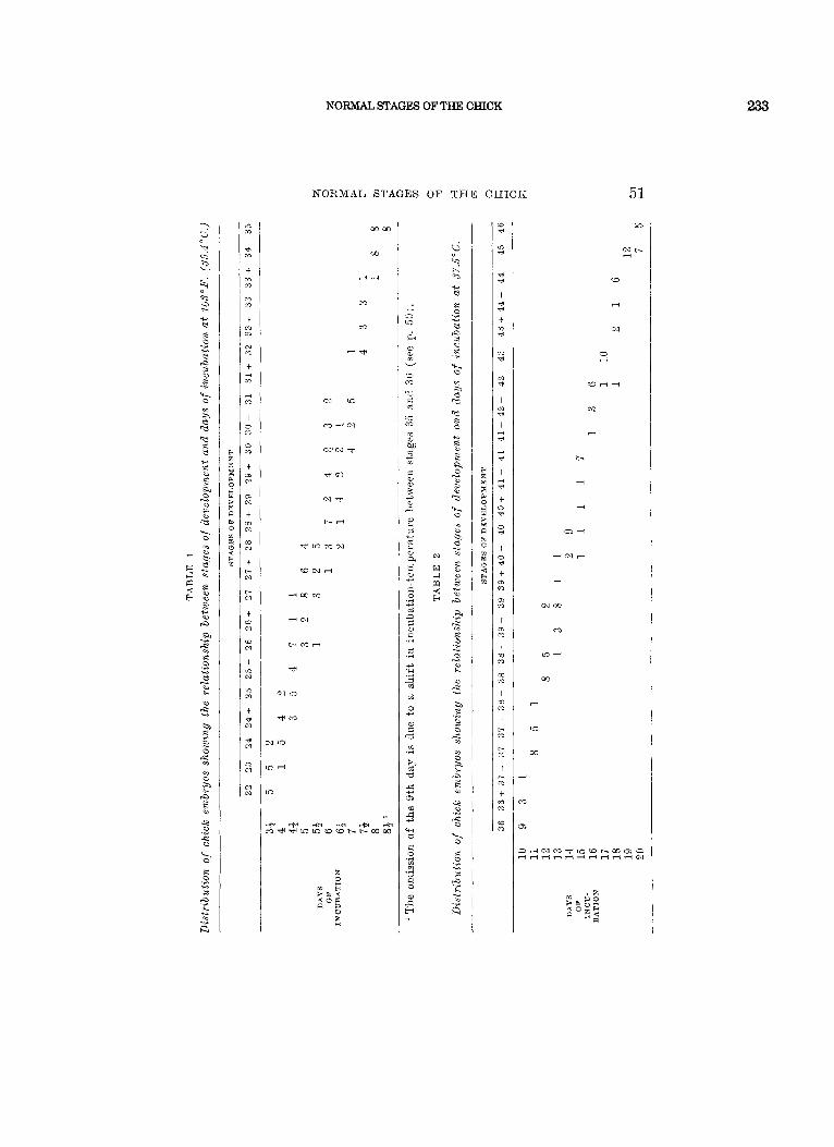
The wide variations in external form which occur a t any given chronological age are clearly seen in tables 1 and 2 which show the distribution of 296 embryos from the 4th day until hatching when classified according to our series of stages. F o r example, a “6-day” embryo may range anywhere from stage 27 + to stage 31 (table 1). It will also be noted that the data in table 1 are based on an incubation-temperature of 103°F. (ca. 39.4”C.) whereas those in table 2 are based on a temperature of 375°C. This difference has resulted in the skipping of the “9-day” embryo altogether! It is not sur- prising, therefore, that the use of chronology with its lack of precision in the designation of embryos has actually led to misunderstandings and controversies which could readily have been avoided by the use of an adequate series of morpho- logical stages.
Keibel and Abraham (1900) worked out a series of stages of the chick embryo based on morphological characters. This series never became popular and it has been rarely used and quoted. Among its shortcomings are its inadequate illustra- tions which often make the identification of an embryo difficult, the incomplete coverage of older stages, and perhaps also the format and relative inaccessibility of the Normenta- feln. M. Duval’s masterful Atlas d’EmbryoZo,gie (1889) with its artistically perfect drawings is unfortunately incomplete in that i t does not go beyond the 8th day of incubation.
Our own work covers the entire period of incubation. I ts aim is to serve the practical purpose of identifying and desig- nating embryos on the basis of external characters. The uii-
c d
E rl
? e
L 3
v1 a 4 m
La m
di m
t m c? - .- 3
t N c?
N m
t 3 .* ti m
t 0 c2
0 m
t 3) N
m N
t zi N
m N
t c N
,- N
t c2 N
(0 N
t u3 31
3 . 3 Y
t d N
d N
-1 N
N N
NORMAL STAGES OF THE CHICK
NORMAL STAGES O F THE C H I C K 51
w w
W
d d
m
M
h e
‘a P
tn d
P
I P
t
d n
- P
N d
I N P
t P ti
rl P
I
P
t 0 P
a d
3
I 0 d
t m n
m n
I n m
t Ii r‘3
m
I n m
c3
t t- 3
t- 7
I c P
t a m
(0 m
M
Nr- rl
c3
.-
r-
ri
ri
olm
m
cfl i
m
n
n
233
234 HAMBURGER AND HAMILTON
52 V. HAMBURGER A N D 13. L. I lAMILTON
exc.ellec1 series of stages of Ainblystoma by Harrison lias served as a model. Our series is independent of chronological age and of size of embryos, as is the ,4?r&Zystoma series. The photographs and drawings show most of the diagnostic criteria ; this, we hope, will facilitate a rapid identification. A brief text is added, in which the distinguishing criteria are listed for each stage.
We are aware of the complications which derive from the iiidepeiideiit variations of different characters. For instance, the progress of differentiation in the visceral arches may lag behind that in the limb-buds, when compared with an average sequence. F o r this reason, the amnion and allantois, and the iiuinber of pairs of somites beyond 22 a re of no diagnostic value. We have tried to establish average or “standard” types by comparing a considerable number of embryos in each stage, aiid me have selected for illustrations those embryos which appeared typical.
Dixi-iiig the different phases of development, different char- acters become prominent, and theref ore particularly useful for the diagnosis. For the second day of incubation we have adopted the conventional designation of embryos according to numbers of pairs of somites. We have chosen intervals of three somites as “stages”; this makes it possible to desig- nate embryos with intermediate numbers of somites by a + or - sign. Somites mere not counted unless fully formed and coiiipletely separated by clefts from the adjacent mesoderm. The first somite was not included in the counts beyond stage 10 when it begins to dwindle away.
During the third day of incubation, or, more precisely, from the stage of 22 somites oiiwai-d (stage 14), the rapid progress in development of the limbs provides the most com venient diagnostic criteria. Preliminary \vork oii these stages has lieen done by Hamburger (’38, ’42) and by Saiinders (’48). Our stages 15 to 21 are identical with stages 1 to 7 of these authors. The original work was carefully rechecked and detailed descriptions of all characters were added. Stages 8 and 9 of Xawiders a r e combined in 0111’ stage 22;
NORMAL STUES OF THE CHICK
NORMAL STAGES O F THE C E I C K 53
235
stage 10 of Saunders is identical with our stage 23. The de- velopmental phase between 4 and 9 days of incubation is characterized by rapid changes in the wings, legs, and visceral arches. From the 8th to the 12th days, feather-germs and eyelids provide the most useful criteria. The designation of stages during the last phase of incubation is difficult because practically no new structures are formed and there is mainly just growth of what almready exists. Hence, we have had to make use of measurements of the lengths of the beak and of the toes.
The senior author is responsible f o r stages 14 to 35 and the junior author for all the others.
All illustrations and descriptions are based on material fixed in Bouin's solution or formalin. It is possible that minor distortions have occurred due to differential shrink- age, for instance in the amnion. The embryos used for stages 14 to 35 came from a flock of White Leghorns at St. Louis. They were incubated in a small size Buckeye incubator ( for 350 eggs) without forced draft, at a temperature of 1Q3"F. (ca. 39.4"C.). The embryos used for the other stages were of several breeds (White Leghorn, Barred Plymouth Rock, and Rhode Island Red) from the Iowa State College Poultry Farm, and were incubated in a forced-draft incubator at a constant temperature of 375°C. During the course of this work several hundred embryos have been examined and clas- sified from the second day of incubation until hatching.
We wish to express our great appreciation of the expert advice and help which Dr. Mary E. Rawles, Johns Hopkins University, and Dr. Nelson T. Spratt, University of Minne- sota, have given us in the difficult matter of selecting stages 1 to 6. Dr. Rawles has generously supplied data on the range of time within which a given stage may usually be obtained, based on records of 7 0 embryos incubated a t 38°C. Her data are included iii the text f o r stages 5-14 and 22. Dr. Spratt has supplied photographs and slides for illustrating
236 HAMBURGER AND HAMILTON
54 V. HSMBUKGEB AND H. L. HAMILTON
the prc-somitie stages and has given estimates of incubation- time for stages 2-4.
The photographic work for stages 22 to 35 was done by Mr. L. Pinkers and Mr. D. Bucklin at Washington University, and that for the remaining stages by Mi-. John Staby of the Iowa State College Experiment Station. All drawings were made by Mix. Elsie Herbold Froeschner of Ames, Iowa. Additional assistance was given by Miss Thelma Dunnebacke and lzliss Mary Lee Winkler, both of Washington University. We wish t o thank all our helpers for their efficient and h inti ring cooper- ation. The work was supported, in part , by a Research Grant of the Rockefel’ler Foundation to the Department of Zoiilogy of Washington University, and by the Industrial Science Research Institute of Iowa State College.
The description which follows should be used in conjunc- tion with the ill’ustrations (plates 1-14) which are numbered according to stages.
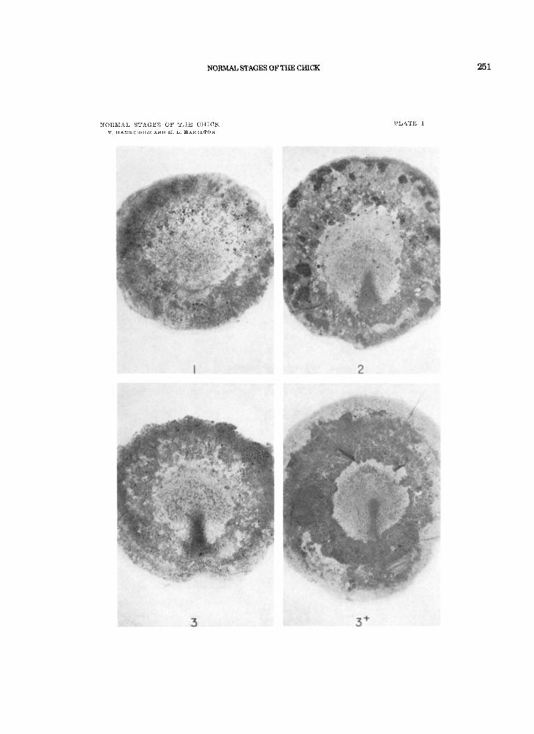
X i a g e I . Pre-Streak: Prior to the appearance of the primitive streak. A n “embryonic shield” may be visible, due to the ac- cumulation of cells toward thc posterior half of the blastoderm. (See Spratt , ’42, pp. 71-72.)
Rtage 2. Initial Streak: (‘( Short-Sroad beginning-streak” of Spratt , ’42). A rather transitory stage in which the pr imitke streak first appears as a short, conical thickening, almost as broad as long (0.3-0.5 mm in length), a t the posterior border of the pellucid area. Usually obtained after 6-7 hours of incubation.
Sfage 3. Intermediate Xtreak: (12-13 hrs.). The primitive streak extends from the posterior margin to approximately the center of the pellucid area. The streak is relatively broad throughout its length, and is flared out where it touches the opaque area. No primitive groove.
Stage 4. Definitive Xtreak: (18-19 hrs.) . The primitive streak has reached its maximal length (average length = 1.88 mm, Spratt , ’46). The primitive groove, primitive pit, and Hensen’s node are present. The area pellucida has become pear-shaped and the streak extends over two-thirds to three-fourths of its length.
Stage 5. Head-Process: (19-22 hrs.) . The notochord or head- process is visible as a rod of condensed mesoderm extending
NORMAL STAGES OF THE CHICK
NORMAL STAGES O F TEE C H I C K 55
237
forward from the anterior edge of Hensen’s node. The head-fold has not yet appeared. Since the length of the notochord increases during this stage, it is suggested that the length of the notochord in millimeters be appended to the number of the stage for further precision (e.g., “Stage 5-0.2” would designate a notochordal blastoderm with notochord 0.2 mm in length).
HeabPold: (23-25 hrs.). A definite fold of the blastoderm anterior to the notochord now marks the anterior end of the embryo proper. No somites have yet appeared in the mesoderin lateral to the notochord. This is a transitory stage, since the head-fold and the first pair of somites develop rather clos~ly in time.
Stages 7 to 14 are based primarily on the numbers of pairs of somites which are clearly visible. The number of somites appears t o be the simplest criterion for staging this phase of development, and it is sufficiently accurate fo r practical purposes. A stage is assigned t o every third pair of somites which is added; embryos with in- between numbers of somites are designated by adding a + o r - sign to the appropriate stage. Thus, stage 7 designates an embryo with one pair of somites; stage 7 + = two pairs; stage 8 - = three pairs; stage 8 = four pairs; etc. (See plates 2 and 3.) Stage 7 . One somite: (23-26 hrs.). This is actually the second
somite of the series; number one is not yet clearly defined. Neural folds are visible in the region of the head.
Stage 8. Pour somaites: (26-29 hrs.). Neural folds meet a t level of midbrain. Blood-islands are present in posterior half of blastoderm.
Stage 9. Seven. somites: (29-33 hrs.). Primary optic vesicles are present,. Paired primordia of heart begin t o fuse.
Stage 10. Ten somites: (33-38 hrs.). The first somite is becoming dispersed ; it is not included in the counts far subsequent stages.’ First indication of cranial flexure. Three primary brain-vesicles are clearly visible. Optic vesicles not constricted a t bases. Heart bent slightly to right.
Stage 11. Tlawteen somites: ( 4 0 4 5 hrs.) Slight cranial flexarr Five nenronieres of hindbrain are distinct. Anterior neuropore is closing. Optic vesicles are constricted at bases. Heart bent to right.
Btage 6.
’ It is suggested tha t embryos which haye gained oiie soiiiite beyond Stage 10, but have lost s. 1 in the meantime, be designated as Stage 10 2 ; Stage 10 + would then Itale 11 s., no t counting the rudimentaiy one; stage 11 - = 12 s., not couiitiiig the rudimentary one, etc.
238
56 V. HAMBURGER AND H. L. HAMILTON
X t a g e 12. Sixteen somites: (45-49 hrs.). Head is turning onto le f t side. Anterior nenropore closeil. Telencephalon indicated. Pri- mary optic vesicles and optic stalk well established. Auditory pit is deep, but wide open. Heart is slightly S-shaped. Head- fold of amnion covers entire region of forebrain.
S t a g e 13. Nineteen s o m i t e s : (48-52 hrs.). Head is partly to fully turned to the left. Cranial and cervical flexures make broad cnrves. Distinct enlargement of telencephalon. Slight narrow- ing of opening to deep auditory pit. No indication of hp- pophysis. Atrio-ventricular canal indicated by constriction. Head-fold of amnion covers forebrain, midbrain, and anterior part of hindbrain.
Flexures aad rotatim. Cranial flexure: axes of forebrain and hindbrain form abont a right angle. Cervical flexure a broad curve. Rotation of body back as far as somites 7-9. Behind this level, a slight flexure makes its appearance which will be referred t o as “trunk-flexure.”
T7isceral arches 1 and 2, and clefts 1 and 2 are distinct. Posterior arches not distinct.
Primary optic vesicle begins to invaginate ; lens-placode is formed. Opening of nuatory p i t constricted. Ratkke’s pouch can be recognized. Ventricular loop of heart now ventral to atrio-ventricular canal. Anamhz extends to somites 7-10.
Beyond stage 14 the number of somites becomes increasingly difficult to determine with accuracy. This is due in port to the dis- persal of the mesoderm of the anteriormost somites, and, in later stages, to the curvature of the tail. Total somite-counts given for the following stages are typical, but sufficiently variable m as not to be diagnostic. For these reasons, the limb-buds, visceral arches, and other externally visible structures are used as identifying criteria from stage 15 onward. Stage 15. (Hamburger, ’38 ; Sannders, ’48, stage 1 ; ca. 50-55 hrs.) .
1. Lateral body-folds extend t o anterior end of wing-level (somites 15-17).
2. Limb-prinaordia: prospective limb-areas flat, not yet demar- cated. Inconspicuous condensation of mesoderm in wing-level.
3. Smaites: 24-27. 4. Amnion extends to somites 7-14. 5. Flexures a n d rotatiom. Cranial flexure: axes of forebrain
and hindbrain form an acute angle. The ventral contours of forebrain and hindbrain are nearly parallel. Cervical flexure
Siage 14. Twenty-two ~sowites: (50-53 hra.).
NORMAL STAGES OF THE CHICK
NORMAL STAGES O F THE C H I C K 57
239
a broad curve. The t runk is distinct. Rotation extends to somites 11 to 13.
6. V%soeral arches: Visceral arch 3 and cleft 3 are distinct. The latter is shorter than cleft 2 and usually oval in shape.
7. Eye: Optic cup is completely formed; double contour dis- tinct in region of iris.
Stage 16. 1.
2.
(Hamburger-Sannders stage 2 ; ca. 51-56 hrs.) . Latera l body-folds extend to somites 17-20, between levels
of wings a n d legs. L i m b s . Wing is lifted off blastoderm by infolding of lateral
body-fold. It is represented by a thickened ridge. Primordinm of leg is. still flat; represented by a condensation of nieso- derm.
3. flomites: 26-28. 4. Amnion extends to somites 10-18. 5. Plexures and rotai ion: All flexures a re more accentiiated
6. Tail-bwd a short, straight cone, delimited from blastoderm. 7. Vislceral arches: Third cleft still oval in shape. 8. Forebrain lengthened ; constrictions between brain-parts are
than in stage 15. Rotation extends to somites 14-15.
deepened. Epiphysis indistinct or not yet formed. Xtage 17. (Hamburger-Saunders stage 3 ; ca. 52-64 hrs.).
1.
2.
Latera l body- fo lds extend around the entire circumference of the body.
L i m b - b u d s : both wing- and leg-buds lifted off blastoderill by infolding of the body-folds. Both are distinct swellings of approximately equal size (see plate 5) .
3. Xomdtes: 29-32 4. A m n i o n : Considerable variability, ranging from a condition
in which posterior t runk and tail, from approximately somite 26, are uncovered, to complete closure except for an oval hole over somites 28-36. Intermediate stages with an anterior fold covering as fa r back as somite 25 and a posterior fold covering par t of the tail are common.
5. Plextcres and rotat ion: Cranial flexure is unchanged. Cer- vical flexure is more sharply bent than in preceding stages, but its angle is still larger than 90". Trunk-flexure is distinct in brachial level. Rotation extends to somites 17-18.
6. T a d - b u d bent ventrad. Its mesodern1 unsegmented. 7. Epiphysis: a distinct knob. Indication of nasal pzts. 8. A l lan to is : not yet formed.
240 HAMBURGERANDHAMILTON
58 V. HAMBURGEE AND H. L. HA1\!tILTON
Stage 28. 1.
(Haiiiburger-Sa~iiiders stage 4 ; ca. 6 5-69 hrs.). Linab-B~l s enlarged ; leg-buds slightly larger than wing-buds
(see plates 4 and 5). L/W of wing = 6 or < 6 (L = length = anterior-posterior dimension as measured along the body-wall ; W = width = distance from body-wall to apex; see stage 20, plate 3 ) .
2. 3.
4.
Xomites: 30-36; extend beyond level of leg-bud. A m n i o n : Usually closed; occasionally an oval hole in lumbar
region. Flexures and rs ta t ion: At the cervical flexure, the axis of
the medulla forms approximately a right angle to the axis of the posterior trunli. The trunk-flexure has shifteldl t o the lumbar region. The rotation extends now to the posterior part of the body ; hence, the leg-buds are no longer in the horizontal plane.
5. The tail-Dud is turned t o the right, a t about an angle of 90” to the axis of the posterior trunk.
6. Visceral arches: Maxillary process absent or inconspicuous. Fourth visceral cleft indistinct o r absent.
‘7. Allamtois: A short, thicli-walled pocket ; not yet vesicular.
1. Linab-buds: Enlarged, symmetrical. Leg-buds slightly larger and bulkier than wing-buds (see plate 5). L/W of wing- buds = 4-6.
Sonaites: 3 7 4 0 ; extend into tail; but the end of the tail which is directed forward is unsegmented.
Flexup-es and rotat ion: I n the cervical flexure the axis of the medulla forms an acute angle with the axis of the trunk. The trunk-flexure has nearly or entirely disappeared due t o the rotation of the entire body. The contour of the posterior part of the trunk is straight to the base of the tail.
Sfage 19. (Hamburger-Saunders stage 5 ; ca: 68- 72 hrs.).
2.
3.
4 5
T a d - b ~ d curved, its tip pointing forward. Tizsce~-al arches: The maxillary process is a distinct swelling
of approximately the same length as the mandibular process. The first visceral cleft is an open narrow slit at its dorsal part. It continues into a ‘shallow furrow. The second arch projects slightly over the surface. The 4th cleft is a fairly distinct slit a t its dorsal part and continues ventrally as a shallow groove. It does not perforate into the pharynx as a true (open) cleft, but is, nevertheless, IiomoiogoLis to the other three clefts.
Al lanto is : A small pocket of variable size; not yet vesicular. 6. ‘7. E y e s unpigmentecl.
NORMAL STAGES OF THE CHICK
NOlX1RIAL STAGES O F THE C H I C K 59
241
S t a g e 20. 1.
(Hamburger-Saunders stage 6 ; ca. 70 - 7 2 hrs.) . LimB-buds enlarged ; leg-buds are (distinctly larger from n o ~ v
on than wing-buds. The wing-buds are still approximately symmetrical ; the leg-buds are slightly asymmetrical (see plate 5 ) . L/W of wing = 3-3.9 ; L/W of leg = 3-2.3.
2. 3. F l e x u r e s and r o t a t i o n : Cervical flexure more accentuated
than in stage 19. The bend in the tail-region begins to extend forward into the lunibo-sacral region. Contour of mid-trunk a straight line. Rotation completed.
4. Visceral arches: Maxillary process distinct, equals or ex- ceeds the mandibular process in length. Second arch projects over surface. Fourth arch less prominent and smaller than third arch. Fourth cleft shorter than third cleft; a narrow slit a t its dorsal part, continuing into a shallow groove.
A l l a n t o i s : Vesicular, variable in size; 011 the average of the size of the midbrain.
E y e - p i g m e n t . A faint grayish hue.
S o m i t e s : 40-43 ; tip of tail still unsegmented.
5 .
6.
1. Linzbs: Enlarged; both wing- and leg-buds are slightly asymmetrical ; their proximo-distal axes are directed caudad, and the apex of the bud lies posterior t o the midline bisecting the base of the bud. The posterior contours of wing- and leg-buds are steeper than the anterior contours; they meet the baseline a t an angle of approximately 90". L/W of wing = 2.3-2.7; L/W of leg = 2.0-2.5.
S t a g e 21. (Saunders stage 7 ; ca. 34 days).
2. 3.
Somztes: 43-44; extreme tip of tail unsegmented. F l e x u r e s : The posterior c-urvatore includes the lumbo-sacral
region. The dorsal contour of the trunk is straight or slightly bent.
Visceral arches: Maxillary process is defiiiitely longer than mandibular process, extending approximately to the middle of the eye. The second arch extends distinctly over the sur- face and overlaps the third arch ventrally. Fourth arch distinot; 4th cleft visible as a slit.
Alla?ztois: Variable, usually larger than in stage 20; may extend to head.
4.
5.
6 . E y e - p i g m e n t a t i o n : Faint. (Saunders stages 8 and 9 combined ; ca. 34 days).
1. Linzbs: Elongated buds, pointing caudad. The anterior and posterior contours are nearly parallel a t their bases (see plate 7) . L/W of wing = 1.5-2; L/W of leg = 1.3-1.8.
S t a g e 22.
242 HAMBURGER AND HAMILTON
60 V. TIAMBURGER A N D H. L. HAMILTON
2. S o n d e s : Extend to t ip of tail. 3. Plexures: Little change. The dorsal contour of the trunk is
a straight line o r curved. 4. Visceral arches: Little change compared with stage 21.
Maxillary process enlarged; 4th cleft distinct as a slit. 5. Allantois: Variable in size; extends t o head and may overlap
the forebrain. 6. Eye-pigmentcrtion: Distinct.
1. Stage 23. (Saunders stage 10 ; ca. 3+ 4 days).
Limbs: Longer than in stage 22 ; particularly the proximal parts in which anterior and posterior contours run parallel are lengthened ; otherwise, little change in shape. Both wing- and leg-buds approximately as long as they are wide.
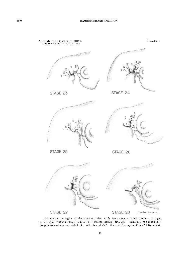
Visceral arches (see plates 7 and 8) : Maxillary process is lengthened further. The first visceral cleft is represented by a broken line. I ts dorsal par t is a distinct slit. A slight protuber- ance (“a’ , ) is noticeable anterior to the dorsal slit. The claudal part of the second arch is distinctly elevated over the surface. Arches 3 and 4 are still completely exposed. Visceral cleft 3 is a distinct groove, and cleft 4 is reduced to a narrow oval pit a t its dorsal end.
3. Flexures: The dorsal contour from hindbrain to tail is a curved line.
Stage 24. (ca. 4 days).
2 .
1. Limbs: Wing- and leg-buds distinctly longer than wide. Digital plate in wing not yet demarcated. Toe-plate in leg-bud distinct. Toes not yet demarcated.
Visceral arches (see plates 7 and 8) : First visceral cleft a distinct curved line. Slight intdication of two protuberances (‘ ‘ a, ” “b’ ’) on mandibular process and of three protuberances
on second arch. Par t “c” of mandibular process is receding. Second arch longer ventrally (a t “ f ’,) and much wider than mandibular process. Third arch reduced and partly overgrown by second arch; 4th arch flattened. Both are sunk beneath the surface. Thirld visceral cleft is an elongated groove. Fourth visceral cleft reduced to a siiiall pit.
2.
( l , , , , , C L e , ” C l f , , )
Staye 25. (ca. 4 t days). 1. Liinibs: Elbow and knee-joints distinct ( in dorsal o r ventral
view). Digital plate in wing distinct, but no demarcation of digits. Indication of faint grooves demarcating the third toe on leg.
Visceral al-ches (see plates 7 and 8) : Maxillary process length- ened; i t meets the wall of the nasal groove (notice the notch a t
2.
NORMAL STAGES O F THE C H I C K 61
point of fusion). Three protuberances on each side of first vis- ceral cleft ( “a” to “ f ” ) . I n dorsal view, “a ,” “b,” and “ d ” appear as round knobs, and c 1 c 7 7 as a flat ridge. Par t “ f ” is conspicuous and projects distinctly over the surface. It mill be referred to as the “collar.” Dorsal part of third arch still visible. Third and 4th visceral clefts reduced to small circular pits.
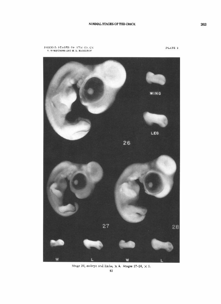
S t a g e 26. (ca. 43-5 days). 1. Limbs: Considerably lengthened. Contour of digital plate
rounded. Indication of faint groove between second and third digit. Demarcation of the first three toes distinct.
Visceral nrrhps (see plates 8 and 9) : Contour of maxillary process a broken line. Mandibular process lengthened ventrally. Protuberances “ a ” and “ b ” project over the surface. The middle protuberance (11 b” ) is subdivided by a shallow groove. A small knob is distinct a t the dorsal edge of l 1 c. ” On the sec- ond arch, protuberances l 1 d” and “ e ” are only slightly elevated over the surfaoe. The ( “ f ” ) has broadened and over- grown visceral arches I11 and IV. A deep groove separates “ f ” from “c.” The two pits represent,ing the 3rd and 4th visceral clefts are no longer visible.
2.
Stage 27. (ca. 5 days). 1. Limbs: Contour of digital plate angular in region o€ first
digit. Grooves between first, second, and third digits indi- cated. Grooves between toes are distinct on outer and inner surfaces of toe-plate. First toe projects over the tibia1 part at an obtuse angle. Tip of third toe not yet pointed.
Visceral at-ches (see plates 8 and 9 ) : Contour of maxillary process is a curved, broken line. Mandibular process has broad- ened ventrally (at “c” ) and grown forward. Protuberances “ a ” and “ b ” project over the surface. Parts “ d ” and “ e ” are flat. Protuberances “ b ” and “e” are close to fusion, but a separating line is still distinct. The ‘‘ collar” (‘ ‘ f ’ ’) has broad- ened and continned its growth backward. It rises conspicuously above the surface. The groove between “c” and “ f ” has w i deiic d
2 .
3. Beak: Barely recognizable.
1. Sfage 28. (ca. 56 days).
L i m b s : Second digit and third toe longer than others, which gives the digital and toe-plates a pointed contour. Three digits and 4 toes distinct. No indication of 5th toe.
Visceral arclzes (see plates 8 and 9 ) : Protnberance “ a ” still projects over the surface. Mandibular process has lengthened
2.
244 HAMBURGER AND HAMILTON
62 T7. H A M B U R G E R AND JT. L. H A M I L T O N
and grown forward. Parts “b” and “ e r r have fused; a fine suture line is occasionally still visible. Parts “b,” “d,” and “e” no longer project above the surface. External auditory opening is now very distinct between “a , ” “b,” and “d.” l 1 Collar ’ ? ( ‘ ‘ f ”) projects distinctly over the surface. The neck between “collar ” and mandible has lrngthenril
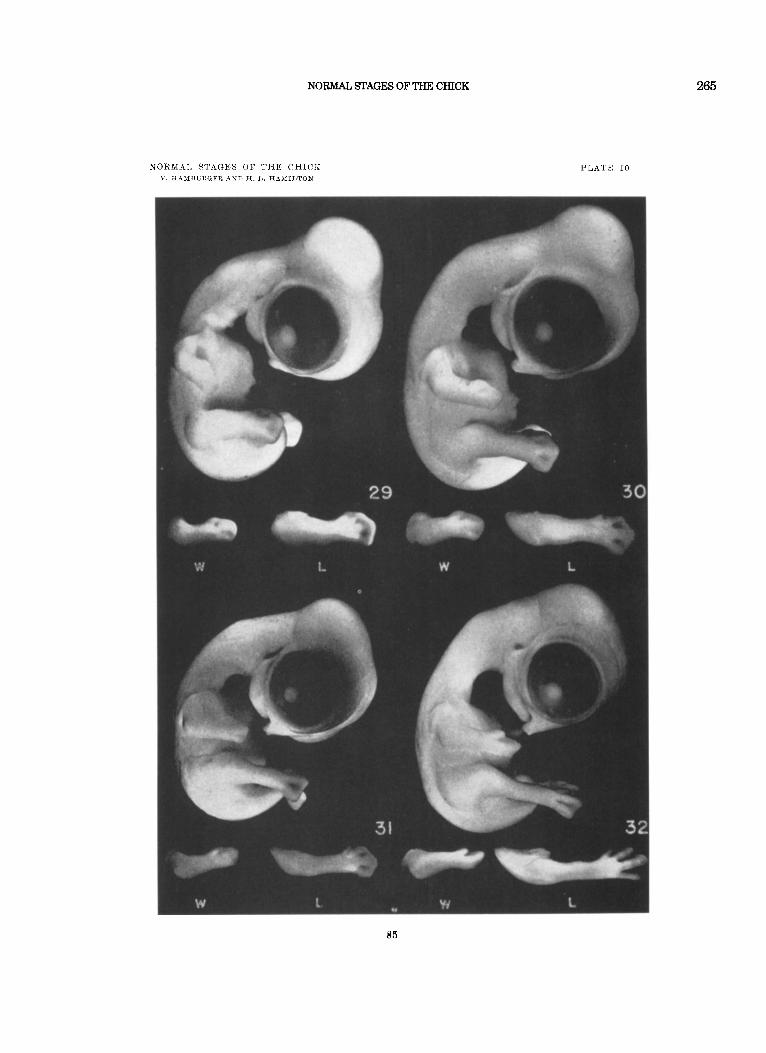
B e a k : A distinct outgrowth is visible in profile. 3. S t a g e 29. (aa. 6 days).
1. Limbs: Wing bent in elbow. Second digit distinctly longer than the others. Shallow grooves between first, second, and third digits. Second to 4th toes stand out a s ridges separatrd by distinct grooves, and u. ith iiidiclations of webs between them. Distal contours of w3bs are straight lines, occasionally with indication of convexity. Rudiment of 5th toe visible.
2. V i s c e r a l arches: Mandib~ilar process lengthened (compare with stage 28). Mandibular process and secoiid arch are broadly fused. Auditory ineatus distinct at dorsal end of fusion. All protuberances have flattened. Neck between “collar 7 7 and mandibular process has lengthened. “Collar” stands out conspicuously.
3. B e a k : More prominent than in stage 28. No egg-tooth visible as yet.
S t a g e 30. ( c a . 64 days). 1. L i m b s : The threc major segments of wing and leg are
clearly demarcated. Wing bent in elbow-joint. Leg bent in knee-joint. Distinct grooves between first and second digits. Contours of webs between first two digits and between all toes are slightly curved concave lines.
2. V i s c e r a l arches : The mandibular process approaches the beak, but the gap between the two is still conspicuous. TJengthening of neck between l 1 collar ’ ’ and mandible is very conspicuous. “ Collar’ begins to flatten.
F e a t h e r - g e r m s : Two dorsal rows to either side of the spinal cord at the brachial level. Three rows a t the level of the legs; they are rather indistinct at thoracic level. None on thigh.
4. Sc le ra l pap i l lae : One on either side of choroid fissure; soine- times indistinct but never more than two.
5. E g g - t o o t h distinct, slightly protruding. Beak more pro- noanced than iii previous stage.
3.
S t a g r 31. ( c a . ‘i days). 1. Limbs: Indication of a web between first and secoiid digits.
Rudinient of 5th toe still distinct.
NORMAL STAGES OF THE CHICK
NORMAL STAGES O F THE CHICK 63
2.
3.
Visceral arches: The gap between mandible and beak has narrowed t o a small notch. “Collar” inconspicuous or absent.
Peather-germs: On dorsal surface, continuous from brachial to lumbo-sacral level. Approximately 7 rows at limbo-sacral level. Distinct feather papillae on thigh. One indistinct row on each lateral edge of the tail.
Xcleral papillae: Usually 6 ; 4 on the dorsal side near the choroid fissure, and two on the opposite side.
4.
Stage 32. (ca . ‘it days). 1. Limbs: All digits and 4 toes have lengthened conspicuously.
Rudiment of 5th toe has disappeared. Webs between digits and toes are thin and their contours are concave. Differences in size of individual digits and toes become conspicuous.
Visceral arches: Anterior tip of mandible has reached the beak. “ Collar” has disappeared o r is faintly recognizable.
Peather-germs: Eleven rows o r more on dorsal surface a t level of the legs. One row on tail distinct, second row in- Idistinct. Scapular and flight feather-germs barely perceptible a t optimal illumination o r absent.
4. Scleral papi l lae: Six to 8, in two groups; one group on dorsal and one on ventral side. Circle not yet closed.
2.
3.
Stage 33. (ca. 74-8 days). 1.
2.
Limbs : Web on radial margin of arm and first digit becomes discernible. All digits and toes lengthened.
Visceral arches: Mandible and neck have lengthened con- spicuously. (Compare the ventral contour of boldy, from heart- region, along neck to tip of mandible, in this and the preceding stages.)
Peather-germs: Scapular and flight feather-germs not much advanced over stage 32. Tail: three rows distinct, the middle row considerably larger than the others.
4. Scleral papillae: Thirteen, forming an almost complete circle, with gap for one missing papilla a t a ventral point near the middle of the jaw.
3.
Stage 34. (ca . 8 (days). 1. Limbs : Differential growth of second digit and third toe
conspicuous. Contours of webs between digits and toes are concave and arched.
Visceral arches: Lengthening of mandible and of neck con- tinues (see previous stage).
Peather-germs: On scapula, on ventral side of neck, on pro- coracoid, and posterior (flight) edge of wing, feather-germs are visible under good illumination. Feather-germs next to
2.
3.
HAMBURGER AND HAMILTON
64 T. HA1\4BURGER AXD H. L. H A M I L T O N
dorsal midline, particularly a t limbo-sacral level, extend slightly over surface when viewed i n profile. Feather-germs on thigh protrude conspicuously. One roK on inner side of each eye. None around umbiIim.1 cord.
4 5 N i c t i t a t i n g waenbbrane extends halfway between outer rim
Sclera-1 pap i l lae : Thirteen o r 14.
of eye (eyelid) and scleral papillae. S t a g e 35. (ca. 8-9 days).
1. L i m b s : webs between digits and toes become inconspicuous. A transitory protuberance on the ulnar side of the second digit is probably a remnant of the web. Phalanges in toes are distinct.
2 V i s c e r a l arch~s: Lengthening of beak continues. Compare the distance between the eye and the t ip of the beak, in this and the preceding stages.
3 F e a t h e r - g e r m s : All are more conspicuous. Mid-dorsal line stands out distinctly in profile view. A t least 4 rows on inner side of each eye. New appearance of feather-germs near mid- ventral line, close to sternum, and extending to both sides of umbilical cord.
4 N i c t i t a t i n g m e m b r a n e has grown conspicuously and ap- proaches the outer scleral papillae. Eyelids (external to nic- titating membrane) have extended towards the beak and have began to overgrow the eye-ball. The circuniference of the eye- lids has become ellipsoidal.
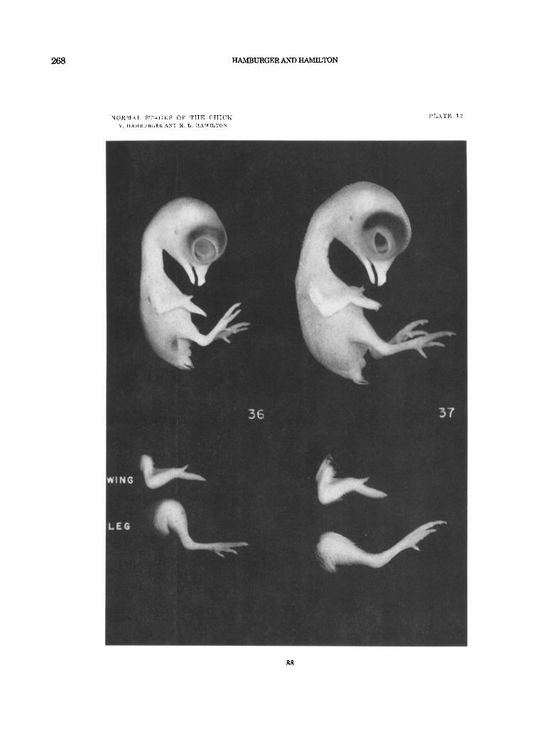
S t a g e 36. (cal. 10 days). 1. Limhs: Distal segments of both wing and leg are proportion-
ately much longer. Length of third toe, from its t ip to the middle of its metatarsal joint = 5.4 i- 0 3 mm. Tapering pri- mordia of claws are just visible on termini of the toes and on digit 1 of the wing. Protuberance on posterior side of digit 2 of wing is missing.
Viscera l a rches : Primordium of the comb appears as a promi- nent ridge with slightly serrated edge along the dorsal mid- line of the beak. A horizontal groove (the “labial groove”) is clearly visible a t the t ip of the upper jaw, hut is barely indicated on the t ip of the mandible. Nostril has narrowed to a slit. Length of beak from anterior aiigle of nostril to t ip of bill = 2.5 nun.
F e a t h e r - g e r m s : Flight-feathers are conspicuous ; coverts are jubt visible in web of wing. Feather-germs now cover the tibio- fihular portion of the leg. At least 9-10 rows of feather-germs between each upper eyelid and the dorsal midline. Sternal tracts prominent, with 3-4 rows on each side of ventral mid-
2
3
NORMAL STAGES OF THE CHICK
NORMAL STAGES O F THE C H I C K 65
247
line when counted i n anterior par t of sternum, merging into many rows around the umbilicus.
4. EyeZids: Nictitating membrane covers anteriormost scleral papillae and approaches cornea. Lower l id has grown upward to level of cornea. Circumference of lids is a narrowing ellipse with its ventral edge flattened.
Stage 37. (ca. 11 days). 1. Lzmbs: Claws of toes are flattened laterally and curved ven-
trally ; dorsal tips are opaque. indicating onset of cornification. Tip of claw on wing is also opaque. Pads on plantar surface of foot are conspicuous. Transverse ridges along the superior surfaoes of the metatarsus and phalanges are first indication of scales. Length of third toe = 7.4 & 0 3 mm.
Visceral arches: Labial groove on mandible is now clearly marked off. The comb is more prominent and clearly serrated. Length of beak from anterior angle of nostril to t ip of bill = 3.0 mm.
Feather-gei-ms: Much more numerous, and in most-advanced tracts (e.g., along back and on tail) elongated into long, much-tapered cones. External auditory meatus is nearly sur- rounded by feather-germs. Circumference of eyelids is bor- dered by a single row of just-visible primordia; none on remainder of lids. Sternal tracts contain 5-6 prominent rows when counted a t anterior end of sternum.
4. Eyel ids: Nictitating inembrane has reached anterior edge of cornea. Upper lid has reached dorsal edge of cornea. Lower lid has covered one-third to one-half of cornea. Circumference of lids now bounds a much-narrowed and ventrally-flattened biconvex area
X f a g e 38. (ca. 12 days).
2.
3.
1. 1,zwzbs: Primordia of scales are marked off over entire sur- face of leg; ridges have not 5-et grown out to overlap surface. Tips of toes show a ventral center of cornification as well as the more extensive dorsal one. Main plantar pad is ridged when seen in profile. Length of third toe = 8.4 i 0.3 mm.
Visceral arches: Labial groove marlred off by a deep furrow a t the end of each jaw. Length of beak from anterior angle of nostril to t ip of bill = 3.1 mm.
Feather-gernzs: Coverts of web of wing are beconiing coni- cal. External auditory meatus is surrounded by feather-germs. Sternum is covered with feather-germs except along midline. IJpper eyelid is covered with newlyformed feather-germs ; lower lid is naked except for 2-3 rows a t its edge.
2 .
3.
248 HAMBURGER AND HAMILTON
66 V. HAMBURGER AND H. L. IIAMILTON
4. Eyelids: Lower lid covers two-thipds to three-fourths of cornea. Opening between lids is much reduced.
Stage 39. (ca. 13 days). 1. Limbs: Scales overlapping on superior surface of leg. Major
pads of phalanges corered with papillae ; minor pads are smooth. Length of third toe = 9.8 f 0 3 mm.
2. Visceral a w h e s : Mandible and maxilla cornified (opaque) back as fa r as level of proximal edge of “egg-tooth.” The channel of the auditory meatus can be seen only a t the PO<- terior edge of its shallow external opening. Length of beak from anterior angle of nostril to t ip of bill = 3.5 mm.
Feather-gernas: Coverts of web of wing are verv long taper- ing cones. Note great increase in length of feather-germs in major tracts. Four to 5 rows of feather-germs at edge of lower eyelid.
4. Eyelids: Opening between lids reduced to a thin crescent. Stages 40 to 44 are based mainly on the length of the beak and on
the length of the third (longest) toe, since other external features have lost their diagnostic value. Of these two criteria, the length of the beak is the better, because i t is more easily and accurately meas- ured (with calipers) and shows less variability. Stage 40. (ca. 14 days).
3.
1. Visceral arches: Length of beak from anterior edge of nos- tril to t ip of bill = 4.0 mm. The main channel of the auditory ineatixs is not visible in strictly Iateral view of its external chamber.
Liwalis: Length of third toe = 12.7 t 0.5 mm. Scales over- lapping on inferior as well as superior snrfaces of leg. Dorsal and ventral loci of cornification extend to base of exposed portion of toe-nail. Entire plantar surface of phalanges is covered with well-developed papillae.
2 .
Stage 41. (ca. 15 days). 1.
2.
1.
2.
1.
Beak: Length from anterior angle of nostril to tip of upper
Thbd t o e : Length = 14.9 z 0.8 mm.
Beak: Length from anterior angle of nostril to t ip of upper
Third toe: Length = 16.7 k 0 . 8 mm.
Beak: Length from anterior angle of nostril to tip of upper bill = 5.0 mm. “Labial grooves” are reduced t o a white graii- ular crust a t the edge of each jaw; that of the lower jaw may be partially o r completely sloughed off.
bill = 4.5 mm.
Stage 42. ( c a . 16 days).
bill == 4.8 mm.
Stage 43. (ca. 17 days).
NORMAL STAGES OF THE CHICK 249
NORMAL STAGES O F THZ C H I C K 67
2.
1.
Thi id toe: Length =18 6 I. 0.8 mm.
Beak: Length from anterior angle of nostril to tip of upper bill = 5.7 mm. The translucent peridermal covering of the beak is starting to peel off proximally.
2. Third toe: Length = 20 4 t 0.8 mm.
1.
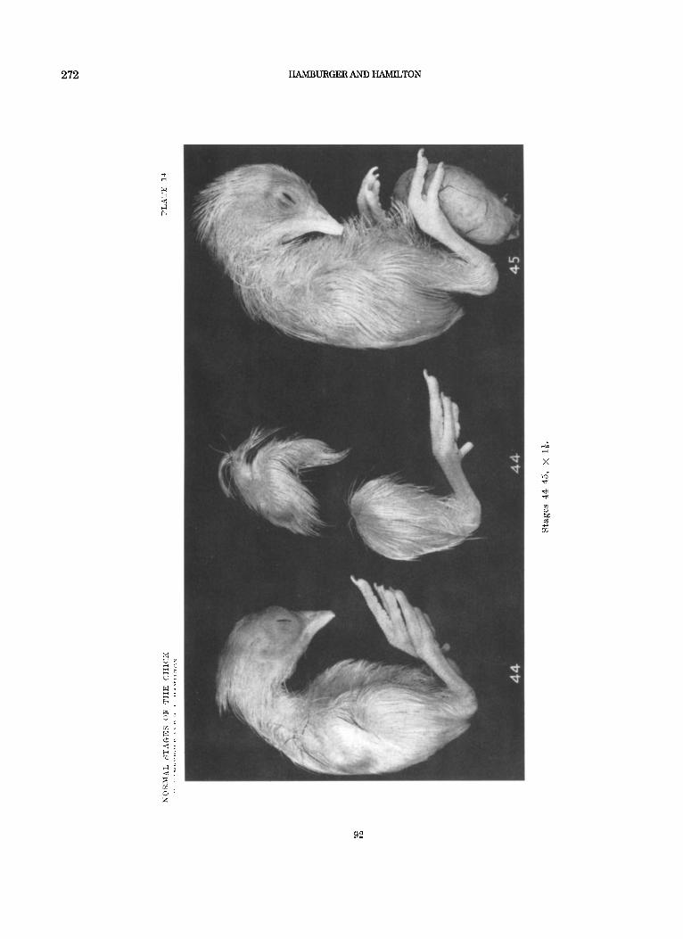
Stage 44. (ca . 18 days),
Btage 45. (ca. 19-20 days). Beak: Length is no longer diagnostic; in fact, the beak is
usually shorter than in stage 44, due to a loss (by sloughing off) of its entire peridermal covering. As a consequence, the beak is now shiny all over and more blunt a t its tip. Both labial grooves have disappeared with the periderm.
2. Third toe: Average length is essentially unchanged from that of stage 44, except in those breeds with a longer period of incubation (21 days) and a heavier build of body. For these latter, length of third toe = ca. 21.4 i 0.8 mm.
3. Extra-embryonic membranes: Yolk-sac is half-enclosed in body-cavity. Chorio-allantoic membrane contains less blood and is “sticky” in the living embryo.
Stage 46. Newly-hatched chick (20-21 days).
REFERENCES
DUVAL, MATHIAS 1889 Atlas d’Embryologie. (116 pp., 40 plates). Paris. HAMBURGER, VIKTOR 1938 Morphogenetic and axial self-differentiation of
transplanted limb primordia of 2-day chick embryos. J. Exp. Zool., 7 7 : 379-399.
1942 A Manual of Experiinental Embryology. 213 pp. University of Chicago Press.
IIELBEL, F., AND K. ABRAHAM 1900 Normentaf el zur Entwicklungsgeschiclite des Huhnes (Gallus domesticus). 132 pp. Jena.
SPRATT, NELSON T., JR. Location of organ-specific regions and their re- lationship to the development of the primitive streak in the early chick blastoderm. J. Exp. Zool., 89: 69-101.
_~ 1946 Formcation of the primitive streak in the explanted chick blastoderm marked with carbon particles.. J. Exp. Zool., 103: 259- 304.
SAUNDERS, JOHN W., JR. 1948 The proximo-distal sequence of origin of the parts of the chick wing and the role of the ectoderm. J. Exp. Zool., 108: 363403.
1942
EXPLANATION O F PLATES
All numbers in the following plates refer to the corresponding stage numbers in the text. The description of each stage should be consulted for a more complete explanation of the figures.
PLATE 1
EXPLANATION O F FIGURES
Stages 1-3+, illustrated by photographs provided by Dr. Nelson T. Spratt, J r . (Stages 1 and 2 are published in J. Exp. Zool., 103: 265 and 274.) X 20.
68
NORMAL STAGES OF THE CHICK
NOItMAL S T A G E S O F TJIE C H l C K V. HAMBURGER AND 1%. L. H A A I I L T O N
PLATE 1
251
252 HAMBURGER AND HAMILTON
PLATE 2
IIXPLANATION O F FJCr1JRF.S
S tages 4-9! x 20. Stage 10, x 1%. (Stages -L> 5, a i i d 8- were pliotographed froin slides provided by Dr. Nelson 7'. Rpratt,, .Ti,. A l l otlievs n r e froill the Iowa State College collec.tion.)
N

254 HAMBURGER AND HAMILTON
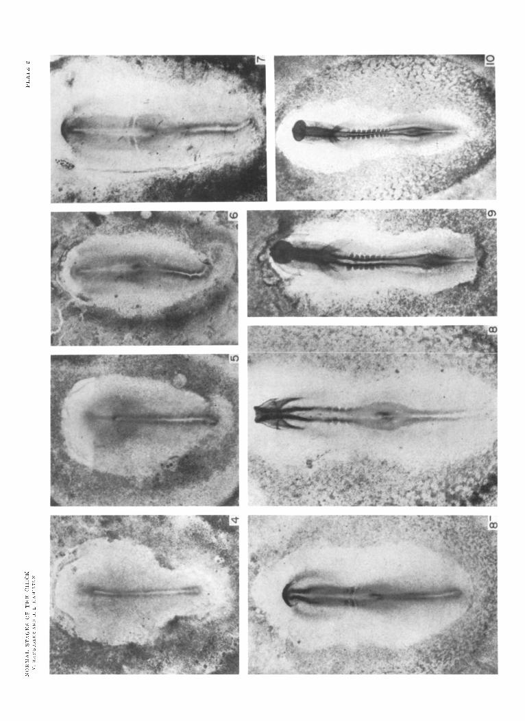
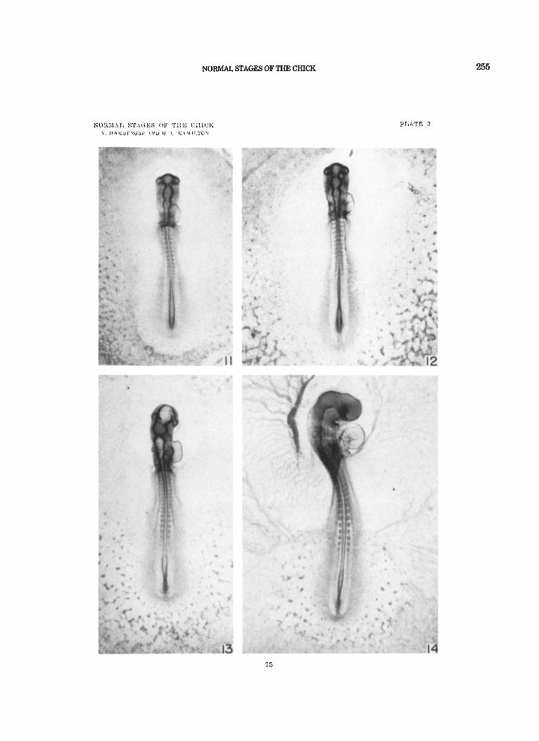
I’l,i\TK 3
ESPLAXATIOX O F FIGDRliS
Stagcs 11-14, X I!!.
7 4
NORMAL STAGES OF THE CHICK 255
PLATE 3
75

256 HAMBURGER AND HAMILTON
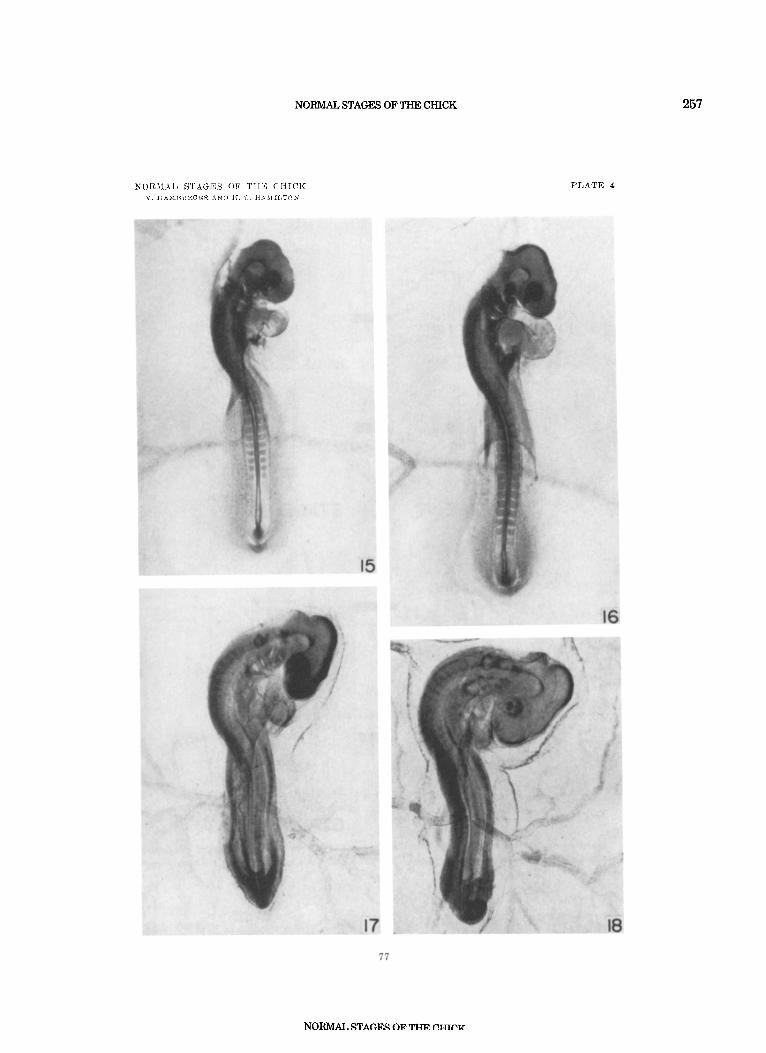
PLATE 4
EXPLANAlION O F FIGURES
Stages 15-18, x 12. Contours of limbs for stages 1 7 and 18 are shown in the drawings on plate 5.
76
NORMAL STAGES OF THE CHICK 257
PLATE 4
258
;) . ..:. 1 i: ..
...... ..... ,<:, . .- .:;.
HAMBURGER AND HAMILTON
j .... ...> . . .
... . .i : . : :. .... .;;. . , ,, .,
.;. ... 2,.
WINGS
STAGE 17 .::) . ..','. ...... . , . .,. . . I ,.. .
..... . .:: .:>'. .. :.., . . . . .....
. . . . . . ... . . . . . :..
:;::i:l: .....:I. .... :,. .... ...... . I . '
..... .....
....... ') t:.:. . . . I . / . ...... . ..... : I::
. ,.::*:
. .: jj.; . ;.::$p
. .._. ..:-. ........ .. .:i .... : >.
.. .!..?<* ,..cx
.;:;?. ..(' ..I.. ...
STAGE 19
LEGS
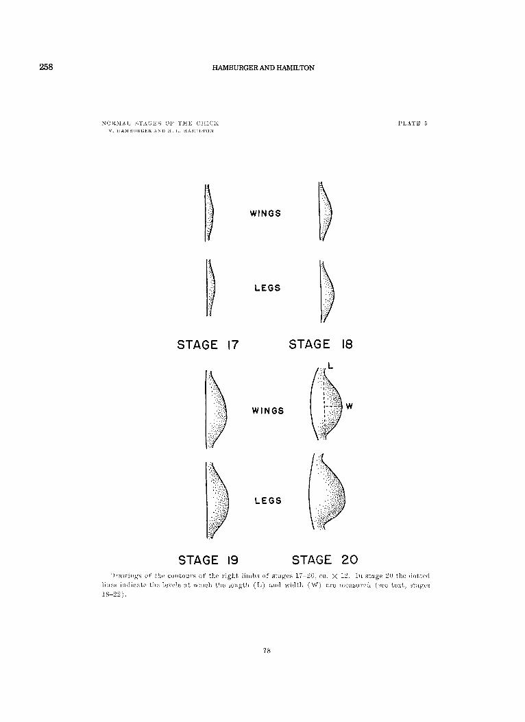
P L A T E 5
STAGE 18
LEGS
STAGE 20 1)ixwiiigs of the con tour s of the ~ i g l i t liiiihs of stages 17-2U, ca. x 12. In stage 2U tlii: ilottctl
lilies iiltlicatc: thc lerels at which the Icngtlt ( I d ) :m(1 width ('A') art! iiie:isui.c:d ( see text; st:isw 18-32).
78
NORMAL STAGES OF THE CHICK
NORRIAL STAGES OF TI-IE CIIICli \,. H A X U L R G I R 4XD H. L. I I \ l I I , T O N
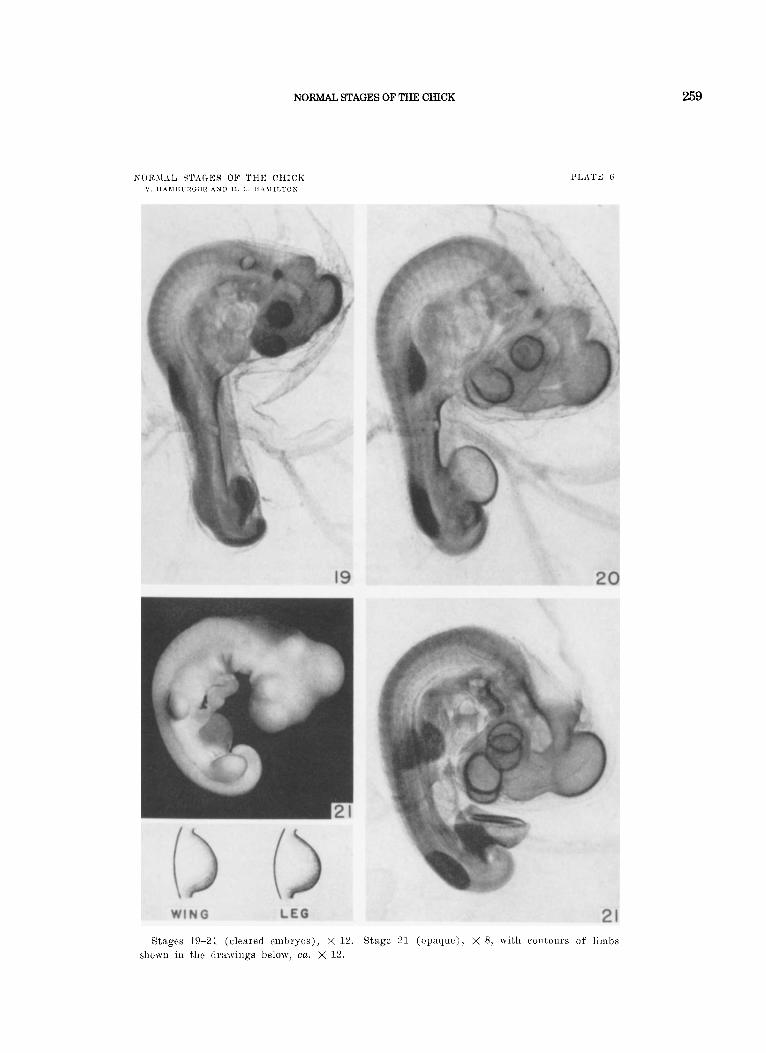
Stages 19-21 (clenied embiyos), X 12. Stage 21 ( o l x t q ~ t e ) ) , X 8, nit11 contours of linibs slio\vn in the clruwiilys below, ca. X 12.
259
260 HAMBURGER AND HAMILTON
I’1,AT‘d i
E M ’ T A N AT1 0 f; 0 L” PIG I’ KE S
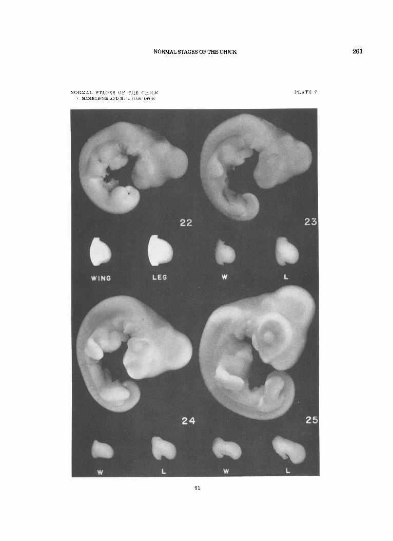
Stages 22-25, x S. Tl ic limbs f o r stage 2 2 are tlr:lwi1igs, cn. x 1 2 ; all others are pliotograplis, x 8. E’or details of visceunl arclics of stages 23-25, see plate 8.
80
NORMAL STAGES OF THE CHICK 261
PLATE 7
81
262 HAMBURGER AND HAMILTON
1'1-ATE 8
Drawings of the region of the visceral arclies, iiiade from camera lueida tracings. S t a g e s 23-25, x 7. Stages 26-28, X 4.2. I-IV = visceral arches; mx., md. = maxillary and manrlibu- lar processes of visceral arch I; 4 = 4th visceral cleft. see text for explallation of letters a - f .
82
NORMAL STAGES OF THE CHICK
NORMAL STAGES O F THE C H I C K V. HAMBURGER AND H. L. H A M I L T O N
Stage 26, embryo and limbs, X 8. Stages 27-28, x 5. 83
263
P L A T E 9

264 HAMBURGER AND HAMILTON
PLATE 1 0
EXPLANATION O F FIGURES
Stages 29-30, X 5. Stages 31-32, x 4.
84
NORMAL STAGES OF THE CHICK
NOIZMA4L STAGES OF. TIIE C H I C K V. H I M B L R G E R A X D H. L. H A H I L T O N
265
PLATE 10
85

266 HAMBURGER AND HAMILTON

NORMAL STAGES OF THE CHICK 267
87
268 HAMBUFtGERAND HAMILTON
I’TATI? 1 2
NORMAL STAGES OF THE CHICK
NORMAL STBGES O F THE C I I I C K V . IXAMBURUBR .4XD H. L. IIAALILTON
269
PLATE 12
Rtnges 36-39, X 2. A9

270
NORMAI, STAGES O F THE ( ' I I ICK V. H A M H ~ R G L R AND H I H ~ M I I I T O N
HAMBURGER AND HAMILTON
PLATE 13
00
NORMAL STAGES OF THE CHICK
NORMAL STAGES O F THE C H I C K V. HAMBURGEX AND 71, L. HAMIMTON
PLATE 13
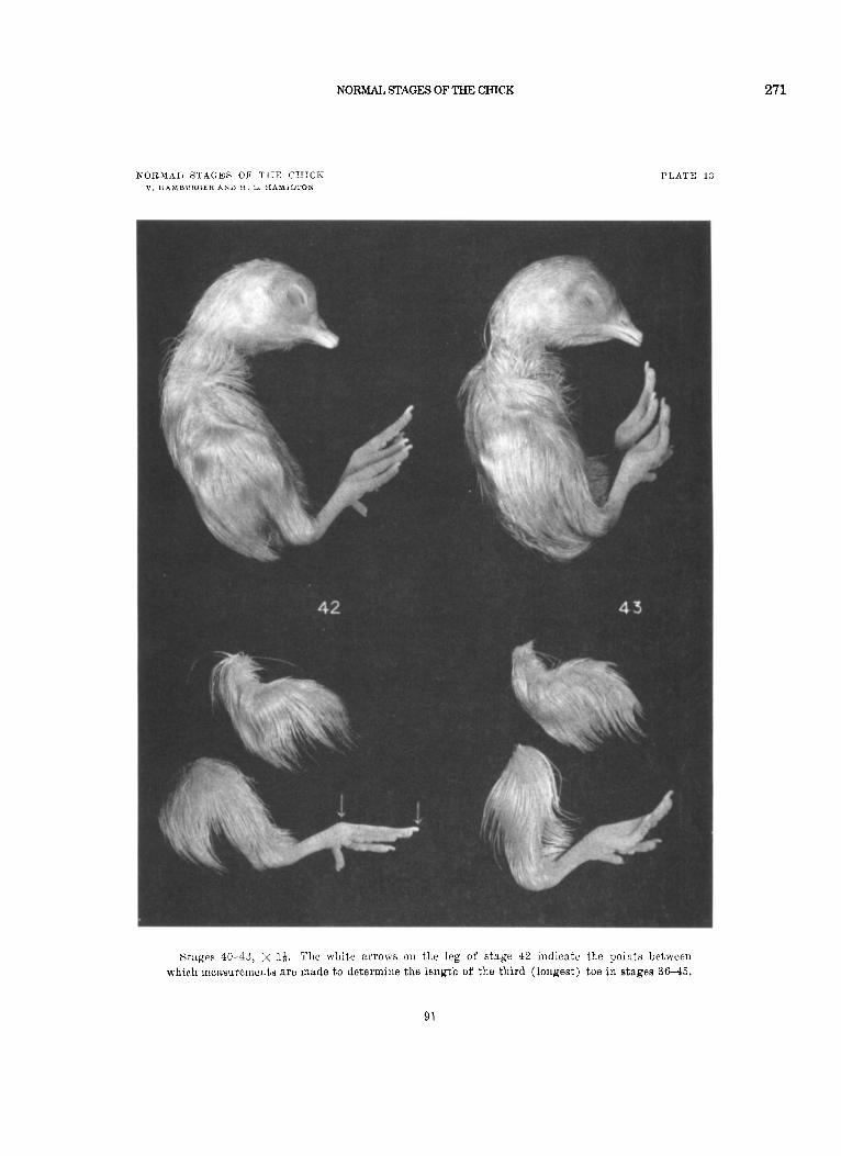
Stages 40-43, x I$. The white arrows on the leg of stage 42 indicate the points between which mensurenients a re made to determine the leiigth of the third (longest) toe in stages 36-45.
271
91
272 HAMBURGER AND HAMILTON
92