Embed Size (px)
Citation preview

J. Am. Chem. SOC. 1995, 117, 5179-5197 5 179
A Second Generation Force Field for the Simulation of Proteins, Nucleic Acids, and Organic Molecules
Wendy D. Cornell? Piotr Cieplak,’ Christopher I. Bayly,s Ian R. Gould,l Kenneth M. Merz, Jr.,” David M. Ferguson,& David C. Spellmeyer: Thomas Fox, James W. Caldwell, and Peter A. Kollman*
Contribution from the Department of Pharmaceutical Chemistry, University of California, San Francisco, California 94143
Received November IO, 1994@
Abstract: We present the derivation of a new molecular mechanical force field for simulating the structures, conformational energies, and interaction energies of proteins, nucleic acids, and many related organic molecules in condensed phases. This effective two-body force field is the successor to the Weiner et al. force field and was developed with some of the same philosophies, such as the use of a simple diagonal potential function and electrostatic potential fit atom centered charges. The need for a 10-12 function for representing hydrogen bonds is no longer necessary due to the improved performance of the new charge model and new van der Waals parameters. These new charges are determined using a 6-31G* basis set and restrained electrostatic potential (RESP) fitting and have been shown to reproduce interaction energies, free energies of solvation, and conformational energies of simple small molecules to a good degree of accuracy. Furthermore, the new RESP charges exhibit less variability as a function of the molecular conformation used in the charge determination. The new van der Waals parameters have been derived from liquid simulations and include hydrogen parameters which take into account the effects of any geminal electronegative atoms. The bonded parameters developed by Weiner et al. were modified as necessary to reproduce experimental vibrational frequencies and structures. Most of the simple dihedral parameters have been retained from Weiner et al., but a complex set of 4 and yj parameters which do a good job of reproducing the energies of the low-energy conformations of glycyl and alanyl dipeptides has been developed for the peptide backbone.
Introduction The application of computer-based models using analytical
potential energy functions within the framework of classical mechanics has proven to be an increasingly powerful tool for studying molecules of biochemical and organic chemical interest. These applications of molecular mechanics have employed energy minimization, molecular dynamics, and Monte Carlo methods to move on the analytical potential energy surfaces. Such methods have been used to study a wide variety of phenomena, including intrinsic strain of organic molecules, structure and dynamics of simple and complex liquids, ther- modynamics of ligand binding to proteins, and conformational transitions in nucleic acids. In principle, they are capable of giving insight into the entire spectrum of non-covalent interac- tions between molecules, and, when combined with quantum mechanical electronic structure calculations, modeling covalent bonding changes, essentially all molecular reactions and interac- tions. Given their importance, much effort has gone into consideration of both the functional form and the parameters that must be established in order to apply such analytical potential energy functions (or “force fields”).
t Graduate Group in Biophysics, University of California, San Francisco. * Permanent address: Department of Chemistry, University of Warsaw,
8 Current address: Merck Frosst Canada, Inc., C.P. 1005 Pointe Claire-
Current address: Deuartment of Chemism. Universitv of Manchester.
Pasteura 1, 02-093, Warsaw, Poland.
Domal, Quebec H9R 4P8, Canada.
Lancs M13 9PL, U.K. Current address: Department of Chemistry, The Pennsylvania State
University, State College; PA 16802.
Minnesota, Minneapolis, MN 55455. Bi Current address: Department of Medicinal Chemistry, University of
# Current address: Chiron Corporation, Emeryville, CA 94608. *Author to whom correspondence and reprint requests should be
@ Abstract published in Advance ACS Abstracts, April 15, 1995. addressed.
In the area of organic molecules, the book by Allinger and Burkert’ provides a thorough review pre-1982 and the subse- quent further development of the MM2* and MM33 force fields by Allinger and co-workers has dominated the landscape in this area. The number of force fields developed for application to biologically interesting molecules is considerably greater, prob- ably because of the greater complexity of the interactions which involve ionic and polar groups in aqueous solution and the difficulty of finding an unequivocal test set to evaluate such force fields. Many of these force fields developed prior to 1987 are described briefly by McCammon and Harvey.4
Given the complexities and subjective decisions inherent in such biological force fields, we have attempted to put the development of the force field parameters on a more explicitly stated algorithmic basis than done previously, so that the force field could be extended by ourselves and others to molecules and functional groups not considered in the initial development. This is important, because, if the assumptions, approximations, and inevitable imperfections in a force field are at least known, one can strive for some cancellation of errors.
Approximately a decade ago, Weiner et al.596 developed a force field for proteins and nucleic acids which has been widely
(1) Burke& U.; Allinger, N. J. Molecular Mechanics; American Chemical Society: Washington, DC, 1982.
(2)Allinger, N. L. J. Am. Chem. SOC. 1977, 99, 8127-8134 and subsequent versions, e.g. MM2-87, MM2-89, MM2-91.
(3) Allinger, N. L.; Yuh, Y. H.; Lii, J.-H. J. Am. Chem. SOC. 1989,ll I ,
(4) McCammon, J. A.; Harvey, S . C. Dynamics of Proteins and Nucleic Acids; Cambridge University Press: Cambridge, 1987.
( 5 ) Weiner, S. J.; Kollman, P. A,; Case, D. A,; Singh, U. C.; Ghio, C.; Alagona, G.; Profeta, S., Jr.; Weiner, P. J. Am. Chem. SOC. 1984,106, 765- 784.
(6) Weiner, S. J.; Kollman, P. A.; Nguyen, D. T.; Case, D. A. J. Comp. Chem. 1986, 7, 230-252.
8551 -8566, 8566-8576, 8576-8582.
0002-7863/95/1517-5179$09.00/0 0 1995 American Chemical Society
Dow
nloa
ded
by U
NIV
ER
SIT
AT
WIE
N B
IBL
IOT
HE
KS
on J
uly
15, 2
009
Publ
ishe
d on
May
1, 2
002
on h
ttp://
pubs
.acs
.org
| do
i: 10
.102
1/ja
0012
4a00
2

5180 J. Am. Chem. SOC., Vol. 117, No. 19, 1995 Comell et al.
used. Important independent tests of this force field were performed by Pavitt and Hall for peptides’ and Nilsson and Karplus8 for nucleic acids and it was found to be quite effective. Nonetheless, it was developed in the era before one could routinely study complex molecules in explicit solvent. Weiner et al. attempted to deal with this issue by showing that the same force field parameters could be effectively used both without explicit solvent (using a distance-dependent dielectric constant ( E = Ru)) and with explicit solvent ( E = 1) on model systems. Further support for this approach was provided by molecular dynamics simulations of proteins9-” and DNAl2.l3 which compared the implicit and explicit solvent representations.
As computer power has grown, it has become possible to carry out more realistic simulations which employ explicit solvent representations. It is therefore appropriate that any new force field for biomolecules focus on systems modeled in the presence of an explicit solvent representation. This approach has been pioneered by Jorgensen and co-workers in their OPLS (Optimized Potentials for Liquid Simulations) m0de1.I~ In particular, the development of parameters which reproduce the enthalpy and density of neat organic liquids as an essential element ensures the appropriate condensed phase behavior. The OPLS non-bonded parameters have been combined with the Weiner et al. bond, angle, and dihedral parameters to create the OPLS/Amber force field for peptides and proteins,I5 which has also been effectively used in many systems.I6
We have been influenced by the OPLS philosophy of balanced solvent-solvent and solute-solvent interactions in our thoughts about a second-generation force field to follow that of Weiner et aL5v6 The Weiner et al. force field used quantum mechanical calculations to derive electrostatic potential (ESP) fit atomic centered charges, whereas the OPLS charges were derived empirically, using mainly the liquid properties as a guide. For computational expediency, Weiner et al. relied principally on the STO-3G basis set for their charge derivation. This basis set leads to dipole moments that are approximately equal to or smaller than the gas-phase moment but tends to underestimate quadrupole moments. Thus, it is not well balanced with the commonly used water models (SPC/E,” TIP3P,I8 TIP4PI8) which have dipole moments that are about 20% higher than the gas-phase value for water. These water models, which have empirically derived charges, include condensed-phase electronic polarization implicitly. Kuyper et aZ.l9 suggested that the logical choice of a basis set for deriving ESP-fit partial charges for use in condensed phases is the 6-3 lG* basis set, which uniformly overestimates molecular polarity. Standard ESP charges derived with that basis set were shown
~
(7) Pavitt, N.; Hall, D. J. Compur. Chem. 1984, 5, 441-450 (8) Nilsson, L.; Karplus, M. J . Comput. Chem. 1986, 7, 591-616. (9) Tilton, R. F.; Singh, U. C.; Weiner, S. J.; Connolly, M. L.; Kuntz, I.
D., Jr.; Kollman, P. A.; Max, N.; Case, D. J. Mol. Eiol. 1986, 192, 443- 456.
(10) Guenot, J. M.; Kollman, P. A. Prorein Sci. 1992, 1, 1185-1205. ( 1 1) York, D. M.; Wlodawer, A.; Redersen, L.; Darden, T. A. Proc. Narl.
(12) Sinsh. U. C.: Weiner. S. J.: Kollman. P. A. Proc. Natl. Acad. Sci. Acad. Sci. U.S.A. 1994, 91, 8715-8718.
U.S.A’l983, 82, 755-759. ’
(13) Seibel, G. L.; Singh, U. C.; Kollman, P. A. Proc. Natl. Acad. Sci. U.S.A. 1985, 82, 6537-6340.
(14) Jorgensen, W. L.; Pranata, J. J . Am. Chem. SOC. 1990, 112,2008- 2010.
(15) Jorgensen, W. L.; Tirado-Rives, J. J. Am. Chem. SOC. 1988, 110, 1657 - 1666.
(16) (a) Tirado-Rives, J.; Jorgensen, W. L. J . Am. Chem. SOC. 1990,112, 2773-2781. (b) Orozco, M.; Tirado-Rives, J.; Jorgensen, W. L. Eiochem- istry 1993, 32, 12864-12874.
(17) Berendsen, H. J. C.; Grigera, J . R.; Straatsma, T. P. J . Phys. Chem. 1987, 91, 6269-6271.
(18) Jorgensen, W. L.; Chandreskhar, J.; Madura, J. D.; Impey, R. W.; Klein, M. L. J. Chem. Phys. 1982, 79, 926-935.
(19) Kuyper, L.; Ashton, D.; Men, K. M., Jr.; Kollman, P. A. J. Phys. Chem. 1991, 95, 6661-6666.
to lead to excellent relative free energies of solvation for benzene, anisole, and trimethoxyani~ole.’~
A 6-3 1G* based ESP-fit charge model, like the OPLS model, is capable of giving an excellent reproduction of condensed- phase inter molecular properties such as liquid enthalpies and densities and free energies of solvation.20 A major difference between such a model and most others is the magnitude of the charges on hydrocarbons. For example, 6-3 lG* standard ESP charges derived from the trans conformation of butane have values of -0.344 for the methyl carbon and 0.078 for the methyl hydrogen. In both cases, however, the carbon and hydrogen charges offset each other, resulting in small net charges on the methyl groups of -0.1 10 and -0.059 for the trans and gauche charges, respectively. Furthermore, free energy perturbation calculations involving the perturbation of methane with standard ESP charges (qc = -0.464 and q H = 0.116) to methane with charges of 0.0 in solution yield essentially no change in free energy.21 The standard ESP charges also result in conforma- tional energies for butane which are in reasonable agreement with experiment, when used with a 1-4 electrostatic scale factor of m.2.20
Nevertheless, the 6-3 lG* standard ESP charges are less than ideal for two reasons. First, when charges generated using different conformations of a molecule are compared, there is often considerable variation seen. This was demonstrated by Williams, who studied the conformational variation of ESP-fit charges in alanyl dipeptide for 12 different conformations.22 Butane is another example, where charges from the gauche conformation have values of -0,197 and 0.046 for the methyl carbon and hydrogen, respectively. Another example is pro- pylamine, which was studied at length by Comell et aL2O Five low-energy conformations can be identified for propylamine, and the 6-31G* standard ESP charges calculated for each conformation show significant variation. The average and standard deviation for the charge on a given atom over the five conformations are as follows: a-carbon qav = 0.339 and IJ = 0.059, /3-carbon qav = 0.033 and u = 0.060, and y-carbon qav = -0.205 and u = 0.146. This inconsistency is potentially problematic in terms of deriving other force field parameters which may be sensitive to the variation. Furthermore, it reduces the reproducibility of a particular calculation, which is not a problem in other force fields where the charges are assigned empirically.
The second reason that the 6-3 lG* standard ESP charges are less than ideal is that the charges on “buried” atoms (such as the sp3 carbons described above for butane and propylamine) are statistically underdetermined and often assume unexpectedly large values for nonpolar atoms. Bayly et aLZ3 found that the electrostatic potential of methanol could be fit almost equally well using either the standard ESP charges determined by the linear least-squares fit or an altemative set of charges derived with the methyl carbon constrained to have a much smaller value.
Considering the problems associated with the standard ESP charge model, it might seem tempting to adopt the OPLS approach of empirically derived charges. However, any empiri- cally derived charge model cannot easily describe transition states and excited states, as can an electrostatic potential fit
(20) Cornell, W.; Cieplak, P.; Bayly, C.; Kollman, P. A. J. Am. Chem. SOC. 1993, 115, 9620-9631.
(21) (a) Sun, Y. X.; Spellmeyer, D.; Pearlman, D. A.; Kollman, P. A. J. Am. Chem. SOC. 1992, 114, 6798-6801. (b) Sun, Y. X.; Kollman, P. A. Hydrophobic Solvation of Methane and Nonbond Parameters of the TIP3P Water Model. J. Cornput. Chem., in press. Pang, Y. P.; Kollman, P. A., unpublished.
(22) Williams, D. E. Biopolymers 1990, 29, 1367-1386. (23) Bayly, C.; Cieplak, P.; Comell, W.; Kollman, P. A. J. Phys. Chem.
1993, 97, 10269-10280.
Dow
nloa
ded
by U
NIV
ER
SIT
AT
WIE
N B
IBL
IOT
HE
KS
on J
uly
15, 2
009
Publ
ishe
d on
May
1, 2
002
on h
ttp://
pubs
.acs
.org
| do
i: 10
.102
1/ja
0012
4a00
2

Simulation of Proteins and Nucleic Acids
model. Furthermore, the conformational dependence of N- methylacetamide ( M A ) is better represented with an ESP-fit
Finally, the requirement of Monte Carlo calculations on requisite liquids including appropriate fragments makes it more problematic to make an empirical charge model that will cover most or all of chemicalhiochemical functionality.
Given the above-mentioned deficiencies in the standard ESP model, along with the desire to retain the general strategy of fitting charges to the electrostatic potential, Bayly et were motivated to develop the RESP (restrained ESP-fit) charge model. The RESP model still involves a least-squares fit of the charges to the electrostatic potential, but with the addition of hyperbolic restraints on charges on non-hydrogen atoms. These restraints serve to reduce the charges on atoms which can be reduced without impacting the fit, such as buried carbons. The final RESP model requires a two-stage fit, with the second stage needed to fit methyl groups which require equivalent charges on hydrogen atoms which are not equivalent by molecular symmetry. The new charge model has been shown to perform well at reproducing interaction energies and free energies of solvation. When used with a 1-4 electrostatic scale factor of U1.2 (as opposed to the scale factor of 1/2 employed by Weiner et al.), both the RESP (and standard ESP) charges also result in good conformational energies for many of the small molecules studied to date without the necessity for an elaborate dihedral potential.20
In addition to the new charges which have been tailored for condensed phase simulations, new van der Waals (VDW) parameters have also been adopted and developed which are optimized for reproducing liquid properties. The VDW param- eters in the Weiner et al.5.6 force field are primarily a modification of a set originally proposed by Hagler-Euler- Lifs01-1,~~ which were fit to lattice energies and crystal structures of amides. The new VDW parameters for aliphatic and aromatic hydrogens take into account the effects of any vicinal elec- tronegative atom^.^^.^^
High-level quantum mechanical data are now available on the conformational energies of the glycyl and alanyl dipeptides28 and these data are critical for developing 4 and q dihedral parameters for the peptide backbone. Because such high-level data were unavailable at the time the Weiner et al. force field was developed, torsional parameters for the 4 and q angles were left as 0.0 kcdmol since the resulting molecular mechanical energies seemed to be in reasonable agreement with the best theoretical data available at that time. That force field led to conformational energies for glycyl dipeptide where the C5 extended conformation was about 1 kcdmol too high in energy and for alanyl dipeptide where the C5 conformation was nearly 2 kcal/mol too high in energy but the C7ax conformation was about 1 kcal/mol too low in energy. The error in the alanyl dipeptide C7,, energy is not critical since it is rarely found in proteins29 (only in y-turns), but the errors in the energies of the C5 conformations are more important since that is the confor- mation found in P-sheets. Any errors in the energies of the C5 conformations are multiplied by the length of the secondary structure. The new force field includes VI, V2, V3, and V4
J. Am. Chem. SOC., Vol. 117, No. 19, 1995 5181
dihedral parameters for 4 and q which result in good agreement between the molecular mechanical and quantum mechanical energies of the dipeptides.
Finally, the benzene molecule as modeled by the Weiner et al. all-atom force field has been shown to possess excessive flexibility for out-of-plane distortion^.^^ This was caused by the use of the V2 potential derived for the united atom model. This underestimate of the benzene V2 parameter is noteworthy, because it affects not only the flexibility of benzene and benzene-like moieties but also the interpolation scheme used for determining the VZ barriers for X-C-N-X and X-C- C-X dihedrals in conjugated rings. These V2 parameters are determined by interpolating according to the bond length either between a pure single bond and a partial double bond (benzene) or between a partial double bond and a pure double bond. The excessive out-of-plane motion of benzene has been easily fixed by adjusting the V2 parameter from 5.5 to 14.5 kcaVmol to match the experimental normal mode frequencies.
(24) Cieplak, P.; Kollman, P. A. J. Comput. Chem. 1991, 12, 1232- 1236.
(25) Hagler, A,; Euler, E.; Lifson, S. J. Am. Chem. Soc. 1974,96,5319- 5327.
(26) Gough, C.; DeBolt, S.; Kollman, P. A. J. Compur. Chem. 1992, 13, 963-970.
(27) Veenstra, D.; Ferguson, D.; Kollman, P. A. J. Compur. Chem. 1992, 13, 97 1-978.
(28) (a) Gould, I. R.; Kollman, P. A. J. Phys. Chem. 1992, 96, 9255- 9258. (b) Gould, I. R.; Comell, W. D.; Hillier, I. H. J. Am. Chem. Soc. 1994, 116, 9250-9256.
(29) Creighton, T. E. Proreins, 2nd. ed.; W. H. Freeman: New York, 1984.
General Description of the Model
The model presented here (eq 1) can be described as “minimalist” in its functional form, with the bond and angles represented by a simple diagonal harmonic expression, the VDW interaction represented by a 6- 12 potential, electrostatic interac- tions modeled by a Coulombic interaction of atom-centered point charges, and dihedral energies represented (in most cases) with a simple set of parameters, often only specified by the two central atoms. Electrostatic and van der Waals interactions are only calculated between atoms in different molecules or for atoms in the same molecule separated by at least three bonds. Those non-bonded interactions separated by exactly three bonds (“1-4 interactions”) are reduced by the application of a scale factor.
bonds angles
Our assumption is that such a simple representation of bond and angle energies is adequate for modeling most unstrained systems. The goal of this force field is to accurately model conformational energies and intermolecular interactions involv- ing proteins, nucleic acids, and other molecules with related functional groups which are of interest in organic and biological chemistry.
A. Atom Types. The atom types employed are similar to those defined previously and are given in Table 1. The one significant departure is the definition of new atom types for hydrogens bonded to carbons which are themselves bonded to one or more electronegative atoms. This is similar in spirit to the electronegativity based bond length correction used in MM2 and MM3.
B. Bond and Angle Parameters. The req, e,,, K,, and Ke values5s6 were used as starting values and adjusted as necessary to reproduce experimental normal mode frequencies. These values were initially derived by fitting to structural and vibrational frequency data on small molecular fragments that make up proteins and nucleic acids. For example, in complex fragments such as the nucleic acid bases, the req and e,, values have been taken from X-ray structural data, the Kr values
(30) Lipkowitz, K. B.; Peterson, M. A. J. Compur. Chem. 1993.14, 121- 125.
Dow
nloa
ded
by U
NIV
ER
SIT
AT
WIE
N B
IBL
IOT
HE
KS
on J
uly
15, 2
009
Publ
ishe
d on
May
1, 2
002
on h
ttp://
pubs
.acs
.org
| do
i: 10
.102
1/ja
0012
4a00
2

5182 J. Am. Chem. SOC., Vol. 117, No. 19, 1995
Table 1. List of Atom Types“ atom type description
Come11 et al.
carbon CT C CA CM cc cv cw CR
CB
C*
CN
CK
CQ
nitrogen N NA
NB
NC
N*
N2
N3 oxygen OW
OH
os 0 0 2
sulfur S SH
phosphorus P hydrogen H
Hw HO HS HA HC
H1
H2
H3
HP
H4
H5
any sp3 carbon any carbonyl sp2 carbon any aromatic sp2 carbon and (CE of Arg) any sp2 carbon, double bonded sp2 aromatic in 5-membered ring with one
substituent + next to nitrogen ( C y in His) sp2 aromatic in 5-membered ring next to carbon
and lone pair nitrogen (e.g. C6 in His (6)) sp2 aromatic in 5-membered ring next to carbon
and NH (e.g. C6 in His ( E ) and in Trp) sp2 aromatic in 5-membered ring next to
two nitrogens ( C y and C E in His) sp2 aromatic at junction of 5- and 6-membered
rings (C6 in Trp) and both junction atoms in Ade and Gua
sp2 aromatic in 5-membered ring next to two carbons (e.g. C y in Trp)
sp2 junction between 5- and 6-membered rings and bonded to CH and NH (Ce in Trp)
sp2 carbon in 5-membered aromatic between N and N-R (C8 in purines)
sp2 carbon in 6-membered ring between lone pair nitrogens (e.g. C2 in purines)
sp2 nitrogen in amides sp2 nitrogen in aromatic rings with hydrogen
attached (e.g. protonated His, Gua, Trp) sp2 nitrogen in 5-membered ring with lone pair
(e.g. N7 in purines) sp2 nitrogen in 6-membered ring with lone pair
(e.g. N3 in purines) sp2 nitrogen in 5-membered ring with carbon
substituent (in purine nucleosides) sp2 nitrogen of aromatic amines and
guanidinium ions sp3 nitrogen sp3 oxygen in TIP3P water sp3 oxygen in alcohols, tyrosine, and
sp3 oxygen in ethers sp2 oxygen in amides sp2 oxygen in anionic acids sulfur in methionine and cysteine sulfur in cysteine phosphorus in phosphates H attached to N H in TIP3P water H in alcohols and acids H attached to sulfur H attached to aromatic carbon H attached to aliphatic carbon with
H attached to aliphatic carbon with
H attached to aliphatic carbon with
H attached to aliphatic carbon with
H attached to carbon directly bonded to
protonated carboxylic acids
no electron-withdrawing substituents
one electron-withdrawing substituent
two electron-withdrawing substituents
three electron-withdrawing substituents
formally positive atoms (e.g. C next to NH3+ of lysine)
electronegative neighbor (e.g. hydrogemon C5 of Trp, C6 of Thy)
electronegative neighbors (e.g. H8 of Ade and Gua and H2 of Ade)
H attached to aromatic carbon with one
H attached to aromatic carbon with two
a See refs 5 and 6.
determined by linear interpolation between pure single and double bond values using the observed bond distances and the KO value taken from vibrational analysis of a simple sp2 atom containing fragments such as benzene and NMA. That this approach was reasonably successful is supported by the reason- able agreement found in nucleic acid base vibrational analysis
and suggested by the critical analysis of Halgren of the diagonal force constants used in different force fields.31
One “difficulty” arose in the development of this new force field compared to that of Weiner et al. which was related to the switch to the 6-31G* basis set for charge derivation. With 6-31G* standard ESP charges and a 1-4 electrostatic scale factor of M . 2 rather than U2.0 (see below), we found that the exocyclic -NH2 groups of the bases moved considerably away from their res and 8, values upon energy minimization. This problem was considerably reduced with RESP charges and a 1-4 electrostatic scale factor of M.2, so we chose not to selectively increase the KO values around the - N H 2 group to force it to more “canonical” geometries.
In general, however, one might have resorted to a more complex optimization of re,.,, Oeq, K,, and KO to ensure that the geometries of simple fragments were as close as possible to experiment after energy minimization, rather than taking r, and 8, from experiment and assuming little distortion would occur (which is generally the case, with the slight exception of the case of the -NH2 groups noted above). We chose not to undertake a more time-consuming iterative self-consistent derivation of geometrical parameters, because of our assumption that any such errors which we were making were of much smaller consequence for accurately representing conformations and intermolecular interactions than the inaccuracies remaining in the dihedral and non-bonded (charge and VDW) parameters.
C. Dihedral Parameters. Weiner et ~ 1 . ~ 9 ~ developed a limited set of general and specific dihedral parameters which were appropriate for the functionalities found in proteins and DNA and calibrated to adjust the energies of small model compounds. In this strategy, a dihedral parameter is optimized on the simplest molecule possible and then applied to larger and more complex molecules. This approach is in contrast to one employed by many other force field developers where the parameters are optimized to best reproduce the conformational energies of a large number of molecules. An advantage of our approach is the lack of dependence of the resulting parameters on the particular molecules chosen for the test set.
For the most part, a minimalist approach has been retained with regards to dihedral parameters. For example, we have only a 3-fold Fourier component (V3) for dihedrals around -C-C- bonds, with the exception of cases such as E-C-C-E’ where E and E’ are electronegative atoms like 0 or F. In these cases, there is a “gauche” effect which stabilizes the gauche conforma- tion over the trans and this can be modeled with a 2-fold Fourier component (V2). The rotation around phosphorus-ester bonds (CT-OS-P-OS) also requires a 2-fold component. In these cases, we have been able to go beyond the Weiner et al. force field by making use of reasonably high level ab initio models (MW6-31G*) to fit the values of such V2 Fourier components.
Two exceptions were made to the principle of adding extra Fourier terms to the dihedral energies only in the presence of a compelling physical basis. These exceptions are the dipeptide v and 4 and the nucleoside x dihedrals. Here we used additional Fourier components to try to reproduce as well as possible the relative energies of the alanyl and glycyl dipeptides and a model nucleoside fragment calculated at a high level of theory without the requirement of “a physical picture’’. An altemative approach would be to empirically adjust the atomic partial charges to achieve the same aim. Given the power of the RESP methodol- ogy for deriving atomic partial charges which lead to good representations of intermolecular interactions and the importance of maintaining an accurate balance between intra- and inter- molecular interactions, we chose to empirically adjust the terms in the Fourier series for v and 4 as well as x.
(31)Halgren, T. A. J. Am. Chem. SOC. 1990, 112, 4710-4723.
Dow
nloa
ded
by U
NIV
ER
SIT
AT
WIE
N B
IBL
IOT
HE
KS
on J
uly
15, 2
009
Publ
ishe
d on
May
1, 2
002
on h
ttp://
pubs
.acs
.org
| do
i: 10
.102
1/ja
0012
4a00
2

Simulation of Proteins and Nucleic Acids
Table 2. Standardized Parameters for Scaling Algorithms
J. Am. Chem. SOC., Vol. 117, No. 19, 1995 5183
unavailable for the OPLS force field. These Monte Carlo simulations were the f i s t calculations carried out as part of the development of this new force field, and as such employed 6-31G* standard ESP charges. The electrostatic contribution for the n-alkanes was very small regardless of the charge model-at most a few tenths of a kcdmol. We note that the standard ESP charges for benzene (qc= -0.145 and qH= 0.145) accurately reproduce the quadrupole moment of that molecule.
We have taken most of the remaining VDW parameters from the OPLS modelI5-sp2 and sp3 N; sp2 0, ether ester (OS), hydroxyl (OH) and TIP3P water (OW) sp3 oxygens; and sulfur (SH and S)-since it has been optimized for reproducing liquid properties. The Weiner et ~ 1 . ~ 9 ~ phosphorus (P) parameters were not re-optimized since that atom is most frequently found buried inside of four other heavy atoms.
The VDW model is minimalist as well, with some exceptions. A standard VDW parameter is used for a given atom and hybridization, e.g. all sp2 carbons have the same VDW parameters. The only heavy atom exceptions are sp3 0, where oxygens in water (OW), alcohol (OH), and ether (OS) have slightly different parameters, as found in OPLS. We suspect that this is due to the use of a zero VDW radius on hydrogens bound to oxygen, so that an effectively larger R* is required for a water oxygen than alcohol than ether.
A significant departure has been made from the previous model in the treatment of hydrogens. The current model does not employ 10-12 hydrogen bonding Ha .X parameters, although these are still supported within the AMBER software. The original Hagler et and OPLS a p p r o a ~ h ' ~ ? ' ~ suggested a zero R* and 6 for hydrogen binding hydrogens. Thus the TIP3P water model has R* and 6 equal to 0.0 for its hydrogen (HW). We opted not to develop a new water model, but to use the TP3P one.
Hydrogen and helium are unique in the periodic table in not having an inner shell of electrons. Consequently, it makes physical sense for the hydrogen VDW radius, unlike other atoms, to be very sensitive to its bonding environment. This has been extensively analyzed for the hydrogen R* in X-C-H systems by Gough et al. and Veenstra et al.,26,27 who demon- strated the sensitivity of R* to the electron-withdrawing proper- ties of X. For example, a ''normal" C-H has VDW R* = 1.487 8,; whereas in CF3-H it is -0.3 A shorter and in CH3NH3+ it is -0.4 8, shorter still.
We have employed the following approach here. A C-H has R* = 1.487 8, and, based on nucleic and base pairing energy minimization, an N-H has R* = 0.6 8,. This qualitative dependence on electronegativity makes physical sense. Based on the Veenstra et aL2I studies we have chosen to reduce the R* on sp3 C-H atoms by 0.1 8, for each electronegative (0, N, F, S) substituent. The hydrogen atom types are then defined as H1, H2, and H3 for 1, 2, and 3 electronegative groups, respectively. The hydrogen R* is reduced by 0.4 8, for each neighboring positively charged group (atom type HP). For sp2 C-H, R* has been reduced by 0.05 8, for each electronegative neighbor (atom types H4 and H5).
Given our retention of the simplicity of a 6-12 rather than a 6-exponential VDW representation, we have continued to reduce 1-4 VDW interactions since the 6- 12 approximation and the lack of polarization in the model both will lead to exaggerated short-range repulsion. It is difficult to determine the scale factor unambiguously so we have retained the value of 112.0 used by Weiner et a1.s.6
E. Electrostatic Energies. In Cornel1 et aL20 and Cieplak et we have extensively analyzed the development of our
~~
bond r," KP
pure C=C 1.336e 57of pure C-N 1.4499 337h pure C=N 1.273' 570,
pure C-C 1.507' 317d
torsion re( v2k
pure X-C-C-X 1.507c 0.0' partial X-C=C-X 1.397" 14.5" pure X-C=C-X 1.336' 30.0" pure X-C-N-X 1.4499 0.w partial X-C=N-X 1.3354 10.0' pure X-C=N-X 1.273' 30.0"
a In A. In kcaV(mo1 A2). Microwave data from acetone (ref 32). Value taken from MM2, ref 2. e Microwave data from propene (ref
32). f Default from NMA normal mode analysis for carbonyl force constant. 9 Benedetti structural data (ref 33). * Value derived from normal mode analysis on NMA. ' Microwave data from methylenimine (ref 32). J Default value, see footnote f. I; In kcaVmo1. Assumed free rotation about pure C-C single bond. Structural data from benzene (ref 32). From normal modes analysis of benzene. Approximate rotational barrier of ethylene is -60 kcaVmol (see ref 34). p Assumed free rotation about a pure single C-N bond. 4 Benedetti structural data (ref 33). ' Reference 35. Calculated rotational barrier in methylenimine is 57.5 kcaUmol (see ref 36).
In our previous force field, the bond length and V2 parameters for X-C-N-X and X-C-C-X fragments involving sp2 hybridized atoms were determined by a linear interpolation approach (according to the experimental bond length) between the known barriers of pure single, pure double, and partial double bonded systems (benzene for X-C-C-X and NMA for X-C-N-X). We have used the same approach here, but have adjusted the V2 term of benzene to more accurately describe its out-of-plane frequencies (Weiner et ~ 1 . ~ 3 ~ had used the V2 derived for a united atom model of benzene, which was significantly different). Table 2 presents the parameters used. For example, given a C(sp2)-C(sp2) bond length, its bond stretching force constant is linearly interpolated between the values for pure single bond and double bond given in Table 2. Its V2 torsional potential is interpolated between the values for pure double and partial double or between partial double and single, dependin on whether the bond length is greater or less than the 1.397 f of benzene. This is exactly the procedure used by Weiner et ~ 1 . ~ 9 ~
D. VDW Parameters. Given the success of the OPLS approach in modeling liquids, we have developed all-atom sp3 carbon and aliphatic hydrogen VDW parameters by canying out Monte Carlo simulations on C b , C2H6, C3H8, and C4H10 liquids and empirically adjusting R* and E for the C and H to reproduce the densities and enthalpies of vaporization of these
Such parameters have also been employed in calcula- tions of relative free energies of solvation of CQ, C2H6, and C3H8.21,38 We also derived VDW parameters for sp2 C and aromatic H employing Monte Carlo simulations on benzene liquid and adjusting the R* and 6 of these atoms to reproduce the density and enthalpy of liquid benzene.37 At the time these parameters were developed, such all-atom parameters were
(32) Harmonv. M.: Laurie. V.: Kuezkowski. R.: Schwendeman. R.: Ramsay, D.; Livas, F.; Lafferty, W.; Maki, A. J. Phys. Chem. Re$ Data 1979, 8, 619-721.
(33) Benedetti. E. In Peutides-Proceedings of the 5th American Peutide Symposium; Goodman, MI, Meienhofer, J.,-Edi.; J. Wiley and Co.: 'New York, 1977; pp 257-273.
(34) Douglas, J.; Rabinovich, B. S.; Looney, F. J. Chem. Phys. 1955, 23. 315-323.
Symposium; Goodman, MI, Meienhofer, J.,-Edi.; J. Wiley and Co.: 'New York, 1977; pp 257-273.
(34) Doudas, J.; Rabinovich, B. S.; Looney, F. J. Chem. Phys. 1955, 23. 315-323.
(35) Momany, F.; McGuire, R.; Burgess, A,; Scheraga, H. J. Phys. Chem. 1975, 79, 2361-2381.
(36) Lehn, J.; Munsch, B.; Millie, P. H. Theor. Chim. Acta 1970, 16,
(37) Spellmeyer, D., unpublished. 35 1-372.
(38) Sun, Y.; Kollman, P. A. Hydrophobic Solvation of Methane and Nonbond Parameters of the TIP3P Water Model. J. Comput. Chem., accepted for publication.
Dow
nloa
ded
by U
NIV
ER
SIT
AT
WIE
N B
IBL
IOT
HE
KS
on J
uly
15, 2
009
Publ
ishe
d on
May
1, 2
002
on h
ttp://
pubs
.acs
.org
| do
i: 10
.102
1/ja
0012
4a00
2
5184 J. Am. Chem. SOC., Vol. 117, No. 19, 1995
electrostatic model, which relies on the use of 6-31G* derived electrostatic potentials, multiple molecules, multiple conforma- tions, and the RESP fitting approach. The multiple molecules/ conformations and RESP fitting all serve to reduce the problem of statistically under-determined charges on buried atoms. We have further validated these models in their ability to calculate liquid enthalpies and densities40 and free energies of solvationz0 of the prototypal polar molecules methanol and NMA in good agreement with experiment. We have not used lone pairs on sulfur in the new force field, despite their importance in hydrogen bond d i r e ~ t i o n a l i t y ~ . ~ because of the PDB analysis of Gregoret et al., which showed that neutral sulfur functions only extremely rarely as a proton acceptor in proteins!'
The new RESP charge model employs a scale factor of 111.2 for 1-4 electrostatics, which was calibrated on 1,2-ethanediol and also performed well on tests on simple alcohols, amines, and butane.20 The RESP and standard ESP charge models were shown by Howard et al. to perform better than MM2 and MM3 in the conformational analysis of substituted 1,3 requiring only the addition of a single dihedral parameter optimized on 2,4-dioxapentane.
Methods
ESP and RESP charges were calculated from electrostatic potentials derived using the Gaussian 90 and Gaussian 92 programs.43 These programs were also employed for a b initio calculations of conforma- tional energies. All minimization and normal mode calculations reported for this work were carried out using the AMBER package.@ Scale factors of U1.2 and 112 were applied to 1-4 electrostatic and VDW interactions, respectively.
Free energy perturbation calculations for perturbing methanethiol to methanol and dimethyl thioether to dimethyl ether were carried out using the AMBER program and the slow growth meth0d.4~ Simulations were run for 200 ps with a time step of 2 fs. SHAKEM was applied to constrain all bonds and perturbed bonds were shrunk. Only the solution perturbation was carried out (with TIP3P waterI8 and periodic boundary conditions) and the intramolecular components were not included. Calculations were carried out in both the forward and reverse directions. The PMF correction was included to account for the free energy change associated with perturbed bonds.47
Free energy perturbation calculations for the perturbation of 9-meth- yladenine to methane were carried out using the SPASMS4s module
(39) Cieplak, P.; Comell, W. D.; Bayly, C.; Kollman, P. A. Application of the Multimolecule and Multiconformation RESP Methodology to Bio- polymers: Derivation for DNA, RNA and Proteins. J. Comput. Chem., in press.
(40) Caldwell, J.; Kollman, P. The Structures and Properties of Neat Liquids Using Nonadditive Molecular Dynamics: Water, Methanol, and N-Methyl Acetamide. J. Phys. Chem., in press.
(41) Gregoret, L. M.; Rader, S. D.; Fletterick, R. J.; Cohen, F. E. Proteins- Struct. Funct. Genet. 1991, 9, 99-107.
(42) Howard, A,; Cieplak, P.; Kollman, P. A. A Molecular Mechanical Model that Reproduces the Relative Energies for Chair and Twist-Boat Conformations of 1,3- Dioxanes. J. Comput. Chem., in press.
(43) (a) Frisch, M. J.; Head-Gordon, M.; Trucks, G. W.; Foresman, J. B.; Schlegel, H. B.; Raghavachari, K.; Robb, M. A.; Binkley, J. S.; Gonzalez, C.; Defrees, D. J.; Fox, D. J.; Whiteside, R. A.; Seger, R.; Melius, C. F.; Baker, J.; Martin, L. R.; Kahn, L. R.; Stewart, J. J. P.; Topiol, S.; Pople, J. A. Gaussian 90; Gaussian, Inc.: Pittsburgh, PA, 1990. (b) Frisch, M. J.; Trucks, G. W.; Head-Gordon, M.; Gill, P. M. W.; Wong, M. W.; Foresman, J. B.; Johnson, B. G.; Schlegel, H. B.; Robb, M. A.; Replogle, E. S.; Gomperts, R.; Andres, J. L.; Raghavachari, K.; Binkley, J. S.; Gonzalez, C.; Martin, R. L.; Fox, D. J.; Defrees, D. J.; Baker, J.; Stewart, J. J. P.; Pople, J. A. Gaussian 92, Revision A; Gaussian, Inc.: Pittsburgh, PA, 1992.
(44) Pearlman, D. A.; Case, D. A.; Caldwell, J. W.; Seibel, G. L.; Singh, U. C.; Weiner, P. A,; Kollman, P. A. AMBER 4.0 (UCSF); Department of Pharmaceutical Chemistry, University of California: San Francisco, CA, 1991.
145) van Gunsteren. W. F.: Berendsen. H. J. C. J. Comuur. Aided Mol.
Comell et al.
Des. 1987, 1, 171-176. (46) (a) van Gunsteren, W. F.; Berendsen, H. J. C. Mol. Phys. 1977,34,
13 11 - 1327. (b) Ryckaert, J. P.; Cicconi, G.; Berendsen, H. J. C. J. Comput. Phys. 1977, 23, 327-341.
(47) Pearlman, D. A.; Kollman, P. A. J. Chem. Phys. 1991, 94, 4532- 4545.
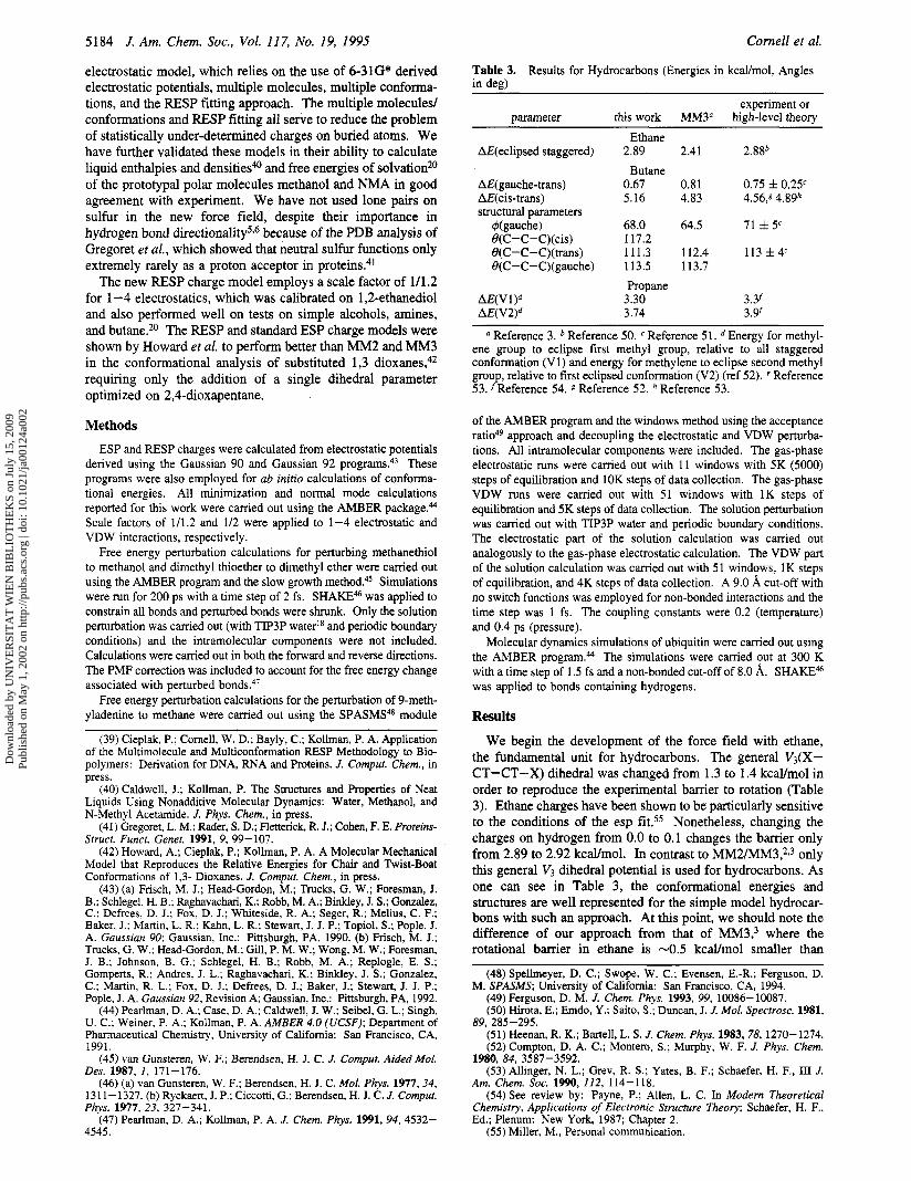
Table 3. Results for Hydrocarbons (Energies in kcaymol, Angles in deg)
experiment or this work MM3" high-level theory parameter
AE(ec1ipsed staggered)
AE(gauche-trans) AE(cis-trans) structural parameters
+(gauche) O(C-C-C)(cis) B(C-C-C)(trans) O(C-C-C)(gauche)
Ethane
Butane 2.89
0.67 5.16
68.0 117.2 111.3 113.5
Propane 3.30 3.74
2.41 2.88b
0.81 0.75 f 0.25' 4.83 4.56,s 4.89h
64.5 71 f 5'
112.4 113 f 4' 113.7
3.Y 3 . 9
Reference 3. Reference 50. Reference 51. Energy for methyl- ene group to eclipse first methyl group, relative to all staggered conformation (Vl) and energy for methylene to eclipse second methyl group, relative to first eclipsed conformation (V2) (ref 52). e Reference 53. fReference 54. 5 Reference 52. Reference 53.
of the AMBER program and the windows method using the acceptance ratio49 approach and decoupling the electrostatic and VDW perturba- tions. All intramolecular components were included. The gas-phase electrostatic runs were carried out with 11 windows with 5K (5000) steps of equilibration and 10K steps of data collection. The gas-phase VDW runs were carried out with 51 windows with 1K steps of equilibration and 5K steps of data collection. The solution perturbation was carried out with TIP3P water and periodic boundary conditions. The electrostatic part of the solution calculation was carried out analogously to the gas-phase electrostatic calculation. The VDW part of the solution calculation was carried out with 51 windows, 1K steps of equilibration, and 4K steps of data collection. A 9.0 A cut-off with no switch functions was employed for non-bonded interactions and the time step was 1 fs. The coupling constants were 0.2 (temperature) and 0.4 ps (pressure).
Molecular dynamics simulations of ubiquitin were carried out using the AMBER program.@ The simulations were carried out at 300 K with a time step of 1.5 fs and a non-bonded cut-off of 8.0 A. SHAKEM was applied to bonds containing hydrogens.
Results
We begin the development of the force field with ethane, the fundamental unit for hydrocarbons. The general V3(X- CT-CT-X) dihedral was changed from 1.3 to 1.4 kcal/mol in order to reproduce the experimental barrier to rotation (Table 3). Ethane charges have been shown to be particularly sensitive to the conditions of the esp fit.55 Nonetheless, changing the charges on hydrogen from 0.0 to 0.1 changes the barrier only from 2.89 to 2.92 kcdmol. In contrast to MM2/MM3,2,3 only this general V3 dihedral potential is used for hydrocarbons. As one can see in Table 3, the conformational energies and structures are well represented for the simple model hydrocar- bons with such an approach. A t this point, we should note the difference of our approach from that of MM3: where the rotational barrier in ethane is -0.5 kcaYmol smaller than
(48) Spellmeyer, D. C.; Swope, W. C.; Evensen, E.-R.; Ferguson, D. M. SPASMS; University of California: San Francisco, CA, 1994.
(49) Ferguson, D. M. J. Chem. Phys. 1993, 99, 10086-10087. (50) Hirota, E.; Emdo, Y.; Saito, S.; Duncan, J. J. Mol. Spectrosc. 1981,
(51) Heenan, R. K.; Bartell, L. S. J. Chem. Phys. 1983,78, 1270-1274. (52) Compton, D. A. C.; Montero, S.; Murphy, W. F. J. Phys. Chem.
(53) Allinger, N. L.; Grev, R. S.: Yates, B. F.; Schaefer, H. F., III J. Am. Chem. SOC. 1990, 112, 114-118.
(54) See review by: Payne, P.; Allen, L. C. In Modem Theoretical Chemistry, Applications of Electronic Structure Theory; Schaefer, H. F., Ed.; Plenum: New York, 1987; Chapter 2.
89, 285-295.
1980, 84, 3587-3592.
(55) Miller, M., Personal communication.
Dow
nloa
ded
by U
NIV
ER
SIT
AT
WIE
N B
IBL
IOT
HE
KS
on J
uly
15, 2
009
Publ
ishe
d on
May
1, 2
002
on h
ttp://
pubs
.acs
.org
| do
i: 10
.102
1/ja
0012
4a00
2
Simulation of Proteins and Nucleic Acids
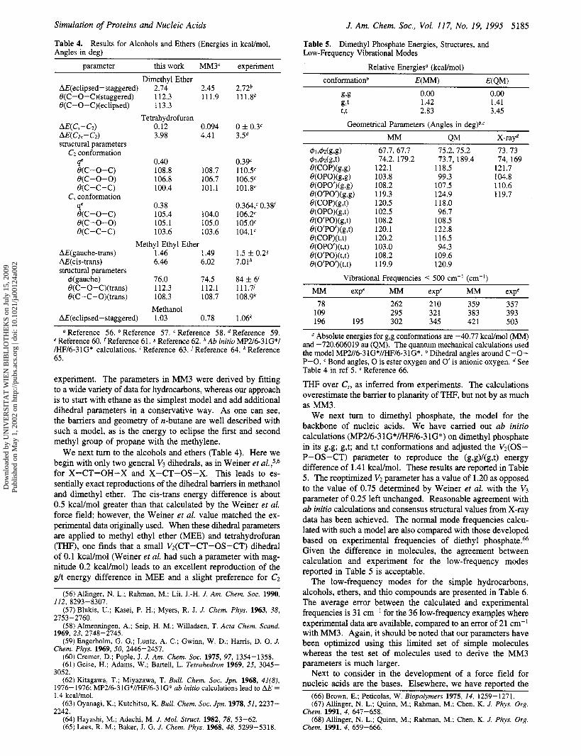
Table 4. Results for Alcohols and Ethers (Energies in kcal/mol, Angles in deg)
parameter this work MM3" experiment
J. Am. Chem. Soc., Vol. 117, No. 19, 1995 5185
Dimethyl Ether AE(ec1ipsed-staggered) 2.74 2.45 O(C-0-C)(staggered) 112.3 111.9 B(C-0-C)(eclipsed) 113.3
AE(Cs-Cz) 0.12 0.094 AE(c2v-c~) 3.98 4.41 structural parameters
C2 conformation
Tetrahydrofuran
qe 0.40 e(c-o-c) 108.8 108.7 e(c -0 -0) 106.8 106.7 e(c-c-c) 100.4 101.1
qe 0.38 e(c-o-c) 105.4 104.0 e(c-0-0) 105.1 105 .O e(c-c-c) 103.6 103.6
C, conformation
Methyl Ethyl Ether AE(gauche-trans) 1.46 1.49
4(gauche) 76.0 74.5 B(C-0-C)(trans) 112.3 112.1
AE(cis-trans) 6.46 6.02 structural parameters
O(C-C-O)(trans) 108.3 108.7 Methanol
AE(ec1ipsed-staggered) 1.03 0.78
2.72b 111.8b
0 f 0.3" 3.5d
0.39' 110.5' 106.5' 101.8'
0.364,' 0.38f 106.2' 105.0' 104.1'
1.5 f 0.28 7.01h
84 f 6' 111.7j 108.9k
1.06' a Reference 56. Reference 57. Reference 58. Reference 59.
e Reference 60. fReference 61. g Reference 62. hAb initio MF'2/6-31G*/ /HF/6-31G* calculations. Reference 63. Reference 64. Reference 65.
experiment. The parameters in MM3 were derived by fitting to a wide variety of data for hydrocarbons, whereas our approach is to start with ethane as the simplest model and add additional dihedral parameters in a conservative way. As one can see, the barriers and geometry of n-butane are well described with such a model, as is the energy to eclipse the first and second methyl group of propane with the methylene.
We next tum to the alcohols and ethers (Table 4). Here we begin with only two general V3 dihedrals, as in Weiner et al.,5X6 for X-CT-OH-X and X-CT-OS-X. This leads to es- sentially exact reproductions of the dihedral barriers in methanol and dimethyl ether. The cis-trans energy difference is about 0.5 kcdmol greater than that calculated by the Weiner et al. force field; however, the Weiner et al. value matched the ex- perimental data originally used. When these dihedral parameters are applied to methyl ethyl ether (MEE) and tetrahydrofuran (THF), one finds that a small Vz(CT-CT-OS-CT) dihedral of 0.1 kcdmol (Weiner et al. had such a parameter with mag- nitude 0.2 kcal/mol) leads to an excellent reproduction of the g/t energy difference in MEE and a slight preference for C2
(56) Allinger, N. L.; Rahman, M.; Lii, J.-H. J. Am. Chem. SOC. 1990,
(57) Blukis, U.; Kasei, P. H.; Myers, R. J. J. Chem. Phys. 1963, 38,
(58 ) Almenningen, A,; Seip, H. M.; Willadsen, T. Acta Chem. Scand.
(59) Engerholm, G. G.; Luntz, A. C.; Gwinn, W. D.; Harris, D. 0. J.
(60) Cremer, D.; Pople, J. J. Am. Chem. SOC. 1975, 97, 1354-1358. (61) Geise, H.; Adams, W.; Bartell, L. Tetrahedron 1969, 25, 3045-
3052. (62) Kitagawa, T.; Miyazawa, T. Bull. Chem. SOC. Jpn. 1968, 41(8),
1976-1976; MP2/6-31G*//HF/6-31G* ab initio calculations lead to AE = 1.4 kcal/mol.
(63) Oyanagi, K.; Kutchitsu, K. Bull. Chem. SOC. Jpn. 1978,51,2237- 2242.
(64) Hayashi, M.; Adachi, M. J. Mol. Struct. 1982, 78, 53-62. (65) Lees, R. M.; Baker, J. G. J. Chem. Phys. 1968, 48, 5299-5318.
112, 8293-8307.
2753-2760.
1969, 23, 2748-2745.
Chem. Phys. 1969, 50, 2446-2457.
Table 5. Low-Frequency Vibrational Modes
Dimethyl Phosphate Energies, Structures, and
Relative Energies" (kcaVmo1) conformationb E(MM) E(QM)
0.00 0.00 1.42 1.41 2.83 3.45
Geometrical Parameters (Angles in dedb.' ~~
MM QM X-rayd $%42z(g,g) 67.7, 67.7 75.2, 75.2 73.73 4Jlr42(grt)
e(opo')(g,g) 108.2
73.7, 189.4 74, 169 74.2, 179.2 e(cop)(g,g) 122.1 118.5 121.7 e(opo)(g,g) 103.8 99.3 104.8
107.5 110.6 e(o'po')(g,g) 119.3 124.9 119.7 e(cop)(g,t) 120.5 118.0 e(oPO)(g,t) 102.5 96.7 e(o'po)(g,t) 108.2 108.5 e(o'Po')(g,t) 120.1 122.8 e(cop)(t,t) 120.2 116.5
103.0 94.3 108.2 109.6 119.9 120.9
Vibrational Frequencies < 500 cm-' (cm-I)
e(opo')(t,t) e(o'Po)(t,t) e(o'po')(t,t)
MM exp' MM expe MM expe 78 262 210 359 357
109 295 321 383 393 196 195 302 345 42 1 503
a Absolute energies for g,g conformations are -40.77 kcaVmol (MM) and -720.606019 au (QM). The quantum mechanical calculations used the model MP2//6-31G*//HF/6-31G*. Dihedral angles around C-O- P-0. Bond angles, 0 is ester oxygen and 0' is anionic oxygen. See Table 4 in ref 5. e Reference 66.
THF over C,, as inferred from experiments. The calculations overestimate the barrier to planarity of THF, but not by as much as MM3.
We next turn to dimethyl phosphate, the model for the backbone of nucleic acids. We have carried out ab initio calculations (MP2/6-3 1G*//HF/6-3 1G*) on dimethyl phosphate in its g,g; g,t; and t,t conformations and adjusted the Vz(0S- P-OS-CT) parameter to reproduce the (g,g)/(g,t) energy difference of 1.41 kcdmol. These results are reported in Table 5 . The reoptimized VZ parameter has a value of 1.20 as opposed to the value of 0.75 determined by Weiner et al. with the V3 parameter of 0.25 left unchanged. Reasonable agreement with ab initio calculations and consensus structural values from X-ray data has been achieved. The normal mode frequencies calcu- lated with such a model are also compared with those developed based on experimental frequencies of diethyl phosphate.& Given the difference in molecules, the agreement between calculation and experiment for the low-frequency modes reported in Table 5 is acceptable.
The low-frequency modes for the simple hydrocarbons, alcohols, ethers, and thio compounds are presented in Table 6. The average error between the calculated and experimental frequencies is 31 cm-' for the 36 low-frequency examples where experimental data are available, compared to an error of 21 cm-' with MM3. Again, it should be noted that our parameters have been optimized using this limited set of simple molecules whereas the test set of molecules used to derive the MM3 parameters is much larger.
Next to consider in the development of a force field for nucleic acids are the bases. Elsewhere, we have reported the
(66) Brown, E.; Peticolas, W. Biopolymers 1975, 14, 1259-1271. (67) Allinger, N. L.; Quinn, M.; Rahman, M.; Chen, K. J . Phys. Org.
(68) Allinger, N. L.; Quinn, M.; Rahman, M.; Chen, K. J. Phys. Org. Chem. 1991, 4 , 647-658.
Chem. 1991, 4, 659-666.
Dow
nloa
ded
by U
NIV
ER
SIT
AT
WIE
N B
IBL
IOT
HE
KS
on J
uly
15, 2
009
Publ
ishe
d on
May
1, 2
002
on h
ttp://
pubs
.acs
.org
| do
i: 10
.102
1/ja
0012
4a00
2
5186 J. Am. Chem. SOC., Vol. 117, No. 19, 1995
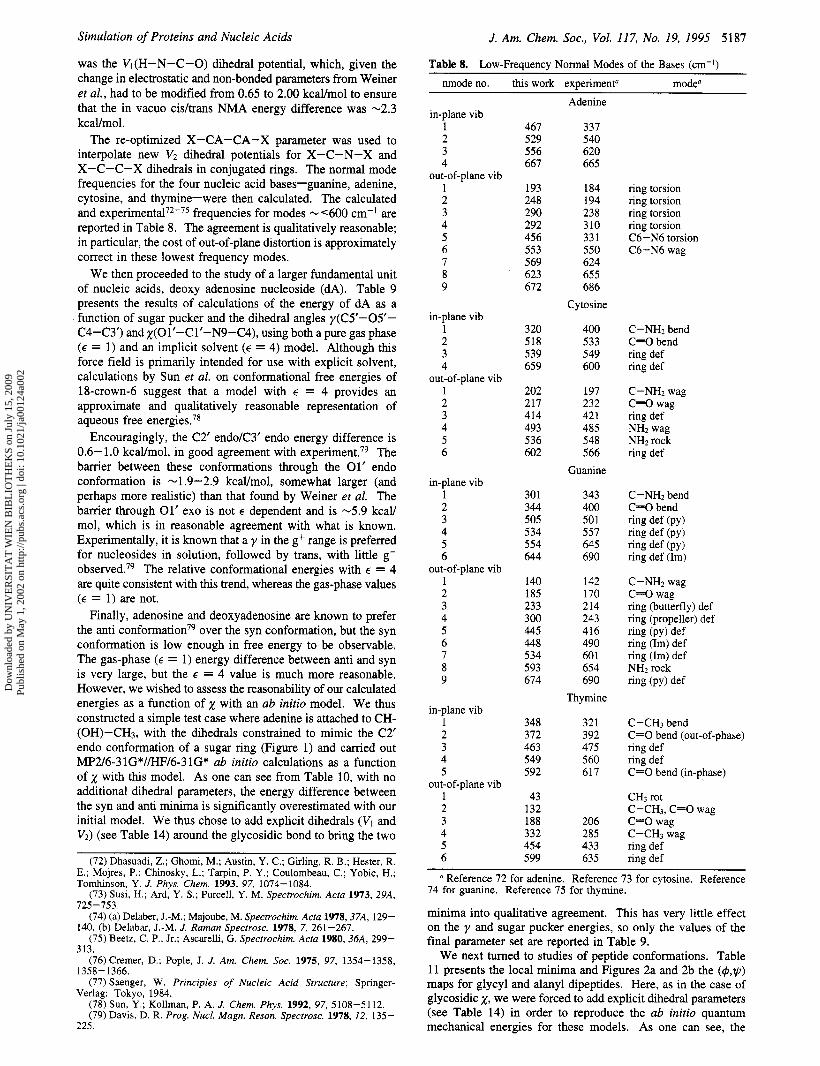
Table 6. Low-Frequency (< 1000 cm-I) Vibrational Modes for Small Hydrocarbons, Ethers, Alcohols, and Sulfur Compounds
symm @(this work) O(MM3). @(exp)b,c moded trans-NMA
Come11 et al.
Table 7. Normal Modes of trans-NMA and Benzene (cm-I)
nmode no. symm this work experimenta mode
312 811 811 898
23 1 275 356 733 809
866
877
127 236 27 1 272
364
297 867
212 279 416 798
123 225
27 1
283
404
755
806
707 80 1
279 69 1 720
105 236 275 509 710 713
Ethane 279 283 CH3-CH3 torsion 908 822 CH3 asym rocking 908 822 CH3 asym rocking 962 995 C-C stretch
208 217 CH3-CH2 torsion 255 265 CH3-CH2 torsion 375 379 C-C-C bend 803 748 CH2 rock + CH3 def 850 868 CH3 rock +
sym C-C str/str 938 921 CH3 rock +
asym C-C str/str 961 899 CH2 twist + CH3 def
122 121 CHI-CH~ torsion 216 CH3-CH2 torsion 245 266 CH3-CH2 torsion 287 asym C-C-C bend +
C-C -C bend 394 427 sym C-C-C bend +
C-C-C bend
Propane
Butane
Methanol 263 270 CH3-0 torsion
1052 1034 C-0 stretch
Dimethyl Ether 188 198 CH3-O sym torsion 273 242 CH3-0 asym torsion 400 424 C - 0 - C bend 924 918 C-0 sym stretch Methyl Ethyl Ether 114 C2H5-0 torsion 216 CH3-C torsion +
CH3-O torsion 257 238 CH3-0 torsion +
CH3-C torsion 296 308 C-0-C bend +
C-C-0 bend 420 472 C-C-0 bend +
C-0-C bend 870 820 CH3 rock + CH2 rock +
CH2 twist 897 855 C-0 str + CH3 wag +
C-C str Methanethiol
695 704 c-s 823 803 C-S-H
Dimethyl Sulfide 285 282(285) C-S-C 683 691 (683) S-C sym 702 741 (704) S-C asym Dimethyl Disulfide 116 102 (106) C-S-S-C torsion 241 239 (242) S-S-C bend 279 272 S-S-C bend
1 A” 44 2 A” 97 3 A“ 184 192 4 A’ 286 289 5 A’ 440 439 6 A” 587 600 7 A’ 591 628 8 A” 696 725 9 A’ 80 1 883
10 A’ 963 99 1 11 A” 1037 1044 12 A” 1046 13 A’ 1075 1114 14 A‘ 1082 1161 15 A‘ 1209 1300 16 A‘ 1395 1374 17 A’ 1398 1414 18 A” 1402 1441 19 A” 1407 1451 20 A’ 1428 1458 21 A’ 1516 1471 22 A’ 1614 1569 23 A’ 1693 1660 24 A’ 2868 2935 25 A’ 2869 2935 26 A” 2980 298 1 27 A’ 2982 298 1 28 A’ 2982 2994 29 A’ 2983 2994 30 A’ 3304 3307
1 e2u 410 410 2 e2g 609 606 3 a2u 66 1 673 4 b2g 704 703 5 e lg 900 849 6 e2u 979 975 7 alg 941 992
9 blu 1167 1010 10 elu 1124 1038 11 b2u 1194 1150 12 e2g 1129 1178 13 b2u 1331 1310 14 a2g 1729 1326 15 elu 1493 1486 16 e2g 1706 1596
18 alg 3062 3062 19 elu 3064 3063 20 blu 3068 3068
Benzene
8 b2g 947 995
17 e2g 3064 3047
ring def ring def CH bend ring def CH bend CH bend ring stretch (breathing) CH bend ring def CH bend CH bend CH bend ring stretch (kekule) CH bend ring stretch + def ring stretch CH stretch CH stretch CH stretch CH stretch
a Reference 70 for trans-NMA. Reference 71 for benzene.
the bases is the dihedral potential for out-of-plane motion, as discussed by Weiner et al. As in the development of our previ- ous force field, normal mode analyses of benzene and NMA are imuortant. The results for the normal mode analyses auulied
514 509 (514) S-S stretch to thesk molecules are presented in Table 7. We ha& readjisted 701 689 (694) S-C stretch 703 (694) S-C stretch
a References 2, 56,67, and 68. See references 2,5,56,67, and 68 for experimental frequencies. Experimental frequencies given in parentheses refer to those used as reference for MM3 values. See references 2, 5, 56, 67, and 68 for’ the mode assignments.
hydrogen bond energies and structures of A:T and G:C pairs and these appear to be in good agreement with the highest level of ab initio data currently available.69 However, a critical element in the development of planar functionalities such as
(69) Gould, I. R.; Kollman, P. A. J. Am. Chem. SOC. 1994, 116, 2493- 2499.
the X-CA-CA-X V 2 value and the improper out-ofiplane dihedral X-X-CA-HA to ensure correct representation of the lowest frequency modes of benzene, with the four lowest modes (1700 cm-’) in good agreement with experiment.”
We next turn to NMA, the model for the peptide backbone. With a few adjustments to the Weiner et al.53b bonded parameters, the agreement with experiment70 for the six lowest frequency modes is again excellent. In NMA, a key adjustment
(70) Rey-Lafon, M.; Ford, M. T.; Garrigen-Lagrange, C. Specrrochim. Acta, Part A 1973, 29A, 471-486.
(7 1) Shimananchi, T. Tables of Molecular Vibrational Frequencies; National Stand. Ref. Data Ser.; National Bureau of Standards: Washington, DC, 1967; Parts 1-3.
Dow
nloa
ded
by U
NIV
ER
SIT
AT
WIE
N B
IBL
IOT
HE
KS
on J
uly
15, 2
009
Publ
ishe
d on
May
1, 2
002
on h
ttp://
pubs
.acs
.org
| do
i: 10
.102
1/ja
0012
4a00
2
Simulation of Proteins and Nucleic Acids J. Am. Chem. SOC., Vol. 117, No. 19, 1995 5187
Table 8. Low-Frequency Normal Modes of the Bases (cm-I)
nmode no. this work experimentn modea
was the Vl(H-N-C-0) dihedral potential, which, given the change in electrostatic and non-bonded parameters from Weiner et al., had to be modified from 0.65 to 2.00 kcallmol to ensure that the in vacuo cidtrans NMA energy difference was -2.3 kcal/mol.
The re-optimized X-CA-CA-X parameter was used to interpolate new V2 dihedral potentials for X-C-N-X and X-C-C-X dihedrals in conjugated rings. The normal mode frequencies for the four nucleic acid bases-guanine, adenine, cytosine, and thymine-were then calculated. The calculated and e~pe r imen ta l~*-~~ frequencies for modes - (600 cm-’ are reported in Table 8. The agreement is qualitatively reasonable; in particular, the cost of out-of-plane distortion is approximately correct in these lowest frequency modes.
We then proceeded to the study of a larger fundamental unit of nucleic acids, deoxy adenosine nucleoside (dA). Table 9 presents the results of calculations of the energy of dA as a function of sugar pucker and the dihedral angles y(CS-05’- C4-C3’) andx(01’-Clf-N9-C4), using both a pure gas phase ( E = 1) and an implicit solvent ( E = 4) model. Although this force field is primarily intended for use with explicit solvent, calculations by Sun et al. on conformational free energies of 18-crown-6 suggest that a model with E = 4 provides an approximate and qualitatively reasonable representation of aqueous free energiesS7*
Encouragingly, the C2’ endo/C3’ endo energy difference is 0.6-1.0 kcal/mol, in good agreement with e~pe r imen t .~~ The barrier between these conformations through the 01’ endo conformation is -1.9-2.9 kcal/mol, somewhat larger (and perhaps more realistic) than that found by Weiner et al. The barrier through 01’ exo is not E dependent and is -5.9 kcal/ mol, which is in reasonable agreement with what is known. Experimentally, it is known that a y in the g+ range is preferred for nucleosides in solution, followed by trans, with little g- observed.79 The relative conformational energies with E = 4 are quite consistent with this trend, whereas the gas-phase values ( E = 1) are not.
Finally, adenosine and deoxyadenosine are known to prefer the anti c~nformat ion~~ over the syn conformation, but the syn conformation is low enough in free energy to be observable. The gas-phase ( E = 1) energy difference between anti and syn is very large, but the E = 4 value is much more reasonable. However, we wished to assess the reasonability of our calculated energies as a function of x with an ab initio model. We thus constructed a simple test case where adenine is attached to CH- (OH)-CH3, with the dihedrals constrained to mimic the C2’ endo conformation of a sugar ring (Figure 1) and carried out MP2/6-31G*//HF/6-3 lG* ab initio calculations as a function of x with this model. As one can see from Table 10, with no additional dihedral parameters, the energy difference between the syn and anti minima is significantly overestimated with our initial model. We thus chose to add explicit dihedrals (VI and V2) (see Table 14) around the glycosidic bond to bring the two
(72) Dhasuadi, Z.; Ghomi, M.; Austin, Y. C.; Girling, R. B.; Hester, R. E.; Mojres, P.; Chinosky, L.; Tarpin, P. Y.; Coulombeau, C.; Yobic, H.; Tomhinson, Y. J. Phys. Chem. 1993, 97, 1074-1084.
(73) Susi, H.; Ard, Y. S.; Purcell, Y. M. Spectrochim. Acta 1973, 29A, 725-753.
(74) (a) Delaber, J.-M.; Majoube, M. Spectrochim Acta 1978,37A, 129-
(75) Beetz, C. P., Jr.; Ascarelli, G. Spectrochim. Acta 1980, 36A, 299-
(76) Cremer, D.; Pople, J. J. Am. Chem. SOC. 1975, 97, 1354-1358,
(77) Saenger, W. Principles of Nucleic Acid Structure; Springer-
(78) Sun, Y.; Kollman, P. A. J. Chem. Phys. 1992, 97, 5108-5112. (79) Davis, D. R. Prog. NucE. Magn. Reson. Spectrosc. 1978, 12, 135-
140. (b) Delabar, J.-M. J. Raman Spectrosc. 1978, 7, 261-267.
313.
1358- 1366.
Verlag: Tokyo, 1984.
225.
in-plane vib 1 2 3 4
1 2 3 4 5 6 7 8 9
out-of-plane vib
in-plane vib 1 2 3 4
1 2 3 4 5 6
out-of-plane vib
in-plane vib 1 2 3 4 5 6
1 2 3 4 5 6 7 8 9
out-of-plane vib
in-plane vib 1 2 3 4 5
1 2 3 4 5 6
out-of-plane vib
467 529 556 667
193 248 290 292 456 553 569 623 672
320 518 539 659
202 217 414 493 536 602
301 344 505 534 554 644
140 185 233 300 445 448 534 593 674
348 372 463 549 592
43 132 188 332 454 599
Adenine
337 540 620 665
184 194 238 310 33 1 550 624 655 686
Cytosine
400 533 549 600
197 232 421 485 548 566
Guanine
343 400 501 557 645 690
142 170 214 243 416 490 60 1 654 690
Thymine
32 1 392 475 560 617
206 285 433 635
ring torsion ring torsion ring torsion ring torsion C6-N6 torsion C6-N6 wag
C-NH2 bend C=O bend ring def ring def
C-NH2 wag C=O wag ring def NH2 wag NH2 rock ring def
C-NH2 bend C=O bend ring def (PY) ring def (PY) ring def (PY 1 ring def (Im)
C-NH2 wag C=O wag ring (butterfly) def ring (propeller) def ring (PY) def ring (Im) def ring (Im) def NH2 rock ring (PY) def
C-CH, bend C=O bend (out-of-phase) ring def ring def C=O bend (in-phase)
CH3 rot C-CH3, C=O wag C=O wag C-CH3 wag ring def ring def
~
Reference 72 for adenine. Reference 73 for cytosine. Reference 74 for guanine. Reference 75 for thymine.
minima into qualitative agreement. This has very little effect on the y and sugar pucker energies, so only the values of the final parameter set are reported in Table 9.
We next turned to studies of peptide conformations. Table 11 presents the local minima and Figures 2a and 2b the (@,q) maps for glycyl and alanyl dipeptides. Here, as in the case of glycosidic 2, we were forced to add explicit dihedral parameters (see Table 14) in order to reproduce the ab initio quantum mechanical energies for these models. As one can see, the
Dow
nloa
ded
by U
NIV
ER
SIT
AT
WIE
N B
IBL
IOT
HE
KS
on J
uly
15, 2
009
Publ
ishe
d on
May
1, 2
002
on h
ttp://
pubs
.acs
.org
| do
i: 10
.102
1/ja
0012
4a00
2

5188 J. Am. Chem. Soc., Vol. 117, No. 19, 1995
Table 9. Conformational Energies for Deoxyadenosine (Angles in deg, Energies in kcal/mol)”
pucker qa wd yb zb 3‘OHC SOHd Ee A d Sugar Pucker Profile
E = 18 C2’endo 0.40 146.1 51.3 -158.4 176.5 171.4 -52.40 0 C3’endo 0.37 5.7 56.9 -162.3 -178.3 -179.3 -51.87 0.63 04’endo 0.38 65.3 54.1 -156.7 -175.5 -175.4 -49.53 2.87 O4’exo 0.29 276.6 42.6 -178.7 175.8 178.5 -46.54 5.86
E = 48 C2’endo 0.39 144.9 55.3 -153.1 176.2 -179.2 -1.65 0 C3’endo 0.38 14.1 56.3 -156.1 178.1 179.9 -0.61 1.04 04’endo 0.39 56.6 55.6 -153.1 179.1 179.5 0.21 1.86 04’exo 0.30 285.0 42.7 176.9 177.3 179.8 4.03 5.68
Gamma Dependence E = 18
C2’endo 0.40 146.1 51.3 -158.4 176.5 171.4 -52.40 0 C2’endo 0.42 141.9 -168.6 -168.6 179.6 -179.9 -50.39 2.01 C2’endo 0.42 151.7 -62.3 -169.9 -179.7 -179.7 -50.92 1.48
E = 48 C2’endo 0.39 144.9 55.5 -153.1 176.2 179.2 -1.65 0 C2’endo 0.40 148.0 -172.9 -162.4 180.0 -179.9 -1.31 0.34 C2’endo 0.40 150.0 -66.6 -169.9 -179.9 -179.9 -0.13 1.52
x Dependence E = 18
C2’endo 0.40 146.1 51.3 -158.4 176.5 171.4 -47.46 0 C2’endo 0.40 166.7 63.3 60.8 -179.1 179.8 -41.52 5.94
E = 48 C2’endo 0.39 145.4 55.3 -141.6 176.5 179.2 3.24 0 C2’endo 0.40 144.0 52.6 37.6 180.0 179.6 1.84 -1.40
a q and W defined in ref 76. y and x defined in ref 77. Above, the first entry corresponds to an “anti” conformation for x, the second to “syn”. 3’OH refers to C4’-C3’-03’-H03’ dihedral. 5’OH refers to H05’-05’-C5’-C4’ dihedral. e Absolute molecular mechanical energy. f Relative conformational energy. g E is the dielectric screening factor used in eq 1.
Cornel1 et al.
H61\ N H 6 2 N6
HE-CE
H04’-04’-Cl ‘-HI ’
HZ’l-CZt-H2’2
H2’3
Figure 1. Model of deoxyadenosine employed in the quantum mech- anical and molecular mechanical conformational studies reported in Table 10. In the quantum mechanical calculations, the H04’-04’- Cl’-N9 and H2’3-C2’-Cl’-N9 dihedrals were held fixed at values characteristic of a C2’-endo sugar, in order to mimic the conformation of the sugar ring. In the molecular mechanical calculations, the dihedrals were restrained to those values with dihedral restraints of 500 k c d mol.
agreement with high-level ab initio data is very good for all but alanyl dipeptide C7ax and glycyl dipeptide a R . The ala C,, conformation is rarely found in proteins and gly occurs relatively infrequently in a-helices, due to the loss of conformational entropy, so these conformations were reasonable ones in which to tolerate any error.
(80) Ben Naim, A.; Marcus, Y. J . Chem. Phys. 1984, 81, 2016-2027. (81) Wolfenden, R. Biochemisrry 1978, 17, 201-204.
Table 10. x Angle Profile for Base with Sugar Fragment (kcdmol)
AMBER (E = 1)
ab initio no specific with specific pb MP2/6-31G*//HF/6-31G* dihedral dihedral‘
60 min 120 180 min 210 240 300 360
60 min 120 180 min 210 240 300 360
Model of Deoxyadenosine 0.94 4.63 0.63 (74.7°)d 4.62 (61.1°)d 3.37 5.48 0.06 0.38 0.00 (198.2°)d 0.00 (196.7°)d 0.22 0.20 1.45 1.58 5.33 6.57
9.68
2.27 6.00 2.02 (72.3°)d 5.99 (61.0°)d 7.02 8.48 0.74 1.37 0.00 (210.00)d 0.00 (205.2°)d 0.00 0.05 1.29 1.77 8.15 8.94
13.11
Model of Deoxythymidine
1.53
5.07 0.40 0.00 (197.2°)d 0.15 1.20 3.61 4.72
1.45 (54.50)d
2.94 2.83 (55.4°)d 8.05 1.43 0.00 (205.5°)d 0.03 1.40 5.82 8.18
Reference 77. Degrees. Specific VI and V2 dihedral terms were added for OS-CT-N*-CK (purines) and OS-CT-N*-CM (pyri- midines) dihedral angles. Minimized value of x. Table 11. Conformational Energies of Glycyl and Alanyl Dipeptides (kcdmol)
glycyl dipeptide alanyl dipeptide
E(MM) UQMY E(MM) E(QM)”
C I 0.0 0.0 c 7 q 0.0 0.0 c 5 1.9 2.0 c7, 1.5 2.1 aR 6.0 4.0 c 5 1.5 1.5
aR 3.9 3.9
Quantum mechanical energies calculated at the MP2/TZP//HF/6- 31G* level on methyl-blocked versions of the dipeptides. See ref 28 for further details.
One of the important features in our force field is the attempt to reproduce the solvation free energies of a representative set of molecules. In Table 12, we present such a representative set. As one can see, the absolute solvation free energy of methane is somewhat (0.5 kcdmol) too large with our model, but the relative solvation free energies of methane, ethane, and propane are within 0.3 kcdmol of experiment. For our protypal polar molecules, methanol and NMA, the agreement with experimental solvation free energies is within -0.5 kcdmol. We wished also to assess the solvation free energies for sulfur compounds and the relative solvation free energies of those are in reasonable agreement with experiment (again within 0.5 kcaV mol). The calculated free energy of 9-methyladenine is a prediction, because there are no precise experiment^,^^ but the relative free energies of this force field and that of Weiner et al.536 suggest that the experimental determination of this quantity would be of great interest. Turning to the ionic molecules, our results make clear that a typical two-body additive force field will tend to overestimate ion solvation (when corrected for long- range cut-off) unless its parameters are significantly modified, but fully non-additive calculations with exactly the same parameters reproduce experiment very well.
(82) Meng, E.; Cieplak, P.; Caldwell, J.; Kollman, P. Accurate Solvation Free Energies of Acetate and Methylammonium Calculated with a Polarized Water Model. J . Am. Chem. Soc., in press.
(83) Analyzed by Cramer, C. J.; Truhlar, D. G. J. Am. Chem. Soc. 1991, 113, 8305-8311.
(84) Hine, J.; Mookejee, P. K. J. Org. Chem. 1975, 40, 292-298. (85) Ferguson, D. M.; Pearlman, D. A.; Swope, W. C.; Kollman, P. A.
J. Comput. Chem. 1992, 13, 362-370.
Dow
nloa
ded
by U
NIV
ER
SIT
AT
WIE
N B
IBL
IOT
HE
KS
on J
uly
15, 2
009
Publ
ishe
d on
May
1, 2
002
on h
ttp://
pubs
.acs
.org
| do
i: 10
.102
1/ja
0012
4a00
2

Simulation of Proteins and Nucleic Acids J. Am. Chem. SOC., Vol. 117, No. 19, 1995 5189
180
120
60
0 PSI
-60
-120
-180
-180 -120 -60 0 60 120 180
PHI 180
120
60
0 PSI
-60
-120
-180
-180 -120 -60 0 6 0 120 180
PHI Figure 2. (a) The molecular mechanical (4.v) map for methyl-blocked glycyl dipeptide generated using the force field presented here. Contours are drawn every 2 kcdmol. (b) The molecular mechanical (4,q) map for methyl-blocked alanyl dipeptide generated using the force field presented here. Contours are drawn every 2 kcal/mol.
Table 12. (kcal/mol’,
Solvation Free Energies for Model Compounds
molecule AAG(ca1c) AAG(exp)
C& - nothing
C3Hs - C2H6
NMA - CHq CH3NH3+ - nothing
CH3C02- - nothing
C2H6 - c& CH30H - CH3CH3
CH3SCH3 - CH3OCH3 CH30H A CHsSH 9-CH3 adenine - CHq
-2.5 f 0.1‘ -0.1 f 0.1‘ -0.2 f 0.1c
6.9 f O.ld 11.6 f 0.2d 87.6 f 2.0
(75.4 f 1.7)’ 87.1 f 1.2
(71.6 f 1.0)’ 0.9 f 0.1” 3.5 f 0.1”
18.3 f 2.6, 13.9 f 0.4’
-2 .P 0.26
-0.26 6.9*
12.1e 77-798
70-71h
0.4‘ 3.7‘
15.6 2r 1.1)
a Reference 21b. Reference 80. Reference 21a. Because of the uncertainty in the electrostatic potential derived charges for ethane, the average of the free energies for the electrostatic potential derived and Mulliken charges for these free energy calculations are presented.
Reference 20. Reference 81. f Reference 82, additive potential, values in parentheses are for nonadditive potential. 8 Reference 83. This paper. Reference 84. j See Note Added in Proof.
The results described above were obtained on model systems that were relatively very simple3’ (neat liquids) andor small (dipeptides and nucleosides). In order to test the performance of the new force field on a more complex system, we carried
1 Comell et al. -
0.0 60.0 120.0 180.0 time [ps]
Figure 3. RMS deviation (A) between the crystal structure of ubiquitin and structures along an MD trajectory as modeled by the Weiner et aL6 and Cornell et al. (this work) force fields. The lower lines correspond to the RMS deviation of the heavy backbone atoms only and the upper lines to the RMS deviation for all heavy atoms.
out an MD simulation of ubiquitin in water with periodic boundary conditions. The RMS difference was calculated for structures along the trajectory relative to the crystal structures6 for (1) the backbone atoms and (2) all of the heavy atoms. These results were then compared to those obtained with the Weiner et ~ 1 . ~ 3 ~ force field (Figure 3). The RMS values are reported for the first 72 residues only, since the four residues of the carboxy terminus were mobile. The behavior of the new force field presented here is better in two ways. First, the protein structure seems to have stabilized after 50 ps of simulation with the new force field, while the RMS deviation continues to increase throughout the trajectory with the Weiner et al.536 force field. Second, the RMS deviation for all of the heavy atoms after 180 ps of simulation is about 2.0 8, with the force field presented here and about 2.5 8, with the Weiner et al.5.6 force field. Alonso and Daggett have also reported the results of a long MD simulation of ubiquitin, and they found a backbone RMS deviation of 1.4 8, from the crystal structure, comparable to the deviation found here.87 A referee has pointed out that smaller deviation from a crystal structure could simply be a consequence of an “unrealistically stiff’ force field. We cannot rule this out, but stress that we did not, in our force field derivation on the fragments described above attempt to add “stiffness”.
Even closer agreement with a protein crystal structure has been obtained by York et al.,” who carried out a 1000 ps MD simulation of BPTI with the long-range electrostatic forces of the crystal environment treated using the particle mesh Ewald method and the Weiner et ~ 1 . ~ 5 ~ force field. With this model they obtained an RMS deviation from the crystal structure of 0.33 8, for backbone atoms. These results serve to illustrate the difference between errors arising from the force field itself and those arising from its implementation in a given calculation. Currently, most MD simulations employ an 8 or 9 8, cutoff for nonbonded interactions in order to reduce this rate-determining part of the calculation. In systems where long-range electrostat- ics play an important role, this approximation is clearly inadequate. Although the Ewald method is only fully appropri- ate for periodic crystal systems, other methods also exist which allow for the more accurate treatment of long-range electrostat-
(86) Vijay-Kumar, S.; Bugg, C. E.; Cook, W. J. J. Mol. B i d . 1987,194, 531-544.
(87) Alonso, D. 0. V.; Daggett, V. Molecular Dynamics Studies of Partially Unfolded Conformations of Ubiquitin in Methanol and Their Refolding in Water, submitted for publication.
Dow
nloa
ded
by U
NIV
ER
SIT
AT
WIE
N B
IBL
IOT
HE
KS
on J
uly
15, 2
009
Publ
ishe
d on
May
1, 2
002
on h
ttp://
pubs
.acs
.org
| do
i: 10
.102
1/ja
0012
4a00
2

5190 J. Am. Chem. Soc.. Vol. 117, No. 19, 1995 Come11 et al.
0.1 105 0.2936H H1 0-0.5819
I I 0.5366 N-CT-C-
-0.5163 - I 0'0397
I o'0560
4.01 73 HC - CT - HC -0.01 73
0.0823 02719 H H1 0-0.5679
0.0698 0.2719 H H I 04.5679
-0.4157 I I 0.5973 - N-CT-C-
I -0'0z5z H1 0.0698
GLY
0.0880 0.2936 H H I 0-0.5819
I I 0.5366 N-CT-C-
-0.5163 - I O'OSB1
I -O.O3O3
-o.oizz HC - CT - HC -0.01 22
C 0.7994
-0.8014 02 '02 -0.8014
ASP
-0.41 57 I I I 0.5973 N-CT-C- -
I 0.0337
00603 HC - C'T - HC 0.0603
HC 0.0603
ALA 0.0869
0,2719 H H1 0-0.5679
N-CT- -0.41 57 -
-0.0597 I -0.3204/HC o'088z 0.0187 HC - CT0= CT - HC 0.0882 I \HC 0.0882
0.0236 HC - CT - HC 0.0236
I -0'0430
I -o.0660
0.0186 HC-CT-HC 0.0186
HC 0.0186
ILE
0.2719 H H1 0.0976
-0.41 57 I I -0.1 490 - N-CT-I41 0.0976
-0.0425 HC - CT - HC -0.0425
I 0'0136
-0.81 88 02 0.0922
0.2719 H H1 0-0.5679
I I 0.5973 N-CT-C-
-0.41 57 - I -0.0518
0.0457 HC - CT - HC 0.0457
GLU 0.0850
0.2719H H1 0 -0.5679
-0.4157 I I 0.5973 - N-CT-C-
0,1048
O.2719H H l 04 ,5679
I I I 0.5973 N-CT-C-
-0.4157 - I O'Olr3
I
0.0797 HC - CT - HC 0 0797
/ C y / / " I -o'2041 0.4196
4.5931 0 N -0.9191
H 0.4196
ASN
0,0171 HC-CT-HC 0.0171
1-0.0036 0.1000 HC-CT-HC 0.1000
I -0'41 i
0.0352 HC - CT - HC 0.0352
I -0.0645
HC 0 . 1 W
LEU
/ C y 1 / H 0.4251
-0.6086 0 N -0.9407
H 0.4251
GLN 0,1360
0.2719 H H1 0-0.5679
-0.4157 I I I 0.5973 N-CT-C- -
I -0'0581
0.0969
0.2719 H H1 0-0.5679 I l l 0.0881
0.2719 H H1 0-0.5679 -0,4157 I I I 0.5973
NME - N-CT-C- 4.0875 I -0.31g/HC 0'0791
-0.0297 HC - CT - CT - HC 0.0791 I 0'298s \HC 0.0791 O . l l 2 3 H C 0-0.5679
I I
H1 0.0976
-0.4157 I I 0.5973 N-CT-C- -
I o'0188
I -0'0462
\ I -0'3811
0.0402 HC - CT - HC 0.0402
CC 4.0266 0.1147 H4. CV / 0.1 292 \NA/H 0.3649
0.0367 HC - CT - HC 0.0367
0.1862 H4 \ C W -0.2207 NB -0.5432
0,1123 H C 4 . 3 6 6 & - A m 0.0791 HC - CT - HC 0.0791
I 0.1123 HC
I -031g2 HC 0.0791
VAL ACE \ I -0.2795 NA - CR 0 1635
H5 01435 / \
0 3339 H
HIE
-0.5727 NE- Ck 0,2057
\ 0.1007
O.2719H H1 0-0.5679 H5 0.1392
HID 0.0843
0.2719H H I 0-0.5679
I 1 I 0.5973 N-CT-C-
-0.4157 - -0,4157 I I I 0.5973 - N-CT-C- I 4 0 3 8 9
0.1212
0.2747 H H l 0-0.5894
-0.3479 I 1 1 0.7341 N-CT-C- -
1 -0.6761 0.0043 H1- CT - OH-HO 0.4102
I 0'3654 0.0642 HC - CT - HC 0.0642
I -0.2438 HC 0.0642
THR
0.1124
0.2719 H H1 0-0.5679 I l l
0,0352 H1- CT - H1 0.0352
I O 2 l I 7
-0.6546 OH-HO 0.4275
SER 0.0810 HC - CT -HC 0.0810
I -0'041' CC-o.oor2
C W 6 14)NA-G; 0'3866 0.2317 H4
\ I 0.0766
02719 H H l 0-0.5679
1 I I 0.5973 N-CT-C-
-0.41 57 - I 0'0429
I -0'0790
0.0910H1-~-H10.0910
s -0.1081
CYX
0.0880
4.1718NA-CR -0.0170
/ H5 0.2681
\ -0.4157 I I I 0,5973 - N-CT-C-
I 0'0213
0.3911 H'
HIP 0,1112 Hl-CT-HI 0.1112
I -0.1231 -0.3119 SH-HS 0,1933
CYS
0,1560 0.2747 H H1 0-0.5894
I I I 0.7341 -0.3479 N-CT-C- -
I -0.2637
I -0'0007
0.0327 HC - CT - HC 0.0327
0.1426 0.2747H H1 0 4 . 5 8 9 4
-0.3479 I I I 0.7341 N -CT-C- -
I -0'2400 0.0362 HC - CT - HC 0.0362
I -O'Oog4
I 0'0187
I 4'0479
I -0'0143
I -0'3854
0.0103 HC-CT-HC 0.0103
0.0621 HC - CT - HC 0.0621
0,1135 HP-CT-HP 0.1135
0.3400 H-N3-H 0.3400
H 0.3400
0.2719H H1 0-0.5679
I I I 0.5973 N-CT -C-
-0.4157 - 0.0641 H1 0-05748
I 1 0.5896 CT-C-
-0.0266 -0 2548 N -
0.0285 HC - CT - HC 0.0285
I 0'0390 0.0687 H1- CT - H1 0.0687 0.0241 HC - CT - HC 0.0241
I 0'0342
I O o 0 l 8
0.0440Hl-CT-Hl 0.0440
I I 1 -0.0070 0,0391 HI-CT 0.0192 CT-HC 0.0253
/ \ / \ CA0.8076 -0.8627 / \ -0.8627 0.0391 H1 HC 0.0253
,4478 H - NZ NZ - H 0.4478
-0.5295 NZ - H 0.3456
i-0,2737
I 0.0684 Hl-CT-Hl 0.0684
I -0.0536 H1 0.0684
MET PRO
ARG LYS
Dow
nloa
ded
by U
NIV
ER
SIT
AT
WIE
N B
IBL
IOT
HE
KS
on J
uly
15, 2
009
Publ
ishe
d on
May
1, 2
002
on h
ttp://
pubs
.acs
.org
| do
i: 10
.102
1/ja
0012
4a00
2
Simulation of Proteins and Nucleic Acids
0.0978 0.0876
J. Am. Chem. SOC., Vol. 117, No. 19, 1995 5191
0.2719 H H1 0-0.5679
-0.4157 I I I 0.5973 - N-CT-C-
I -0'0024
0.2719 H Hl 0-0.5679
-0.41 57 I 1 I 0.5973 N-CT-C- -
I -o'w14
0.1 123 0.2719 H h1 0-0.5679
0.1116 HP 0 -0.5713
0.1984 M \ 0.1592 1 10,6123
0.1984 H- N3 - CT - C-
0.1984 H ' I o'0221
-0.4157 I I i 0.5973 - N-CT-C-
0.0295 HC - CT - HC 0.0295
0.1330HA
0.0295 HC - CT - HC 0.0295
0.1699 HA
0.0125 HC-CT-HC 0,0125
I 0'0865
I 0'0334
I I -0'0341
0.0292 H1- CT - H1 0.0292
S-0.2774
0.0597 H1-CT-Hl 0.0597
-0.1704CA CA -0.1 704
0.1430HA ' \CAY \ H A 0.1430
I -0'1072
-0.2341CA CA -0 2341
0.1656 HA / \ c / \ H A 0 1 6 5 6
I 0'3226 I HA 0.1572 H 0.3412 HA 0.1 297 -0.5579 OH.
HO 0.3992 Hl 0.0597
PHE TYR TRP MET-nt
0.1056
0.2681 H H1
-0,382, I I /02 -0'7855 - N-CT- I - 0 . 2 ~ 9 : ~ ' ~
02 -0.7855 H1 0.0698
GLY-ct
Figure 4. Charges for the peptide fragments. All the charges for the non-terminal amino acids are presented. For histidine, the three protonation states are presented (HID, HIE, HIP). For cysteine, both the disulfide bonded forms (CYX) and the reduced form (CYS) are presented. The N terminal and C terminal blocking groups are presented (ACE and NME, respectively). The N and C terminal amino acids for ubiquitin (MET-nt and GLY-ct) are shown; the remaining N and C terminal residues are available by anonymous ftp as are the protonated forms of GLU and ASP and the deprotonated form of LYS. See ref 39 for a description of how these charges were derived.
A B \ / H 0.4167
0.4167 H
N2-0.9123
I -0.6175
0.1877 H5 \ / C A Y
0.1607 CK 0.0725 CB NC -0.7624 I I I
0.4314 H
N2-0.9773
I CA 0.8439 0.1 963HA d , 5 k C r ' 'NC-0.7748
I I CQ 0.5716
I -0.0268 N*- ' CB
0'3800 \NC/ \H5 0.0598 -0.7417
ADE
-0.0183& 0.7959
0.2293H4 ' \y-i39'0 -0.6548
0.0754 H1. -0.0069 Base
0.0754 HINCT\ 7; / H2
,CTO.l629 0.1176 H1
\0.0713 (0,0854 ,CT-CT
0.0985 H1
C M 0 -0.5699
I -0.5725
C 0,4918 H 0.3520 \ / \ /
0.1997 H5
0.0736 c,K 0.1 991 c r NA-0.5053
I I HC 0.0718
0.0577 pi' - CB CA 0.7432 H 0.4235 0.1814 LNC/ \ /
N2-0.9230
7 - H 2 0.1746 H 0.4235 I (or\ 1 o,0358 -0.6636
OH -0.6549
HO 0.4396 \
D
-0'5232 O r
C
I GLIA
THY
Figure 5. Charges for DNA. The four bases and the C1' and H1' charges are shown separately. These are combined with any of four combinations of sugarhackbone charges. A nucleoside corresponds to fragments A and D with the sugar. A 5' terminal residue corresponds to fragments A and C and the sugar; a 3' terminal residue corresponds to combining B and D with the sugar and a central residue corresponds to combining B and C with the sugar. See ref 39 for how these charges were derived.
ics.88 Thus, it appears that the way electrostatic interactions are handled is significantly more important than the detailed force field parameters in ensuring that a molecular dynamics trajectory stays near an experimental (X-ray or NMR) structure. We suggest, however, that comparing two force fields with the same cutoff protocol can be illustrative and we conclude, on that basis, that the new force field performs at least as well as, or slightly better than, that of Weiner et ~ 1 . ~ 3 ~ for full solution simulations.
Discussion
We have presented the development and the description of a new force field for proteins, nucleic acids, and organic molecules. Previously, we have attempted to give a coherent description of the underlying basis for the Weiner et al. force field,5s6 in order that it could be extended by others as well as ourselves for studies of molecular interactions and conforma- tions. We should emphasize again that our goal is to describe molecular conformational energies and structures as accurately as possible in condensed phases with a simple, transferable, and (88) Saito, M. J. Chem. Phys. 1994, 101, 4055-4061.
Dow
nloa
ded
by U
NIV
ER
SIT
AT
WIE
N B
IBL
IOT
HE
KS
on J
uly
15, 2
009
Publ
ishe
d on
May
1, 2
002
on h
ttp://
pubs
.acs
.org
| do
i: 10
.102
1/ja
0012
4a00
2

5192 J. Am. Chem. SOC., Vol. 117, No. 19, 1995 Comell et al.
A B
I l.l 662 -0.7760 02 - P - 02 -0.7760
I 0.4295 HO
OS -0.4989 \
-0.6223 OH
0.0679 H1.CT0.0558 Base
0.0679 H1’ \ 7; / ,CT 0.1061
0.1174Hl \ \cTo.z02z 10.0670
00615 H1’ I - T L H l 0.0972
1 OH -0.61 39. HO 0.41 86
-0.0251 N*- CB CQ 0.5875
0’3053 \NC’ \H50.0473 (J‘ \cT0,0394 l H 2 0.2007
ADE
0 -0.5197
4.1709 I 0.1640 H5, ,C.y77o,H 0.3424
o,l 374 c,K 0.1 744c,@ NA -0.4787
I 0.0492 N’ - CB CA 0.7657 H 0.4364
0.1222 \ N C / \ / N24.9672
I \c~0.0191 -0’6323 (M ) . H2 0.2006 H 0.4364
I GUA
0.4234 H
N2-0.9530
I C- 0.8185 0.1928 HA
-o,5kC,M’ \NC4.7S84 I 0.0053 CM C 0.7538 / \ * / \
0.1958 H4
CYT
0 -0.5761
C D
Figure 6. Charges for RNA. Notation is the same as in Figure 5 .
Table 13. Comparison of Cornell et aL, Weiner et aL, CHARMm, OPLSIAMBER, and GROMOS Force Fields
I C 0.5952 H 0.31 54
NA-0.3549 \ / \ /
0.1811 HA
-0’3635 ‘I* I -0.1 126 CM c 0.4687
0.2188 H 4 ’ \yA18‘o -0.5477
URA
force field electrostatics van der Waals VDW combining rules“ torsions
CHARMm93 (1983) empirical fit to QM dimers empirical (x-tals) R* arithmetic mean; E geometric mean single bond pathb GROMOS92 empirical empirical (x-tals) A and B “non-standard” geometric meanC user specified OPLS/AMBERIS (1990) empirical (MC on liqs) empirical (liquids) A and B geometric means equal division among
Weiner et a1.s,6 ESP fit (STO-3G) empirical (x-tals) R* arithmetic mean; E geometric mean equal division among
this work E S P fit (6-31G*) empirical (liquids) R* arithmetic mean: E geometric mean equal division among
equiv bond paths
equiv bond paths
equiv bond paths
A = E R * ’ ~ and B = ~ E R * ~ . In CHARMm22, the torsion representation was changed to the more commonly used equal division of the energy along equivalent bond paths. GROMOS employs the geometric mean method for calculating VDW interactions, but for water-methyl interactions, for example, a smaller VDW radius is assumed for the water since it is no longer in a hydrogen bonding interaction. This has been shown to result in a “too hydrophilic” methyl g r o ~ p . ~ ~ , ~ ~
general model. This goal has framed our approach, which has been to focus mainly on the electrostatic, VDW, and dihedral energies and use both ab initio calculations, empirical liquid and solvation data, and experiment to calibrate the model. However, our approach differs significantly from that of many in building from the ground up with the simplest model and defining relatively few general principles, which are elucidated in the section General Description of the Model above.
We will attempt to summarize the salient features of some of the more commonly used force fields here, in order to compare and contrast our approach with theirs. They can be roughly grouped into four different categories, depending on the nature and complexity of the force field equation: (1) those with rigid or partially rigid geometries, (2) those without electrostatics, (3) simple diagonal force fields, and (4) more complex force fields.
The ECEPP force field of Scheragas9 employs rigid internal geometries which allow a more efficient exploration of con- formational space. This approach has the disadvantage that it can cause certain conformations and conformational barriers to be too high in energy. A second force field which uses only partially rigid geometries is developed by Lavery
(89) (a) Roterman, I. K.; Lambert, M. H.; Gibson, K. D.; Scheraga, H. A. J. Biomol. Srruct. Dyn. 1989, 7, 421-453. (b) See also: Kollman, P. A.: Dill, K. A. 1991, 8, 1103- 1107. Gibson, K. D.; Scheraga, J. Biomol. Struct. Dyn. 1991, 8, 1109-1111.
(90) Lavery, R.; Hartmann, B. Biophys. Chem. 1994, 50, 33-45.
and co-workers. This force field has been developed for nucleic acids and allows flexibility in the sugar ring but uses mainly internal geometries and keeps the bases rigid.
The SYBYL force field9’ has been developed for the calculation of internal geometries and conformational energies. Because it contains no electrostatic term, it is inappropriate for studying detailed condensed-phase properties. The YETI force field?2 developed by Vedani and Huhta, is a modification of the Weiner et al. force field with highly damped electrostatics and an angular dependent hydrogen bond (and metal ligation) potential added. This approach could be valuable in some modeling situations, where large and difficult to handle elec- trostatic energies are present, but it is also unlikely to be general and extendable to condensed-phase phenomena.
The category of simple diagonal force fields includes the Weiner et ~ l . , ~ , ~ GROMOS,93 CHARMm?4 and OPLS/AM- BERI5 force fields. All of these force fields employ a simple harmonic diagonal representation for the bond and angle terms. Descriptions of the nonbonded and dihedral energies are given in Table 13. The Weiner et al. force field derived charges from
(91) Clark, M.; Cramer, R. D.; van Oppenbosch, N. J. Comput. Chem.
(92)Vedani, A,; Huhta, D. A. J. Am. Chem. SOC. 1990, 112, 4759-
(93) van Gunsteren, W. F.; Berendsen, H. J. C. Groningen Molecular
(94) Brooks, B. R.; Brucoleri, R. E.; Olafson, B. D.; Slater, D. J.;
1989, 10, 982-1012.
4767.
Simulations (GROMOS) Library Manual; Biomos: Groningen, 1987.
Swaminathan, S . ; Karplus, M. J. Compur. Chem. 1983, 4, 187-217.
Dow
nloa
ded
by U
NIV
ER
SIT
AT
WIE
N B
IBL
IOT
HE
KS
on J
uly
15, 2
009
Publ
ishe
d on
May
1, 2
002
on h
ttp://
pubs
.acs
.org
| do
i: 10
.102
1/ja
0012
4a00
2

Simulation of Proteins and Nucleic Acids
fits to the electrostatic potential of a molecule whereas the other two force fields used empirical fits to interaction energies ( C W m ) or liquid and solid state data (GROMOS). The Weiner et al., CHARMm, and GROMOS force fields all employ VDW parameters derived from crystal data, whereas the VDW parameters in the OPLS/AMBER and Comell et al. force fields are derived from liquid simulations. (The OPLS/AMBER and GROMOS force fields specify values for “A’ and “B”, the repulsive and attractive coefficients, respectively, whereas Weiner et al., Cornell et al., and CHARMm specify values for R* and E . Some force fields use “C” instead of “B”. See Table 13 for the relationship between A, B, E * , and R*.) For heteronuclear interactions, the OPLS/AMBER and GROMOS force fields determine values for A and B using geometric mean combining rules. By comparison, Weiner et al., Cornell et al., and CHARMm employ arithmetic mean combining rules for R* and geometric mean combining rules for E . GROMOS makes the further distinction of using different values for A and B for a particular atom type, depending on the second atom involved in the interaction. This has been shown to result sometimes in anomalous b e h a ~ i o r . ~ ~ , ~ ~
Two new sets of CHARMm hydrocarbon VDW parameters have recently been p ~ b l i s h e d ~ ~ ~ ~ * and tested by Kaminski et aLw for their ability to reproduce condensed-phase properties. The CHARMm9297 parameters resulted in a density for liquid butane which was 63% in error. The CHARMm94% parameters performed much better, reproducing the density and heat of vaporization of butane with an average error of 3.2% and 4.5%, comparable to the results obtained with the AMBER parameters reported in ref 21 and used here, where the average error for butane was 1.7% and 3.0%.37J00 Nonetheless, the CHARMm94 model is more complex, using a different R* and E for CH2 and CH3 carbons. Kaminski et al. also reported new all-atom VDW parameters for the OPLS force field, and these were shown to result in average errors of 0.9% and 1.7% for the density and heat of vaporization of ethane, propane, and butane. The OPLS all-atom parameters also performed better at reproducing the relative free energies of solvation of methane, ethane, and propane than the Sun et al. parameters.21 It should be noted that while the OPLS parameters result in the lowest overall error for the systems describedhncluded above, this is achieved at the expense of fitting the neat liquid properties of methane (errors in density and AH vaporization -10%). The main difference between the Sun et a1.*’ and Kaminski et parameters is the van der Waals well depths for hydrogen (0.0157 and 0.030 kcaumol, respectively) and carbon (0.1094 and 0.060 kcal/mol, respectively), with compensating differences between the van der Waals radii. The Sun et al. values for carbon are more in line with the magnitude of the well depth for the other first row atoms in the force field presented here. However, we wish to stress that both the OPLS and the Sun et al. parameters are appropriate and effective models to use in condensed-phase studies of organic molecules that are not highly strained or have very short nonbonded distances involving hydrogen.
While all five force fields employ a simple Fourier expansion to represent the dihedral energy, some variation is also seen in the assignment of that energy, with Weiner et al., Comell et
(95) Mark, A. E.; van Helden, S.; Smith, P. E.; Janssen, L. H. M.; van
(96) Aqvist, J.; Medina, C.; Samuellson, J. E. Protein Eng. 1994, 385-
(97) Smith, J. C.; Karplus, M. J. Am. Chem. SOC. 1992, 114, 801-812. (98) Woolf, T. B.; Roux, B. J. Am. Chem. SOC. 1994, 116.5916-5926. (99) Kaminski, G.; Duffy, E. M.; Matsui, T.; Jorgensen, W. L. J. Phys.
Chem. 1994, 98, 13077-13082. (100) Nagy, J.; Weaver, D. F.; Smith, V. A. A Comprehensive Study of
Alkane Non-Bonded Empirical Force Fields. Suggestions for Improved Parameter Sets, submitted for publication.
Gunsteren, W. J. Am. Chem. SOC. 1994, 116, 6293-6302.
391.
J. Am. Chem. SOC., Vol. 117, No. 19, 1995 5193
al., OPLS/AMBER, and later versions of CHARMm distributing the energy equally among equivalent bond paths (such as the nine HC-CT-CT-HC dihedrals in ethane), and GROMOS allowing user specification of that parameter. In earlier versions of CHARh4m the dihedral energy was assigned to only one specific bond path (quartet of atoms).
Finally, the category of “more complex” force fields includes not only the MM2 and MM3 force fields for small molecule^^^^ but also two other force fields. These force fields go beyond the simple diagonal potential function in their inclusion of higher order terms as well as cross-terms for representing bonds and angles. The MM3 force field is the state-of-the-art for modeling organic molecules in the gas phase and has been carefully calibrated to reproduce many properties of these molecules. The focus of MM3 is quite different from that of the force field presented here in that it is not oriented toward the representation of polar and ionic molecules in condensed phases, although, for example, some crystal minimizations were used to calibrate some of the nonbonded parameters. Its complex functional form is necessary for reproducing vibrational frequencies and subtle- ties of molecular geometries. The use of a 6-exponential nonbonded potential is more accurate than the 6- 12 used here, particularly for close contacts such as those found in highly strained organic molecules. The MM2/MM3 model uses a point dipole approach for electrostatic interactions which has often worked well for modeling intramolecular properties but has not been rigorously established as a general model for modeling intermolecular interactions. MM2MM3 has a large number of dihedral parameters specific to four-atom bond quartets which have been fit to a large set of data.
A second complex force field is the “Class 11” one under development by Hagler and co-workers.I0’ This force field has a functional form of similar complexity to that of MM2MM3, but it differs in the extensive use of quantum mechanical energies and gradients for its calibration. The developers of this force field are pioneering new ways of deriving parameters and analyzing molecular interactions. This force field currently suffers, however, from the lack of a general charge model of the same caliber as the other parameters.
The third complex force field is the Merck Molecular Force Field (MMFF) under development by Halgren.Io2 The stated purpose of this force field is to be able to handle all of the functional groups of interest in pharmaceutical design. The nonbonded function is a “buffered” 7-14 potential, which Halgren found to give the best fit to rare gas interactions, and an empirical bond dipole model is used to assign partial charges. The key calibration test set is a series of conformational energies calculated at a very high level of ab initio theory (MP4SDQ/ TZP//MP2/6-3 lG*). Thus far, no condensed-phase simulations have been carried out, but they are planned. This approach has the advantage of generality to a large number of molecules, but at the expense of the use of a simple, empirical, generic charge model and a large number of dihedral parameters.
Conclusion
second generation force field for the simulation of proteins, DNA, and organic molecules primarily in the condensed phase. The strengths of the approach presented here are: (1) the general and algorithmic strategy employed to develop the force field; (2) the emphasis
We have described the development of
(101) (a) Maple, J. R.; Dinur, U.; Hagler, A. T. Proc. Natl. Acad. Sci. U.S.A. 1988, 85, 5350-5354. (b) Dinur, U.; Hagler, A. T. J . Chem. Phys. 1989. 91. 2949-2958. (c) Made, J. R.; Hwang, M. J.; Stockfisch, T. P.; Dinur, U.; Waldman, M.; Ewig, C. S.; Hagler, A. T. J. Comput. Chem.
(102) Halgren, T. J. Am. Chem. SOC. 1992, 114, 7827-7843. Halgren. 1994, 15, 162-182.
T., results to-be submitted for publication.
Dow
nloa
ded
by U
NIV
ER
SIT
AT
WIE
N B
IBL
IOT
HE
KS
on J
uly
15, 2
009
Publ
ishe
d on
May
1, 2
002
on h
ttp://
pubs
.acs
.org
| do
i: 10
.102
1/ja
0012
4a00
2
5194 J. Am. Chem. SOC., Vol. 117, No. 19, 1995
Table 14. Molecular Mechanical Parameters"
Bond Parameters
Come11 et al.
C-CA C-CB C-CM C-CT C-N C-N* C-NA C-NC c-0 c - 0 2 C-OH C*-CB C*-CT c * - c w C*-HC CA-CA CA-CB CA-CM CA-CN CA-CT CA-H4
469.0 447.0 410.0 317.0 490.0 424.0 418.0 457.0 570.0 656.0 450.0 388.0 317.0 546.0 367.0 469.0 469.0 427.0 469.0 317.0 367.0
1.409 1.419 1.444 1.522 1.335 1.383 1.388 1.358 1.229 1.250 1.364 1.459 1.495 1.352 1.080 1.400 1.404 1.433 1.400 1.5 10 1.080
CA-HA CA-N2 CA-NA CA-NC CB-CB CB-CN CB-N* CB-NB CB-NC CC-CT cc-cv cc-cw CC-NA CC-NB CK-H5 CK-N* CK-NB CM-CM CM-CT CM-H4 CM-H5
367.0 481.0 427.0 483.0 520.0 447.0 436.0 414.0 461.0 317.0 512.0 518.0 422.0 410.0 367.0 440.0 529.0 549.0 317.0 367.0 367.0
1.080 CM-HA 1.340 CM-N* 1.381 CN-NA 1.339 CQ-H5 1.370 CQ-NC 1.419 CR-H5 1.374 CR-NA 1.391 CR-NB 1.354 CT-CT 1.504 CT-F 1.375 CT-H1 1.371 CT-H2 1.385 CT-H3 1.394 CT-HC 1.080 CT-HP 1.371 CT-N 1.304 CT-N* 1.350 CT-N2 1.510 CT-N3 1.080 CT-OH 1.080 CT-OS
Angle Parameters
367.0 448.0 428.0 367.0 502.0 367.0 477.0 488.0 310.0 367.0 340.0 340.0 340.0 340.0 340.0 337.0 337.0 337.0 367.0 320.0 320.0
1.080 1.365 1.380 1.080 1.324 1.080 1.343 1.335 1.526 1.380 1.090 1.090 1.090 1.090 1.090 1.449 1.475 1.463 1.47 1 1.410 1.410
CT-S CT-SH CV-H4 CV-NB CW-H4 CW-NA H-N H-N* H-N2 H-N3 H-NA HO-OH HO-OS HS-SH 02-P OH-P o s - P o w - H w s-s
227.0 1.810 237.0 1.810 367.0 1.080 410.0 1.394 367.0 1.080 427.0 1.381 434.0 1.010 434.0 1.010 434.0 1.010 434.0 1.010 434.0 1.010 553.0 0.960 553.0 0.960 274.0 1.336 525.0 1.480 230.0 1.610 230.0 1.610 553.0 0.9572 166.0 2.038
"
angle Ked @eo' angle Ksd @eae angle Ked angle Ked OeQe C-CA-CA C-CA-HA C-CB-CB C-CB-NB C-CM-CM C-CM-CT C-CM-H4 C-CM-HA C-CT-CT C-CT-H1 C-CT-HC C-CT-HP C-CT-N C-CT-N3 C-N-CT C-N-H C-N*-CM C-N*-CT C-N*-H C-NA-C C-NA-CA C-NA-H C-NC-CA C-OH-HO C*-CB-CA C*-CB-CN C* -CT-CT C * -CT-HC C * -CW -H4 C* -CW -NA CA-C-CA CA-C-OH CA-CA-CA CA-CA-CB CA-CA-CN CA-CA-CT CA-CA-H4 CA-CA-HA CA-CB-CB CA-CB-CN CA-CB-NB CA-CM-CM CA-CM-H4 CA-CM-HA CA-CN-CB CA-CN-NA
63.0 35.0 63.0 70.0 63.0 70.0 35.0 35.0 63.0 50.0 50.0 50.0 63.0 80.0 50.0 30.0 70.0 70.0 30.0 70.0 70.0 30.0 70.0 35.0 63.0 63.0 63.0 50.0 35.0 70.0 63.0 70.0 63.0 63.0 63.0 70.0 35.0 35.0 63.0 63.0 70.0 63.0 35.0 35.0 63.0 70.0
120.00 120.00 119.20 130.00 120.70 119.70 119.70 119.70 111.10 109.50 109.50 109.50 110.10 111.20 121.90 120.00 121.60 117.60 119.20 126.40 125.20 116.80 120.50 113.00 134.90 108.80 115.60 109.50 120.00 108.70 120.00 120.00 120.00 120.00 120.00 120.00 120.00 120.00 117.30 116.20 132.40 117.00 123.30 123.30 122.70 132.80
CA-CT-HC CA-N2-CT CA-N2-H CA-NA-H CA-NC-CB CA-NC-CQ CB-C-NA CB-C-0 CB-C*-CT CB-C*-CW CB-CA-H4 CB-CA-HA CB-CA-N2 CB-CA-NC CB-CB-N* CB-CB-NB CB-CB-NC CB-CN-NA CB-N*-CK CB-N*-CT CB-N*-H CB -NB -CK CB -NC-CQ CC-CT-CT CC-CT-HC CC-CV-H4 CC-CV-NB CC -CW -H4 CC -CW -NA CC -NA-CR CC-NA-H CC-NB -CR CK-N*-CT CK-N*-H CM-C-NA CM-C-0 CM-CA-N2 CM-CA-NC CM-CM-CT CM-CM-H4 CM-CM-HA CM-CM-N* CM-CT-HC CM-N* -CT CM-N*-H CN-CA-HA
50.0 50.0 35.0 30.0 70.0 70.0 70.0 80.0 70.0 63.0 35.0 35.0 70.0 70.0 70.0 70.0 70.0 70.0 70.0 70.0 30.0 70.0 70.0 63.0 50.0 35.0 70.0 35.0 70.0 70.0 30.0 70.0 70.0 30.0 70.0 80.0 70.0 70.0 70.0 35.0 35.0 70.0 50.0 70.0 30.0 35.0
109.50 123.20 120.00 118.00 112.20 118.60 111.30 128.80 128.60 106.40 120.00 120.00 123.50 117.30 106.20 110.40 127.70 104.40 105.40 125.80 125.80 103.80 111.00 113.10 109.50 120.00 120.00 120.00 120.00 120.00 120.00 117.00 128.80 128.80 114.10 125.30 120.10 121.50 119.70 119.70 119.70 121.20 109.50 121.20 121.20 120.00
CN-NA-H CR-NA-CW CR-NA-H CR-NB-CV CT-C-N CT-C-0 CT-C-02 CT-C*-CW CT-CC-CV CT-CC-CW CT-CC-NA CT-CC-NB CT-CT-CT CT-CT-H 1 CT-CT-H2 CT-CT-HC CT-CT-HP CT-CT-N CT-CT-N* CT-CT-N2 CT-CT-N3 CT-CT-OH CT-CT-OS CT-CT-S CT-CT-SH CT-N-CT CT-N-H CT-N2-H CT-N3-H CT-OH-HO CT-OS-CT CT-OS-P CT-S-CT CT-S-S CT-SH-HS CV -CC -NA CW -CC-NA CW-CC-NB CW-NA-H F-CT-F F-CT-H1 H-N-H H-N2-H H-N3-H HI-CT-H1 H1 -CT-N
30.0 70.0 30.0 70.0 70.0 80.0 70.0 70.0 70.0 70.0 70.0 70.0 40.0 50.0 50.0 50.0 50.0 80.0 50.0 80.0 80.0 50.0 50.0 50.0 50.0 50.0 30.0 35.0 50.0 55.0 60.0
100.0 62.0 68.0 43.0 70.0 70.0 70.0 30.0 77.0 35.0 35.0 35.0 35.0 35.0 50.0
123.10 120.00 120.00 117.00 116.60 120.40 117.00 125.00 120.00 120.00 120.00 120.00 109.50 109.50 109.50 109.50 109.50 109.70 109.50 111.20 111.20 109.50 109.50 114.70 108.60 118.00 118.04 118.40 109.50 108.50 109.50 120.50 98.90
103.70 96.00
120.00 120.00 120.00 120.00 109.10 109.50 120.00 120.00 109.50 109.50 109.50
CA-CT-CT 63.0 114.00 CN-NA-CW 70.0 111.60 HI-CT-N* 50.0 109.50
H1 -CT-N2 H1 -CT-OH H1-CT-OS H1-CT-S H1-CT-SH H2-CT-H2 H2-CT-N* H2-CT-OS H4 - CM - N* H4-CV-NB H4-CW-NA H5-CK-N* H5 - C K - NB H5 -CQ-NC H5-CR-NA H5 -CR-NB HC - CT - HC HO-OH-P HP-CT-HP HP-CT-N3 HS-SH-HS HW-OW-HW N-C-0 N*-C-NA N*-C-NC N*-C-0 N* -CB-NC N*-CK-NB N*-CT-OS N2-CA-N2 N2-CA-NA N2-CA-NC NA-C-0 NA-CA-NC NA-CR-NA NA-CR-NB NC-C-0 NC-CQ-NC 0-c-0 02-c-02 02-P-02 02-P-OH 02-P-os OH-P-OS o s - P - o s P- o s -P
50.0 109.50 50.0 109.50 50.0 109.50 50.0 109.50 50.0 109.50 35.0 109.50 50.0 109.50 50.0 109.50 35.0 119.10 35.0 120.00 35.0 120.00 35.0 123.05 35.0 123.05 35.0 115.45 35.0 120.00 35.0 120.00 35.0 109.50 45.0 108.50 35.0 109.50 50.0 109.50 35.0 92.07
100.0 104.52 80.0 122.90 70.0 115.40 70.0 118.60 80.0 120.90 70.0 126.20 70.0 113.90 50.0 109.50 70.0 120.00 70.0 116.00 70.0 119.30 80.0 120.60 70.0 123.30 70.0 120.00 70.0 120.00 80.0 122.50 70.0 129.10 80.0 126.00 80.0 126.00
140.0 119.90 45.0 108.23
100.0 108.23 45.0 102.60 45.0 102.60
100.0 120.50
Dow
nloa
ded
by U
NIV
ER
SIT
AT
WIE
N B
IBL
IOT
HE
KS
on J
uly
15, 2
009
Publ
ishe
d on
May
1, 2
002
on h
ttp://
pubs
.acs
.org
| do
i: 10
.102
1/ja
0012
4a00
2

Simulation of Proteins and Nucleic Acids
Table 14 (Continued)
J. Am. Chem. Soc., Vol. 117, No. 19, 1995 5195
Torsional Parameters
torsion no. of pathd VJ2g Y h n' torsion no. of pathd' V,,l2g Y h n' X-C-CA-X X-C-CB-X X-C-CM-X X-C-CT-X X-C-N-X X-C-N*-X X-C-NA-X X-C-NC-X X-C-OH-X X-C* -CB -X X-C*-CT-X x-c* -cw-x X-CA-CA-X X-CA-CB-X X-CA-CM-X X-CA-CN-X X-CA-CT-X X-CA-N2-X X-CA-NA-X X-CA-NC-X X-CB-CB-X X-CB-CN-X X-CB-N*-X X-CB-NB-X X-CB-NC-X X-CC-CT-X x-cc-cv-x x-cc-cw-x X-CC-NA-X X-CC-NB-X X-CK-N*-X X-CK-NB-X X-CM-CM-X X-CM-CT-X X-CM-N*-X X-CN-NA-X X-CQ-NC-X X-CR-NA-X X-CR-NB-X X-CT-CT-X X-CT-N-X X-CT-N*-X X-CT-N2-X X-CT-N3-X
4 4 4 4 4 4 4 2 2 4 6 4 4 4 4 4 6 4 4 2 4 4 4 2 2 6 4 4 4 2 4 2 4 6 4 4 2 4 2 9 6 6 6 9
14.50 12.00 8.70 0.00
10.00 5.80 5.40 8.00 1.80 6.70 0.00
26.10 14.50 14.00 10.20 14.50 0.00 9.60 6.00 9.60
21.80 12.00 6.60 5.10 8.30 0.00
20.60 21.50 5.60 4.80 6.80
20.00 26.60 0.00 7.40 6.10
13.60 9.30
10.00 1.40 0.00 0.00 0.00 1.40
180.0 180.0 80.0 0.0
180.0 180.0 180.0 180.0 180.0 180.0
0.0 180.0 180.0 180.0 180.0 180.0
0.0 180.0 180.0 180.0 180.0 180.0 180.0 180.0 180.0
0.0 180.0 180.0 180.0 180.0 180.0 180.0 180.0
0.0 180.0 180.0 180.0 180.0 180.0
0.0 0.0 0.0 0.0 0.0
2.0 2.0 2.0 2.0 2.0 2.0 2.0 2.0 2.0 2.0 2.0 2.0 2.0 2.0 2.0 2.0 2.0 2.0 2.0 2.0 2.0 2.0 2.0 2.0 2.0 2.0 2.0 2.0 2.0 2.0 2.0 2.0 2.0 3.0 2.0 2.0 2.0 2.0 2.0 3.0 2.0 2.0 3.0 3.0
X-CT-OH-X X-CT-OS-X X-CT-S-X X-CT-SH-X X-CV-NB-X X-CW-NA-X X-OH-P-X x-os-P-x C-N-CT-C C-N-CT-C C-N-CT-C C-N-CT-C CT-CT-C-N CT-CT-C-N CT-CT-C-N CT - CT- C - N CT-CT-N-C CT-CT-N-C CT-CT-N-C CT-CT-N-C CT-CT-OS -CT CT-CT-OS-CT CT- S -S -CT CT-S -S -CT H-N-C-0 H-N-C-0 N-CT-C-N N-CT-C-N N-CT-C-N N-CT-C-N OH-CT-CT-OH OH-CT-CT-OH OH-P-OS-CT OH-P-OS-CT OS-CT-CT-OH OS-CT-CT-OH OS-CT-CT-OS OS-CT-CT-OS OS-CT-N*-CK OS-CT-N*-CK OS-CT-N*-CM OS-P-OS-CT OS-P-OS-CT S -CT-N*-CM
Improper Torsions
3 3 3 3 2 4 3 3 1 1 1 1 1 1 1 1 1 1 1 1 1 1 1 1 1 1 1 1 1 1 1 1 1 1 1 1 1 1 1 1 1 1 1 1
0.50 1.15 1 .OO 0.75 4.80 6.00 0.75 0.75 0.0 0.0 0.20 0.00 0.100 0.000 0.07 0.000 0.50 0.15 0.00 0.53 0.383 0.1 0.60 3.50 2.50 2.00 0.40 0.0 1.35 0.75 0.144 1 .oo 0.25 1.20 0.144 1 .oo 0.144 1 .oo 0.50 2.50 0.50 0.25 1.20 2.50
0.0 0.0 0.0 0.0
180.0 180.0
0.0 0.0 0.0
180.0 180.0 180.0
0.0 0.0 0.0
180.0 180.0 180.0 180.0
0.0 0.0
180.0 0.0 0.0
180.0 0.0
180.0 0.0
180.0 180.0
0.0 0.0 0.0 0.0 0.0 0.0 0.0 0.0
180.0 0.0
180.0 0.0 0.0 0.0
3.0 3.0 3.0 3.0 2.0 2.0 3.0 3.0
-4.0 -3.0 -2.0
1.0 -4.0 -3.0 -2.0
1 .o -4.0 -3.0 -2.0
1 .o -3.0
2.0 3.0
-2.0 -2.0
1 .o -4.0 -3.0 -2.0
1 .o -3.0
2.0 -3.0
2.0 -3.0
2.0 -3.0
2.0 -2.0
1 .o -2.0 -3.0
2.0 1 .o
torsion VJ29 n' torsion X-CT-N-CT X-N2-CA-N2 x-02-c-02 x-x-c-0 X-X-CA-H4 X-X-CA-H5 X-X-CA-HA X-X-CK-H5 X-X-CM-H4 X-X-CM-HA
1 .o 10.5 10.5 10.5 1.1 1.1 1.1 1.1 1.1 1.1
180.0 180.0 180.0 180.0 180.0 180.0 180.0 180.0 180.0 180.0
2.0 2.0 2.0 2.0 2.0 2.0 2.0 2.0 2.0 2.0
X-X-CQ-H5 X-X-CR-H5 X-X-CV-H4 X-X-CW-H4 X-X-N-H X-X-N2-H X-X-NA-H CA-CA-C-OH CA-CA-CA-CT CB -NC-CA-N2
VJ28 1.1 1.1 1.1 1.1 1 .o 1 .o 1 .o 1.1 1.1 1.1
- Y h ni torsion 180.0 180.0 180.0 180.0 180.0 180.0 180.0 180.0 180.0 180.0
2.0 2.0 2.0 2.0 2.0 2.0 2.0 2.0 2.0 2.0
~~
CK-CB-N*-CT CM-C-CM-CT CM-C-N*-CT CT-CM-CM-C CW-CB -C*-CT NC-CM-CA-N2 NA-CV-CC-CT NA-CW-CC-CT NA-NC-CA-N2 NB-CW-CC-CT
VJ29 1 .o 1.1 1 .o 1.1 1.1 1.1 1.1 1.1 1.1 1.1
Y h 180.0 180.0 180.0 180.0 180.0 180.0 180.0 180.0 180.0 180.0
ni
2.0 2.0 2.0 2.0 2.0 2.0 2.0 2.0 2.0 2.0
-
Van der Waals Parameters atom type R*J €k atom type R*I ek atom type R*I €k atom type R*J €k
C' CA CM c s CT F H H1
1.9080 1.9080 1.9080 3.3950 1.9080 1.75 0.6000 1.3870
0.0860 H2 0.0860 H3 0.0860 H4 0.0000806 H5 0.1094 HA 0.061 HC 0.0157 HO 0.0157 HP
1.2870 1.1870 1.4090 1.3590 1.4590 1.4870 0.0000 1.1000
0.0157 0.0157 0.0150 0.0150 0.0150 0.0157 0.0000 0.0157
HS Hw IP K Li N" N3" 0
0.6000 O.oo00 1.8680 2.6580 1.1370 1.8240 1.875 1.6612
0.0157 0.0000 0.00277 0.000328 0.0183 0.1700 0.1700 0.2100
0 2 OH os ow P Rb S SH
1.6612 1.7210 1.6837 1.7683 2.1000 2.9560 2 . m 2.oo00
0.2100 0.2104 0.1700 0.1520 0.2000 0.00017 0.2500 0.2500
Dow
nloa
ded
by U
NIV
ER
SIT
AT
WIE
N B
IBL
IOT
HE
KS
on J
uly
15, 2
009
Publ
ishe
d on
May
1, 2
002
on h
ttp://
pubs
.acs
.org
| do
i: 10
.102
1/ja
0012
4a00
2

5196 J. Am. Chem. SOC., Vol. 117, No. 19, 1995 Come11 et al.
Table 14 (Footnotes)
See eq 1. kcal/(mol A2). A. kcal/(mol radian2). e deg. f Number of bond paths that the total VJ2 is divided into. This is equal to the product of the number of bonds to each of the middle two atoms. For example, since X-C-CA-X has 4 bond paths, each of them has a Vn/2 of 14.5/4 kcal/mol assigned to it. g Magnitude of torsion in kcal/mol. Phase offset in deg. ’ The periodicity of the torsion. A negative value is not used in the calculation but signifies more than one component around a given bond. J van der Waals R* for a given atom in A. The value used in eq 1 for an interaction of atom i and atom j is R,]* = R,* + Rl*. van der Waals well depth for a given atom in kcavmol. The value used in eq 1 for an intersection between atoms i and j = el/ = ( E ~ E , ) ” ~ . Note that A , = E,*(R,*J’2 and B, = ~ * E , * ( R , * ) ~ . All sp2 carbons have these parameters. All sp2 nitrogens have these parameters. sp3 nitrogen parameters, see ref 109; these parameters were derived subsequent to the ubiquitin simulation, which used the common parameter for sp2 and sp3 parameters of atom type N.
on the accurate reproduction of electrostatic interactions-a demonstrated strength of the Weiner et al. force field;596 (3) the use of a new approach for deriving electrostatic potential fit charges (multiconformer RESP) which are better behaved than the previous standard ESP model; (4) general and algorithmic approaches to describe the nonbonded interactions, particularly for hydrogens; and (5) a minimalist approach to adding dihedral potentials to the energy function. Through our approach we have minimized the coupling between the different terms in the force field equation. Although only the total energy can be compared directly with experiment, the force field has the potential of providing additional qualitative insight when the results agree with
How can one extend this model to new organic molecules? First, one must carry out quantum mechanical calculations at the 6-31G* level to derive restrained electrostatic potential (RESP) charges, ideally with multiple conformation^^^^^^^ to minimize statistical errors. Secondly, one can use the van der Waals parameters presented here, or from the OPLS model, if appropriate liquids have been simulated involving the requisite atom types. With a few exceptions, most of the van der Waals parameters are likely to be already available. The bond, angle, and dihedral parameters can come from experimental data, using initially “generic” torsional parameters such as X-C(sp3)- C(sp3)-X, as suggested above. Then, 6-31G*/MP2 quantum mechanical conformational analysis can be carried out on appropriate flexible fragments of the molecule(s) of interest. By comparison with the energies calculated with the molecular mechanical model of these fragments, additional specific torsional potentials can be added to ensure as accurate a representation of the intrinsic conformational energies as possible. Based on our experience, additional explicit torsional potentials are likely to be required for well-understood “ano- meric effects”, such as in 1,3 dioxanes$2 or in cases of large internal electrostatic interactionshtramolecular H-bonds such as those involved with the peptide qj,$ or the nucleoside x angles.
Further applications will be required to assess how successful the new model is. In the studies described above, the major weakness was the necessity of adding dihedral potentials for the qj and 4 of peptides and x of nucleic acids without obvious physical justification. This effect is at least partially due to the somewhat too large polarity of the 6-3 lG* RESP model, which is needed to accurately simulate solvation at the effective two- body level. The magnitude of the re-optimized qj and 4 dihedral parameters is considerably reduced in a non-additive force field with reduced gas-phase-like polarity,IM and the magnitudes are slightly reduced for x.Io5 A better behaved set of charges which yielded more accurate conformational energies and still repro- duced solvation free energies could possibly be derived through empirical adjustment. But then the generality and simplicity of the model would be sacrificed. These examples do emphasize the degree to which the nonbonded and dihedral terms dominate
(103) Reynolds, C. A.; Essex, J. W.; Richards, W. C. J. Am. Chem. SOC.
(104) Cornell, W. D.; Caldwell, J. W.; Kollman, P. A,, manuscript in
(105) Cieplak, P.; Kollman, P. A., unpublished results.
1992, 114, 9075-9079.
preparation.
any complex intramolecular function, particularly when the charges are optimized for an effective two-body model to reproduce the energies of polar and ionic molecules in solution.
This new force field has retained some of the features of the Weiner et al. force field?.6 with its emphasis on the accurate representation of electrostatics and simple representation of bond and angle energies, while offering electrostatic and VDW parameters which are optimized for state-of-the-art condensed- phase simulations. Further work is being carried out in this laboratory to investigate the improved performance of models which incorporate either off-center charges (lone pairs)IM or electronic polarization.40~82~~M~’05~~07~’08 It is our belief, however, that with this new force field we have reached the limit for accurately representing biomolecular systems with an effective two-body additive potential employing quantum mechanically derived atom centered charges.”
Acknowledgment. When this article appears in print, we will provide an anonymous FIT address for anyone to download the complete set of topology files for amino acids and nucleic acids and the complete parameter file (see Note Added in Proof). P.A.K. is pleased to acknowledge research support from the
W.D.C. was the recipient of a Rosenberg graduate fellowship from the University of California at San Francisco. P.C. acknowledges the support of DARPA (MDA-9 1 -Y- 10 13) and The Polish Committee for Scientific Research. We are grateful to the UCSF Computer Graphics Lab (RR-1081, T. Ferrin, P.1) for graphics support and to the San Diego and Pittsburgh Supercomputer Centers. We are grateful to Allison Howard, Steve DeBolt, Craig Gough, and David Veenstra for their contributions to this work. We thank J. L. Miller and J. Nagy for their unpublished data.
Note Added in Proof. Computer readable files for all the parameters of this force field and any others stored in the amberldat directory can be retrieved by anonymous ftp from ftp.amber.ucsf.edu or by visiting the WWW page at http:// www.amber.ucsf.edu.
We have carried out further calculations on the relative free energy of solvation of 9-methyladenine and methane (Table 12) using the methodologies in AMBER 4.0 (Gibbs) to compare with those reported using the SPASMS module (J. L. Miller and P. Kollman, studies in progress). The calculated free energies for the electrostatic part of the perturbation are in excellent agreement with each other (12.16 kcaVmol with SPASMS and 11.94 kcaVmol with GIBBS for the relative solvation free energy of 9-methyladenine and methane) and
NIH (GM-29072 and CA-25644) and NSF (CHE-91-13472).
(106) Dixon, R.; Kollman, P. A., work in progress. [ 107) Caldwell, J. W.; Kollman, P. A. Cation-x Interactions. Non-additive
Effects are Critical in Their Accurate Representation. J. Am. Chem. Soc., accepted for publication.
(108) Sun, Y. C.; Caldwell, J. W.; Kollman, P. A. Molecular Dynamics and Free Energy Perturbation Study of Spherand Complexation with Metal Ions Employing Additive and Non-Additive Force Fields. J. Phys. Chem., accepted for publication.
(109) Morgantini, P.-Y.; Kollman, P. A. Solvation Free Energies of Amides and Amines: Disagreement Between Free Energy Calculations and Experiment. J. Am. Chem. Soc., accepted for publication.
Dow
nloa
ded
by U
NIV
ER
SIT
AT
WIE
N B
IBL
IOT
HE
KS
on J
uly
15, 2
009
Publ
ishe
d on
May
1, 2
002
on h
ttp://
pubs
.acs
.org
| do
i: 10
.102
1/ja
0012
4a00
2

Simulation of Proteins and Nucleic Acids
involve simulations up to 800 ps in length. On the other hand, the second leg of the simulation, which involves the disappear- ance of the van der Waals interactions of the base atoms and changing the N-C bond to the H-C bond of methane, gives very different free energies for the two protocols. The calculated value is 6.16 kcdmol with SPASMS and 1.94 kcdmol with GIBBS, for simulations as long as 500 ps (with SPASMS) and 800 ps (with GIBBS). This leads to the values of 18.3 and 13.9 reported in Table 12. Using the calculated free energy of solvation of methane (2.5 kcdmol), this leads to an absolute
J. Am. Chem. SOC., Vol. 117, No. 19, I995 5197
solvation free energy for 9-methyladenine of 15.8 kcdmol with SPASMS and 11.4 kcdmol with GIBBS. Interestingly, these two values bracket the extrapolated experimental value of 13.6 f 1.1 kcdmol (see Ferguson et al. (Ferguson, D. M.; Pearlman, D. A.; Swope, W. C.; Kollman P. A. J. Comp. Chem. 1992, 13,362-372) for a discussion on how this "experimental" value was determined). Further calculations are required to sort out this issue.
JA943664F
Dow
nloa
ded
by U
NIV
ER
SIT
AT
WIE
N B
IBL
IOT
HE
KS
on J
uly
15, 2
009
Publ
ishe
d on
May
1, 2
002
on h
ttp://
pubs
.acs
.org
| do
i: 10
.102
1/ja
0012
4a00
2


![Spaces of Analytical Functions and …arXiv:math/0204018v1 [math.CV] 1 Apr 2002 Spaces of Analytical Functions and Wavelets Lecture Notes Vladimir V. Kisil February 1, 2008 Abstract](https://img.pdfslide.us/doc/110x75/5e32286a9182dd69b2739586/spaces-of-analytical-functions-and-arxivmath0204018v1-mathcv-1-apr-2002-spaces.jpg)