Embed Size (px)
Citation preview

www.wjpr.net Vol 8, Issue 8, 2019.
Alam et al. World Journal of Pharmaceutical Research
384
A SCIENTIFIC RECENT TREND FOR THE MANAGEMENT AND
TREATMENT OF FILARIASIS (DAUL FEEL)
Mahe Alam1*, Mustehasan
2, Hakimuddin Khan
3 and Misbahuddin Azhar
4
1Central Council for Research in Unani Medicine, Janakpuri-58, New Delhi, India.
2Central Council for Research in Unani Medicine, Janakpuri-58, New Delhi, India.
3Regional Research Institute of Unani Medicine, Bhadrak-756100, Odisha, India.
4Regional Research Institute of Unani Medicine, Aligarh-202002, Uttar Pradesh, India.
ABSTRACT
Filariasis (Daul feel) is the name for a group of tropical diseases
caused by various thread like parasite round worms (nematodes) and
their larvae. The larvae transmit the disease to humans through a
mosquito bite. Filariasis is characterized by fever, chills, headache, and
skin lesions in the early stages and if untreated, can progress to include
gross enlargement of limbs and genitalia in a condition called
elephantiasis, while filariasis is rarely fatal. The infections have a
significant economic and psychological impact in endemic areas,
disfiguring, as more than 40 million persons worldwide are physically
incapacitated and disfigured by chronic lymphatic filariasis, it is also a
disease that prevents patients from having a normal working life. Thus, the fight to eliminate
lymphatic filariasis is also a fight against poverty. The world Health organization (WHO) has
named filariasis one of only six “potentially eradicable” infectious diseases, and has
embarked upon a 290 years campaign to eradicate the disease. Strategy for Elimination of
Lymphatic Filariasis Lymphatic filariasis seems to be eradicated with the advent of cost-
effective control strategies. Hence, the World Health Organization (WHO) launched a Global
Programme to Eliminate Lymphatic Filariasis as a public health problem by the year 2020
and India is a signatory to it. India has set its target for national elimination by the year 2015.
According to the Unani System of Medicine, this disease cause by the derangement of
humours like, Bulgham, Safra and Sauda. Many Unani drugs have been used in the treatment
of Daul Feel with very low degree of successes. As no comprehensive drugs are available to
supplement such problem.
World Journal of Pharmaceutical Research SJIF Impact Factor 8.074
Volume 8, Issue 8, 384-397. Review Article ISSN 2277– 7105
Article Received on
02 May 2019,
Revised on 22 May 2019,
Accepted on 12 June 2019,
DOI: 10.20959/wjpr20198-15297
*Corresponding Author
Mahe Alam
Central Council for Research
in Unani Medicine,
Janakpuri-58, New Delhi,
India.

www.wjpr.net Vol 8, Issue 8, 2019.
Alam et al. World Journal of Pharmaceutical Research
385
KEYWORDS: Daul feel, Filariasis, Diethylcarbamazine (DEC), Diagnosis and Prevention.
INTRODUCTION
History of Filariasis
Lymphatic filariasis is thought to have affected humans since approximately 4000 years ago.
Africans from ancient Egypt (2000BC) and the Nok civilization in West Africa (5000) show
possible elephantiasis symptoms. The first clear reference to the diseases occurs in ancient
Greek literature, where scholars differentiated the similar symptoms of lymphatic filariasis
from those of leprosy. The first documentation of symptoms occurred in the 16th
century,
when Jan Huyghen van Linschoten wrote about the disease during the exploration of Goa.
In 1866, Timothy Lewis, building on the work of Jean Nicolas Demarquay and Otto Henry
Wucherer, made the connection between microfilariae and elephantiasis, establishing the
course of research that would ultimately explain the disease. In 1876, Joseph Bancroft
discovered the adult form of the worm. In 1877, the life cycle involving an arthropod vector
was theorized by Patrik Mason, who proceeded to demonstrate the presence of the worms in
mosquitoes. Manson incorrectly hypothesized that the disease was transmitted through skin
contact with vector in which the mosquito had laid egg. In 1900, Gorge Carmichael low
determined the actual transmission method by discovering the presence of the worm in the
proboscis of the mosquito vector.[1-2]
UNANI CONCEPT OF LYMPHATIC FILARIASIS
Lymphatic filariasis is known as „Daul Feel’ in Unani system of medicine. Daul Feel is
defined as a swelling of feet and calf in which the affected leg becomes hugely swollen in
advanced stage which resembles the leg of an elephant. Therefore, it is termed Daul Feel.
AETIOLOGY
According to Sadidee, Daul Feel is caused by abnormal black bile (Sauda Ghair Tabai)
which is of two types.
1. Sauda-e-Muharreqa
2. Sauda-e-Balgham
PATHOGENESIS
Daul Feel is produced by Soo-e-Mizaj Maddi in which there is a derangement of humours in
the body leading to the production of abnormal Sauda which accumulates in the affected part.

www.wjpr.net Vol 8, Issue 8, 2019.
Alam et al. World Journal of Pharmaceutical Research
386
This abnormal Sauda may be Sauda-e-Muharreqa or Sauda-e-Balghami. Balgham-e-Ghaleez
may be converted into Sauda which is known as Sauda-e-Balghami.
CLINICAL FEATURES: DAUL FEEL DUE TO SAUDA-E-MUHARREQA
Daul Feel caused by Sauda-e-Muharreqa is characterized by the following features:
i. Swelling (Oedema) The affected part is swollen.
ii. Skin over the affected part is
Hot on palpation
Red in colour.
iii. Fissuring of skin may occur.
iv. Ulceration may occur.
v. Oozing
vi. Pus may ooze from chronic ulceration in the affected part.
DAUL FEEL DUE TO SAUDA-E-BALGHAMI
In this type of disease, the affected part is swollen. The skin over the affected part is usually
cold on palpation and the colour of skin remains unchanged. There may be fissuring of skin
and formation of ulcers in the affected part. When the disease becomes chronic, the sensation
is lost in the affected part.[3-5]
MODERN CONCEPT FOR FILARIASIS (DAUL FEEL)
Filariasis (Daul feel) is the name for a group of tropical diseases caused by various thread like
parasite round worms (nematodes) and their larvae. The larvae transmit the disease to humans
through a mosquito bite. Filariasis is characterized by fever, chills, headache, and skin lesions
in the early stages and if untreated, can progress to include gross enlargement of limbs and
genitalia in a condition called elephantiasis, while filariasis is rarely fatal.
The infections have a significant economic and psychological impact in endemic areas,
disfiguring, as more than 40 million persons worldwide are physically incapacitated and
disfigured by chronic lymphatic filariasis, it is also a disease that prevents patients from
having a normal working life. Thus, the fight to eliminate lymphatic filariasis is also a fight
against poverty.[6]
The world Health organization (WHO) has named filariasis one of only six “potentially
eradicable” infectious diseases, and has embarked upon a 290 years campaign to eradicate the

www.wjpr.net Vol 8, Issue 8, 2019.
Alam et al. World Journal of Pharmaceutical Research
387
disease. “Strategy for Elimination of Lymphatic Filariasis” Lymphatic filariasis seems to be
eradicated with the advent of cost-effective control strategies. Hence, the World Health
Organization (WHO) launched a Global Programme to Eliminate Lymphatic Filariasis as a
public health problem by the year 2020 and India is a signatory to it. India has set its target
for national elimination by the year 2015.[7-9]
CAUSES OF THE FILARIASIS: (LYMPHATIC FILARIASIS)
Mosquitoes of the genera- aedes Anopheles, Culex, or Mansonia are the intermediate hosts
and the vectors of all species that cause lymphatic filariasis. Acute lymphatic filariasis is
related to larval molting and adult maturation to fifth stage larvae. Adult worms are found in
lymph nodes and lymphatic vessels distal to the nodes. Females measure 80-100 mm in
length and male are 40 mm. the most common affected nodes are the femoral and
epitrochlear regions. Abscess formation may occur at the nodes or anywhere along the distal
vessel. Infection with B. timori appears to result in more abscesses than infection with
B.malayi and W. bancrofti.
Cellular invasion, with plasma cells, eosiophils and macrophages, together with hyperplasia
of the lymphatic endothelium, occurs with repeated inflammatory episodes. The consequence
is lymphatic damage and chronic leakage of protein-rich lymph in the tissues, thickening and
verrucous changes of the skin and chronic streptococcal and fungal infections, which all
contribute to the appearance of elephantiasis. B. malayi elephantiasis is more likely to affect
the upper and lower limbs, with genital pathology and chyuria.
Table 1: Lists the parasite and filarial disease caused.
S.No FILARIAL PARASITE FILARIAL DISEASE
1. Onchocerca volvulus Onchocerciasis
2. Wuchereria bancrofti Bancroftian filariasis (lymphatic filariasis)
3. Brugia malayi and brugia timori Malayan filariasis (Lymphatic filariasis)
4. Loa loa Loiasis
5. Mansonella species Mansonelliasis
6. Dirofilaria species Dirofilasiasis

www.wjpr.net Vol 8, Issue 8, 2019.
Alam et al. World Journal of Pharmaceutical Research
388
Table 2: Filarial infections cause some type of skin problems in addition to systemic
manifestations.
S. No DISEASE PARASITE VECTOR
1. Onchocerciasis Onchocerca volvulus Blackflies: Smulium species
2. Bancroftian filariasis
(lymphatic filariasis) Wuchereria bancrofti
Mosquitoes: Anopheles Aedes
Culex and Mansonia Species
3. Malayan filariasis
(Lymphatic filariasis)
Brugia malayi and
brugia timori
Mosquitoes: Anopheles Aedes
Culex and Mansonia Species
4. Loiasis Loa loa Red flies: Chrysops species
5. Mansonelliasis Mansonella species Midges: Culicoides species
6. Dirofilasiasis Dirofilaria species Mosquitoes: Culex species
SIGNS AND SYMPTOMS: Adult worms live in the lymph vessels and nodes, while the
younger forms are found primarily in the blood. The symptoms are seen four to twelve
months after infection, and usually begin with swelling and inflammation in the genitals or
extremities. Other symptoms include fever, pain and swelling of lymph glands, headache and
inflammation of lymph drainage areas, swelling of the scrotum, skin rashes and blindness.
Progression of the diseases often causes enlargement of the legs resulting in a condition
called elephantiasis or lymphatic filariasis. This enlargement occurs due to the presence of
lymphodema or presence of fluid in the tissue spaces that may begin to accumulate in the first
24 hours. The skin becomes thick and rough and the increase in the size and eight of the
affected parts lead to disability.[10-13]
DIAGNOSIS
PHYSICAL EXAMINATION
It may show characteristic painful/swollen lymph nodes (lymphadenitis), which is most sever
at the affected node and decreases in intensity with distance from the node (retrograde). In
later stages, involvement of the peritoneal lymphatics will also occur in a retrograde fashion.
Abdominal palpation (examination by pressing on the abdomen) may reveal a swollen spleen
or liver. In chronic cases, swelling of the scrotum and enlarged lymphatic vessels are
characteristic physical symptoms. The infected area, commonly a limb or the skin becomes
enormously enlarged and the skin becomes thick, coarse and cracked (fissured). Hard mass
may accumulate in the breast, legs hands or testicles. A milky, white substance (chyle) may
appear in the urine as a result of lymphatic vessels rupture into the urinary tract.[14]

www.wjpr.net Vol 8, Issue 8, 2019.
Alam et al. World Journal of Pharmaceutical Research
389
TEST
The diagnosis of filariasis is confirmed by microscopic examinations of blood or lymph for
the presence of microfilariae. Blood tests will show immature worms in the blood after six to
twelve month of infection. It may take two to three years for worms to develop in the blood
of indigenous persons. Worms may also be present in fluid drawn from swollen areas. Each
species of filarial nematode will show characteristic structure and morphology under
microscopic examination. Microscopic (histologic) examination of the skin in areas of mass
accumulation will show hardening and loss of elasticity. Lymph nodes may become fibrotic
and secondary bacterial infection may be detected. In blood studies (serology), chronic
infections may show high filarial antibody titer and IgE level. Lung infection will show high
eosiniphil counts (Eosinophilia). In occult disease, worms are not present in the blood. Adult
worms may be detected in tissues, using Ultasonography, X-rays may shows scattered small
nodular lesions on the lungs. There may be increased evidence of vascular damage on the
chest films.[15]
NEW ADVANCES IN DIAGNOSIS OF LYMPHATIC FILARIASIS
The recent developments in the diagnosis of lymphatic filariasis are given below, which have
heralded changes in the management strategies.
1. MEMBRANE FILTRATION METHOD FOR MICROFILARIA DETECTION
venous blood drawn at night and filtered through millepore membrane filters, enables
easy detection of microfilaria and to qualify the load of infection. They are seen early
stages of the diseases before clinical manifestations develop. Once lymphodema develops
microfilaria generally absent in the peripheral blood. The Quantitative blood count (QBC)
methods also can be used to identify the microfilaria and to study their morphology in the
blood drawn at night. Thought this can be performed quickly, it is no more sensitive than
examination of the conventional blood smear.[16-19]
2. ULTRASONOGRAPHY: Recently ultrasonography using a 7.5 or 10 MHz probe has
helped to locate and visualize the movements of living adult filarial worms of W.
Bancrifti in the scrotal lymphatics as asymptomatic males with microfilaraemia. The
constant thrashing movements of the adult worms in their „nests‟ in the scrotal lymphatics
is described as the “filarial dance sign”. The lymphatic vessels lodging the parasite are
dilated and this condition in snot seen to revert to normal even after the worms are killed
by administration of Diethylcarbamazine (DEC). Ultrasound has been used to study the

www.wjpr.net Vol 8, Issue 8, 2019.
Alam et al. World Journal of Pharmaceutical Research
390
effect of drugs on the adult worms and to retrieve them surgically from the dilated scrotal
lymphatics. Ultrasonography is not useful in patients with filarial lymphoedema because
living adult worms are generally not present at this stage of the disease/ similarly
ultrasonography has not helped in locating the adult worms of B. malayi in the scrotal
lymphatic since they do not involve in the genitalia.[20-22]
3. LYMPHOSCINTIGRAPHY: The structure and function of the lymphatics of the
involved limb can be assessed by lymphoscintigraphy. After infection radio-labeled
albumin or dextran in the web space of the toe, the structural changes are imaged using a
gamma camera. Lymphatic dilatation, dermal back flow and obstruction can be directly
demonstrated in the oedematous limbs by this method. Lymphoscintigraphy has shown
that even in the early, clinically asymptomatic stage of the disease, there are lymphatic
abnormalities in the affected limbs of people harboring microfilaria.[23]
4. The Og4C3 (ELISA): Two monoclonal- based enzyme linked immunosorbent assays
(ELISA) that detect circulating W. bancrofti antigens have been developed. The first
assay, based on the monoclonal antibody AD 12, recognizes a 200kD antigen that is
thought to be of adult worm origin. The second assay, which utilizes the monoclonal
antibody Og4C3 is thought by some investigators to recognize only adult worm and the
microfilariae the antigen may be shared by both the adult worm and the microfilariae. A
major advantage of both these assays is that circulating filarial antigen remains diurnally
constant; therefore, blood for diagnosis can be collected during the day.
The Og4C3 assay was the first to become commercially available (as Trop-Ag W. bancrofti,
manufactured by JCU Tropical Biotechnology Pty LT. Townsville, Queensland Australia). In
microfilaraemic persons its sensitivity approaches 100%, although it may be as 72% to 75%
in persons with ultra low microfilarial densities. The assay also has a high specificity 98.6%
to 100%.
The monoclonal antifilarial antibody AD12 has recently been incorporated into a
commercially available rapid format card test by ICT Diagnostics (Balgowalh, New South
Wales, Australia). The assay has a reported sensitivity of 96% to 100% and a specificity of
100%. Sensitivity appears to be lower in infected persons who are microfilaria negative or
those with ultra low microfilarial densities.

www.wjpr.net Vol 8, Issue 8, 2019.
Alam et al. World Journal of Pharmaceutical Research
391
Both antigen detection assays appears to be more sensitive than detection of micropilariae in
1 ml of filtered blood. Thus, they appear promising for diagnostic use. A positive test result
should be interpreted as evidence for the presence of live adult W. bancrofti. Patterns of
filarial antigen clearance after treatment with DEC or ivermectin have been confirmed drug
efficacy against that living adult worms are no longer present. Clearance of adult worm
antigen may take several months; histologic evidence reveals incomplete absorption of adult
worms as long as four months after treatment. Therefore, antigen-detection assay may not be
useful for short term post treatment follow-up.[24-27]
5. IMMUNOCHROMATOGRAPHIC TEST (ICT): Highly sensitive and specific filarial
antigen detection assays, both as card test and in ELISA based format are now available
for the diagnosis of W. bancrofti infection. The card test has the advantage that it can be
performed on blood sample drawn by finger prick at any time of the day. This test is
positive in early stages of the diseases when the adult worms are alive and becomes
negative once they are dead. At present no such test is available for B. malayi filariasis,
where the detection of IgG4 antibodies is helpful the day.[28]
6. MOLECULAR BASE TEST: DNA probes using Polymerase Chain Reaction (PCR)
These tests are of high specificity and sensitivity, where are available to detect parasite
DNA in humans as well as vectors in both bancroftian and brugian filariasis. Though this
method is quick and easy to perform. The disadvantage is that it requires sophisticated
equipment and is available only in very few centers.[29-31]
TREATMENT AND PREVENTION OF FILARIASIS (DAUL FEEL)
DEC (6mg/kg) is the drug registered for use in lymphatic filariasis. Efficacious treatment is
the administration of high-dose DEC. Unfortunately, DEC administration in this fashion
cause adverse effects, which remained a disincentive to its use in many locales. A low dosage
of DEC can be administered to all residents of an endemic area except infants, pregnant
women, elderly persons, and persons with debilitating disorders. Sometimes, low dose DEC
is combined with albendazole. Ivermectin (400mcg/kg//d) is an equally potent
microfilaricide, and the combination of DEC and Ivermectin provides significant synergism.
Tetracycline antibodies kill Wolbachia endosymbionts and have a macrofilaricidal effect in
lymphatic filariasis. Doxycycline at 100 mg/d for 6-8 weeks has demonstrated efficacy
against lymphatic filariasis. For control, the World Health Organization has long
recommended a single, yearly oral dose of ivermectin (400mcg/kg) with DEC (6mg/kg).
www.wjpr.net Vol 8, Issue 8, 2019.
Alam et al. World Journal of Pharmaceutical Research
392
Additionally, patients should use limb elevation, special message technique and elastic
stocking to protect the affected extremity. Patients with severely damaged extremities may
benefit remarkably from surgical decompression of the lymphatic system through
endovenous shunt surgery followed by excision of redundant tissue. Surgical correction or
repeated drainage is the treatment for hydroceles. Surgical correction sometimes is used to
correct chyluria. Interestingly, the diagnostic lymphangiography itself often appears to
terminate the leak of chyluria into the urine, probably because of its sclerosing effects on the
lymphatic vessels that have ruptured into the renal pelvis.[31-33]
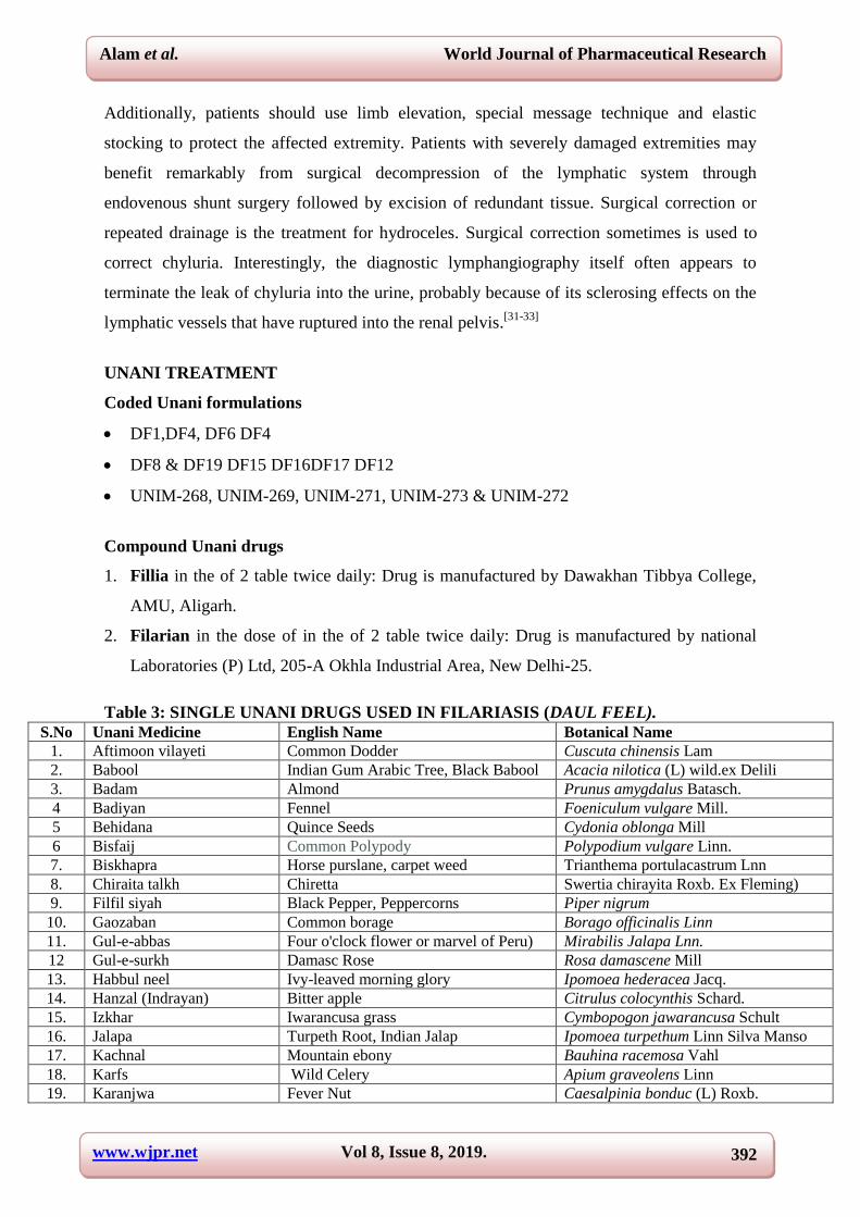
UNANI TREATMENT
Coded Unani formulations
DF1,DF4, DF6 DF4
DF8 & DF19 DF15 DF16DF17 DF12
UNIM-268, UNIM-269, UNIM-271, UNIM-273 & UNIM-272
Compound Unani drugs
1. Fillia in the of 2 table twice daily: Drug is manufactured by Dawakhan Tibbya College,
AMU, Aligarh.
2. Filarian in the dose of in the of 2 table twice daily: Drug is manufactured by national
Laboratories (P) Ltd, 205-A Okhla Industrial Area, New Delhi-25.
Table 3: SINGLE UNANI DRUGS USED IN FILARIASIS (DAUL FEEL).
S.No Unani Medicine English Name Botanical Name
1. Aftimoon vilayeti Common Dodder Cuscuta chinensis Lam
2. Babool Indian Gum Arabic Tree, Black Babool Acacia nilotica (L) wild.ex Delili
3. Badam Almond Prunus amygdalus Batasch.
4 Badiyan Fennel Foeniculum vulgare Mill.
5 Behidana Quince Seeds Cydonia oblonga Mill
6 Bisfaij Common Polypody Polypodium vulgare Linn.
7. Biskhapra Horse purslane, carpet weed Trianthema portulacastrum Lnn
8. Chiraita talkh Chiretta Swertia chirayita Roxb. Ex Fleming)
9. Filfil siyah Black Pepper, Peppercorns Piper nigrum
10. Gaozaban Common borage Borago officinalis Linn
11. Gul-e-abbas Four o'clock flower or marvel of Peru) Mirabilis Jalapa Lnn.
12 Gul-e-surkh Damasc Rose Rosa damascene Mill
13. Habbul neel Ivy-leaved morning glory Ipomoea hederacea Jacq.
14. Hanzal (Indrayan) Bitter apple Citrulus colocynthis Schard.
15. Izkhar Iwarancusa grass Cymbopogon jawarancusa Schult
16. Jalapa Turpeth Root, Indian Jalap Ipomoea turpethum Linn Silva Manso
17. Kachnal Mountain ebony Bauhina racemosa Vahl
18. Karfs Wild Celery Apium graveolens Linn
19. Karanjwa Fever Nut Caesalpinia bonduc (L) Roxb.

www.wjpr.net Vol 8, Issue 8, 2019.
Alam et al. World Journal of Pharmaceutical Research
393
20. Kasni Chicory, Succory, Wild Endive Cichorium intybus Linn
21. Kataikhurd Yellow Berried Nightshade Solanum zanthocarpum Schrad
22. Khare khasak Small Caltrops Tribulus terrestris Linn
23. Khayar Shambar (Amaltas) Indian Laburnum Cassia fistula Linn.
24. Khatmi Marshmallow Althaea officinalis Linn
25. Mundi East Indian Globe Thistle Spnaeranthus indicus Linn
26. Naeem Margosa Tree Aadirachta idica Linn. Juss
27. Sana Senna, Arabian senna Cassia angustifolia Vahl.
28. Sarphoka Wild Indigo Tephrosia purpura Linn.
29. Turbud Indian jalap,Turpeth Merremia turpethum Linn
30. Unnab Jujube Fruit Zizyphus sativa Gaertn.
31. Ushba Indian Sarsaparilla Hemidesmus indicus (L) R.Br.
32. Zaitoon Olive Olea europaea Linn [34]
PREVENTION
Prevention includes giving medicine that kill the microscopic worms to the entire community
in the areas where the infection prevalent. Avoiding mosquito bites is another form of
prevention. These mosquitoes usually bite between the hours of dusk and dawn. One can
follow these steps, if living in an infected area.
Sleep under a mosquito net
Use mosquito repellents in the exposed skin
Take a yearly dose of medicine that kills the worms in the blood.[35-36]
RISK OF FILARIASIS
Over 120 million have already been affected by it, over 40 million of them are seriously
incapacitated and disfigured by the disease. One-third of the people infected with the disease
live in India, one third are in Africa and most of the remainder are South Asia, the Pacific and
America. In communities where the condition is endemic, 10-50% of men and up to 10% of
women can be affected. Through the infection is generally acquired early in childhood, the
disease may take years to manifest itself.[37-38]
CONCLUSION
Filariasis is an infection caused by a parasite worm and is transmitted by insect bite. It is
more prevalent in the tropical and subtropical areas of Africa and Asia Central and South
America. In India, it is common Uttar Pradesh Bihar Orissa Tamil Nadu. Lymphatic filariasis
affects more than 120 million people worldwide, over 40 million of these are seriously
incapacitated and disfigured by the disease. This disease spreads from person to person by
mosquito bites. When a mosquito bites an infected person, microscopic worms circulating in
this blood enter and infect the mosquito. These worms then pass to the other person when this

www.wjpr.net Vol 8, Issue 8, 2019.
Alam et al. World Journal of Pharmaceutical Research
394
infected mosquito bite him. The worms transferred from the mosquito, move through the
skin, and travel to lymph vessels, where they grow into adults, and adult worm lives fro about
seven years. The adult worms mate and release millions of microscopic worms in to the
blood. There are eight different types of this worm, out of which three are responsible for
causing the diseases. 1. Wucheria bancrifti and Brugia malayi cause lymphatic filariasis and
Onchocera volvulus cause Onchocerciasis (River blindness). The medications started at low
doses to prevent reactions caused by large numbers of dying parasites. These medications can
cause severe side effects in up-to 70% of patients. These side effects can be controlled with
anti-histamines and anti- inflammatory drugs (corticosteroids). Rarely, treatment with Diethyl
carbamazine may leads to a fatal inflammation of the brain (encephalitis). The proper
diagnosis is done by new advance technology to filariasis after that the treatment may given
to patient and post treatment evidence also recorded. The present research in filariasis is fully
deepened on old methods of diagnosis and evaluated by one parameter e.g. water
displacement method. In this article we emphasize the research work should be done with
new advance technology for diagnosis and scientifically prove the efficacy of Unani drugs.
ACKNOWLEDGMENT
The authors are thankful to Director General, CCRUM, New Delhi, Ministry of AYUSH
Ayurveda, Yoga & Naturopathy, Unani, Siddha and Homoeopathy (AYUSH), Health and
Family Welfare, Govt. of India, for providing support and all staff members of Regional
Research Institute of Unani Medicine (RRIUM), Bhadrak, Odisha, for cooperation and
support in this study.
REFERENCES
1. Nicolas L, Pichart C, Nguyen LN, Moulia Pelat JP. Reduction of Wuchereria bancrofti
adult worm circulating antigen after annual treatments of diethylcarbamazine combined
with ivermectin in French Poynesia. J infect Dis, 1997; 175: 489.
2. Pani SP, Hoti SL, Vanamail P, Das LK. Comparison of an immunochromatographic card
test with night blood smear examination for detection of Wuchereria bancrofti
microfilaria carriers. Natl Med J India, 2004; 17: 304.
3. Arzani MA. 1929. Tibb-e-Akbar. Munshi Nawal Kishore Press, Lucknow, 2: 167.
4. Kabiruddin M.1980. Tarjuma Sharah-e-Asbab. 9th
Edn. Hikmat Book Depot, Hyderabad,
3: 210-212.
www.wjpr.net Vol 8, Issue 8, 2019.
Alam et al. World Journal of Pharmaceutical Research
395
5. Khan MA. 1885. Ikseer-e-Azam. 2nd
Edn., Munshi Nawal Kishore Press, Liuchow, 4:
11-12.
6. Weil GJ, Lammie PJ, Richards FO, and Eberhard ML. Changes in circulating parasites
antigen levels after treatment of bancroftian filariasis with diethylcarbamazine and
ivermectin. J Infect Dis, 1991; 164: 814-816.
7. WHO Expert Committee on Filariasis. 1984. Lymphatic filariasis fourth report of WHO
Expert Committee on Filariasis. WHO Technical Report Series (World Health
Organization, Geneva), 702: 48-66.
8. WHO Expert Committee on Filariasis. 1992. Lymphatic filariasis the disease and its
control fifth report of the WHO Expert Committee on Filariasis. WHO Technical Report
Series (World Health Organization, Geneva), 821: 8-13, 42-53.
9. World Health Organization. 1994. Strategies for control of lymphatic filariasis infection
and disease: Report of WHO/CTD/TDR consultative meeting held at the University
Sains, Malaysia, Penang 22-24 August 1994 (TDR/CTD/ FIl/PENANG/94.1) World
Health Organization, Geneva.
10. Cook GC, Zumla A. 2003. Manson‟s Tropical Diseases. 21st Edn., Saunders, Elsevier
Sciences, London, 1487-1502.
11. Kasper DL, Braunwald E, Fauci AS, Hauser SL, Longo DL, Jameson JL. 2005.
Harrison‟s Principles of Internal Medicine. 16th
Edn., McGraw-Hill Companies, USA, 1:
1260-1263.
12. Kumar V, Abbas AK, Fausto LD. 2006. Robbins & Cotran Pathologic Basis of Disease,
7th
Edn., Saunders, Elsevier, USA, 409-410.
13. Wyngaarden JB, Smith LH, Bennett JC. 1992. Cecil Textbook of Medicine. 19th
Edn.,
W.B. Saunders Companies, Philadelphia, 2015-2017.
14. Meyrowitsch DW, Simonsen PE, and Makunde WH. Mass Diethylcarbamazine
chemotherapy for control of bancroftian filariasis through community participation:
comparative efficacy of a low monthly dose and medicated salt. Trans R Sco Trop Med
Hyg, 1996; 90: 74.
15. Eberhard ML and Lammie PJ. Laboratory diagnosis of filariasis. Clinical lav Med, 1991;
11: 977-1010.
16. Dreyer G, Pimentel A, Medeiros Z, Beliz F, Moura I, Coutinho A, De Andrade LD,
Rocha A, da Silva LM and Piessens WF. Studies on periodicity and intravascular
distribution of Wuchereria bancrofti microfilaria in paired samples of capillary and
venous blood from Recfic. Brazil Trop Med Int Health, 1996; 1: 264-272.

www.wjpr.net Vol 8, Issue 8, 2019.
Alam et al. World Journal of Pharmaceutical Research
396
17. Sasa M. 1976. Human Filariasis. A global Survey of Epidemiology and control
(University Park Press, Baltimore).
18. Simonsen PE, Neimann L and Meyrowitsch DW. Wuchereria bancrofti in Tanzania
microfilarial periodicity and effect of blood sampling time on microfilarial intensities.
Trop Med Int Hlth, 1997; 2: 153-158.
19. Weil GJ and Liftis F. Identification and partial characterization of a parasite antigen in
sera from humans infected with Wuchereria bancrofti, J Immunol, 1987; 138: 3035-3041.
20. Amaral F, Dreyer G, Figuerodo-Silva J, Noroes J, Cavalcanti A, Samico SC, Santos A
and Coutinho A. Adult worms detected by ultrasonography in human bancroftian filariais.
Amm J Trop Med Hyg, 1994; 50: 753-757.
21. Dreyer G, Amaral F, Noroes J and Medeiros Z. Ultrasonography evidence for stability of
adult worm location in bancroftian filariasis. Trans R Sco Trop Med Hyg, 1994; 88: 558.
22. Dreyer G, Brandao AC, Amaral F, Mederiros Z and Addiss D. Detection by ultrasound of
living adult Wuchereria bancrofti in the female breast. Mem Inst Oswaldo Cruz, 1996;
91: 95-96.
23. More SJ, Copeman DB. A highly specific and sensitive monoclonal antibody based
ELISA for the detection of circulating antigen in bacroftian filariasis. Trop Med
Parasitol, 1990; 41: 403-406.
24. Turner P, Copeman B, Gerisi D, Speare R. A comparison of the Og4C3 antigen capture
ELISA, the Knott test and an IgG4 assay and clinical signs in the diagnosis of bancroftian
filariasis. Trop Med Parasitol, 1993; 44: 45-48.
25. Chanteau S, Moulia-Pelat JP, Glaziou NL. Og4C3 circulating Antigen: A marker of
infection and adult worm burden in Wuchereria bancrofti filariasis. J infect Dis., 1994;
170: 247-250.
26. Lamine PJ, Hightower AW, Eberhard ML. The age specific prevalence of antigenemia in
a Wuchereria bancrofti exposed population. Amm J Trop Med Hyg, 1994; 51: 348-355.
27. Rocha A, Addiss D, Ribiero ME, Noroes J Baliza M, Medeiros Z and Dreyer G.
Evaluations of the Og4C3 ELISA in Wuchereria bancrifti infection: Infected persons
with undeletable or ultra low microfilarial densities. Trop Med Int Hlth, 1996; 1: 859-864.
28. Weil GJ, Lamine PJ, Weiss N. The ICT filariasis Test: A rapid antigen test for diagnosis
of bancroftian filariasis. Parasitol Today, 1997; 13: 401-404.
29. Zhong M, McCarthy J, Bierwert L, Lizott aniewski MR, Chanteau S, Nutman TB,
Ottesen EA and Williams SA. A PCR assay for the detection of the parasite Wuchereria
bancrofti in human blood samples. Am J Trop Mrd Hyg, 1996; 54: 661-662.

www.wjpr.net Vol 8, Issue 8, 2019.
Alam et al. World Journal of Pharmaceutical Research
397
30. Lizotte MR Supali T Partono F, Williams SA. A polymerase chain reaction assay for the
detection of Brugia malayi in blood. Am J Trop Med Hyg, 1994; 51: 314-321.
31. Williams SA, Nicolas L, Lizott-Waniewski M, Plichart C, Luquiaud P, Nguyen NL and
Mouila Pelt JP. Use of a polymerase chain reaction assay for the detection of Wuchereria
bancrofti in blood samples from Tahiti. Trans R Soc Trop Med Hyg, 1996; 89: 225-226.
32. Addiss DG Beach MI, Streit TG. Randomized placebo-controlled comparison of
ivermectin and albendazle alone and in combination for Wuchereria bancrofti
microfilraemia in Haitian Children. Lancet, 1997; 350: 480.
33. Ottesen EA and Campbell WC. Ivermectin in human medicine. J Antimicrob Chemother,
1994; 34: 195.
34. Anonymous. Workshop on filariasis 1992 Workshop Proceeding on Filariasis, Organized
by CCRUM, New Delhi, 20-21 January, Madras.
35. Maizels RM, Bundy DA Selkirk ME. Immunological modulation and evasion by
helminthes parasites in human populations. Nature, 1993; 365: 797.
36. Klion AD, Ottesen EA and Nutman TB. Effectiveness of Diethylcarbamazine in treating
loiasis acquired by expatriate visitors to endemic regions: long term follow-up. J. Infect
Dis., 1994; 169: 604.
37. http://www.cdc.gov/ncidod/dpd/parasites/lymphaticfilariasis/treatment_lymphatic_filar.ht
m.
38. Meyrowitsch DW, Simonsen PE and Makunde WH. Mass Diethylcarbamazine
chemotherapy for control of bancroftian filariasis through community participation:
comparative efficacy of a low monthly dose and medicated salt. Trans R Sco Trop Med
Hyg, 1996; 90: 74.