Embed Size (px)
Citation preview
A REVIEW OF SONOGRAPHIC IDENTIFICATION OF ABDOMINAL BLOOD VESSELS AND JUXTAVASCULAR ORGANS
KATHY A. SPAULDING, DVM
Abdominal vasculature can be evaluated non-invasively using 2-D ultrasound imaging and Doppler u l t r a s o n ~ g r a p h y . ~ , ~ ~ ~ ~ The identification of abdominal vessels using ultrasound is based on knowledge of their normal location, appearance and relationship to specific organs. Because anatomic location of major abdominal vessels is fairly consistent, finding and following vessels is a useful aid when attempt- ing to locate and evaluate juxta-vascular organs. Sonographic vascular evaluation may be beneficial in providing information regarding central cardiovascular abnormalities, detection of abnormalities in- volving the vessel interrogated, or detection of abnormalities of the parenchymal bed that the vessel
Knowledge of the location and appearance of the vessels is necessary for this information to be of diagnostic value.
In this paper, abdominal vessels in the dog are reviewed and a sonographic map of major abdominal vessels is described. This includes the sonographic appearance of the aorta, caudal vena cava, and portal vein, their major branches and their anatomic relationship with adjacent organs.
Identification of the medial iliac lymph nodes, mesenteric lymph nodes, abdominal esophagus, du- odenum, spleen, kidneys, pancreas, adrenal glands and liver lobes are assisted by recognizing specific vascular landmarks. The locations and appearances of pertinent organs as pertains to abdominal vasculature (caudal vena cava and aorta) are described. Veterinary Radiology & Ultrasound, Vol. 38, No. 1, 1997, p p 4-23.
Key Words: abdominal ultrasound, vessels, Doppler, canine.
Introduction bdominal vessels are typically standard in location. A Knowledge of the normal appearance and location of
the abdominal aorta, caudal vena cave, portal vein and their main branches is critical. Abnormalities in organs sup- plied by these vessels often result in changes within the vessel. Variations in the normal course of a vessel may be the result of a congenital anomaly or reflect an abnormality in a surrounding organ. Organs difficult to identify are lo- cated with the aid of standard vessels in close proximity or vessels supplying that organ. Interrogation of these vessels functions in assessing normalcy or alteration of blood flow to specific organs and are useful as landmarks for organ location and congenital vascular variation^.^-'^
The purpose of this article is to describe the sonographic appearance of the course of major abdominal vessels, how
From the Department of Anatomy, Physiological Sciences and Radiol- ogy, College of Veterinary Medicine, North Carolina State University.
Address correspondence and reprint requests to Kathy Spaulding, Col- lege of Veterinary Medicine, North Carolina State University, Raleigh, NC 27606.
Received April 17, 1995; accepted for publication December 1, 1995.
to locate these vessels and how to use these vessels as landmarks for finding specific abdominal organs.
This paper describes the sonographic appearance of the aorta, the caudal vena cava, their major branches and thus provides a map of the abdomen to aid in examining the abdomen. (Fig. 1 and 2). The vessels functioning as land- marks to aid in identification of the medial iliac and mes- enteric lymph nodes, adrenal glands, kidneys, esophagus, and to a lesser extent the spleen, pancreas, intestinal tract and liver lobes are described.
The overall anatomic location and sonographic appear- ance of the vessels is presented as viewed initially from the left in a right lateral decubitus position followed by exam- ination on the right in a left lateral decubitus position. The imaging plane, anatomic location of major abdominal ves- sels, the sonographic description of the vessels and sur- rounding organs, and imaging techniques are reported.
Even though this paper indicates specific views or angles to image a vessel or structure, these should be considered rules of thumb by which to begin the examination. The sonographer may then adapt the imaging planes to the ves- sel being interrogated in a specific situation. Eliminating confusion of vessel identification may be accomplished by knowing the sonographic location and appearance, obtain-
4

VOL. 38, No. 1 SONOGRAPHIC IDENTIFICATION OF ABDOMEN 5
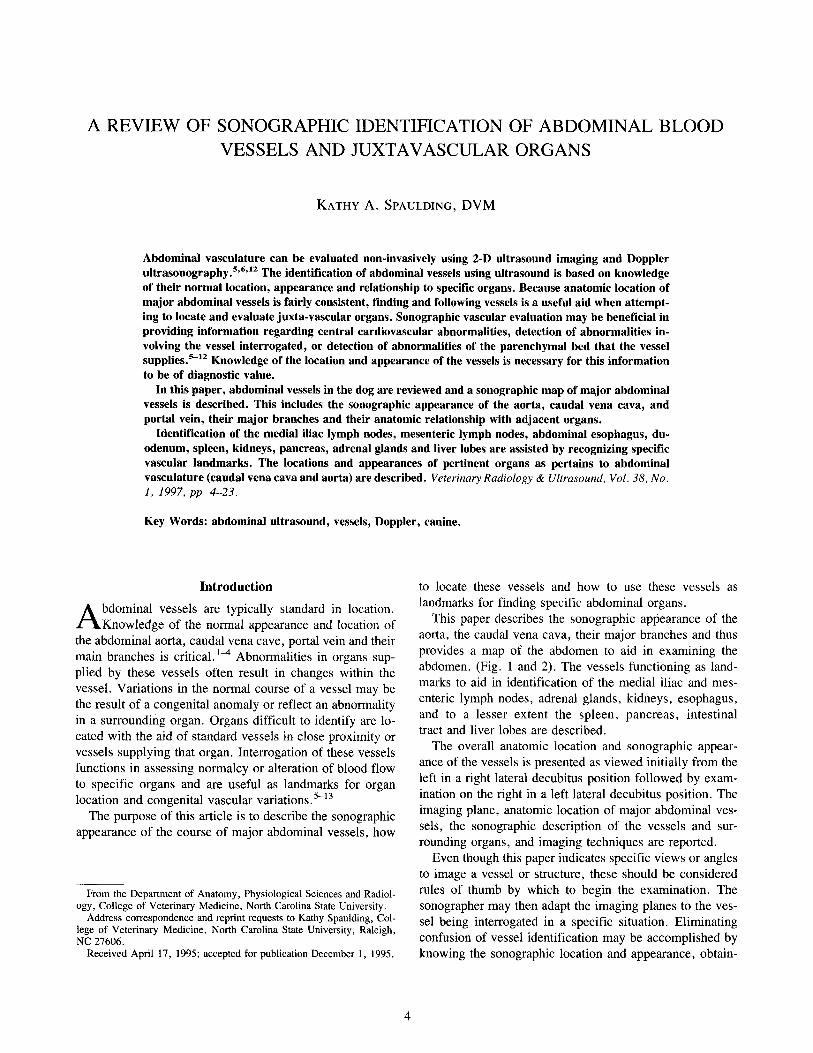
FIG. 1. The caudal vena cava and the aorta with their major branches are portrayed in this image. This lateral radiograph with a rendition of the location and relationships of the major abdominal systemic vessels as they are positioned from a lateral perspective provides a road map to the abdomen to be used as an anatomic reference for imaging a segment of the abdomen with ultrasound.
ing the characteristic arterial or venous Doppler wave-form or signal, noting direction of flow, or by tracing the inter- rogated vessel to the parent aorta or caudal vena cava. Ex- amination of these major vessels should be included in rou- tine abdominal studies.
The Abdominal Systemic Circulation
Sonographic Evaluation
The position of the patient will vary depending on the experience and preference of the sonologist performing the examination. It is most important to form a consistent scan- ning pattern. Developing a routine scanning procedure will be unique for each sonologist.
The lateral decubitus position is preferred by the author as the starting point for the examination. This position is advantageous because of the close proximity of the vessels to the transducer and because intestinal gas is usually not in
the imaging plane. Animals are relatively comfortable in lateral recumbency and rarely sneeze which is more com- mon when they are confined in a ventrodorsal position. In addition, lateral recumbency allows for flexibility and ver- satility in accessing the abdomen dorsally from the retro- peritoneal space to the ventrally located abdominal viscera. Additional oblique or sagittal imaging planes required to fully evaluate abdominal vessels are easily obtained with the dog in lateral recumbency. Imaging both right and left sides ensures all intra-abdominal structures are seen and allows many structures to be interrogated from multiple scan planes.
A thorough examination of each major vessel should be attempted. This is much easier in small dogs or cats with a relaxed abdomen. Strict adherence to a rigid examination procedure will not be possible in every patient. To assure completeness and consistency, it is best to attempt to follow the same examination procedure in each patient. However,
6 SPAULDINC 1997
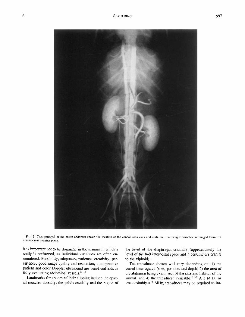
FIG. 2. This portrayal of the entire abdomen shows the location of the caudal vena cava and aorta and their major branches as imaged from this ventrodorsal imaging plane.
it is important not to be dogmatic in the manner in which a study is performed, as individual variations are often en- countered. Flexibility, adeptness, patience, creativity, per- sistence, good image quality and resolution, a cooperative patient and color Doppler ultrasound are beneficial aids in fully evaluating abdominal vessel^.^-'^
Landmarks for abdominal hair clipping include the epax- ial muscles dorsally, the pelvis caudally and the region of
the level of the diaphragm cranially (approximately the level of the 8-9 intercostal space and 5 centimeters cranial to the xiphoid).
The transducer chosen will vary depending on: 1) the vessel interrogated (size, position and depth) 2) the area of the abdomen being examined, 3) the size and habitus of the animal, and 4) the transducer a ~ a i l a b l e . ~ - ' ~ A 5 MHz, or less desirably a 3 MHz, transducer may be required to im-
VOL. 38, No. 1 SONOGRAPHIC IDENTIFICATION OF ABDOMEN 7
age major vessels in very large or obese dogs. However, a frequency of 7 MHz or higher is recommended for best image quality. In general, the highest MHz transducer available that will be able to penetrate to the level of the vessel desired to be interrogated should be used.99" A sec- tor type of transducer may be used; however, a linear array transducer or curved array technology designed for broad, near field imaging provides for better continuity when iden- tifying and following superficial vessels. Sector transducers are often useful for evaluating deep abdominal vessels. Of- ten both sector and linear transducers will be needed to thoroughly examine all desired abdominal vessels. Even though gastrointestinal contents may not prevent a satisfac- tory examination, it is recommended that the patient be fasted for eight to twelve hours prior to sonography as ar- tifacts produced from ingesta and gas within the stomach may impede adequate evaluation. Cleansing enemas or bar- ium studies prior to the examination are not recommended as they result in reflective and resorptive artifacts, respec- tively.
For a dorsal or sagittal plane, the transducer or the image is positioned so that the cranial aspect of the dog (organ) is to the left of image displayed on the monitor. On a trans- verse plane from either the right or left side, the dorsal aspect of the dog (organ) is shown to the left of the image. If the transverse image is made from the midline, the right side of the dog (organ) is shown on the left of the displayed
image. Hard copy images should be sufficiently labeled so that one can easily ascertain the structure depicted.
In general, the appropriate imaging plane is the one which best accentuates the long axis of the interrogated vessel or allows the examiner to follow the vessel to a specific organ.
Sonographic Examination of the Systemic Vasculature from the Left
Aorta, Caudal Vena Cava
The examination is initiated in the caudodorsal abdomen with the animal positioned in a right lateral decubitus posi- tion. The area caudal to the kidneys is generally an acces- sible part of the abdomen where the aorta and caudal vena cava are easily located and thus serves as a consistent region to begin the examination. The descending thoracic aorta becomes the abdominal aorta as it enters the dorsocranial portion of the abdominal cavity through the aortic hiatus, dorsal to the diaphragm and in the mid-sagittal plane of the retroperitoneal cavity. It is enclosed dorsally by the pars lumbalis muscle and ventrally by the left and larger right crura of the diaphragm. As the abdominal aorta continues caudally, it is located in a ventral furrow formed by the right and left iliopsoas muscles. The cranial two thirds of the abdominal aorta is located on midline and to the left of the
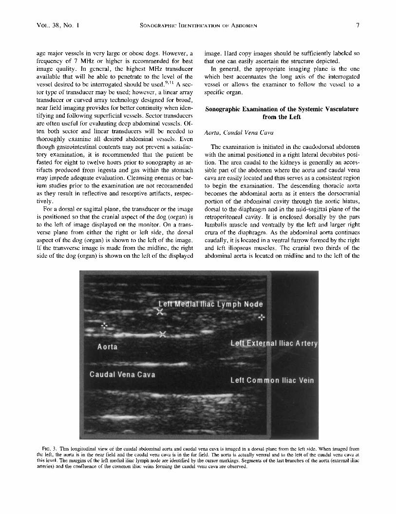
FIG. 3. This longitudinal view of the caudal abdominal aorta and caudal vena cava is imaged in a dorsal plane from the left side. When imaged from the left, the aorta is in the near field and the caudal vena cava is in the far field. The aorta is actually ventral and to the left of the caudal vena cava at this level. The margins of the left medial iliac lymph node are identified by the cursor markings. Segments of the last branches of the aorta (external iliac arteries) and the confluence of the common iliac veins forming the caudal vena cava are observed.
8 SPAULDING 1997
more ventrally located caudal vena cava. The aorta reverses its relative dorsal position with the caudal vena cava near the level of L5-6 intervertebral disc space and origin of the deep circumflex arteries and then courses ventral and slightly to the left of the caudal vena cava. The division of the aorta into the right and left internal iliac arteries is ventral to the left common iliac vein, and though variable, it is usually slightly caudal to the point where the right and left common iliac veins unite to form the caudal vena cava. ' ,3
The major branches originating from the aorta (from cra- nial to caudal) are the celiac, cranial mesenteric, paired phrenicoabdominal, paired renal, testicular/ovarian, paired lumbar, caudal mesenteric, paired deep circumflex iliac, and paired external iliac arteries. Variations in number, ori- entation and location are uncommon but can occur. 1-3
The caudal vena cava is formed by the confluence of the common iliac veins in the caudodorsal abdomen. Beginning
near the level of the caudal pole of the left kidney and coursing cranially, the caudal vena cava assumes a gradual cranioventral course that results in a divergent path from the aorta and lumbar spine. The caudal vena cava passes through the foramen venae cavae of the diaphragm, right of midline, in middorsal segment of the diaphragm. As the caudal vena cava passes through the liver, it is surrounded by the caudate and right lateral hepatic lobes.
The major tributaries contributing to the caudal vena cava (caudal to cranial), in addition to the common iliac veins, include paired deep circumflex iliac, right testicularl ovarian, paired renal and phrenicoabdominal, and multiple hepatic veins.
The caudal two-thirds of the caudal vena cava and aorta can be imaged from either the right or left side. The cranial one-third of the aorta is usually best imaged from the right side, due the presence of gas within the fundus of the stom- ach when the dog is in right lateral recumbency. The caudal
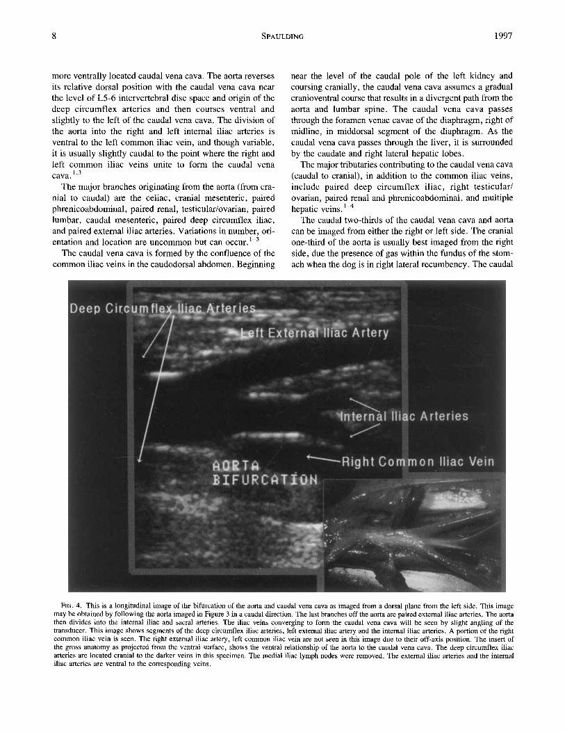
FIG. 4. This is a longitudinal image of the bifurcation of the aorta and caudal vena cava as imaged from a dorsal plane from the left side. This image may be obtained by following the aorta imaged in Figure 3 in a caudal direction. The last branches off the aorta are paired external iliac arteries. The aorta then divides into the internal iliac and sacral arteries. The iliac veins converging to form the caudal vena cava will be seen by slight angling of the transducer. This image shows segments of the deep circumflex iliac arteries, left external iliac artery and the internal iliac arteries. A portion of the right common iliac vein is seen. The right external iliac artery, left common iliac vein are not seen in this image due to their off-axis position. The insert of the gross anatomy as projected from the ventral surface, shows the ventral relationship of the aorta to the caudal vena cava. The deep circumflex iliac arteries are located cranial to the darker veins in this specimen. The medial iliac lymph nodes were removed. The external iliac arteries and the internal iliac arteries are ventral to the corresponding veins.
VOL. 38, No. 1 SONOCRAPHIC IDENTIFICATION OF ABDOMEN 9
vena cava and branches, excluding the left renal and left hepatic veins, are often best imaged from the right side of the animal.
Locating the aorta and caudal vena cava can be accom- plished by positioning the transducer over the caudodorsal abdomen ventral to the epaxial muscles and caudal to the left kidney. Oriented in a dorsal plane, the transducer is slowly fanned in a ventral to dorsal direction until the ves- sels are seen. The vessels will be in close proximity to each other. The transducer is maneuvered to optimize the image of the longitudinal axis of the cranial to caudal orientation of these vessels. Following these vessels often requires con- tinual, slight adjustments in the orientation of the transducer and TGC settings. Fanning (hold the transducer in a fixed position on the skin, maintain contact and use a sweeping motion as an area or organ is examined), sliding (maintain
the transducer in the same plane and with continued contact and imaging of a structure move the fixed position of the transducer), pointing (hold the transducer in a fixed position and angle the transducer in a single, narrow-focused, direc- tion), and rotating (maintain contract, turn the transducer in a clockwise or counterclockwise direction) the transducer are maneuvers performed to optimize the image of the ves- sel and to follow the selected vessel.
When imaging in a dorsal plane from the left caudodorsal abdomen, the aorta will be in the near field and the caudal vena cava will be in the far field. The aorta and caudal vena cava have sonographic characteristics which allow distinc- tion between them.15 The vessels appear as two parallel, tubular structures with an anechoic lumen and hyperechoic walls. The caudal vena cava has approximately the same diameter as the corresponding segment of the aorta (Fig. 3).
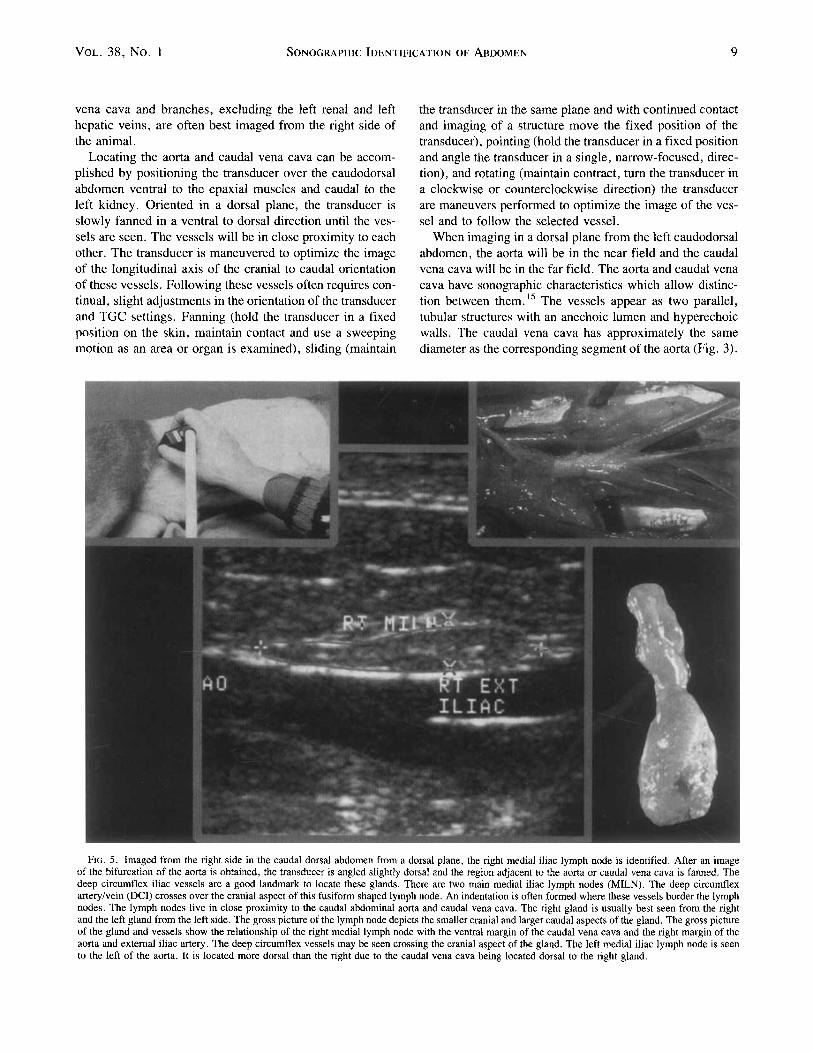
FIG. 5. Imaged from the right side in the caudal dorsal abdomen from a dorsal plane, the right medial iliac lymph node is identified. After an image of the bifurcation of the aorta is obtained, the transducer is angled slightly dorsal and the region adjacent to the aorta or caudal vena cava is fanned. The deep circumflex iliac vessels are a good landmark to locate these glands. There are two main medial iliac lymph nodes (MILN). The deep circumflex arterylvein (DCI) crosses over the cranial aspect of this fusiform shaped lymph node. An indentation is often formed where these vessels border the lymph nodes. The lymph nodes live in close proximity to the caudal abdominal aorta and caudal vena cava. The right gland is usually best seen from the right and the left gland from the left side. The gross picture of the lymph node depicts the smaller cranial and larger caudal aspects of the gland. The gross picture of the gland and vessels show the relationship of the right medial lymph node with the ventral margin of the caudal vena cava and the right margin of the aorta and external iliac artery. The deep circumflex vessels may be seen crossing the cranial aspect of the gland. The left medial iliac lymph node is seen to the left of the aorta. It is located more dorsal than the right due to the caudal vena cava being located dorsal to the right gland.
10 SPAULDING 1997
The caudal vena cava usually has a thinner wall and is more compressible from the aorta. Mobile echoes created by blood flow (laminar flow) within the vessels can be visual- ized with 2D imaging, especially in animals with a slow heart rate. Cardiac induced pulsatile motion is often ob- served in the aorta or iliac arteries. Due to the close prox- imity between the aorta and caudal vena cava, the aortic pusatile motion may be transferred to the caudal vena cava. Flow within the cauda vena cava will be towards the heart (away from the transducer) and flow within the aorta will be directed caudally toward the pelvic limbs (towards the transducer). Doppler (color or spectral) ultrasound is a use- ful aid in following and imaging the abdominal vessels, especially small vessels. Doppler ultrasound can be used to determine if there is blood flow, the direction of flow, ve- locity, and turbulence. Each vessel has a characteristic spectral Doppler pattern, which serves as its signature.
Iliac Vessels
Maintaining the longitudinal view of the aorta in the im- age, the transducer is moved (slid) caudally. The last major
aortic branches, the right and left external iliac arteries, diverge caudolaterally from the aorta at approximately the level of the sixth lumbar vertebrae and course caudolaterally to the respective pelvic limb. The aorta terminates into right and left internal iliac and median sacral a r t e r i e ~ . ” ~ (Fig. 4). The umbilical artery arises from the ventral surface of the internal iliac arteries about 0.5 cm from its origin or from the terminal portion of the aorta. The termination of the aorta and the bifurcation of the external iliac arteries is usually slightly caudal to the formation of the caudal vena cava by the common iliac veins.
Because of the close proximity of the vessels, the identity of the iliac veins and the arteries can become confusing. Tracing these vessels to the aorta or caudal vena cava or using Doppler ultrasound to distinguish between the pulsa- tile triphasic appearance of the arterial flow and the monophasic venous flow are both methods that will dis- criminate between the vessels.
Medial Iliac Lymph Nodes The aorta is located ventral to the caudal vena cava and
slightly to the left of midline in the caudal abdomen. Two
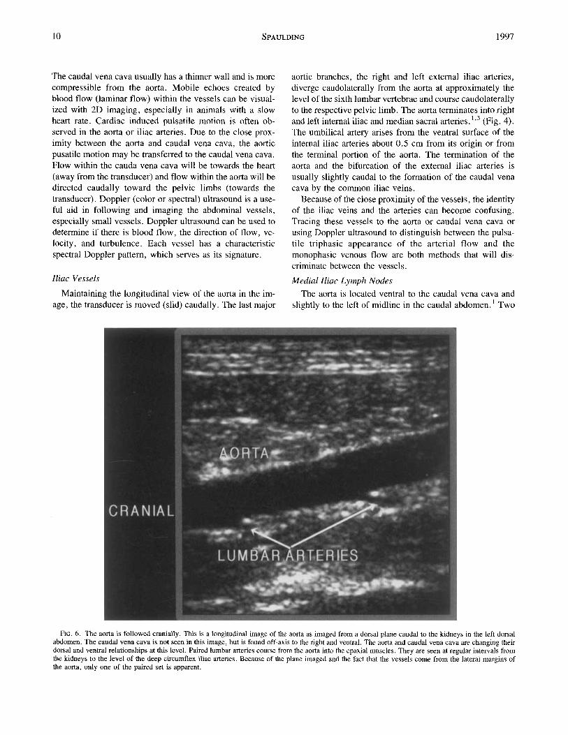
FIG. 6. The aorta is followed cranially. This is a longitudinal image of the aorta as imaged from a dorsal plane caudal to the kidneys in the left dorsal abdomen. The caudal vena cava is not seen in this image, but is found off-axis to the right and ventral. The aorta and caudal vena cava are changing their dorsal and ventral relationships at this level. Paired lumbar arteries course from the aorta into the epaxial muscles. They are seen at regular intervals from the kidneys to the level of the deep circumflex iliac arteries. Because of the plane imaged and the fact that the vessels come from the lateral margins of the aorta, only one of the paired set is apparent.
VOL. 38, No. I SONOGRAPHIC IDENTIFICATION OF ABDOMEN 11
major lymph nodes are identified in the iliosacral lympho- center. The left and right medial iliac lymph nodes are located adjacent to the lateral (abaxial) margins of the caw dal aorta and corresponding external iliac artery. The lymph nodes extend cranial to the deep circumflex iliac vessels and caudal to the level of the branching of the external and internal iliac vessels. The right medial iliac lymph node,
located on the ventrolateral surface of the caudal vena cava and right common lilac vein, is positioned more ventral than the left medial iliac lymph node which lies adjacent to the more dorsal aorta and left external iliac artery (Fig. 5). Normal lymph nodes can be routinely identified. The pa- renchyma of these glands generally has a homogeneous echogenic appearance. The overall echogenicity is mildly
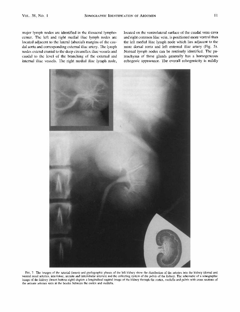
FIG. 7. The images of the arterial (insert) and pyelographic phases of the left kidney show the distribution of the arteries into the kidney (dorsal and ventral renal arteries, interlobar, arcuate and interlobular arteries) and the collecting system of the pelvis of the kidney. The schematic of a sonographic image of the kidney (insert bottom right) depicts a longitudinal sagittal image of the kidney through the cortex, medulla and pelvis with cross sections of the arcuate arteries seen at the border between the cortex and medulla.
12 SPAULDING 1997
hypoechoic relative to the surrounding fat. They have a thin hyperchoic capsule that is best seen when imaged perpen- dicular to the ultrasound beam. A hyperchoic line (stripe) often can be seen coursing from the hilus caudomedially through the node. This hyperchoic stripe is due to fat, fas- cia, lymphatic and vascular vessels and folds of the gland arising from the hilus of the gland. The cranial aspect of both lymph nodes has a markedly narrowed region at the level of the deep circumflex vessels. An isthmus is formed between the larger caudal portion of the lymph node and the smaller segment located cranial to the deep circumflex ves- sels. The lymph nodes are generally fusiform in shape. However, they are flatter dorsoventrally and wider medio- laterally and are often mildly indented dorsomedially as they conform to the shape of the adjacent vessel. The dorsal margin of the gland that contacts the vessels is usually flat- ter than the rounded ventral surface. The less frequently seen hypogastric lymph nodes are small, round, variably numbered, lymph nodes that lie in the angle between the internal iliac vessels and the median sacral artery. Aortic
lymph nodes are located throughout the length of the aorta, but are infrequently identified unless enlarged.
Deep Circumflex Iliac VesselslCaudal Mesenteric Artery
At the level just ventral to the 5-6th lumbar disc space, paired right and left deep circumflex iliac vessels arise from the lateral surface of the aortakaudal vena cava slightly cranial to the origin of the external iliac a r te r ie~ .”~ They course laterally across the ventral surface of the epaxial muscles. These vessels are small, requiring high resolution equipment (7-10 MHz), or color Doppler ultrasound for detection. They are best visualized as they cross the isthmus of the medial iliac lymph nodes. The single caudal mesen- teric artery, which supplies the distal colon, arises from the ventral surface of the aorta slightly cranial to the deep cir- cumflex iliac arteries. Because of its small size, this vessel is inconsistently observed. This vessel may be mistaken for the lumbar vessels or the slightly more lateral and caudal position of the deep circumflex iliac vessels. However, it is
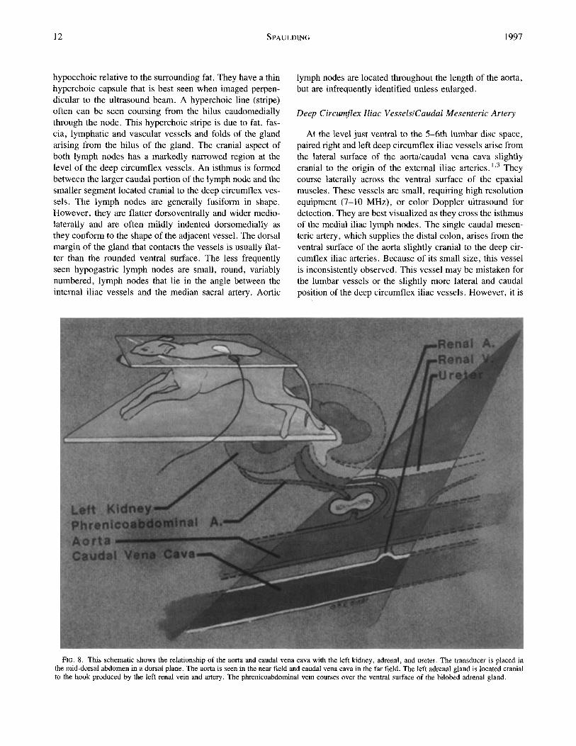
FIG. 8. This schematic shows the relationship of the aorta and caudal vena cava with the left kidney, adrenal, and ureter. The transducer is placed in the mid-dorsal abdomen in a dorsal plane. The aorta is seen in the near field and caudal vena cava in the far field. The left adrenal gland is located cranial to the hook produced by the left renal vein and artery. The phrenicoabdominal vein courses over the ventral surface of the bilobed adrenal gland.
VOL. 38, No. 1 SONOGRAPHIC IDENTIFICATION OF ABDOMEN 13
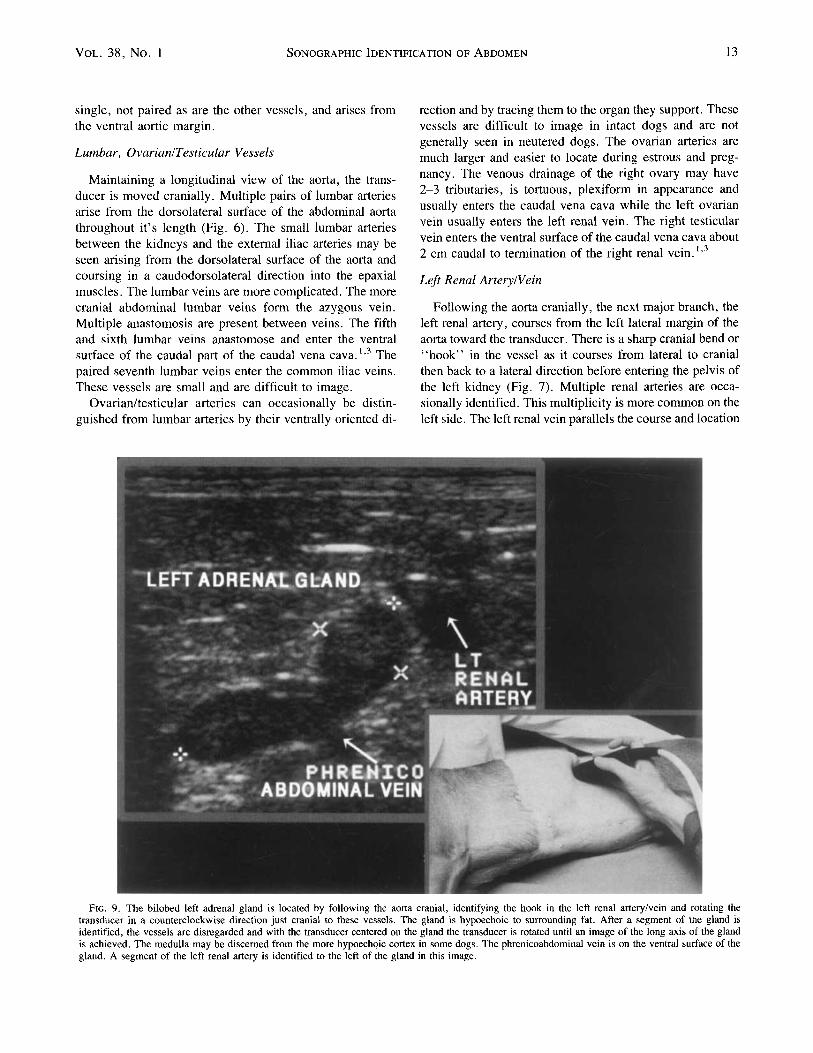
single, not paired as are the other vessels, and arises from the ventral aortic margin.
Lumbar. OvarianlTesticular Vessels
Maintaining a longitudinal view of the aorta, the trans- ducer is moved cranially. Multiple pairs of lumbar arteries arise from the dorsolateral surface of the abdominal aorta throughout it’s length (Fig. 6). The small lumbar arteries between the kidneys and the external iliac arteries may be seen arising from the dorsolateral surface of the aorta and coursing in a caudodorsolateral direction into the epaxial muscles. The lumbar veins are more complicated. The more cranial abdominal lumbar veins form the azygous vein. Multiple anastomosis are present between veins. The fifth and sixth lumbar veins anastomose and enter the ventral surface of the caudal part of the caudal vena ~ a v a . ’ . ~ The paired seventh lumbar veins enter the common iliac veins. These vessels are small and are difficult to image.
Ovarian/testicular arteries can occasionally be distin- guished from lumbar arteries by their ventrally oriented di-
rection and by tracing them to the organ they support. These vessels are difficult to image in intact dogs and are not generally seen in neutered dogs. The ovarian arteries are much larger and easier to locate during estrous and preg- nancy. The venous drainage of the right ovary may have 2-3 tributaries, is tortuous, plexiform in appearance and usually enters the caudal vena cava while the left ovarian vein usually enters the left renal vein. The right testicular vein enters the ventral surface of the caudal vena cava about 2 cm caudal to termination of the right renal vein.’13
Lefl Renal ArterylVein
Following the aorta cranially, the next major branch, the left renal artery, courses from the left lateral margin of the aorta toward the transducer. There is a sharp cranial bend or “hook” in the vessel as it courses from lateral to cranial then back to a lateral direction before entering the pelvis of the left kidney (Fig. 7). Multiple renal arteries are occa- sionally identified. This multiplicity is more common on the left side. The left renal vein parallels the course and location
FIG. 9. The bilobed left adrenal gland is located by following the aorta cranial, identifying the hook in the left renal arteryhein and rotating the transducer in a counterclockwise direction just cranial to these vessels. The gland is hypoechoic to surrounding fat. After a segment of the gland is identified, the vessels are disregarded and with the transducer centered on the gland the transducer is rotated until an image of the long axis of the gland is achieved. The medulla may be discerned from the more hypoechoic cortex in some dogs. The phrenicoabdominal vein is on the ventral surface of the gland. A segment of the left renal artery is identified to the left of the gland in this image.
14 SPAULDING 1997
of the left renal artery. It is longer than the artery, as it must cross the ventral surface of the aorta and proceed right of midline to enter the caudal vena cava. The left renal vein often has a larger diameter than the artery. The left adrenal gland is located cranial to the “hook” in the left renal artery.
Intru-renal Arteries
The renal arteries, before entering the renal hilus, divide into dorsal and ventral branches situated on either side of the renal pelvis and ureter. These primary branches divide into approximately four to six interlobar arteries. The inter- lobar arteries, paralleling the corresponding veins, radiate from the renal pelvis in a uniformly spaced fashion through the medulla with a straight course to the corticomeduallary junction. At the corticomeduallary junction, the interlobar arteries branch into arcuate arteries, which are oriented per- pendicular to the interlobar vessels and follow the contour of the corticomedullary junction. Transverse images of these vessels, located at the border between the cortex and medulla, are often identified on 2-D images by the hyper- echoic, and sometimes shadowing, vessel walls. An end-
on-view of these vessels may mimic foci of parenchymal mineralization. Color Doppler insonation will confirm flow within these vessels. Mineralization within the kidney is usually random in location, while the end-on cross sections of the arcuate arteries are in a fixed, predictable, even dis- tribution at the corticomedullary junction location. The ar- cuate branches divide into multiple interlobular branches and radiate toward the periphery of the renal cortex, to become afferent arterioles. Color Doppler imaging is ben- eficial in differentiating renal vessels from the interlobar vessels distally. The venous drainage of the kidneys is from the interlobular to arcuate veins with drainage into interlo- bar and renal veins and emptying into the caudal vena cava usually slightly caudal to the location of the corresponding renal artery.
Right Renal ArterylVein
The right renal artery arises from the right lateral aspect of the aorta, cranial to the left renal artery. Multiple renal arteries are occasionally identified. The right artery courses into the far field away from the transducer in a right cran- iolateral direction. A hook-shaped course is also present in
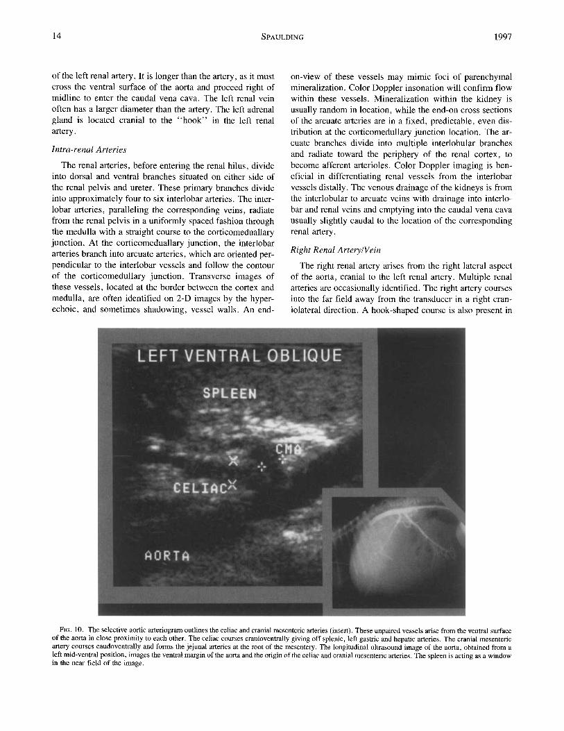
FIG. 10. The selective aortic arteriogram outlines the celiac and cranial mesenteric arteries (insert). These unpaired vessels arise from the ventral surface of the aorta in close proximity to each other. The celiac courses cranioventrally giving off splenic, left gastric and hepatic arteries. The cranial mesenteric artery courses caudoventrally and forms the jejunal arteries at the root of the mesentery. The longitudinal ultrasound image of the aorta, obtained from a left mid-ventral position, images the ventral margin of the aorta and the origin of the celiac and cranial mesenteric arteries. The spleen is acting as a window in the near field of the image.
VOL. 38, No. 1 SONOGRAPHIC IDENTIFICATION OF ABDOMEN 15
the right renal artery, but it is often not as apparent as that observed in the left renal artery. The right renal artery is longer than the right renal vein and the left renal artery, as it must cross the caudal vena cava to reach the kidney. Although the right renal artery may be imaged from the left abdomen in smaller animals, it is often best imaged from the right side. The right renal vein and artery generally follows a similar course through the kidney as on the left.
Left Adrenal Gland
Located cranial to the “hook” in the left renal artery and vein is the left adrenal gland13 (Fig. 8 and 9). To obtain an optimal longitudinal image to the left gland, slow fanning, clockwise rotation or angling of the transducer may be re- quired. Even though the adrenal glands are anatomically located cranial and medial to the kidneys, the left adrenal glands in small dogs, imaged with a linear transducer, ar- tifactually appear to be located caudal to the corresponding kidney. This is due to the oblique image plane used to identify these glands. The adrenal glands, imaged in larger dogs using a sector transducer, are seen cranial and medial to the corresponding kidney. In large dogs, the left kidney and not the left renal artery is often useful as the identifying
landmark for finding the left adrenal gland. The resolution quality is usually best when imaged with the 7.5 MHz linear transducer. However, a 5 MHz sector transducer may be required on large dogs. The left adrenal gland is usually isoechoic relative to the cortex of the kidney and hypo- echoic relative to surrounding fat. When a good quality image is obtained, the cortex and medulla can sometimes be distinguished. When imaged from the left, the left adrenal gland has a “bean” or “peanut” shape with a narrowed mid portion. 1,3,13 In an optimal longitudinal view of the left adrenal gland, sections of the left renal arteryhein are seen caudal and superficial to the caudal pole of the adrenal gland. Segments of the aorta appear in an oblique plane and the left kidney will often not be in the image plane except in large dogs. l 3
Phrenicoabdominal vessels
Multiple small, variable arteries supply the left adrenal gland. The left phrenicoabdominal artery is a small vessel that has a variable point of origin but often arises from the lateral surface of the aorta between the cranial mesenteric and left renal arteries. The left phrenicoabdominal artery crosses the dorsal surface of the narrow, mid-portion of the
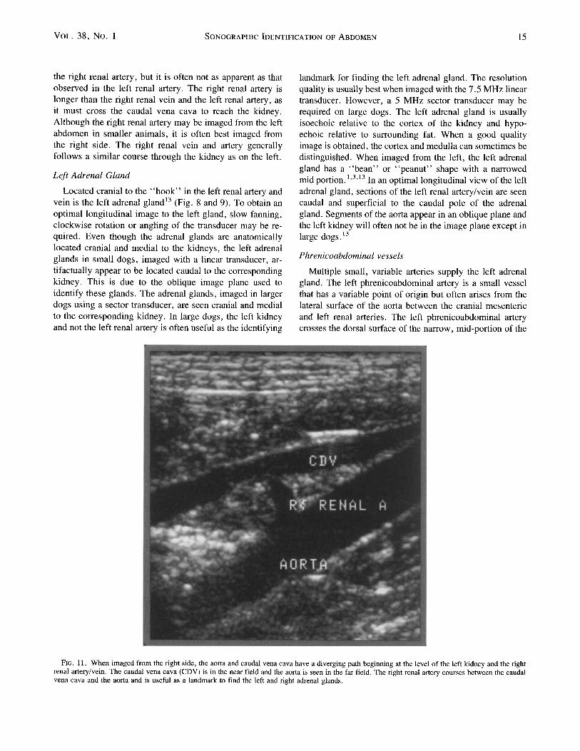
FIG. 11. When imaged from the right side, the aorta and caudal vena cava have a diverging path beginning at the level of the left kidney and the right renal arteryhein. The caudal vena cava (CDV) is in the near field and the aorta is seen in the far field. The right renal artery courses between the caudal vena cava and the aorta and is useful as a landmark to find the left and right adrenal glands.
16 SPAULDING 1997
left adrenal gland, while the corresponding left phrenicoab- dominal vein grooves the ventral, indented surface of the adrenal gland. These vessels are best identified with color Doppler imaging but may be seen on high resolution, gray scale images.
Cranial Mesenteric ArterylCeliac Artery
The cranial mesenteric and celiac arteries are located slightly cranial to the renal arteries. Both unpaired vessels arise from the ventral surface of the aorta with the celiac artery located cranial to the egress of the cranial mesenteric artery. They are in close proximity to each another and, should be identified within the same imaging plane (Fig. 10). Positioning the transducer ventrally over the mid ab- domen using slow fanning, and rotary movement while maintaining dorsal angulation is needed to optimize the lon- gitudinal view of these vessels. The spleen may be used as an imaging window. The cranial mesenteric artery, seen
coursing toward the transducer, continues from the aorta in a caudoventral direction. Its peripheral branches become jejunal arteries and parallel mesenteric veins which form the portal vein at the root of the mesentery. Mesenteric lymph nodes located at the root of the mesentery are seen on both sides of the portal and jejunal veins. These are found just caudal to the plane of the left kidney and in the mid abdo- men. If the transducer is positioned over the ventral mid abdomen and angled dorsally and slightly cranially, while maintaining the image of the aorta, a more longitudinal axis of the cranial mesenteric artery is assumed.
The celiac artery is located a few millimeters cranial to the cranial mesenteric artery. The celiac artery leaves the aorta and courses cranioventrally. There is an acute cranial angulation of the celiac artery within the first centimeter after leaving the aorta. Branches of the celiac artery, in- cluding the splenic, left gastric and especially the hepatic artery, may be followed to the visceral organ they supply, in cooperative, relaxed patients with good resolution Doppler
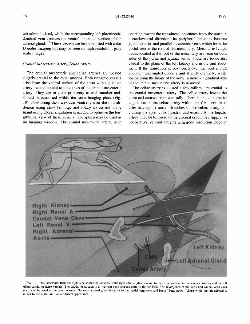
FIG. 12. This schematic from the right side shows the location of the right adrenal gland cranial to the celiac and cranial mesenteric arteries and the left gland caudal to these vessels. The caudal vena cava is in the near field and the aorta in the far field. The divergence of the aorta and caudal vena cava occurs at the level of the renal vessels. The right adrenal gland is closer to the caudal vena cava and has a “bent arrow” shape while the left adrenal is closer to the aorta and has a bilobed appearance.
VOL. 38, No. 1 SONOGRAPHIC IDENTIFICATION OF ABDOMEN 17
equipment and no appreciable gastrointestinal gas in the image plane.
Left lobe of the Pancreas
When assessing the celiac and cranial mesenteric arteries from the left, the stomach may be identified left of the image. The small intestine, with characteristic multiple wall layers, and the spleen, may be identified in the near field. The left lobe of the pancreas is ventral to the celiac artery and often dorsal to the distal aspect of the splenic artery. The cranial pole of the left kidney, the dorsal aspect of the proximal extremity of the spleen, the dorsocaudal margin of the stomach, the transverse image of the proximal descend- ing colon in the right ventral aspect of the image and the splenic vein, are landmarks for identification of the left limb of the pancreas. The left lobe of the pancreas appears as a subtle, slightly hypoechoic linear organ lying adjacent to the caudomedial margin of the spleen, caudal to the splenic vein and extending craniomedially into the far field ventral
to the portal vein. Arterial and venous blood supply, within the left lobe of the pancreas, arise from the pancreatic branch of the splenic arteryhein. These vessels, identified as tubular structures with an anechoic center, a hyperechoic wall are positioned within the center of the gland, parallel the long axis of the gland.
Sonographic Examination of the Systemic Vasculature from the Right Side
The animal is positioned in a left lateral decubitus posi- tion. The examination is begun with the transducer posi- tioned over the dorsocaudal abdomen caudal to the kidneys.
AortalCaudul Venu Cava (AO, CDV)
Once the aorta and caudal vena cava are identified, the transducer is shifted caudally, while the caudal vena cava is maintained in longitudinal view. The two major vessels converging from the pelvic limbs to form the caudal vena cava are the right and left common iliac veins. They parallel
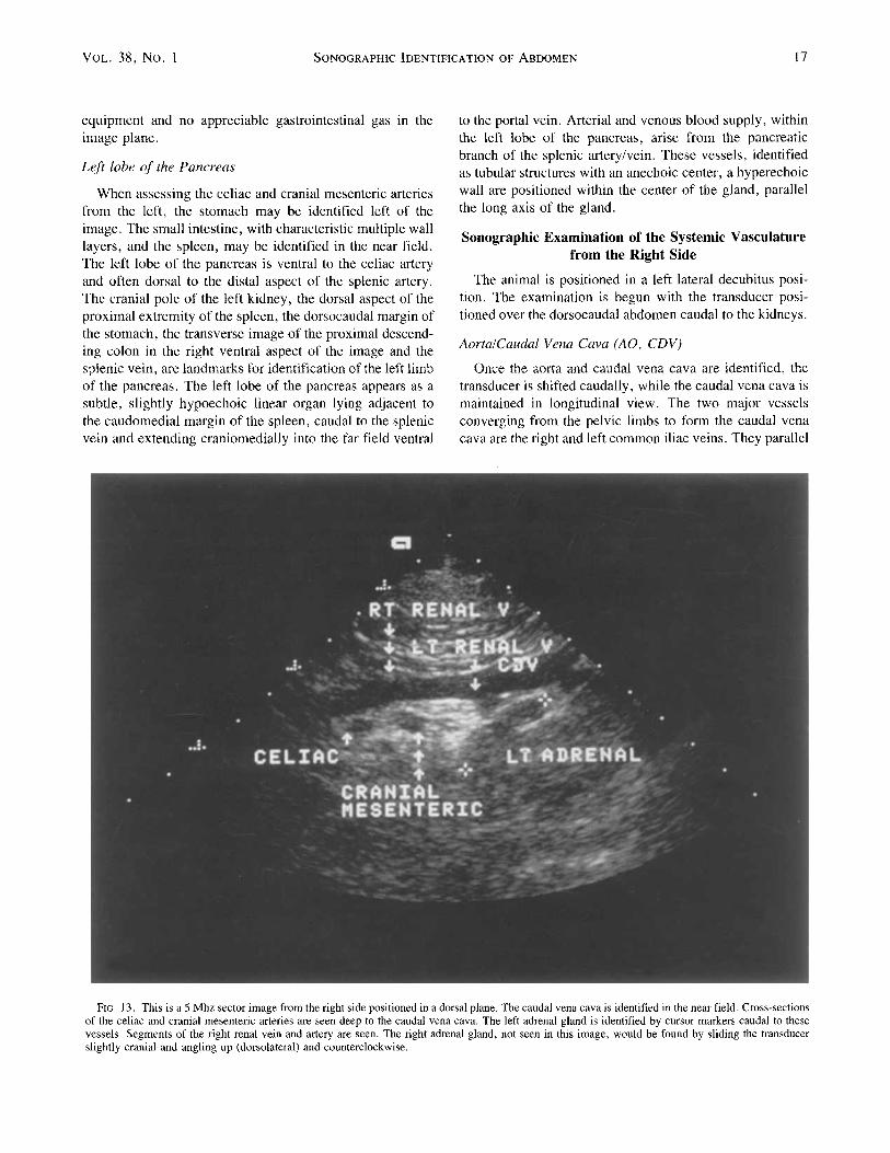
FIG. 13. This is a 5 Mhz sector image from the right side positioned in a dorsal plane. The caudal vena cava is identified in the near field. Cross-sections of the celiac and cranial mesenteric arteries are seen deep to the caudal vena cava. The left adrenal gland is identified by cursor markers caudal to these vessels. Segments of the right renal vein and artery are seen. The right adrenal gland, not seen in this image, would be found by sliding the transducer slightly cranial and angling up (dorsolateral) and counterclockwise.
18 SPAULDING 1997
the external and internal iliac arteries. The caudal vena cava is dorsal and slightly to the right of the caudal aorta, with the left common iliac vein usually directly dorsal to the mid-portion of the caudal aorta. The iliac veins join to form the caudal vena cava, usually slightly cranial to the bifur- cation of the aorta. The right medial iliac lymph node is located lateral to the aorta and right external iliac artery and ventral to the caudal vena cava. The right medial iliac lymph node is often easier to locate than the left. Presum- ably, this is related to its more ventral location, due to the presence of the dorsally positioned caudal vena cava.
Left Rend ArterylVein
As the transducer is moved cranially, the caudal vena cava is maintained in longitudinal view. The diverging pathways of the caudal vena cava and the aorta, beginning at the level of the caudal pole of the left kidneys and ex- tending cranially, is most evident from this right lateral position. At the point where the divergence between the vessels becomes evident, the left renal vein is encountered.
It is located by finding the renal pelvis of the left kidney in the far field. The left renal vein is ventral to the aorta and is detected in its left to right course in the space between the aorta and caudal vena cava. The shorter, smaller, more tortuous path of the left renal artery may be seen in the far field as it traverses from the far sidc or left side of the aorta to enter the left renal pelvis. (Note that the left kidney is now dependent and arterial flow is away from the trans- ducer while venous flow is toward the transducer). The arteries are usually smaller and more cranial and dorsal in location than the corresponding vein. Both vessels are not usually seen simultaneously, as they are located in slightly different planes. The left adrenal gland is identified cranial to the hook in the left renal artery, deep to the aorta, cranial to the left kidney and caudal to the right rcnal, cranial mesenteric, and celiac arteries. The left gland may be im- aged from either the left or right. When it is itnaged from the right, the left adrenal gland may have the typical peanut shape or occasionally, may mimic the right adrenal gland with a bent arrow shape. When imaging from the right, care must be taken not to mistake the left adrenal gland for the
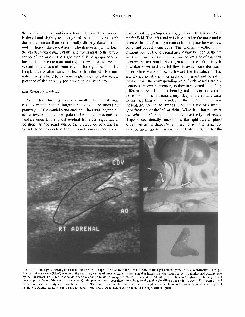
FIG. 14. The right adrenal gland has a “bent arrow” shape. The picture of the dorsal surface of the right adrenal gland shows its characteristic shape. The caudal vcna cava (CDV) is seen in the near field on the ultrasound image. It has a smaller lumen than the aorta duc to its pliability and compression by thc transducer. Often both the caudal vena cava and aorta are not imaged in the same plane as the adrenal gland. The adrenal gland is often angled and overlying the plane of the caudal vena cava. On the picture in the upper right, the right adrenal gland is identified by the white arrows. The adrenal gland is seen in close proximity to the caudal vena cava. The small vessel on the ventral surface of the gland is the phrenicoabdominal vein. A small segment of the lcft adrcnal gland is seen on the lcft sidc of the caudal vend cava slightly caudal to the right adrenal gland.
VOL. 38, No. 1 SONOGRAPHIC IDENTIFICATION OF ABDOMEN 19
right adrenal gland, which is located cranial to the celiac and cranial mesenteric arteries. The celiac and cranial mes- cntcric arteries are good landmarks to use to locate and differentiate between the adrenal glands. The transducer is pointed cranially and fanned dorsally to identify the right adrenal gland. To identify the left adrenal gland, the trans- ducer is slid caudal to the celiac and cranial mesenteric arteries and pointed slightly ventrally (down). The left adre- nal gland is seen cranial to the pole of the left kidney and deep to the caudal vena cava.
Right KidneylRight Renu1 ArterylVein
By sliding the transducer slightly cranially and dorsolat- erally, the right kidney is identified in the near field. The right renal vein may be traced from the right renal pelvis to the right lateral margin of the caudal vena cava. The course of this vessel closely parallels the more dorsally located renal artery. It is often difficult to image both vessels si- multaneously within the same plane. The right renal artery, arising from the aorta in the far field, crosses dorsal to the caudal vena cava and slightly cranial and dorsal to the right renal vein. The right renal artery is longer and often smaller than the corresponding right renal vein. It is best visualized coursing between the diverging aorta and caudal vena cava
cranial to the left renal vessels. The intra-renal vascular appearance is similar to the left kidney (Fig. 1 1).
CeliaclCraniul Mesenteric Arteries
The celiac and cranial mesenteric arteries may be iden- tified from either the right or left side of the abdomen. These unpaired arteries arise from the ventral surface of the aorta and are located cranial to the right renal artery and between the diverging course of the caudal vena cava and aorta. They are a good landmark used to distinguish left and right adrenal glands when imaging from the right side. From a dorsal plane on the right side, cross-sections of the vessels may be identified between the longitudinal axis of the caudal vena cava and the aorta. When the cross sectional image of the vessels is seen, the transducer is rotated to align with the long axis of the vessels. The tortuous path of the vessels may be followed to the aorta or to the visceral organs they supply.
Right Adrenal Gland
The right adrenal gland is observed cranial to the right renal vessels and the celiac and cranial mesenteric arteries.
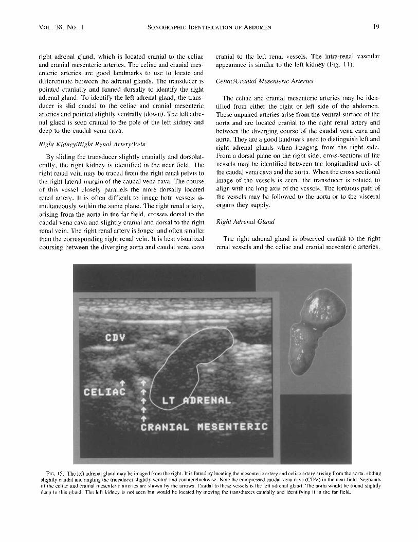
FIG. IS. Thc left adrenal gland may be imaged from the right. It is found by locating the mcsentcric artery and ccliac artcry arising from the aorta, sliding slightly caudal and angling thc transducer slightly ventral and counterclockwise. Note the compressed caudal vena cava (CDV) in the near field. Segments of the celiac and cranial mesenteric arteries arc shown by the arrows. Caudal to these vessels is the lcft adrenal gland. The aorta would be found slightly deep to this gland. The lcft kidncy is not seen but would be located by moving the transducers caudally and identifying it in the far field.
20 SPAULDING 1997
lmaging of this gland is often facilitated by rotating the sternum of the animal dorsally approximately 3045” from a recumbent lateral position. The appearance of the caudal vena cava and the origin of the celiac and cranial mesenteric arteries from the aorta serve as landmarks to find the right adrenal gland (Fig. 12 and 13). When the image of the right adrenal gland is optimized, often the renal image and the images of the major vessels are not optimized, and only segments of these structures may be seen. The caudate lobe of the liver is usually in the image plane cranial to the right adrenal gland, while the crura of the diaphragm are longi- tudinally arranged echogenic structures located dorsally in the far field adjacent to the aorta and caudal vena cava. To find the right adrenal gland, usually the transducer is placed in a subcostal position. In large dogs, an intercostal ap- proach may be necessary. When the caudal vena cava, ce- liac and cranial mesenteric arteries are recognized, the transducer is rotated clockwise in conjunction with a slight fanning of the area while pressure is applied to the trans-
ducer. This is usually a successful technique for locating the right adrenal gland. The sonologist’s ability to find the right adrenal gland is adversely influenced by abdominal muscu- lature rigidity, large body size, obesity, panting, or an oth- erwise uncooperative patient. Tranquilization may be re- quired to ensure abdominal relaxation, as transducer gen- erated abdominal pressure is often required to adequately image the right adrenal gland. The right adrenal gland is shaped like an “L” or a “bent arrow,” with the caudal pole located caudolaterally and the ‘‘bent arrow’ ’ cranial portion of the gland located craniomedially (Fig. 14). In large dogs, the cranio-medial pole of the right kidney adjacent to the caudal vena cava may be used as a useful landmark to locate the right adrenal gland. The adrenal gland often assumes a more “bean shape” in this image plane with a sector trans- ducer.
The left adrenal gland may be imaged from the right side. The left adrenal gland is identified caudal to the celiac and cranial mesenteric arteries. The right adrenal gland is found
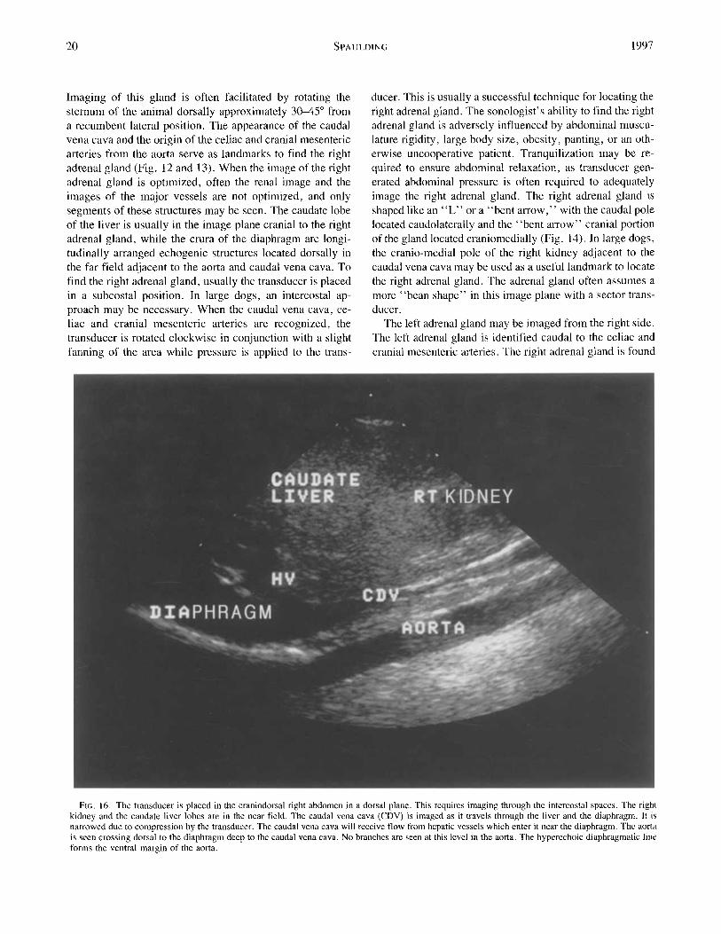
FIG. 16. The transducer is placed in the craniodorsal right abdomen in a dorsal plane. This requires imaging through the intercostal spaces. The right kidney and the caudate liver lobes are in the near field. The caudal vena cava (CDV) is imaged as it travels through the liver and the diaphragm. It is narrowed due to compression by the transducer. The caudal vcna c a w will receive flow from hepatic vessels which enter it near the diaphragm. Thc aorta is seen crossing dorsal to the diaphragm deep to the caudal vena cava. No branches are seen at this level in the aorta. The hyperechoic diaphragmatic line forms thc ventral margin of the aorta.
VOL. 38, No. I SONOGRAPHIC IDENTIFICATION OF ABDOMEN 21
cranial to these vessels. As one locates the beginning di- verging paths of the aorta and caudal vena cava. The left renal artcry courses between the two vessels. By angling the probe slightly ventrally the left adrenal gland is found cra- nial and medial to the left kidney (Fig. 15). The probe is then slid slightly cranially past the celiac and cranial mes- enteric arteries. The probe is angled dorsally and rotated counterclockwise. In this image plane the caudal vena cava is seen but the aorta is often not in the image plane. Occa- sionally, in small, thin dogs and cats, both adrenal glands may be seen in the same image.
Phrenicoubdominal Vessels
The phrenicoabdominal arteries and veins may be de- tected with high-resolution equipment, though recognition is significantly assisted by Doppler color flow ultrasound. The right phrenicoabdominal artery is variable in origin and position and is infrequently recognized. The right phreni-
coabdominal vein is consistently present, indenting the ven- tral surface of the right adrenal gland.
Cranial Aspect qf the Caudal Vena Cuva and Aorta
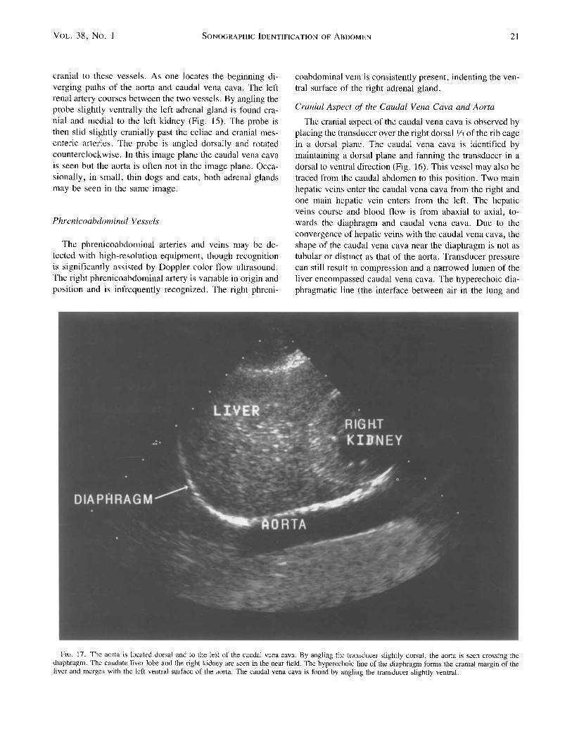
The cranial aspect of the caudal vena cava is observed by placing the transducer over the right dorsal '/3 of the rib cage in a dorsal plane. The caudal vena cava is identified by maintaining a dorsal plane and fanning the transducer in a dorsal to ventral direction (Fig. 16). This vessel may also be traced from the caudal abdomen to this position. Two main hepatic veins enter the caudal vena cava from the right and one main hepatic vein enters from the left. The hepatic veins course and blood flow is from abaxial to axial, to- wards the diaphragm and caudal vena cava. Due to the convergence of hepatic veins with the caudal vena cava, the shape of the caudal vena cava near the diaphragm is not as tubular or distinct as that of the aorta. Transducer pressure can still result in compression and a narrowed lumen of the liver encompassed caudal vena cava. The hyperechoic dia- phragmatic line (the interface between air in the lung and
Fit;. 17. The aorta is located dorsal and to the left of the caudal vena cava. By angling the transducer slightly dorsal, the aorta is seen crossing the diaphragm. The caudate liver lobe and the right kidney are seen in the near field. The hyperechoic line of the diaphragm forms the cranial margin of the liver and merges with the left ventral surface of the aorta. The caudal vena cava is found by angling the transducer slightly ventral.
22 SPAULDINC 1997
the diaphragm) is apparent encircling the caudal vena cava as the caudal vena cava enters the thorax through the caval hiatus in the right mid dorsal diaphragm.
A # r t u j E ~ ~ ~ p ~ ~ ~ u s
Following imaging of the caudal vena cava, with a dorsal plane image maintained, the transducer is pointed slightly dorsally. The aorta should be visible as it courses from the thoracic cavity caudally into the abdominal cavity dorsal to the diaphragm (Fig. 17). No branches from the aorta are seen at this level. Occasionally, the azygous vein is seen with color Doppler ultrasound dorsal and paralleling the aorta. In contrast to the caudal vena cava, the hyperechoic curvilinear diaphragmatic margin is only apparent ventral to the aorta as the aorta does not go through the diaphragm but is located dorsal to it. The diaphragmatic margin appears continuous and blends with the ventral wall of the aorta. The ventral margin of the aorta is at an approximately 75- 90" angle with the diaphragmatic line. Angling the trans-
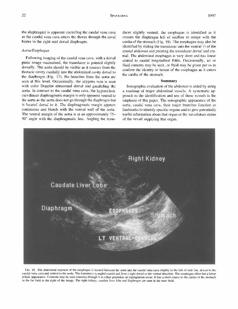
ducer slightly ventral, the esophagus is identified as it crosses the diaphragm left of midline to merge with the cardia of the stomach (Fig. 18). The esophagus may also be identified by sliding the transducer into the ventral Y' of the cranial abdomen and pointing the transducer dorsal and cra- nial. The abdominal esophagus is very short and has linear cranial to caudal longitudinal folds. Occasionally, air or fluid contents may be seen, or fluid may be given per os to confirm the identity or lumen of the esophagus as it enters the cardia of the stomach.
Summary Sonographic evaluation of the abdomen is aided by using
a roadmap of major abdominal vessels. A systematic ap- proach to the identification and use of these vcssels is the emphasis of this paper. The sonographic appcarance of the aorta, caudal vena cava, their major branches function as landmarks to identify specific organs and to givc potentially useful information about that organ or the vasculature status of the vessel supplying that organ.
FIG. 18. The abdominal segment of the esophagus is located hetwccn the aorta and the caudal vena cava slightly to the left of mid-line, dorsal to the caudal vena cava and ventral to the aorta. The transducer is angled cranial and from a right dorsal to left ventral direction. The esophagus often has a linear echoic appcarance. Contents may he seen coursing through it as either peristalsis or regurgitation occur. It has a short course to the cardia of thc stomach in the far field to the right of the image. The right kidney, caudate liver lobe and diaphragm are seen in the near field.
Vor.. 38, No. 1 SONOGRAPHIC IDENTIFICATION or: ABDOMEN 23
Imaging from the left side (right decubitus position) the aorta and caudal vena cava are followed cranially from the iliac vessels. The left and right medial iliac lymph nodes, located adjacent to the aorta, caudal vena cava and the deep circumflex iliac vessels, are found. Progressing cranially, the left renal artery and vein are followed to the left kidney. The vessels within the left kidney are interrogated. The left adrenal gland with the phrenicoabdominal vein coursing across it is found using the left renal artery as a landmark. I n situations of specific interest, the caudal mesenteric, the lumbar and the ovarianltesticular vessels may be located and followed from their origin to the organ of interest. The ccliac and cranial mesenteric arteries are located at their aortic origin and may be followed to the organs of interest.
From the right side (left decubitus position) the caudal vena cava and aorta are traced again from the iliac vessels cranially. The medial iliac lymph nodes are again imaged. Coursing cranially the left renal arteriesivein and the right renal artery and vein and right kidney are located. The cranial mesenteric and the celiac arteries are located as they exit from the aorta. Using these vessels, the right and left
adrenal glands are identified. The cranial mesenteric artery may be traced to the root of the mesentery where the con- fluence forming the portal vein and the mesenteric lymph nodes are found. The three branches of the celiac artery may be traced respectively to the stomach, the spleen and the liver. The aorta and caudal vena cava are traced cranially to the diaphragm. The esophagus coursing through the dia- phragm to the stomach is located using the aorta and caudal vena cava as landmarks. The aorta and caudal vena cava are also useful in locating and identifying hepatic veins, portal veins and organs such as pancreas, spleen, and segments of the gastrointestinal tract (not covered in this discourse).
Using the abdominal vessels as a visual roadmap to the abdomen will afford a systematic approach to abdominal scanning that will effect an easier and more complete ex- amination.
ACKNOWLEDGMENTS
The author acknowledges with gratitude the artistic and technical assis- tance qf Mr. Jacob Johnson and Brenda Bunch MA,MS in rendering thc drawings and images.
REFERENCES
I . Evans HE, Christenscn GC. Miller’s Anatomy of the Dog. 2nd Ed. Philadelphia: W.B. Saundcrs Company, 1979.
2. Sleight DR, Thomford NR. Gross Anatomy of the Blood Supply and Biliary Drainage of the Canine Liver. Anat Rec 1970;166:153-160.
3. Popesko P. Atlas of Topographical Anatomy of the Domestic An- imals. 3rd cd. Philadelphia: W.B. Saundcrs Company, 1979.
4. Schmidt S, Sutcr PF. Angiography of the Hepatic and Portal Ve- nous System in the Dog and Cat: An Investigative Method. Vet Radiol 1980;2 1(2):57-77.
5 . Maslak SH, Freund JG. Color Doppler Instrumentation. Vascular Imaging by Color Doppler and Magnetic Resonance. Lanzcr, P. (Hrsg.) Springer-Verlag, Berlin, Heidelberg, 199 1 ;87- 122.
6. Powis RL. Color Flow Imaging: Understanding Its Science and Technology. J Diag Med Sonography 1988;4:236245.
7 . Grant EG, Schillcr VL, Millener P , et al. Review Article: Color Doppler Imaging of the Hepatic Vasculature. Am Jour Radiol 1992;159: 943-950.
8. Smith HJ, Grottum P, Simonsen S. Ultrasonic Assessment of Ab- dominal Venous Return 11. Volume Blood Flow in the Inferior Vcna Cava and Portal Vein. Acta Rddiologica Diagnosis 1986;27:23-27.
9. Merritt CRB. Doppler Color Flow Imaging. J Clin Ultrasound 1987; 15:591-597.
10. Ralls PW. Color Doppler Sonography of the Hepatic Artery and Portal Venous System. Amer Jour Roentgenol 1990;155:5 17-525.
I I . Mittelstaedt CA. In: Abdominal Ultrasound. New York: Churchill Livingstonc, 1987:441-500.
12. Gooding CAW. The Abdominal Great Vessels. In: Rumack, C.M., S.R. Wilson, J.W. Charboneau. Diagnostic Ultrasound. St. Louis: Mosby Year Book, 1991;335-352.
13. Schclling CG. Editors Kaplan P.M. Ultrasonography of the Adrenal Gland. In: Problems in Veterinary Medicine: Ultrasound. J . B. Lippincott Co. Philadelphia. 199 1 ;Vo1.3(4):604-618.
14. Nyiand DG, Park RD. Hepatic Ultrasonography in the Dog. Vet Radiol 1983;24(2):7484.
15. Spaulding KA. Helpful Hints in Identifying the Caudal Abdominal Aorta and Caudal Vcna Cava. Vet Radio & Ultrasound 1992;33:9(&92.
16. Sadanaga K, Schulman A. An unusual Portosystcinic Shunt in a Dog. J Am Vet Med Assoc 1987;190(5):594-551.
17. Lim JH, Ryu KN, Ko YT, I,cc DH. Anatomic Relationship of Intrahepatic Bile Ducts to Portal Veins. J Ultrasound Med 1990;9(3):137- 143.
18. Bjorling DE, Prasse KW, Holmes RA. Partial Hepatectomy in Dogs. Compend Contin Ed Pract 1980:25&265.