Embed Size (px)
Citation preview
J Neurosurg / Volume 114 / March 2011
J Neurosurg 114:633–639, 2011
633
Brain surgery in an unanesthetized patient was practiced well before the introduction of anesthe-sia,2 but the modern era of craniotomies using CS
techniques began in the 1920s and 1930s with the semi-nal work of Wilder Penfield on cerebral localization for epilepsy surgery7 and was further refined in the 1950s by Herbert Olivecrona, mainly for the treatment of brain tumors.16 In the following years, the introduction of im-age-guided surgery with minimal-sized craniotomies and the use of novel anesthetic protocols have contributed to a CS craniotomy better tolerated by the patient and the neurosurgical team.3,8
Although resection during CS was initially used for lesions located in eloquent areas, the avoidance of GEA and the possible association with a more rapid resump-
tion of patient daily activities have led to the use of CS in the surgical treatment of different brain pathologies, regardless of cerebral location.20,23,24 In fact, same-day surgery for brain tumor resection has been advocated.1,7 However, it should be noted that increased complications related to CS surgery as compared with traditional ap-proaches have been reported by some.5
As patient and socioeconomic demands increase to force surgical practices to become more efficient and less resource intensive, the avoidance of GEA in elective su-pratentorial craniotomies for gliomas will result in the improved utilization of such resources and decrease the inpatient stay. To validate this hypothesis, we retrospec-tively analyzed the outcome of 22 cases matched to a co-hort of 22 similar cases. Our data suggest that craniotomy during CS significantly decreases the use of resources as well as inpatient stay.
MethodsPatient Population
Two cohorts were selected from a database of pa-
A retrospective cohort-matched comparison of conscious sedation versus general anesthesia for supratentorial glioma resection
Clinical articlePierPaolo Peruzzi, M.D.,1 Sergio D. BergeSe, M.D.,2 aDolfo Viloria, M.D.,2 erika g. Puente, M.D.,2 MahMouD aBDel-raSoul, M.P.h.,3 anD e. antonio ChioCCa, M.D., Ph.D.1
Departments of 1Neurological Surgery and 2Anesthesiology, The Ohio State University College of Medicine and James Cancer Hospital; and 3Center for Biostatistics, The Ohio State University, Columbus, Ohio
Object. Glioma resection under conscious (“awake”) sedation (CS) is used for eloquent areas of the brain to minimize postoperative neurological deficits. The objective of this study was to compare the duration of hospital stay, overall hospital cost, perioperative morbidity, and postoperative patient functional status in patients whose gliomas were resected using CS versus general endotracheal anesthesia (GEA).
Methods. Twenty-two cases in 20 patients who underwent surgery for cerebral gliomas under CS and a matched cohort of 22 cases in 19 patients who underwent surgery under GEA over a 3-year period were retrospectively evaluated. Criteria for inclusion in the study were as follows: 1) a single cerebral lesion; 2) gross-total resection as evidenced by postoperative Gd-enhanced MR imaging within 48 hours of surgery; 3) a WHO Grade II, III, or IV glioma; 4) a supratentorial lesion location; 5) a Karnofsky Performance Scale score ≥ 70; 6) an operation performed by the same neurosurgeon; and 7) an elective procedure.
Results. The average hospital stay was significantly different between the 2 groups: 3.5 days for patients who underwent CS and 4.6 days for those who underwent GEA. This result translated into a significant decrease in the average inpatient cost after intensive care unit (ICU) care for the CS group compared with the GEA group. Other variables were not significantly different.
Conclusions. Patients undergoing glioma resection using CS techniques have a significantly shorter hospital stay with reduced inpatient hospital expenses after postoperative ICU care. (DOI: 10.3171/2010.5.JNS1041)
key WorDS • craniotomy • conscious sedation • general anesthesia • glioma
633
Abbreviations used in this paper: BIS = Bispectral Index; CS = conscious sedation; GEA = general endotracheal anesthesia; GTR = gross-total resection; ICU = intensive care unit; KPS = Karnofsky Performance Scale; LOS = length of stay; MRC = Medical Re search Council; NICU = neurosurgical ICU; OAA/S = Observer’s As sess-ment of Alertness/Sedation Scale.
P. Peruzzi et al.
634 J Neurosurg / Volume 114 / March 2011
tients with supratentorial gliomas surgically treated using either CS or GEA at The Ohio State University Medical Center and James Cancer Hospital, Columbus, Ohio. All patients were treated between January 2006 and Decem-ber 2008. The only difference between the 2 cohorts was that CS was reserved for patients with a lesion located in or close to speech or motor areas, as identified by patient symptomatology and anatomical localization on MR im-aging or functional MR imaging. The institutional review board of The Ohio State University Medical Center ap-proved this retrospective chart analysis.
Criteria for inclusion in the cohort were as follows: 1) an isolated glioma without evidence of multicentric or multifocal enhancement; 2) radiological evidence of GTR on MR images obtained within 48 hours postoperatively, as indicated by a lack of nodular enhancement for Gd-enhancing lesions or T2 or FLAIR hyperintensity for nonenhancing lesions; 3) a final pathological diagnosis of glioma (WHO Grades II–IV); 4) a supratentorial lesion location; 5) a preoperative KPS score ≥ 70; 6) an opera-tion performed by the same neurosurgeon (E.A.C.); and 7) a procedure performed electively.
Patients were excluded from the study if they had sig-nificant comorbidities unrelated to the primary brain pa-thology that would require additional hospital stay, such as renal failure, acute myocardial infarction of < 1 year’s duration, cerebrovascular accident of < 1 year’s dura-tion, hepatic failure, lung disease, or active inflammatory/infectious processes. No cases of emergent operations were included. Therefore, among 32 cases in which CS had been used in the study period, 4 cases (12.5%) were excluded because the lesions were incompatible with the histological inclusion criteria: 1 (3%) because the tumor was sampled for biopsy only, and 5 (16%) because the tumor was partially resected. Thus, the final number of cases meeting the inclusion criteria was 22. Because the number of patients who underwent craniotomy while under GEA in the same study period was disproportion-ately higher, a matched selection of cases from the en-tire operative pool was performed based on patient age, sex, tumor volume, KPS score, tumor grade, GTR, lack of major comorbidities, elective nature of the procedure, and procedure performed by the same surgeon (Table 1). Thus, 22 cases were included in the GEA-matched co-hort. As expected, the only significant difference between the CS and GEA groups was tumor location given that the former group consisted of cases with gliomas directly in or close to eloquent brain areas, whereas the latter group did not. In the CS cohort, 2 individuals underwent sur-gery twice during the study period, whereas 3 persons in the GEA cohort underwent surgery twice. The number of enrolled patients was therefore 20 and 19 for the CS and GEA groups, respectively. There was no crossover between groups, meaning that no individuals underwent craniotomy under both CS and GEA. No patient whose craniotomy was started under CS was switched to GEA during the procedure. Moreover, patients were selected for CS if their tumors were located within the sensorimo-tor or speech cortex (Broca areas, Wernicke area, and/or dominant temporal lobe, such as the superior and middle temporal gyri 4–5 cm posterior to the anterior pole of the
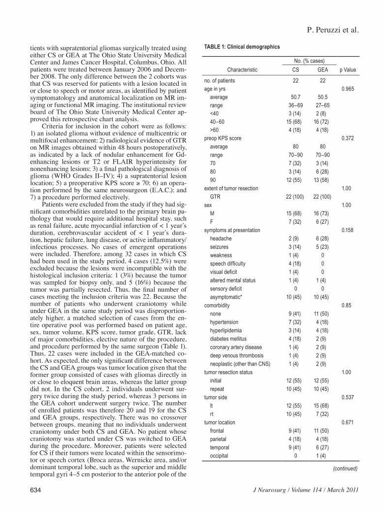
TABLE 1: Clinical demographics
No. (% cases)Characteristic CS GEA p Value
no. of patients 22 22age in yrs 0.965 average 50.7 50.5 range 36–69 27–65 <40 3 (14) 2 (8) 40–60 15 (68) 16 (72) >60 4 (18) 4 (18)preop KPS score 0.372 average 80 80 range 70–90 70–90 70 7 (32) 3 (14) 80 3 (14) 6 (28) 90 12 (55) 13 (58)extent of tumor resection 1.00 GTR 22 (100) 22 (100)sex 1.00 M 15 (68) 16 (73) F 7 (32) 6 (27)symptoms at presentation 0.158 headache 2 (9) 6 (28) seizures 3 (14) 5 (23) weakness 1 (4) 0 speech difficulty 4 (18) 0 visual deficit 1 (4) 0 altered mental status 1 (4) 1 (4) sensory deficit 0 0 asymptomatic* 10 (45) 10 (45)comorbidity 0.85 none 9 (41) 11 (50) hypertension 7 (32) 4 (18) hyperlipidemia 3 (14) 4 (18) diabetes mellitus 4 (18) 2 (9) coronary artery disease 1 (4) 2 (9) deep venous thrombosis 1 (4) 2 (9) neoplastic (other than CNS) 1 (4) 2 (9)tumor resection status 1.00 initial 12 (55) 12 (55) repeat 10 (45) 10 (45)tumor side 0.537 lt 12 (55) 15 (68) rt 10 (45) 7 (32)tumor location 0.671 frontal 9 (41) 11 (50) parietal 4 (18) 4 (18) temporal 9 (41) 6 (27) occipital 0 1 (4)
(continued)
J Neurosurg / Volume 114 / March 2011
Conscious-sedation craniotomy for gliomas
635
temporal lobe and/or the posterior mesiotemporal lobe involving the hippocampus and parahippocampal gyri), and patients had no significant motor or speech deficits preoperatively. During the same time period, 9 gliomas in eloquent cortex and causing significant preoperative defi-cits were treated while the patient was under GEA. In 3 cases in which the glioma was located in eloquent cortex and the patient was relatively intact preoperatively, GEA was induced for other reasons. 1) One patient did not dem-onstrate speech impediments during a CS craniotomy in which subtotal resection was the outcome; this patient un-derwent reoperation soon thereafter while under GEA to obtain a GTR. 2) The neuroanesthesiology team judged CS to be inappropriate for a tumor in the left motor strip in a 33-year-old patient because of anxiety. 3) In a case in which a small glioma recurred on the internal surface of a large cyst near the right motor cortex, it was believed that intracystic removal would be unlikely to affect motor function. None of these cases was included in the GEA group that was used as a control in our analysis.
Anesthesia and Surgical Technique
Conscious-Sedation Craniotomy. The patients were brought to the preoperative holding area for anesthesia assessment, intravenous access, and invasive line place-ment (arterial line and central venous catheter). Once in-travenous access was achieved and the vital signs monitor was connected, the patient was transported to the operat-ing room and a continuous infusion of dexmedetomidine (0.1–0.7 µg/kg/hr) was started. To attain proper titration of dexmedetomidine and adequate sedation, the level of consciousness was continuously assessed using the
OAA/S. If low doses of dexmedetomidine alone did not provide adequate sedation, a second agent such as mid-azolam (0.01 mg/kg) was provided as an adjuvant. Tradi-tionally, preoxygenation was provided and anesthesia was induced with Diprivan (100 µg/kg, administered slowly). As soon as consciousness was lost, a nasal trumpet was placed in 1 of the nares, attached to an 8-mm endotracheal tube connector, and plugged into the anesthesia machine circuit, enabling oxygen delivery, capnography, and gentle bagging, if required for breathing assistance. To maintain anesthesia, the regimen consisted of Diprivan (40–120 µg/kg/min) titrated to a BIS of 50–60 along with the pre-viously started continuous infusion of dexmedetomidine (0.1–0.7 µg/kg/hr). Initially, a higher dose of Diprivan was needed to achieve the same BIS levels as the first regimen. To minimize the vasodilatory and respiratory depression effect caused by Diprivan, sevoflurane was added to the regimen (0.5%–1%), allowing a reduction in the Diprivan dose for anesthesia maintenance. Once the patient was under deep sedation and after 3 ml of lidocaine chloride (1%) with epinephrine had locally infiltrated each of the pin sites, a Mayfield headrest was placed. The surgical in-cision was also locally infiltrated with lidocaine (1%) and epinephrine, and after the surgical incision, dissection, and opening of the dura mater, Diprivan in both groups and sevoflurane were discontinued, allowing for intraop-erative awakening. Patients regained consciousness, and then language and/or motor mapping was initiated before tumor resection. During lesion removal, patients were carefully monitored and assessed for pain and discom-fort (Verbal Response Scale and OAA/S). Small boluses of fentanyl (12.5–25 µg) were administered as needed to treat pain. The dexmedetomidine drip was titrated slowly if deeper sedation was needed. As the patient awakened, standard neurosurgical techniques for mapping function (electrocortical stimulation and monitoring of speech and motor function) were applied to resect the glioma. After tumor resection and in preparation for craniotomy clo-sure, deeper sedation was reinduced using Diprivan, as described above. On completion of the surgery, Diprivan and sevoflurane were discontinued. Patients were again awakened, all sedating agents were discontinued except for low doses of dexmedetomidine, the nasal trumpet was removed, and patients were transported to the NICU.
General Endotracheal Anesthesia Craniotomy. In the GEA group of patients, preoperative sedation was avoided as a general rule; if deemed necessary, however, an anxi-olytic such as midazolam (1- to 2-mg intravenous push) was administered. After invasive lines were placed, the patients were transported to the operating room, placed on the operating room table, and, per American Society of Anesthesiology guidelines, patient monitoring devices and a brain monitor were attached in a manner similar to that in the CS craniotomy group. Fentanyl (1.5–2.5 µg/kg) and Diprivan (2–2.5 mg/kg) were used for the induc-tion of anesthesia. As muscle relaxants, either vecuronium (0.08–0.12 mg/kg) in 3 cases (14%) or rocuronium (0.4–0.9 mg/kg) in 15 cases (68%) was used for endotracheal intubation. In 3 cases (14%) in which a difficult airway was encountered and no severe increase in intra cranial
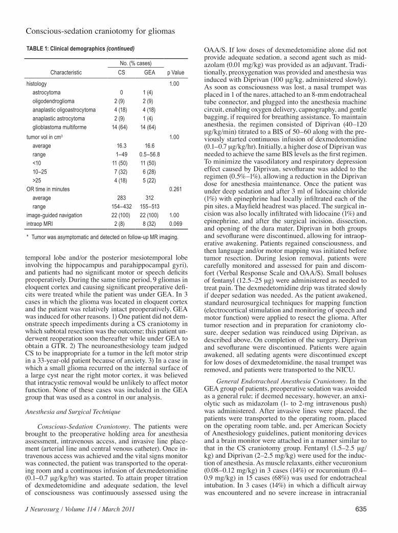
TABLE 1: Clinical demographics (continued)
No. (% cases)Characteristic CS GEA p Value
histology 1.00 astrocytoma 0 1 (4) oligodendroglioma 2 (9) 2 (9) anaplastic oligoastrocytoma 4 (18) 4 (18) anaplastic astrocytoma 2 (9) 1 (4) glioblastoma multiforme 14 (64) 14 (64)tumor vol in cm3 1.00 average 16.3 16.6 range 1–49 0.5–56.8 <10 11 (50) 11 (50) 10–25 7 (32) 6 (28) >25 4 (18) 5 (22)OR time in minutes 0.261 average 283 312 range 154–432 155–513image-guided navigation 22 (100) 22 (100) 1.00intraop MRI 2 (8) 8 (32) 0.069
* Tumor was asymptomatic and detected on follow-up MR imaging.
P. Peruzzi et al.
636 J Neurosurg / Volume 114 / March 2011
pressure was observed, succinylcholine (1–1.5 mg/kg) was used. In 1 case no muscle relaxant was used at in-duction. Finally, the trachea was intubated and general anesthesia was started. To maintain general anesthesia, a combination of intravenous and inhalation agents was used according to the anesthesiologist’s preference. The intravenous anesthetic component consisted of remifenta-nil (0.05–0.1 µg/kg/min) in 18 cases (82%), and a combi-nation of remifentanil (0.05 µg/kg/min) and dexmedeto-midine (0.2–0.3 µg/kg/hr) in the remaining 4 cases (18%). Anesthesiologists chose the inhalation agents: isoflurane in 5 cases (23%), desflurane in 7 cases (32%), or sevoflu-rane in 10 cases (45%). These agents were administered at 0.5–0.8 minimum alveolar concentration levels and titrated to a BIS of 50–60. On completion of the surgi-cal procedure, all intravenous and inhalation agents were discontinued, with the exception of dexmedetomidine, which was briefly maintained without change only in patients who had been on this regimen. With continuous vital signs monitoring, patients were extubated and, after meeting the criteria, were transported to the NICU.
Postoperative Management Patients who underwent surgery while under GEA
were assessed for extubation by the anesthesiologist at the end of the case while still in the operating room. If extubation was not feasible because of delayed arousal, neurological deficit, or other medical reasons, patients were transferred to the NICU while still intubated. Re-lease from the NICU to the floor occurred when patients were deemed neurologically (alert and oriented to self, place, and date) and medically stable by the resident, nursing staff, and attending neurosurgeon. The extent of tumor resection was evaluated via MR imaging with and without Gd within 48 hours postoperatively. Physical and occupational therapy assessment was performed on post-operative Day 1 for all patients to determine the need for acute rehabilitation. Patients were discharged when they were able to feed themselves, ambulate, and void; were taking oral medications only; and had no issues with pain control. The decision to discharge was made before we intended to conduct the current retrospective study. The decision to discharge was made by the attending neu-rosurgeon with consultation and input by the residents, nurses, and physical/occupational therapists. The need for inpatient or outpatient rehabilitation was determined by physical medicine and rehabilitation physician consul-tants, and also was made before we intended to conduct the current retrospective study.
Follow-Up AnalysisEach patient was reevaluated within 2 weeks after
discharge. The findings on physical and neurological ex-amination were compared with the preoperative assess-ments to determine if and how much the recent surgery had changed each patient’s functional status and if new neurological deficits occurred postoperatively.
Hospital CostsThe Ohio State University Administration Office
provided information on costs related to the surgical pro-cedure and hospital stay. For each patient, we identified 4 primary direct expense categories—namely, costs for the operating room, anesthesia, NICU, and ward—which we anticipated would better reflect possible differences between our 2 patient cohorts and for which specific costs were obtained. These direct costs can clearly be directly allocated to a specific patient and his or her inpatient stay. In addition, each patient and procedure is allocated sev-eral variable (indirect) costs; that is, expenses not directly attributable to the surgery but allocated by the hospital’s accounting department. For example, variable costs that get allocated to each surgery would include salaries of various hospital personnel and administrators, costs of equipment purchased, bad debt accrued, interest on build-ings, and so forth. Because these expenses are highly vari-able, change significantly over time, are allocated based on different accounting methods at different times, and appear to be attributed to each surgery on a semi-random basis, they were not included in the analysis.
Statistical Analysis
Clinical Characteristics. Categorical clinical charac-teristic variables of patients with gliomas were compared between the 2 groups using the Fisher exact test (Table 1). Continuous characteristics were compared between the CS and GEA groups using 2-sample t-tests.
Clinical Outcomes. The clinical outcome variables of interest were duration of hospital stay (3 days vs > 3 days), time in NICU (< 24 hours vs > 24 hours), direct hospital costs, postoperative status (stable vs other), and discharge (home vs rehabilitation center; Tables 2 and 3). The relationships between 3 different dichotomized outcome variables (duration of stay, postoperative sta-tus, and discharge) and type of surgery as the predictor were examined using univariate logistic regression mod-els and the likelihood ratio test. The crude relationships were then adjusted for potential confounders while taking care not to over-fit the model. The variables checked for confounding effects included sex, KPS score, primary re-section, tumor volume, surgery time, diagnosis, location, intraoperative MR imaging, and age. Any variables that altered the final model coefficients by 20% were adjusted for as potential confounders. Odds ratios and their corre-sponding 95% CIs were calculated for the variables in the adjusted models. The Hosmer-Lemeshow goodness-of-fit test6 was conducted on the final model. The Fisher exact test was used to assess the relationship between the type of surgery and time in the NICU. A logistic regression model could not be used for this analysis because all of the patients in the CS group were released from the NICU in < 24 hours. All statistical analyses were performed us-ing Stata 10.0 for Windows (Stata Corp.).
ResultsPatient Demographics
The CS and GEA cohorts were matched for variables that could affect the LOS, number of surgeries, sex, mean
J Neurosurg / Volume 114 / March 2011
Conscious-sedation craniotomy for gliomas
637
age, median preoperative KPS score, symptoms, extent of tumor resection, comorbidities, new versus recurrent resections, tumor histology, tumor location, histology, tu-mor volume, average operating room time, use of image guidance, and use of intraoperative MR imaging (Table 1). Except for tumor location, there were no significant differences between the 2 groups.
Patient Outcomes
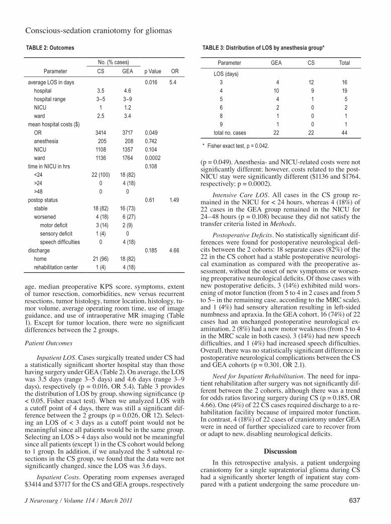
Inpatient LOS. Cases surgically treated under CS had a statistically significant shorter hospital stay than those having surgery under GEA (Table 2). On average, the LOS was 3.5 days (range 3–5 days) and 4.6 days (range 3–9 days), respectively (p = 0.016, OR 5.4). Table 3 provides the distribution of LOS by group, showing significance (p < 0.05, Fisher exact test). When we analyzed LOS with a cutoff point of 4 days, there was still a significant dif-ference between the 2 groups (p = 0.026, OR 12). Select-ing an LOS of < 3 days as a cutoff point would not be meaningful since all patients would be in the same group. Selecting an LOS > 4 days also would not be meaningful since all patients (except 1) in the CS cohort would belong to 1 group. In addition, if we analyzed the 5 subtotal re-sections in the CS group, we found that the data were not significantly changed, since the LOS was 3.6 days.
Inpatient Costs. Operating room expenses averaged $3414 and $3717 for the CS and GEA groups, respectively
(p = 0.049). Anesthesia- and NICU-related costs were not significantly different; however, costs related to the post-NICU stay were significantly different ($1136 and $1764, respectively; p = 0.0002).
Intensive Care LOS. All cases in the CS group re-mained in the NICU for < 24 hours, whereas 4 (18%) of 22 cases in the GEA group remained in the NICU for 24–48 hours (p = 0.108) because they did not satisfy the transfer criteria listed in Methods.
Postoperative Deficits. No statistically significant dif-ferences were found for postoperative neurological defi-cits between the 2 cohorts: 18 separate cases (82%) of the 22 in the CS cohort had a stable postoperative neurologi-cal examination as compared with the preoperative as-sessment, without the onset of new symptoms or worsen-ing preoperative neurological deficits. Of those cases with new postoperative deficits, 3 (14%) exhibited mild wors-ening of motor function (from 5 to 4 in 2 cases and from 5 to 5− in the remaining case, according to the MRC scale), and 1 (4%) had sensory alteration resulting in left-sided numbness and apraxia. In the GEA cohort, 16 (74%) of 22 cases had an unchanged postoperative neurological ex-amination, 2 (8%) had a new motor weakness (from 5 to 4 in the MRC scale in both cases), 3 (14%) had new speech difficulties, and 1 (4%) had increased speech difficulties. Overall, there was no statistically significant difference in postoperative neurological complications between the CS and GEA cohorts (p = 0.301, OR 2.1).
Need for Inpatient Rehabilitation. The need for inpa-tient rehabilitation after surgery was not significantly dif-ferent between the 2 cohorts, although there was a trend for odds ratios favoring surgery during CS (p = 0.185, OR 4.66). One (4%) of 22 CS cases required discharge to a re-habilitation facility because of impaired motor function. In contrast, 4 (18%) of 22 cases of craniotomy under GEA were in need of further specialized care to recover from or adapt to new, disabling neurological deficits.
DiscussionIn this retrospective analysis, a patient undergoing
craniotomy for a single supratentorial glioma during CS had a significantly shorter length of inpatient stay com-pared with a patient undergoing the same procedure un-
TABLE 2: Outcomes
No. (% cases)Parameter CS GEA p Value OR
average LOS in days 0.016 5.4 hospital 3.5 4.6 hospital range 3–5 3–9 NICU 1 1.2 ward 2.5 3.4mean hospital costs ($) OR 3414 3717 0.049 anesthesia 205 208 0.742 NICU 1108 1357 0.104 ward 1136 1764 0.0002time in NICU in hrs 0.108 <24 22 (100) 18 (82) >24 0 4 (18) >48 0 0postop status 0.61 1.49 stable 18 (82) 16 (73) worsened 4 (18) 6 (27) motor deficit 3 (14) 2 (9) sensory deficit 1 (4) 0 speech difficulties 0 4 (18)discharge 0.185 4.66 home 21 (96) 18 (82) rehabilitation center 1 (4) 4 (18)
TABLE 3: Distribution of LOS by anesthesia group*
Parameter GEA CS Total
LOS (days) 3 4 12 16 4 10 9 19 5 4 1 5 6 2 0 2 8 1 0 1 9 1 0 1total no. cases 22 22 44
* Fisher exact test, p = 0.042.
P. Peruzzi et al.
638 J Neurosurg / Volume 114 / March 2011
der GEA. This result translated into a significant decrease in post-ICU inpatient expenses. Note, however, that there was no significant difference in postoperative neurologi-cal deficits, operating room time, NICU stay, or need for inpatient rehabilitation. These results suggest that CS craniotomies could result in a shorter hospital stay with reduced expenses compared with GEA craniotomies. Of note, both patient cohorts were matched for preoperative variables that could have affected LOS and cost.
Craniotomies performed with the patient consciously sedated7,10,11 are widely used for the resection of lesions near or within eloquent cerebral areas. Authors of several reports have described the use of awake craniotomy for the treatment of many intracranial pathologies regardless of their anatomical localization and nature.1,20,23 In an in-teresting prospective study published in 1999 and again in 2007 with an expanded series, patients who underwent brain surgery while under CS for tumors—either primary or metastatic—or other intracranial pathologies could be discharged home as early as the same day surgery was performed.20,23 This report did not establish if CS surgery led to improved short-term recovery times as compared with GEA surgery. In fact, Whittle et al.26 reported the opposite: in a series of 25 patients undergoing awake sur-gery for tumor resection in eloquent areas, 5 (20%) expe-rienced significant postoperative deficits, a much higher incidence compared with the 2.5% “unexpected” postop-erative deficit in patients whose intraoperative monitoring was “uneventful” in the study by Taylor and Bernstein.23 Gupta et al.5 also reported worse outcomes in patients undergoing awake surgery for brain tumors in eloquent areas, as compared with those undergoing craniotomy under GEA. In their prospective randomized study of 53 patients, these authors reported a longer hospital stay, an inferior extent of resection, and an overall increase in postoperative neurological deficits in patients undergo-ing surgery with CS, compared with patients receiving GEA. In their study, different pathological entities were included and more gliomas were treated under CS, pos-sibly skewing the results toward a worse outcome. There-fore, controversy remains as to whether CS surgery leads to improved outcomes and faster patient recovery times when compared with GEA surgeries. Our data appear to significantly expand and agree with the findings of Taylor and Bernstein.
In our study, patients from both cohorts suffered mild postoperative worsening of previous neurological deficits or the occurrence of new deficits. Since patients in the CS cohort harbored gliomas in eloquent cortex, one might have predicted that neurological outcomes could have been worse compared with those in patients in the GEA cohort whose tumors were in noneloquent brain, but this did not appear to be the case in our study compared with the study by Gupta et al.5,14,15
When the need for inpatient rehabilitation is taken into account, even though our results did not reach statis-tical significance, an OR of 4.6 between the 2 cohorts in-dicates that patients undergoing craniotomy during GEA are 4.6 times more likely than those undergoing awake craniotomy to need inpatient rehabilitation.
Overall, the reduced LOS also resulted in a reduction
of inpatient costs, primarily for the post-NICU period. As expected, NICU and anesthesia costs were similar and operating room costs were slightly reduced in the CS group, possibly because of the higher number of GEA cases in which intraoperative MR imaging was used to determine the extent of glioma resection (data not shown) and whose cost was included in the cost of the operating room. We did not include an analysis of total costs, which would also include variable (indirect) costs given that the latter are allocated in a relatively subjective manner and depend on factors unrelated to the specific surgery or in-patient stay.
It is interesting to speculate the reasons for a pro-longed LOS in the GEA group. The process of GEA may be related to postoperative cognitive dysfunction and therefore to an increased LOS. Postoperative cog-nitive dysfunction has been reported not only to affect neurocognitive status early on, but also to be associated with causing prolonged effects with an increased mor-tality rate, premature leave from the labor market, and ultimately increasing dependence on welfare.4,12,13,18,21,22 Therefore, the significant difference in the LOS between the GEA and CS groups could be predominantly related to the neurological status of the patients. The depth of an-esthesia is related to postoperative cognitive dysfunction and affects recovery. Studies on memory impairment in rat models have shown that the use of general anesthesia produces persistent memory effects compared with none observed in rats that received intravenous propofol anes-thesia (general anesthetics have different receptor mecha-nisms of action). Therefore, the choice of anesthetic may play a part in late cognitive outcome. And thus, it appears that general anesthesia–induced memory impairment may be a function of the agent rather than the anesthesia state itself.9,17,19,25
The major limits of this study are its retrospective nature and the relatively small number of enrolled pa-tients. Sources of bias could include the preoperative decision regarding CS versus GEA. This source of bias was minimized by using data from a single neurosurgeon who would apply a relatively consistent philosophy. An-other source of bias could relate to the decision to transfer or discharge. This factor was minimized given that this decision was made before there was an intent to retro-spectively analyze the outcome of CS. To conclusively determine if CS craniotomy leads to a reduced LOS and inpatient expenses, a prospective randomized clinical tri-al would be necessary.
Conclusions Craniotomy for glioma resection under CS is associ-
ated with a significantly shorter hospital stay and reduced inpatient expenses as compared with craniotomy under GEA. If future studies confirm these data, the role of awake craniotomies in neurooncology should be revisited and expanded.
Disclosure
The authors report no conflict of interest concerning the mate-
J Neurosurg / Volume 114 / March 2011
Conscious-sedation craniotomy for gliomas
639
rials or methods used in this study or the findings specified in this paper.
Author contributions to the study and manuscript preparation include the following. Conception and design: Chiocca. Acquisition of data: Chiocca, Peruzzi, Bergese, Viloria, Puente. Analysis and interpretation of data: Chiocca, Peruzzi, Bergese, Viloria, Puente. Drafting the article: Chiocca, Peruzzi, Viloria, Puente. Critically revising the article: Chiocca, Peruzzi, Bergese. Reviewed final ver-sion of the manuscript and approved it for submission: all authors. Statistical analysis: Peruzzi, Abdel-Rasoul. Administrative/techni-cal/material support: Chiocca, Bergese. Study supervision: Chiocca, Bergese.
References
1. Boulton M, Bernstein M: Outpatient brain tumor surgery: in-novation in surgical neurooncology. J Neurosurg 108:649–654, 2008
2. Bulsara KR, Johnson J, Villavicencio AT: Improvements in brain tumor surgery: the modern history of awake cranioto-mies. Neurosurg Focus 18(4):e5, 2005
3. Conte V, Baratta P, Tomaselli P, Songa V, Magni L, Stocchetti N: Awake neurosurgery: an update. Minerva Anestesiol 74: 289–292, 2008
4. Fleisher LA, Anderson GF: Perioperative risk: how can we study the influence of provider characteristics? Anesthesiol-ogy 96:1039–1041, 2002
5. Gupta DK, Chandra PS, Ojha BK, Sharma BS, Mahapatra AK, Mehta VS: Awake craniotomy versus surgery under gen-eral anesthesia for resection of intrinsic lesions of eloquent cortex—a prospective randomised study. Clin Neurol Neuro-surg 109:335–343, 2007
6. Hosmer DW, Lemeshow S: Applied Logistic Regression. New York: John Wiley & Sons, 1989
7. July J, Manninen P, Lai J, Yao Z, Bernstein M: The history of awake craniotomy for brain tumor and its spread into Asia. Surg Neurol 71:621–625, 2009
8. Lanier WL: Brain tumor resection in the awake patient. Mayo Clin Proc 76:670–672, 2001
9. Lee IH, Culley DJ, Baxter MG, Xie Z, Tanzi RE, Crosby G: Spatial memory is intact in aged rats after propofol anesthe-sia. Anesth Analg 107:1211–1215, 2008
10. Louis DN, Ohgaki H, Wiestler OD, Cavenee WK, Burger PC, Jouvet A, et al: The 2007 WHO classification of tumours of the central nervous system. Acta Neuropathol 114:97–109, 2007
11. Meyer FB, Bates LM, Goerss SJ, Friedman JA, Windschitl WL, Duffy JR, et al: Awake craniotomy for aggressive resec-tion of primary gliomas located in eloquent brain. Mayo Clin Proc 76:677–687, 2001
12. Monk TG, Saini V, Weldon BC, Sigl JC: Anesthetic manage-ment and one-year mortality after noncardiac surgery. Anesth Analg 100:4–10, 2005
13. Monk TG, Weldon BC, Garvan CW, Dede DE, van der Aa MT, Heilman KM, et al: Predictors of cognitive dysfunction
after major noncardiac surgery. Anesthesiology 108:18–30, 2008
14. Ohgaki H: Epidemiology of brain tumors. Methods Mol Biol 472:323–342, 2009
15. Ohgaki H, Kleihues P: Population-based studies on incidence, survival rates, and genetic alterations in astrocytic and oli-godendroglial gliomas. J Neuropathol Exp Neurol 64:479–489, 2005
16. Otani N, Bjeljac M, Muroi C, Weniger D, Khan N, Wieser HG, et al: Awake surgery for glioma resection in eloquent areas—Zurich’s experience and review. Neurol Med Chir (Tokyo) 45:501–511, 2005
17. Pandharipande PP, Pun BT, Herr DL, Maze M, Girard TD, Miller RR, et al: Effect of sedation with dexmedetomidine vs lorazepam on acute brain dysfunction in mechanically ventilated patients: the MENDS randomized controlled trial. JAMA 298:2644–2653, 2007
18. Price CC, Garvan CW, Monk TG: Type and severity of cogni-tive decline in older adults after noncardiac surgery. Anesthe-siology 108:8–17, 2008
19. Riker RR, Shehabi Y, Bokesch PM, Ceraso D, Wisemandle W, Koura F, et al: Dexmedetomidine vs midazolam for sedation of critically ill patients: a randomized trial. JAMA 301:489–499, 2009
20. Serletis D, Bernstein M: Prospective study of awake craniot-omy used routinely and nonselectively for supratentorial tu-mors. J Neurosurg 107:1–6, 2007
21. Silber JH, Kennedy SK, Even-Shoshan O, Chen W, Mosher RE, Showan AM, et al: Anesthesiologist board certification and patient outcomes. Anesthesiology 96:1044–1052, 2002
22. Steinmetz J, Christensen KB, Lund T, Lohse N, Rasmussen LS: Long-term consequences of postoperative cognitive dys-function. Anesthesiology 110:548–555, 2009
23. Taylor MD, Bernstein M: Awake craniotomy with brain map-ping as the routine surgical approach to treating patients with supratentorial intraaxial tumors: a prospective trial of 200 cases. J Neurosurg 90:35–41, 1999
24. Tonn JC: Awake craniotomy for monitoring of language func-tion: benefits and limits. Acta Neurochir (Wien) 149:1197–1198, 2007
25. Wei H, Liang G, Yang H, Wang Q, Hawkins B, Madesh M, et al: The common inhalational anesthetic isoflurane induces apoptosis via activation of inositol 1,4,5-trisphosphate recep-tors. Anesthesiology 108:251–260, 2008
26. Whittle IR, Borthwick S, Haq N: Brain dysfunction following ‘awake’ craniotomy, brain mapping and resection of glioma. Br J Neurosurg 17:130–137, 2003
Manuscript submitted January 9, 2010.Accepted May 18, 2010.Please include this information when citing this paper: published
online June 18, 2010; DOI: 10.3171/2010.5.JNS1041.Address correspondence to: E. Antonio Chiocca, M.D., Ph.D.,
De partment of Neurological Surgery, N-1017 Doan Hall, The Ohio State University Medical Center, 410 West Tenth Avenue, Colum-bus, Ohio 43210. email: [email protected].