Embed Size (px)
Citation preview
Analytical Biochemistry 381 (2008) 205–213
00
do
Contents lists available at ScienceDirect
Analytical Biochemistry
journal homepage: www.elsevier.com/ locate /yabio
an
D
a
m
Ba Db S
a
Ar
Re
Av
Ke
M
El
M
Ol
Po
In
ni
or
za
PP
M
DP
ca
cy
tra
sp
ual polarity accurate mass calibration for electrospray ionization
nd matrix-assisted laser desorption/ionization mass spectrometry using
altooligosaccharides
rian H. Clowers a, Eric D. Dodds a, Richard R. Seipert a, Carlito B. Lebrilla a,b,*
epartment of Chemistry, University of California, Davis, Davis, CA 95616, USA
chool of Medicine, University of California, Davis, Davis, CA 95616, USA
r t i c l e i n f o a b s t r a c t
ticle history:
ceived 22 March 2008
ailable online 16 July 2008
In view of the fact that memory effects associated with instrument calibration hinder the use of many
mass-to-charge (m/z) ratios and tuning standards, identification of robust, comprehensive, inexpensive,
and memory-free calibration standards is of particular interest to the mass spectrometry community.
Glucose and its isomers are known to have a residue mass of 162.05282 Da; therefore, both linear and
branched forms of polyhexose oligosaccharides possess well-defined masses, making them ideal can-
didates for mass calibration. Using a wide range of maltooligosaccharides (MOSs) derived from com-
mercially available beers, ions with m/z ratios from approximately 500 to 2500 Da or more have been
observed using Fourier transform ion cyclotron resonance mass spectrometry (FT–ICR–MS) and time-
of-flight mass spectrometry (TOF–MS). The MOS mixtures were further characterized using infrared mul-
tiphoton dissociation (IRMPD) and nano-liquid chromatography/mass spectrometry (nano-LC/MS). In
addition to providing well-defined series of positive and negative calibrant ions using either electrospray
ionization (ESI) or matrix-assisted laser desorption/ionization (MALDI), the MOSs are not encumbered by
memory effects and, thus, are well-suited mass calibration and instrument tuning standards for carbohy-
drate analysis.
ywords:
ass calibration
ectrospray ionization
atrix-assisted laser desorption/ionization
igosaccharide
rous-graphitized carbon
frared multiphoton dissociation
© 2008 Elsevier Inc. All rights reserved.
Accurate and precise mass-to-charge (m/z)1 calibration is the
fundamental tenet from which all subsequent mass spectrometry
(MS) measurements and results are derived. Therefore, it is of great
importance that compounds used for calibration are well character-
ized and produce clearly defined m/z ratios in the range of analyt-
ical interest. When possible, internal calibration is preferred over
external methods; however, in practice, external calibration is
often the only available method. A suitable calibrant behaves chem-
ically in a manner that closely relates to the analyte throughout
all stages of an experiment. An isotopically labeled version of the
03-2697/$ - see front matter © 2008 Elsevier Inc. All rights reserved.
i:10.1016/j.ab.2008.06.041
alyte is ideal, although such compounds are not always available
* Corresponding author. Address: Department of Chemistry, University of Califor-
a, Davis, Davis, CA 95616, USA. Fax: +1 530 754 5609.
E-mail address: cblebrilla@ucdavis.edu (C.B. Lebrilla).1 Abbreviations used: m/z, mass-to-charge; MS, mass spectrometry; PFK, perflu-
okerosene; FC-43, perfluorotributylamine; EI, electron impact; CI, chemical ioni-
tion; FAB, fast atom bombardment; Ultramark 1621, perfluoroalkylphosphazine;
G, polypropylene glycol; PEG, polyethylene glycol; ESI, electrospray ionization;
ALDI, matrix-assisted laser desorption/ionization; MOS, maltooligosaccharide;
, degrees of polymerization; SPE, solid-phase extraction; PGC, porous graphitized
rbon; nano-LC, nano-liquid chromatography; FT–ICR–MS, Fourier transform ion
clotron resonance mass spectrometry; SWIFT, stored waveform inverse Fourier
nsform; IRMPD, infrared multiphoton dissociation; TOF–MS, time-of-flight mass
ectrometry; DHB, 2,5-dihydroxybenzoic acid.
and may be prohibitively expensive. To accommodate the needs of
the many ionization sources compatible with MS, a wide range of
calibration methods and standards have been developed.
Perfluorinated organics, such as perfluorokerosene (PFK) and
perfluorotributylamine (FC-43), have been used extensively with
electron impact (EI) and chemical ionization (CI) and are well
suited to the m/z range associated with typical gas chromato-
graphic analysis (<m/z 1000) [1,2]. However, these compounds are
often plagued by memory effects that interfere with subsequent
analyses. Calibration compounds for fast atom bombardment (FAB)
have also included perfluorinated species, specifically perfluoroal-
kylphosphazine (Ultramark 1621) [3], polypropylene glycol (PPG)
[4], polyethylene glycol (PEG) [5], poly(ethylene/propylene) oxides
[6], and iodide salts [7]. These compounds have an extended mass
range (up to m/z » 2000) but are also encumbered by source con-
tamination following calibration. Although iodide salt solutions
may be tailored to reduce memory effects, these salts must be
clustered with polymeric organics to obtain high m/z ratios (i.e.,
m/z > 10,000) [8]. Unfortunately, these polymeric organics once
again introduce the problems associated with calibrant memory
effects.
The need for broad mass range, dual polarity, carryover-free
calibrants has rapidly expanded with the concurrent rise of
206 Dual polarity accurate mass calibration / B.H. Clowers et al. / Anal. Biochem. 381 (2008) 205–213
electrospray ionization (ESI) [9], matrix-assisted laser desorp-
tion/ionization (MALDI) [10], and efforts focusing on systems
biology using MS. Many of the calibration compounds used
for FAB have been ported to these newer ionization sources,
although their drawbacks have been only slightly mitigated
[8–13]. To mimic actual samples and minimize memory effects,
mixtures of highly purified peptides and proteins have proven
to be extremely effective for higher mass calibration, especially
trypsin autolysis peptides [14]. Although these standards are
adequate for proteomics efforts, the instrumental parameters nec-
essary for their observation are often varied and highly specific.
For example, instrumental operating conditions used for calibra-
tion using water clusters can be drastically different from those
necessary for analysis (e.g., low temperature source conditions
are often necessary to observe water clustering) [15]. Being no
less important than proteomics, the areas of glycomics and glyco-
proteomics also require appropriate standards for mass calibra-
tion and instrument tuning. In contrast to peptides and proteins,
carbohydrates are usually observed as singly charged species
using ESI and MALDI in both positive and negative modes, and
they often require different operating conditions. Despite being
one of the most ubiquitous classes of organic compounds on the
earth, naturally occurring and synthetic glycan standards are
often difficult to prepare and are exceedingly expensive, thereby
reducing their utility as everyday mass and tuning calibrants.
Our efforts to identify suitable carbohydrate standards for mass
calibration and instrumental tuning have identified a commonly
available source of highly specific carbohydrates in beer.
Prior to fermentation the primary sources of carbohydrates in
wort include starch, pentosan, b-glucan, and cell wall fragments;
however, once fermentation begins, these carbohydrates are enzy-
matically digested to form maltooligosaccharides (MOSs) in beer
with degrees of polymerization (DP) ranging from 3 to 40 units
[16,17]. Carbohydrates with DPs of 3 or less are most often metabo-
lized to form the primary products of fermentation, ethyl alcohol,
and carbon dioxide, with the remaining carbohydrates residing in
solution [17,18]. Using a wide variety of analytical approaches, such
as capillary electrophoresis [19], liquid chromatography [16,20],
fluorescence-assisted carbohydrate electrophoresis [21], high-per-
formance anion exchange chromatography, nuclear magnetic reso-
nance spectroscopy [16,22], and MS, it has been long established
that the soluble fraction of beer carbohydrates is composed of a
broad array of hexose polysaccharides. Because glucose and its
isomers possess the same residue mass (theoretical monoisotopic
mass: 162.05282 Da), by extension polymeric molecules built from
these monomers have well-defined masses and are prime selec-
tions for mass calibration.
Using a number of commercially available beers as MOS
sources, this article outlines the general instrumental operating
parameters necessary to use the MOSs derived from beer as mass
calibrants and tuning standards for carbohydrate mass spectrom-
etry. The MOSs found in beer may be observed in both ion polari-
ties using either ESI or MALDI, and they span a broad mass range
(m/z 7 500–2500) that could not otherwise be achieved due to the
commercial unavailability of pure, higher order MOSs. Moreover,
beer MOSs are easily obtained, relatively inexpensive, and consis-
tently distributed across the mass scale, and they do not suffer
from memory effects common with many polymeric or clustering
mass calibrants.
Materials and methods
Chemicals and materials
A total of 12 commercially available beers served as the MOS
sources used in this study. Prior to dilution, each beer sample
was degassed (by either bath sonication or sparging with nitro-
gen gas until no noticeable carbonation remained) and centri-
fuged (maximum speed in a standard analytical centrifuge for
several minutes), and the supernatant was decanted to remove
any particulates remaining from the brewing process. For
infusion experiments and flow injection analysis using nano-
ESI (»200 nl/min), each ale or lager beer was diluted 2500
times (unless otherwise specified) in a 50:50 water/acetoni-
trile solution containing 0.1% formic acid. For MALDI analysis,
each sample was diluted 100 times in a 50:50 water/acetoni-
trile solution. An enriched MOS preparation was also isolated
from beer using solid-phase extraction (SPE) with porous
graphitized carbon (PGC, Thermo HyperSep HyperCarb) [23].
A 100-ll aliquot of degassed beer supernatant was loaded onto
the cartridge and washed with three cartridge volumes of deion-
ized water. The oligosaccharides were eluted with 1 ml of a 50:50
acetonitrile/water solution and further diluted as described
above for ESI and MALDI analysis. To enhance the abundance of
the sodiated molecular ions in positive mode ESI, PGC-purified
MOS mixtures were treated with 10 lM NaCl.
Chromatography and flow injection analysis
When performing nano-liquid chromatography (nano-LC), each
sample was diluted using the initial gradient conditions described
below. Nano-LC was performed at 250 nl/min using a 10-cm cus-
tom-packed PGC stationary phase within a fused silica capillary
column (75 lm i.d. £ 365 lm o.d., Polymicro Technology, Phoenix,
AZ, USA) [24]. The linear gradient using degassed 18 MX water
(solvent A) and acetonitrile (solvent B), both with 0.1% formic acid,
was delivered using an Eksigent one-dimensional nano-LC pump
(Livermore, CA, USA). The gradient profile was as follows: 0 min,
98% A; 10 min, 98% A; 50 min, 60% A; 70 min, 20% A; 80 min, 20%
A. Flow injection analysis was performed with 50% A at a flow rate
of 500 nl/min.
Electrospray ionization mass spectrometry
For analysis by Fourier transform ion cyclotron resonance
mass spectrometry (FT–ICR–MS), a PicoView nano-ESI stage
(New Objective, Woburn, MA, USA) was held at ±1800 to 2400 V
and used to electrospray the beer solutions into a 9.4 Tesla instru-
ment equipped with external ion accumulation and mass filtra-
tion (IonSpec QFT, Irvine, CA, USA). Prior to entering the vacuum
stage of the mass spectrometer, the electrosprayed ions passed
through a sample cone with a 390-lm aperture and traversed a
Z-spray (off-axis) ion source (Waters, Manchester, UK). In addi-
tion to the sample cone potential (±60–115 V), the other key com-
ponent of this ion source was the cone extractor held at ±15 V.
Once through the atmospheric pressure interface, ions were exter-
nally accumulated in the hexapole (operated at 980 kHz, 200 V
base-to-peak amplitude) for up to 4 s prior to injection to the ICR
cell via a quadrupole ion guide (operated at 980 kHz, 275 V base-
to-peak amplitude). After identifying candidate m/z values for tan-
dem MS, the FT–ICR control software was used to trigger stored
waveform inverse Fourier transform (SWIFT) isolation prior to
pulsing a 10.6-lm CO2 laser (Parallax Laser, Waltham, MA, USA)
used for infrared multiphoton dissociation (IRMPD). To accom-
modate the disperse ion cloud, the laser beam was expanded to
0.5 cm using an adjustable beam expander (Synrad Laser, Mukil-
teo, WA, USA) and was directed toward the center of the ICR cell
through a BaF2 window (Bicron, Newbury, OH, USA). Product ion
formation was optimized by varying the IRMPD laser pulse from
500 to 2000 ms. All of the mass spectra shown correspond to the
fast Fourier transformation of a 1.024-s transient signal acquired
using an ADC rate of 1 MHz. The transform was performed after
Dual polarity accurate mass calibration / B.H. Clowers et al. / Anal. Biochem. 381 (2008) 205–213 207
applying a Blackman window for apodization and appending a
one-order zero fill.
Analysis by time-of-flight mass spectrometry (TOF–MS) was
accomplished using an Agilent Technologies 6200 series HPLC
chip/TOF–MS (Santa Clara, CA, USA). MOS samples were delivered
by a syringe pump at a flow rate of 300 nl/min. The sample flow
was interfaced to the nano-ESI ion source using an infusion-only
chip. The ESI capillary potential was held at 1800 V, the fragmentor
potential was held at 425 V, and the skimmer potential was held
at 160 V. Mass analysis was performed by orthogonal acceleration
reflectron TOF in positive ion mode with an acquisition rate of
approximately 1 scan/s.
Matrix-assisted laser desorption/ionization mass spectrometry
MOS samples were co-spotted with the appropriate matrix
to produce either positive or negative ions using MALDI. For the
positive mode, a matrix solution containing 50 lg/ll 2,5-dihy-
droxybenzoic acid (DHB) and 5 lM NaCl was prepared in a 50:50
acetonitrile/water solution, and 1 ll of this mixture was co-spot-
ted with 1 ll of the analyte solution. For the negative mode, spots
were prepared essentially according to Suzuki and coworkers [25]
using harmine (7-methoxy-1-methyl-9H-pyrido[3,4-b]indole,
Sigma–Aldrich, St. Louis, MO, USA) as the matrix (prepared at a
concentration of 5 lg/ll in a 50:50 acetonitrile/water solution)
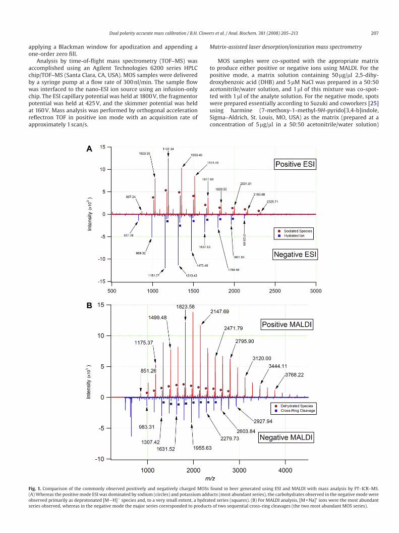
Fig. 1. Comparison of the commonly observed positively and negatively charged MOSs found in beer generated using ESI and MALDI with mass analysis by FT–ICR–MS.
(A) Whereas the positive mode ESI was dominated by sodium (circles) and potassium adducts (most abundant series), the carbohydrates observed in the negative mode were
observed primarily as deprotonated [M¡H]¡ species and, to a very small extent, a hydrated series (squares). (B) For MALDI analysis, [M + Na]+ ions were the most abundant
series observed, whereas in the negative mode the major series corresponded to products of two sequential cross-ring cleavages (the two most abundant MOS series).
208 Dual polarity accurate mass calibration / B.H. Clowers et al. / Anal. Biochem. 381 (2008) 205–213
with a comatrix of NH4Cl (prepared at a concentration of 100 mM,
aqueous). Each 1-ll sample spot was treated with 1 ll of the har-
mine solution and 1 ll of the NH4Cl solution. All MALDI spots were
prepared on a stainless-steel sample target and were vacuum-
dried. MALDI–MS was performed with a 7.0 Tesla, external source
FT–ICR instrument (IonSpec Pro MALDI). Ions from 5 to 25 laser
pulses (Nd:YAG, 355 nm) were accumulated in a hexapole (oper-
ated at 925 kHz, 200 V base-to-peak amplitude) and collisionally
cooled using a controlled leak of nitrogen into the hexapole cham-
ber. Ions were then injected into the ICR cell via a quadrupole ion
guide (operated at 925 kHz, 500 V base-to-peak amplitude). Detec-
tion parameters were essentially the same as those specified for
ESI–FT–ICR analysis.
Results and discussion
Of the 12 beers examined in this study, representative MOS spec-
tra acquired using both nano-ESI and MALDI ionization sources
in both polarities are shown in Fig. 1. These spectra demonstrate
the evenly spaced (Dm/z 162.05282) yet wide range of m/z values
observed for the MOSs in beer. It should be noted that each of the
nano-ESI beer spectra shown (Fig. 1A) was acquired for samples
diluted 2500-fold and required only minimal flushing to eliminate
the MOSs from the system. Given the availability and lack of car-
ryover effects associated with MOSs, the observed signal-to-noise
ratio could be adjusted by simply altering the MOS dilution factor
or by changing the ion accumulation time. To assess the relative
purity of each beer sample and remove any possible interferents, a
carbohydrate enrichment step was performed using SPE with PGC
as described above. Comparison of the enriched fraction with the
diluted beer sample indicated that no marked benefit was afforded
by the SPE enrichment procedure. Consequently, the commercial
beer samples were then used without additional purification.
A well-known feature of carbohydrates is their strong affinity
for alkali earth metals, particularly sodium and potassium [26].
The MOS ions observed in this study were no exception to this
trend, with the two most prominent species observed by positive
ion nano-ESI being sodium [M + Na]+ and potassium [M + K]+
adducts (Fig. 1A, upper trace). For the beer samples examined,
small variations in the relative abundance of the two adducts were
observed and were attributed to the varying mineral content of
the water used during the brewing process. Lower abundance MOS
series were also observed in the positive ESI mode; however, their
restricted m/z range limited their use as a calibration series. The
MOS ions produced by positive mode MALDI were observed almost
exclusively as [M + Na]+ adducts due to the doping of the sample
spots with NaCl (Fig. 1B, upper trace). A previous article examining
beer MOSs reported DP up to 40 (6500.1234 Da, monoisotopic neu-
tral mass) [16,27]. In the current work, the largest MOS observed
using nano-ESI corresponded to a DP of 21 (3421.1198 Da, monoiso-
topic neutral mass), and the largest MOS observed by MALDI cor-
responded to a DP of 27 (4393.4367 Da). The enhanced m/z range
observed using MALDI as compared with ESI was attributed to the
absence of an atmospheric pressure interface and differences in
the ion transfer optics. No doubt, MOSs with higher DP are pres-
ent in beer; however, their low abundance and ion bandpass limita-
tions hindered the observation of higher order MOSs (i.e., DP > 27).
A list of theoretical m/z values and core accurate masses used in
the calculation of MOS m/z is reported in Table 1.
Whereas the positive mode spectra of electrosprayed beer car-
bohydrates were dominated by sodium and potassium adducts,
the MOS species in the negative mode were most commonly depro-
tonated [M¡H]¡, as seen in the lower trace of Fig. 1A. Compared
with the results obtained for the other ionization and source config-
urations, the signal intensity of the negatively charged MOSs using
MALDI with the harmine/NH4Cl matrix was significantly less. Com-
parison of the relative intensities of the major ion peaks in the
positive and negative modes reflects an approximately threefold
difference in sensitivity. To compensate for the lower sensitivity
of negative mode MALDI for MOS analysis, ions were accumulated
from 25 laser shots as opposed to 5 shots for positive mode MALDI.
Although still useful for instrument tuning and calibration, the
observation of intact MOS ions in the negative mode using MALDI
was not attainable due to the relative instability of these com-
pounds in negative mode. Previous studies of negatively charged
nonreducing neutral oligosaccharides have indicated that the
chloride [M + Cl]¡ adducts formed with the use of harmine/NH4Cl
matrix readily decay to form deprotonated [M¡H]¡ ions [28,29].
These deprotonated ions are subject to further in-source and post-
source decay mechanisms, including cross-ring cleavage. Our
experiments provide further confirmation of this behavior given
that the negatively charged beer MOS ions generated by MALDI cor-
responded to two series of cross-ring cleavage products. The first
of these series was equivalent to a deprotonated molecular ion that
had lost 48.0211 Da (i.e., [M–CH2O–H2O–H]¡) and was consistent
with the observations of Yamagaki and coworkers [29]. This series
was represented by the minor signals shown in the lower trace of
Fig. 1B. The second series corresponded to a further degradation
of ions from the first series with the additional loss of 120.0423 Da
to produce [M–C4H8O4–CH2O–H2O–H]¡ fragments. These double-
cleavage products constitute the major series of signals seen in the
lower trace of Fig. 1B.
Using the nomenclature of Domon and Costello [30], the post-
source decay ions can be described as products of successive frag-
mentations of the X and A types. Calibrating the spectrum on the
Table 1
Theoretical monoisotopic masses of beer MOSs
Species Formula Mass (Da)
Hexose residue C6H10O5 162.0528
Water H2O 18.0106
Sodium ion Na+ 22.9892
Potassium ion K+ 38.9632
Proton H+ 1.0073
Cross-ring loss R1 C4H8O4 120.0423
Cross-ring loss R2 CH2O 30.0106
MOS
DP
[M + Na]+ [M + K]+ [M - H]- [M–R1–H]- [M–R2–
H2O–H]-
[M–R1–R2–
H2O–H]-
3 527.1582 543.1322 503.1617 383.1195 455.1406 335.0984
4 689.2111 705.1850 665.2146 545.1723 617.1934 497.1512
5 851.2639 867.2378 827.2674 707.2251 779.2463 659.2040
6 1013.3167 1029.2906 989.3202 869.2779 941.2991 821.2568
7 1175.3695 1191.3435 1151.3730 1031.3308 1103.3519 983.3096
8 1337.4223 1353.3963 1313.4258 1193.3836 1265.4047 1145.3625
9 1499.4752 1515.4491 1475.4787 1355.4364 1427.4575 1307.4153
10 1661.5280 1677.5019 1637.5315 1517.4892 1589.5104 1469.4681
11 1823.5808 1839.5547 1799.5843 1679.5420 1751.5632 1631.5209
12 1985.6336 2001.6076 1961.6371 1841.5949 1913.6160 1793.5737
13 2147.6864 2163.6604 2123.6899 2003.6477 2075.6688 1955.6266
14 2309.7393 2325.7132 2285.7428 2165.7005 2237.7216 2117.6794
15 2471.7921 2487.7660 2447.7956 2327.7533 2399.7745 2279.7322
16 2633.8449 2649.8188 2609.8484 2489.8061 2561.8273 2441.7850
17 2795.8977 2811.8717 2771.9012 2651.8590 2723.8801 2603.8378
18 2957.9505 2973.9245 2933.9540 2813.9118 2885.9329 2765.8907
19 3120.0034 3135.9773 3096.0069 2975.9646 3047.9857 2927.9435
20 3282.0562 3298.0301 3258.0597 3138.0174 3210.0386 3089.9963
21 3444.1090 3460.0829 3420.1125 3300.0702 3372.0914 3252.0491
22 3606.1618 3622.1358 3582.1653 3462.1231 3534.1442 3414.1019
23 3768.2146 3784.1886 3744.2181 3624.1759 3696.1970 3576.1548
24 3930.2675 3946.2414 3906.2710 3786.2287 3858.2498 3738.2076
25 4092.3203 4108.2942 4068.3238 3948.2815 4020.3027 3900.2604
26 4254.3731 4270.3470 4230.3766 4110.3343 4182.3555 4062.3132
27 4416.4259 4432.3999 4392.4294 4272.3872 4344.4083 4224.3660
28 4578.4787 4594.4527 4554.4822 4434.4400 4506.4611 4386.4189
29 4740.5316 4756.5055 4716.5351 4596.4928 4668.5139 4548.4717
30 4902.5844 4918.5583 4878.5879 4758.5456 4830.5668 4710.5245
Dual polarity accurate mass calibration / B.H. Clowers et al. / Anal. Biochem. 381 (2008) 205–213 209
minor series of fragments, the masses of the major ion series were
found to agree with these assignments to within »3 ppm root
mean square mass error, thereby supporting the hypothesis that
the ions are related via cross-ring fragmentation. Although the neg-
atively charged MOS ions produced by MALDI are prone to signifi-
cant fragmentation due to the inherent instability of these species,
their usefulness as mass calibrants is not diminished because the
elemental composition of the observed ions remains known.
The propensity of the negatively charged MOS ions to fragment
was also found to be an important factor in ESI. When probing pep-
tides and related systems, increasing the sample cone potential
typically leads to in-source fragmentation (sometimes referred
to nozzle skimmer dissociation), whereas MOSs tend to remain
intact under the same conditions in the positive mode. This behav-
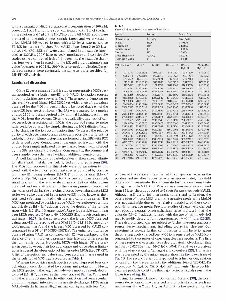
ior is illustrated in the uppermost plot of Fig. 2. At cone potentials
below 75 V, significant losses of carbohydrate signal were experi-
enced in the positive mode, whereas higher cone potentials pro-
duced signals approximately 10 times more intense. Interestingly,
high cone potentials in the negative mode also produced an abun-
dance of signal. However, the primary carbohydrate series did
not correspond to an intact molecular ion. Rather, this series was
the result of in-source fragmentation corresponding to the loss of
120.0423 Da (C4H8O4) from the deprotonated species to yield [M–
C4H8O4–H]¡ as reported by Yamagaki and Nakanishi [31]. These
ions correspond to deprotonated 0,2X or 2,4A fragments of the MOS
ions. Lowering the cone potential to 60 V virtually eliminated the
Fig. 2. Influence of ESI cone voltage on the relative abundance and observed MOS ions in both the positive and negative modes with mass analysis by FT–ICR–MS. (A) In the
positive mode, the type of adduct observed was relatively unchanged with cone potential; however, total ion abundances were highly dependent on this value. (B) Higher
cone potentials in the negative mode induced fragmentation (labeled in smaller font), whereas lower cone potentials yielded the deprotonated species. For the MOS sample
shown, the positive mode was dominated by potassiated adducts, with a smaller series corresponding to the sodiated adducts (circles). Additional series were observed;
however, the relative abundance of these series limits their utility as calibrants.

210 Dual polarity accurate mass calibration / B.H. Clowers et al. / Anal. Biochem. 381 (2008) 205–213
in-source fragmentation and allowed the deprotonated [M¡H]¡
ions of the MOSs to dominate (Fig. 2, lower trace). Overall, the
signal for beer MOSs was maximized using a high cone potential
(»95 V) in the positive mode and a lower cone potential (»60 V) in
Fig. 4. Annotated nano-LC/FT–ICR–MS separation of MOSs derived from beer. Using PGC as the stationary phase, singular m/z values within multiple nano-LC elution peaks
were often observed, indicating the presence of isomeric carbohydrates.
Fig. 3. ESI–FT–ICR tandem MS with IRMPD of selected beer MOSs. The vertical dotted lines provide a visual guide to the parent ion masses. The remaining peaks resulted
from the IRMPD of the precursor ions and represented either the loss of water or cross-ring cleavage (see legend on figure). The m/z region of each spectrum below the par-
ent ion has been magnified for clarity.
Dual polarity accurate mass calibration / B.H. Clowers et al. / Anal. Biochem. 381 (2008) 205–213 211
Fig. 5. Positive ion mode ESI–TOF–MS analysis of PGC-purified beer MOSs with an effective dilution factor of 100. Above m/z 1600, the intensity scale has been expanded by
a factor of 25 for clarity. The major MOS series corresponds to [M + Na]+ ions.
the negative mode. Although the possibility of in-source fragmen-
tation at higher cone potentials cannot be eliminated entirely, this
occurrence seems less likely when considering all data obtained
for each MOS sample in both the positive and negative modes. As
Fig. 2 illustrates, softening the environment of the atmospheric
pressure interface in the negative mode allowed intact molecular
ions to be observed. Using these same source parameters in the
positive mode failed to produce the protonated species or appre-
ciable amounts of metal adducted carbohydrate ions. Because it
was unlikely that higher cone potentials induced the formation
of alkali earth adducts, cone potentials above approximately 75 V
were evidently necessary to adequately desolvate the sodium and
potassium adducts of MOSs prior to mass analysis. Previous work
examining the conformation of metal-coordinated carbohydrates
has demonstrated the ability of the metal ions to coordinate with
multiple oxygen atoms simultaneously [26,32]. This serves to pro-
vide an explanation for the absence of in-source fragmentation in
positive mode ESI of MOS alkali metal adducts.
Although in-source fragmentation was not observed for the
metal-coordinated MOSs produced in positive mode ESI, detailed
tandem MS experiments were possible using IRMPD. As shown in
Fig. 3, the fragments observed for MOSs corresponded to loss of
water, glycosidic bond cleavage, and two subsequent cross-ring
cleavages. For carbohydrates in particular, IRMPD typically pro-
vides a wealth of information that may be used to confirm oligosac-
charide composition. However, these compositional assignments
do not preclude the possibility of isomerism. It is believed that
MOSs derived from beer are composed primarily of linear carbo-
Fig. 6. Infusion of beer MOSs using ESI–FT–ICR–MS. The plot of total ion intensity observed over time illustrates the reproducible nature of five 1-ll beer MOS injections and
the lack of carryover between samples.

212 Dual polarity accurate mass calibration / B.H. Clowers et al. / Anal. Biochem. 381 (2008) 205–213
hydrates, although nano-LC separation using PGC as the stationary
phase illustrates the possibility of isomers.
Fig. 4 shows the annotated nano-LC chromatogram of beer-
derived MOS and the corresponding mass spectrum summed across
the entire chromatographic run. Interestingly, the chromatogram
contains many more peaks than the eight primary m/z ratios found
in the summed mass spectrum for the nano-LC experiment. The
multiplicity of chromatographic peaks in comparison with the mass
spectral signals suggests the presence of isobaric but structurally
distinct carbohydrate species in beer. Despite the presence of MOS
isomers, the utility of these compounds as mass calibrants remains
intact due to the inability of MS to resolve truly isobaric ions.
Although the MOSs found in samples of dilute beer are readily
detected by FT–ICR–MS, the ability to produce and detect these ions
is not unique to that particular mass analyzer. As demonstrated in
Fig. 5, the MOSs are readily observed as sodium adducts in positive
ion mode ESI–TOF–MS. Thus, MOS series not only are useful due to
dual polarity accessibility in both ESI and MALDI, but also provide
calibration and tuning standards well suited to the optimization of
various types of mass spectrometers for carbohydrate analysis. It
should be noted that to increase the detection of these ions with
a good signal-to-noise ratio, the source conditions (voltages) were
altered from those typically used for proteomics.
In addition to their ease of use, compatibility with ESI and
MALDI, and fairly comprehensive mass range in either polarity,
perhaps the most attractive feature of beer MOSs is their lack of
memory effects. Fig. 6 highlights the lack of carryover associated
with the use of beer MOSs as tuning standards for MS. Five 1-ll
injections were performed at 8-min intervals with a flow rate of
500 nl/min. Under these conditions and with the flow rate held
constant, the total ion signal returned to baseline shortly after
injection. No special effort was made to flush the MOSs from
the system. At no time during the course of this study were the
electrospray emitters replaced due to contamination with MOSs.
It should also be noted that through refrigeration, the integrity
of the stock solution was maintained for periods lasting many
months.
Conclusions
Using nano-ESI and MALDI as ionization sources with FT–ICR –MS
and TOF–MS as mass analyzers, beer MOSs have been successfully
used as mass calibrants and tuning standards in both the positive
and negative ion modes. Because the MOSs are derived from beer,
the mass calibration and tuning range are not limited by the com-
mercial unavailability of pure MOSs with sufficiently high DPs. Rel-
atively high cone voltages in positive mode nano-ESI favored the
production of sodium and potassium adducts, whereas the same
settings in the negative mode induced cross-ring cleavage. This in-
source fragmentation was alleviated by lowering the cone potential
to produce the deprotonated molecular ions of beer MOSs. These
source conditions are typical for oligosaccharide analysis using
nano-ESI in our research group. With ionization by MALDI and
using DHB as the matrix, sample spots doped with NaCl almost
exclusively produced MOS ions as sodium adducts in the positive
ion mode. When operating in negative mode MALDI and using har-
mine/NH4Cl as the matrix, the MOSs were observed primarily as
internal fragments resulting from sequential cross-ring cleavages
at both termini, but they remained useful as mass and tuning stan-
dards because the elemental composition of the fragments could
be deduced.
Although MOS content varied slightly with the selected beer,
the overall composition and, more important, the m/z ratios
observed remained constant. The stringent level of quality control
for these products results in highly reproducible MOS distributions
for any given brand. In addition to being amenable to dual polarity
ionization using both ESI and MALDI, beer MOSs are easily enriched,
cost-effective, readily available, amenable for carbohydrate anal-
ysis, and relatively comprehensive calibrants (m/z 7 500–2500)
that do not suffer from memory effects that hinder many tradi-
tional mass and tuning standards.
Acknowledgments
The authors thank Agilent Technologies for use of the 6200 series
HPLC chip/TOF–MS. The following funding sources are also acknowl-
edged: Dairy Management Incorporated California Dairy Research
Foundation (06 LEC-01-NH), University of California Discovery Grant
(05GEB01NHB), and National Institutes of Health (GM 49077).
References
[1] D.L. Lawrence, Accurate mass measurement of positive ions produced by ammonia chemical ionization, Rapid Commun. Mass Spectrom. 4 (1990) 546–549.
[2] K.R. Jonscher, J.R. Yates III, Mixture analysis using a quadrupole mass filter/quadrupole ion trap mass spectrometer, Anal. Chem. 68 (1996) 659–667.
[3] L. Jiang, M. Moini, Ultramark 1621 as a reference compound for positive and negative ion fast-atom bombardment high-resolution mass spectrometry, J. Am. Soc. Mass Spectrom. 3 (1992) (1621) 842–846.
[4] R.P. Lattimer, Fast atom bombardment mass spectrometry of polyglycols, Intl. J. Mass Spectrom. Ion Proc. 55 (1984) 221–232.
[5] L.J. Goad, M.C. Prescott, M.E. Rose, Poly(ethyleneglycol) as a calibrant and sol-vent for fast atom bombardment mass spectrometry: application to carbohy-drates, Org. Mass Spectrom. 19 (1984) 101–104.
[6] R.P. Lattimer, Tandem mass spectrometry of lithium-attachment ions from polyglycols, J. Am. Soc. Mass Spectrom. 3 (1992) 225–234.
[7] K. Vekey, Calibration in positive and negative ion fast atom bombardment using salt mixtures, Org. Mass Spectrom. 24 (1989) 183–185.
[8] S. Konig, H.M. Fales, Calibration of mass ranges up to m/z 10, 000 in electro-spray mass spectrometers, J. Am. Soc. Mass Spectrom. 10 (1999) 273–276.
[9] J.B. Fenn, M. Mann, C.K. Meng, S.F. Wong, C.M. Whitehouse, Electrospray ioniza-tion for mass spectrometry of large biomolecules, Science 246 (1989) 64–71.
[10] K. Tanaka, H. Waki, Y. Ido, S. Akita, Y. Yoshida, T. Yohida, Protein and polymer analyses up to m/z 100,000 by laser ionization time-of-flight mass spectrome-try, Rapid Commun. Mass Spectrom. 2 (1988) 151–153.
[11] M. Moini, Ultramark 1621 as a calibration/reference compound for mass spec-trometry: II. Positive- and negative-ion electrospray ionization, Rapid Com-mun. Mass Spectrom. 8 (1994) (1621) 711–714.
[12] R.B. Cody, J. Tamura, B.D. Musselman, Electrospray ionization magnetic sector mass spectrometry: calibration, resolution, and accurate mass measurements, Anal. Chem. 64 (1992) 1561–1570.
[13] C.N. McEwen, B.S. Larsen, Accurate mass measurement of proteins using elec-trospray ionization on a magnetic sector instrument, Rapid Commun. Mass Spectrom. 6 (1992) 173–178.
[14] W.A. Harris, D.J. Janecki, J.P. Reilly, Use of matrix clusters and trypsin autolysis fragments as mass calibrants in matrix-assisted laser desorption/ionization time-of-flight mass spectrometry, Rapid Commun. Mass Spectrom. 16 (2002) 1714–1722.
[15] S. Konig, H.M. Fales, Formation and decomposition of water clusters as observed in a triple quadrupole mass spectrometer, J. Am. Soc. Mass Spectrom. 9 (1998) 814–822.
[16] E. Vinogradov, K. Bock, Structural determination of some new oligosaccharides and analysis of the branching pattern of isomaltooligosaccharides from beer, Carbohydr. Res. 309 (1998) 57–64.
[17] P. Mauri, M. Minoggio, P. Simonetti, C. Gardana, P. Pietta, Analysis of saccha-rides in beer samples by flow injection with electrospray mass spectrometry, Rapid Commun. Mass Spectrom. 16 (2002) 743–748.
[18] A.S. Araujo, L.L. da Rocha, D.M. Tomazela, A.C.H.F. Sawaya, R.R. Almeida, R.R. Catharino, M.N. Eberlin, Electrospray ionization mass spectrometry finger-printing of beer, Analyst 130 (2005) 884–889.
[19] S. Cortacero-Ramirez, A. Segura-Carretero, C. Cruces-Blanco, M. Hernainz-Ber-mudez de Castro, A. Fernandez-Gutierrez, Analysis of carbohydrates in bever-ages by capillary electrophoresis with precolumn derivatization and UV detec-tion, Food Chem. 87 (2004) 471–476.
[20] L.C. Nogueira, F. Silva, I.M.P.L.V.O. Ferreira, L.C. Trugo, Separation and quantifica-tion of beer carbohydrates by high-performance liquid chromatography with evaporative light scattering detection, J. Chromatogr. A 1065 (2005) 207–210.
[21] K. Bock, T. Dreyer, S. Mueller-Loennies, L. Molskov-Bech, Evaluation of new ana-lytical techniques for the optimization of brewing processes, Proc. Inst. Brew. 24 (1996) 234–238.
[22] I.F. Duarte, M. Spraul, M. Godejohann, U. Braumann, M. Spraul, A.M. Gil, Appli-cation of NMR spectroscopy and LC–NMR/MS to the identification of carbohy-drates in beer, J. Agric. Food Chem. 51 (2003) 4847–4852.
[23] N.H. Packer, M.A. Lawson, D.R. Jardine, J.W. Redmond, A general approach to desalting oligosaccharides released form glycoproteins, Glycoconj. J. 15 (1998) 737–747.

Dual polarity accurate mass calibration / B.H. Clowers et al. / Anal. Biochem. 381 (2008) 205–213 213
[24] M.J. Davies, K.D. Smith, R.A. Carruthers, W. Chai, A.M. Lawson, E.F. Houn-sell, Use of a porous graphitized carbon column for the high-performance liquid chromatography of oligosaccharides, alditols, and glycopeptides with subsequent mass spectrometry analysis, J. Chromatogr. 646 (1993) 317–326.
[25] H. Suzuki, T. Yamagaki, K. Tachibana, Optimization of matrix and amount of ammonium chloride additive for effective ionization of neutral oligosaccha-rides as chloride ion adducts in negative-mode MALDI–TOF mass spectrome-try, J. Mass Spectrom. Soc. Jpn. 53 (2005) 227–229.
[26] M.T. Cancilla, S.G. Penn, J.A. Carroll, C.B. Lebrilla, Coordination of alkali metals to oligosaccharides dictates fragmentation behavior in matrix assisted laser desorption ionization/Fourier transform mass spectrometry, J. Am. Chem. Soc. 118 (1996) 6736–6745.
[27] T.R.I. Cataldi, C. Campa, G.E. de Benedetto, Carbohydrate analysis by high-perfor-mance anion-exchange chromatography with pulsed amperometric detection: the potential is still growing, Fresenius J. Anal. Chem. 368 (2000) 739–758.
[28] R.B. Cole, J. Zhu, Chloride anion attachment in negative ion electrospray ioniza-tion mass spectrometry, Rapid Commun. Mass Spectrom. 13 (1999) 607–611.
[29] T. Yamagaki, H. Suzuki, K. Tachibana, In-source and postsource decay in neg-ative-ion matrix-assisted laser desorption/ionization time-of-flight mass spectrometry of neutral oligosaccharides, Anal. Chem. 77 (2005) 1701–1707.
[30] B. Domon, C.E. Costello, A systematic nomenclature for carbohydrate frag-mentations in FAB–MS/MS spectra of glycoconjugates, Glycoconj. J. 5 (1988) 397–409.
[31] T. Yamagaki, H. Nakanishi, Negative-mode matrix-assisted laser desorption/ionization mass spectrometry of maltoheptaose and cyclomaltooligosaccha-rides, J. Mass Spectrom. Soc. Jpn. 50 (2002) 204–207.
[32] G.E. Hofmeister, Z. Zhou, J.A. Leary, Linkage position determination in lithium-cationized disaccharides: tandem mass spectrometry and semiempirical calcu-lations, J. Am. Chem. Soc. 113 (1991) 5964–5970.