Embed Size (px)
Citation preview

m u l t i
L U M E NM U L T I P L E
C E N T R A L
C A T H E T E R
N u r s i n g
C a r e
G u i d e l i n e s
M U L T I - L U M E NA R R O W
v e n o u s
UNITE D STAT E SArrow International, Inc.2400 Bernville RoadReading, PA 19605, USAPhone: 610-378-0131Toll-free: (800) 523-8446Fax: 610-478-3199Orders-only toll-free fax: (800) 343-2935Email: Customer. S e rv i c e @ a r r o w i n t l . c o m
A F R I C AArrow Africa (Pty) Ltd.Cambridge Commercial Park22 Witkoppen Road, Paulshof Extension Sandton 2054, Republic of South AfricaPhone: (11) 807 4887Fax: (11) 807 4994Email: [email protected]
A U S T R A L A S I AArrow International, Inc.2400 Bernville RoadReading, PA 19605, USAPhone: 610-378-0131Toll-free: (800) 233-3187Fax: 610-374-5360Email: [email protected]
C A N A D AArrow Medical Products, Ltd.2300 Bristol Circle, Unit 1Oakville, Ontario L6H 5S3Phone: (905) 829-9473Toll-free: (800) 387-7819Fax: (905) 829-9414Email: [email protected]
CZECH RE PUBL ICArrow International CR, a.s.Praz̆ská 209500 04 Hradec KrálovéCzech RepublicPhone: 049 575 9111Fax: 049 575 9222Email: [email protected]
EUROP EAN SALES OFFICEFlevolaan 9ANL 1382 JXWeesp, The NetherlandsPhone: +31 294 29900Fax: +31 294 414235Email: [email protected]
F R A N C EArrow France S.A.Atlantic Parc, « Les Pyramides » No. 11Route de Pitoys, P.A. de Maignon64600 Anglet, FrancePhone: 05 59 31 34 90Fax: 05 59 31 34 91Email: [email protected]
G E R M A N YArrow Deutschland GmbHJustus-von-Liebig-Strasse 2D-85435 Erding, GermanyPhone: 08122 9820-0Fax: 08122 40384Email: [email protected]
G R E E C EArrow Hellas A.E.E.230 Kifissias AvenueHalandri, 152 31Athens, GreecePhone: 6777717 / 6777311Fax: 6777911Email: [email protected]
H O L L A N DArrow Holland Medical Products B.V.Flevolaan 9ANL 1382 JXWeesp, The NetherlandsPhone: 294 280 620Fax: 294 412 300Email: [email protected]
I N D I AArrow India9 Demonte StreetSanthome, MylaporeChennai 600004IndiaPhone: 44-495-6769Fax: 44-495-6787Email: [email protected]
J A PA NArrow Japan, Ltd.Nagaoka Bldg.3-16, Kita-Otsuka 3-chomeToshima-ku, Tokyo 170-0004, JapanPhone: 03-5974-1701Fax: 03-5974-1845Email: [email protected]
L AT IN AMER ICAArrow International, Inc.2400 Bernville RoadReading, PA 19605, USAPhone: 610-378-0131Toll-free: (800) 233-3187Fax: 610-374-5360Email: [email protected]
M E X I C OArrow Internacional de Mexico S.A. de C.V.Moliere No. 128, Col. Polanco11560 Mexico City, MexicoPhone: 5281-2291Fax: 5282-1359Email: [email protected]
S L O VA K I AArrow Slovensko Pies̆t’any s.r.o.Valová 49921 01 Pies̆t’any, SlovakiaPhone/Fax: 0838/77 25428Email: [email protected]
S PA I NArrow Iberia S.A.Avenida de Valgrande 1428108 Alcobendas Madrid, SpainPhone: 916 621 267Fax: 916 619 756Email: [email protected]
©1996 Arrow International, Inc. All rights reserved. ML-NG 06/01

“Withou t q ues t i on , in t r avenous
in fus io n therapy has become an
ind i spensab l e t herapeuti c moda l i t y
i n presen t -day med ic i ne . I t has
probab l y saved mo re l i ves than al l
the an t i b io t i c s ever deve loped .” 1
This very powerful statement made over a decade ago isstill valid today as evidenced by the number of intravasculardevices placed in patients annually. With the use of thistechnology has come a plethora of procedures and policies toguide its utilization. Today we find that many of these policiesand procedures are not necessarily based on scientific researchbut rather on practitioner intuition and tradition. Review ofthe literature reveals very few definitive research studies onwhich to base protocols to support device care.
To assist practitioners who are striving to establishpolicies and procedures, Arrow International has compiled anextensive review of the central venous catheter literature. Theintent of providing this information is to give guidance to you,the practitioner, in establishing catheter-related protocols. It isnot meant to dictate nursing or medical practice.
The major areas of current concern have beenaddressed. Please bear in mind that as these words arewritten, research continues. The use of central venouscatheters is an evolving science, and this review will continueto change as the body of knowledge expands.
These educational materials are shared with you, thepractitioner, with hope that they will assist you in the pursuitof excellence in your practice.
Sincerely
Arrow International, Inc.
1 Maki, DG. Preventing infection in intravenous therapy.Anesth Analg. January-February 1977;56:141.
multiple l u m e n c a t h e t e r2 Preface and Table of Contents
3 Types of Catheters
3 Indications for Central Venous Access
4 Catheter Materials and Properties
4 Arrow Multiple-Lumen Catheter Design and Specifications
6 Insertion Sites
6 Site Preparation
8 Catheter Insertion
10 Product Instructions
14 Port Designation
15 Complications
25 Catheter Maintenance
31 Infusions
32 Monitoring
32 Problem Intervention
35 Infection Control
36 Catheter Exchange
36 Catheter Removal
37 Documentation
37 Bibliography
2
types of c a t h e t e r sS ing le-Lumen Ca theter s (S LC)
A SLC consists of a tube or lumen ending in a hub that can be
capped and used for intermittent or continuous infusions of
medication or fluid. The use of a single-lumen central venous
catheter centers around the need for an infusion into a large
central vein. Central venous catheterization is indicated when
there are no peripheral sites available, when a viscous or
hyperosmolar infusion is prescribed, or when a line is needed for
OR support. This type of catheter may be used in an acute setting
or to provide long-term access to a central vein for nutritional
support, antibiotics, or chemotherapy. If the SLC is to be used for
a prolonged period of time, the catheter may be “tunneled” on the
chest for stabilization. Tunneling also reduces the risk of catheter-
related infections.
Mult ip le- Lumen Cathet er s (MLC )
A MLC multiplies the advantages of a single-lumen catheter. The
number of lumens within an MLC can vary from two to four and
allows for many treatments to be performed through one venous
access site. Therapy may be intermittent or constant. The multiple
ports allow for administration of medications, blood infusion and
sampling, fluid replacement, monitoring, and in certain catheters,
visualization of cardio-vascular anatomy. Available literature
shows that catheter design determines whether treatments that are
incompatible may be given at the same time. Multiple-lumen
catheters are found most frequently in the acute care setting;
however, there are double-lumen catheters which have been
developed for the long-term delivery of multiple treatments such
as antibiotics and chemotherapy. As with SLC, these catheters
must be placed in a central vein.
Per i phera l l y Inser t ed Cent ra lCa thet er s ( P ICC)
PICCs are inserted into the arm and advanced until the end of the
catheter is located in a central vein. The catheters contain one or
two lumens and can be used to deliver continuous or intermittent
therapy. PICCs can be used in an acute setting but have been most
popular for long-term venous access to provide nutritional
support, antibiotic therapy or chemotherapy. Because of the arm
placement, specially-trained nurses, in addition to physicians, can
insert the catheters.
Imp lantab le Ca t het ers
When there is a need for prolonged therapy, the central venous
catheters of choice are those which can be implanted or tunneled
under the skin. These specially adapted catheters are placed with
the distal end positioned in a large vein. The proximal end of the
catheter may consist of a hub and pigtails extending from the
single or multiple lumens, or a self-sealing port which is placed
under the skin. The tunneled lines have one or two lumens. They
are sutured into a subcutaneous pocket on the chest or arm and
constitute a completely closed system. Both catheter types may be
used for the long-term infusions of antibiotics, TPN,
chemotherapy or other fluids and medications.
T h e rmodi lu t ion Cathe ter s
Thermodilution catheters, or pulmonary artery catheters, are
used in an acute setting when an accurate assessment of
pulmonary and cardiac status is necessary. The catheter contains
multiple ports which can be used for infusions and monitoring. A
balloon tip is incorporated into the catheter structure to facilitate,
when inflated, wedge placement into a pulmonary artery to
measure left-sided heart function. The catheter also has a
temperature sensing device that can be used to measure cardiac
output by the thermodilution method.
Hemodia ly s i s Ca t hete rs
Hemodialysis catheters are single- or multiple-lumen catheters
that provide temporary vascular access for hemodialysis until a
permanent access is available or until another type of dialysis
therapy is substituted. The multiple lumen catheters contain two
large bore lumens that are connected to the dialysis machine to
form a complete circuit for the removal and return of the patient’s
blood during treatment.
indications for c e n t r a lvenous access• Patients requiring multiple sites for IV access.
• Patients lacking useable peripheral IV sites.
• Patients requiring central venous pressure monitoring.
• Patients requiring total parenteral nutrition.
• Patients receiving incompatible medications.
• Patients requiring multiple infusions of fluids, medications,
or chemotherapy.
• Patients subject to frequent blood sampling or receiving
blood transfusions.
• Patients requiring a temporary access site for hemodialysis.
• Patients receiving infusions that are hypertonic,
hyperosmolar or infusions that have divergent
pH values.
3

catheter materials a n dp ro p e rt i e sIntravascular catheters must be made of biocompatible materials
that will withstand conditions within the vascular system without
deteriorating or causing patient complications. Polymers,
commonly known as plastics, have been used with much success.
Certain properties are sought when developing a product for use
within the vascular system, for example, thromboresistance,
flexibility, smooth surface, lack of kink memory, and reasonable
cost, to name a few. It is also important that the material does not
leach off chemicals used in its manufacture. The six most common
polymers used in vascular catheters are: polyethylene,
fluoropolymer (Teflon®*), polyvinyl chloride (PVC), silicone,
elastomeric hydrogel and polyurethane. 10-17
Catheter Material Advantages Disadvantages
Polyethylene High inherent strength May be stiff
Resistant to fats and oil Exhibits kink memory
High oxygen & carbon
dioxide permeability
Overall good chemical
resistance
Low moisture absorption
Fluoropolymer Resistant to chemicals Exhibits kink memory
(Teflon) Slippery surface due to High incidence of
low surface energy thrombosis
May be stiff
Polyvinyl chloride Stiff on insertion but High absorption of
(PVC) softens within the body certain drugs
High inherent strength, High incidence of
tough thrombosis
Abrasion resistant May leach out
plasticizers
Elastomeric Predictable softening Contact with liquid
hydrogel and size changes on prior to insertion
contact with fluid prevents use
Stiff for insertion
and softens for
biocompatibility
Silicone Most biocompatible May knot
Thromboresistant Poor tolerance to
Slippery surface due to pressure
low surface energy Some formulations
Soft and pliant not easily placed
Resistant to moisture, percutaneously
many chemicals
Catheter Material Advantages Disadvantages
Polyurethane High degree of
biocompatibility
Good tensile strength
Wear resistant
Thromboresistant
Resistant to many chemicals
Kink resistant
Softens within the body
Able to use percutaneous
technique
Thinner wall construction
possible
A rrow m u l t i p l e - l u m e ndesign and specificationsArrow multiple-lumen central venous catheters are radiopaque
catheters available in a variety of lengths and French sizes ranging
from 4 Fr. used in pediatrics to 12 Fr. used for hemodialysis or
trauma. These catheters may have two, three or four separate
lumens running the length of the catheter body. In all adult
catheters the lumens exit at the distal end of the catheter through
individual ports. The lumen exits are placed along the distal
catheter body and are rotated 90° around the catheter
circumference to minimize mixture of infusates. At the proximal
end the lumens are connected to separate color-coded Luer-Lock
extension lines or pigtails. Polyurethane, a compound which is
sufficiently stiff to facilitate percutaneous insertion and softens
inside the body, makes up the body of the catheters. To further
reduce the chance for vessel trauma, Arrow multiple-lumen
catheters have a Blue FlexTip® which is more pliant than the rest
of the catheter. The distal end is tapered to form a dilating tip that
will fit snugly over a .025" or .035" diameter spring wire guide.
The catheters are available in a variety of lengths with various
lumen configurations as illustrated (refer to page 10). External
markings on the body of the catheter are used to aid in proper
anatomical placement of the catheter tip. The catheter body and
pigtails converge at a blue triangular hub which is imprinted with
information about the size, number of lumens and catheter length.
The hub is used as a primary suture site for securing the catheter
to the patient. A separate catheter clamp and fastener are
provided for use on the catheter body as a secondary site for
suturing if a sufficient length of the catheter remains exposed.
Removable slide clamps are provided on the pigtails to aid in
changing Luer-Lock injection caps and tubing. On 12 French
catheters pinch clamps are provided for additional security.
4

Cathet er C ros s-Sec t ions
Pr iming Vo lumes and F low Rates
The amount of space within a catheter lumen from the distal exit
port to the end of the pigtail Luer-Lock hub is the catheter lumen
priming volume. This volume is important for flushing/locking
procedures. When used, the injection cap volumes must be added
to the lumen volumes to determine the amount of fluid needed to
completely fill capped lumens.
The volume of fluid that a catheter can deliver per unit of time is
defined as the flow rate. The flow rate through a tube or catheter
is dependent on several parameters which most importantly
include internal (lumen) diameter, length of catheter, the change
in pressure from inlet to outlet, and the viscosity of the fluid.
Mathematically, the relationship is:
d4 (P1 - P2)16 µ L
In medical practice, a catheter is placed for many reasons with the
most important reason being administration of fluids/flow. It is
i m p o rtant to know how much flow can be infused through the
c a t h e t e r.
The flow rate is often incorrectly assumed to be proportional to
the catheter’s outside diameter (French scale) or gauge size. This
assumption can lead to erroneous conclusions, as the equation
(previous page) indicates. Further, this assumption becomes even
more confusing when the catheter has multiple lumens. Typically,
each individual lumen of a multiple lumen catheter is labeled with
a nominal gauge size derived from the flow rate that a typical
single lumen catheter of the same length would provide. In this
situation, the gauge size does NOT relate to outside diameter or
French scale measurement.
To eliminate any confusion, it is recommended that a clinician
using a multiple lumen catheter know and consider the flow rates
through each separate lumen.
The following table summarizes priming volumes and flow rates
for a selection of Arrow products.
F low Rates and P riming Vo l u m e s —Arrow Mu l t i - Lumen Cathe ter s
Priming Flow Rate (cc/hr)Description Lumens Volume (cc) Gravity (standard tubing)
7 Fr. x 16cm (6") 2 Prox. (14 Ga.) 0.53 5630Distal (18 Ga.) 0.32 1460
7 Fr. x 16cm (6") 2 Prox. (16 Ga.) 0.38 3640Distal (16 Ga.) 0.40 3770
7 Fr. x 16cm (6") 3 Prox. (18 Ga.) 0.37 1900Med. (18 Ga.) 0.35 1800Distal (16 Ga.) 0.39 3380
7 Fr. x 20cm (8") 2 Prox. (16 Ga.) 0.45 3000Distal (16 Ga.) 0.47 3700
7 Fr. x 20cm (8") 2 Prox. (18 Ga.) 0.43 2190Distal (14 Ga.) 0.60 5590
7 Fr. x 20cm (8") 3 Prox. (18 Ga.) 0.39 1590Med. (18 Ga.) 0.39 1510Distal (16 Ga.) 0.44 3110
7 Fr. x 30cm (12") 3 Prox. (18 Ga.) 0.44 1090Med. (18 Ga.) 0.40 993Distal (16 Ga.) 0.49 2320
8 Fr. x 16cm (6") 2 Prox. (14 Ga.) 0.73 5770Distal (14 Ga.) 0.68 6200
8 Fr. x 20cm (8") 2 Prox. (14 Ga.) 0.79 4460Distal (14 Ga.) 0.75 5490
8.5 Fr. x 16cm (6") 4 Prox. (18 Ga.) 0.38 1840Med. 2 (18 Ga.) 0.36 1600Med. 1 (14 Ga.) 0.47 5410Distal (16 Ga.) 0.39 3180
8.5 Fr. x 20cm (8") 4 Prox. (18 Ga.) 0.41 1580Med. 2 (18 Ga.) 0.36 1430Med. 1 (14 Ga.) 0.54 5080Distal (16 Ga.) 0.43 2730
12 Fr. x 16cm (6") 2 Prox. (12 Ga.) 1.3 23700Distal (12 Ga.) 1.3 17400
12 Fr. x 20cm (8") 2 Prox. (12 Ga.) 1.40 19800Dist. (12 Ga.) 1.48 15500
F=
Where: F = flow rate
d = internal diameter
P1 = inlet pressure
P2 = outlet pressure
µ = fluid viscosity
L = length
5
Priming volumes are performed without the injection cap.
Injection cap priming volume=0.17 cc.
Flow rates are performed with normal saline solution at room
temperature and 40" head height. These rates represent
approximate flow capabilities. Priming volumes and flow rates are
printed on the package lidstock.
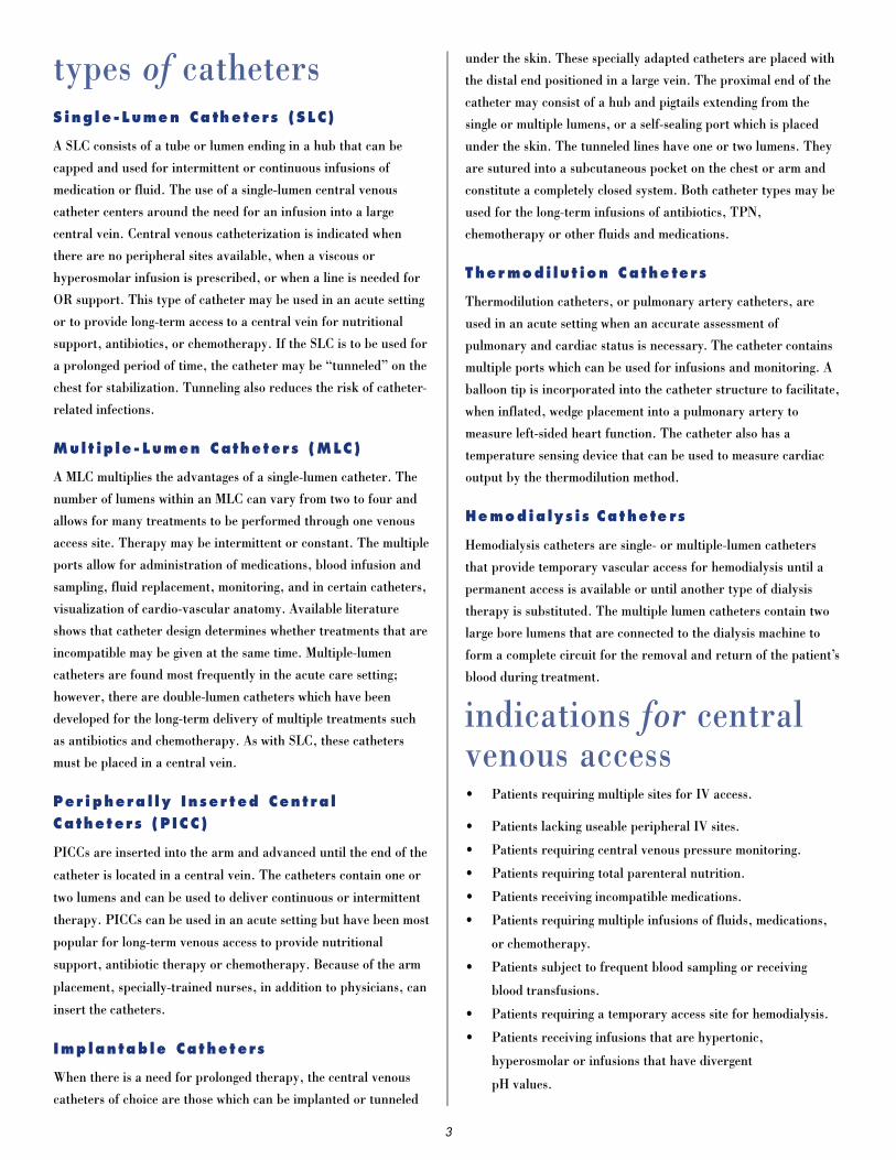
i n s e rtion s i t e sMultiple-lumen catheters are inserted into a large vein and
threaded into the central venous system. To reduce the chance of
complications, the central venous catheter tip should be placed in
the superior vena cava31-35,38-42 above its junction with the right
atrium with the distal catheter parallel to the vessel wall36,42. For
reference, the distal tip should be positioned at a level above
either the azygos vein or the carina of the trachea whichever is
better visualized. The insertion sites most frequently used are
those listed below. (Refer to illustration.)
Site Advantages Disadvantages
Internal jugular Large vessel Uncomfortable for patientEasy to locate Hard to maintain dressingEasy access Close proximity to carotid arteryShort, straight path to Highest infection rate of
vena cava (right side) insertion sitesLow rate of complications Problematic in patients with
tracheotomies
External jugular Easy to locate, visible Difficult to cannulate (rolls,valves, tortuous path)
Higher complication rate than other sites
Hard to maintain dressingUncomfortable for patientProblematic in patients with
tracheotomies
Subclavian Large vessel with high Lies close to the lung apexflow rate (pneumothorax risk)
Lower infection rate Close proximity to subclavian Easy to dress and maintain arterySupra- or infraclavicular Difficult to control bleeding
approaches (noncompressable vessel)Less restricting for patient
Femoral Easy access Decreased patient mobilityLarge vessel Increased rate of thrombosis,Advantageous during phlebitis and infection
resuscitation Risk of femoral artery punctureDressings may be problematic
Brachial Advantageous during Increased incidence of phlebitisresuscitation Longer time for drugs to access
Easy access central circulationCatheter tip movement related to
arm movement
Umbilical Easy access Significant rate of complicationsAccommodates fairly large
catheterRapid placement
Basilic Low incident of thoracic Increased incidence of phlebitiscomplication Catheter tip movement
Direct route into central related to arm movementvenous system with arm at90 degree angle.
Site Advantages Disadvantages
Cephalic Easy access More tortuous than basilic veinLow incidence of thoracic Increased incidence of phlebitis
complications May be compressed with the clavicle by anatomical positioning
Catheter tip movement related to arm movement
site p re p a r a t i o n
Standard procedures for skin preparation prior to the insertion of
central venous catheters include the use of an antiseptic solution
which kills or inhibits the growth of microorganisms. In this way
the numbers of resident and transient organisms on the patient’s
skin are reduced. By reducing the patient’s skin flora, the risks
for developing an infection from the catheter insertion are
decreased. As a rule, antiseptics are also included in routine
follow-up care of the insertion site as designated within each
institution. The choice of antiseptic should be made using
available literature and information about the profile of patients
served by the health care facility.
6
Internal Jugular Vein
Cephalic Vein
Subclavian Vein
External Jugular Vein
Basilic Vein
Femoral Vein

The three antiseptic solutions used most frequently are alcohol,
iodine/iodophor, and chlorhexidine.22
Alcoho l , E thy l (ETOH)
Alcohol provides for the most rapid kill of micro o rganisms of the
t h ree agents listed. The organisms die because alcohol causes
p rotein denaturation to occur. It is very effective against gram-
negative and gram-positive bacteria, and also achieves high levels
of kill for Mycobacterium tuberc u l o s i s , fungi and viruses. Alcohol
is not effective against spores. Alcohol is also effective as a fat
s o l v e n t .
To enable alcohol to denature protein it must be diluted with
water. 70% ETOH is the solution of choice. Other concentrations
are not as effective. To achieve maximum kill alcohol should be
applied to the targeted insertion site with a vigorous rub lasting
one minute during which the site is kept wet with solution. This is
necessary since alcohol has no residual effects once it dries.
The disadvantages to using alcohol are that it is very drying to the
skin and catheter materials with repeated application, and the
solution is flammable.
Iod ine/Iodophor So lu t ions
Iodine solutions achieve the kill of micro o rganisms thro u g h
penetration of the cell wall and intracellular oxidation with re s u l t a n t
release of free iodine within the microbial contents. This pro b a b l y
d i s rupts protein and nucleic acid stru c t u re and synthesis. Iodine
p reparations are effective against gram-positive and gram-negative
b a c t e r i a , M. tuberc u l o s i s , v i ruses and fungi, although pro l o n g e d
contact may be needed to achieve kill against certain fungi and
s p o re s .
The iodine solutions used most frequently are iodophors, a
combination of iodine and a solubizing agent or carrier which
provides a sustained release of free iodine. The most common
iodophor is povidone-iodine, a combination of iodine and
polyvinylpyrolidine. The result of the iodophor combination is a
reduction in toxicity and irritation to the skin. Because the iodine
is released gradually, a contact time of two minutes is necessary to
allow for optimum microbial kill. If adequate time is not routinely
allowed for iodophor action, the use of tincture of iodine may be
considered due to its more rapid action.25
A shortcoming of iodophors is the neutralization of their
antimicrobial properties in the presence of proteinaceous
materials such as blood and pus. There have also been reports of
microbial growth in certain iodophor solutions prompting careful
attention to the proper dilution.23 The available concentrations are
.5%, 2%, 7.5% and 10%.
C h l o r h e x i d i n e
Chlorhexidine is a cationic biguanide that causes microbial death
through cell wall disruption. It is very active against gram-positive
organisms, gram-negative organisms and viruses, less active
against fungi and minimally effective against M. tuberculosis. The
major advantage of chlorhexidine is its ability to bind to skin
protein leaving a residue with persistent antimicrobial effects for
up to 6 hours after application. Organic material has minimal
effect on the action of chlorhexidine. The strength of the solution
which has been tested is 2%, and in at least one study this solution
has been found superior to alcohol (70%) and povidone-iodine
(10%) in preventing IV-related infections.24
Caution must be taken not to introduce chlorhexidine into the ear
due to its known ototoxicity; otherwise, it has few side effects.
Consideration must also be given to the fact that the action of
chlorhexidine is pH dependent (5.5-7), and it can be inactivated
by compounds found in hard water and soap.
Si te Prepa rat ion Guide l ines
• Do not remove hair at the site unless it interferes with
dressing adherence. If necessary, clipping is preferable to
shaving to avoid skin lacerations and disruption of the
epidermal barrier to infection. 21
• Check for patient sensitivity to the prepping solution by
requesting known allergy information or testing on a small
area of skin away from the proposed insertion site.
• Physically clean the skin prior to applying antiseptic solution
and inserting the catheter. Care must be taken to remove all
soap residue.
• Apply the antiseptic in a circular pattern beginning in the
center of the proposed site and moving outward. (Refer to
illustration.)
• Allow the antiseptic solution to air dry prior to inserting the
catheter.
7
catheter i n s e rt i o nP r e p a r a t i o n
Gather the necessary supplies. It is permissible to set up the sterile
field only if the pro c e d u re will follow immediately. Setting up prior
to the pro c e d u re will compromise the sterility of the supplies.
Explain the procedure to the patient and obtain informed consent
as required, if possible. Describe the Valsalva maneuver and therationale for using it. To enhance venous return and increase
intrathoracic pressure, place the patient in Trendelenburg
position with a rolled towel between his/her shoulder blades. Warn
the patient not to move. Describe the placement of sterile drapes
and direct the patient not to disturb the sterile field.
Hands must be washed thoroughly prior to beginning the
procedure. Use sterile technique and follow Universal Blood and
Body Fluid Precautions for all catheter insertions. It is
recommended that personnel directly involved in the catheter
placement use maximal sterile barrier precautions to includemask, cap, sterile gloves, gown and a large sterile drape.272
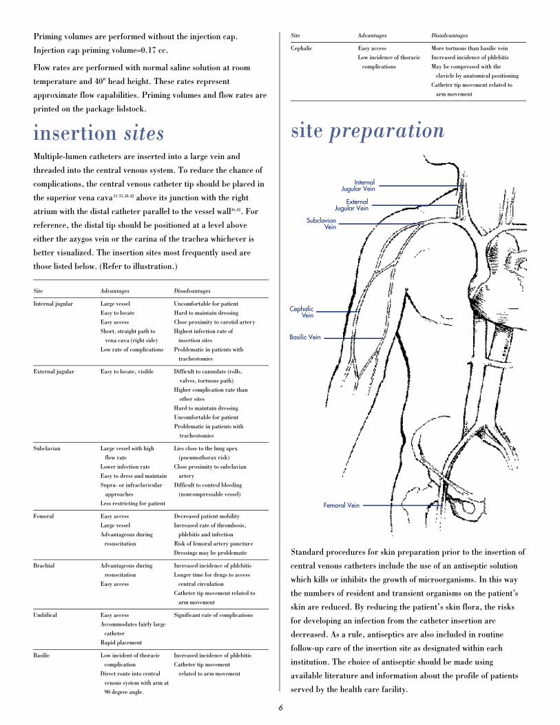
Te c h n i q u e
The Seldinger or modified Seldinger method is the preferred
technique used to insert a central venous catheter.27,29 This
percutaneous technique consists of: (1) locating the appropriatevein by using an introducer needle or a catheter over needle
assembly; (2) introducing a spring-wire guide through the needle
or catheter; and (3) threading a central venous catheter over the
wire to the proper depth.
Percutaneous insertion of central venous catheters has become
widely accepted for a number of reasons that include the speed of
the procedure, preservation of vessel integrity, and a decrease in
the risk of infection. Using a spring-wire guide also allows for the
use of a small needle to ultimately place a much larger catheter.
Spring-wire guides must be strong but flexible enough to conform
to vessel angles. The tips must be soft to prevent damage to the
vessel wall. The end configurations may be straight, for direct
pathways to the superior vena cava, or curved into a J-tip to
facilitate passage through angles in vessels. Marked spring-wire
guides aid practitioners in knowing the depth of the wire
placement.
8
(1) locating the appropriate vein by using an introducer needle
or a catheter over needle assembly;
(2) introducing a spring-wire guide through the needle
or catheter;
(3) threading a central venous catheter over the wire to the
proper depth.

Rauler son Sy ri nge
The Arrow® Raulerson syringe is designed for use as an adjunct in
the placement of a spring-wire guide using the Seldinger
technique. The syringe is designed with a unique hollow
plunger/barrel containing a patented valve system. The spring-
wire guide is inserted into the plunger/barrel, through the
introducer needle and into the vein. The benefits to using this
syringe are the virtual elimination of the potential for air emboli,
less vessel wall trauma, blood containment in the syringe barrel
and stabilization of the needle bevel within the vessel lumen.
Cathet er T ip Pl acement
To reduce the risk of complications, e.g. cardiac tamponade,
vessel wall perforation, or cardiac arrhythmias, the catheter tip
must be located in the superior vena cava 3-4cm above the entry
into the right atrium with the distal catheter positioned parallel to
the vessel wall.31-36,38,42
Prior to insertion, the external anatomy can be used to estimate
the length of catheter needed for proper tip placement. During the
procedure intravascular electrocardiography can be employed to
determine the location of the tip within the central circulatory
system.37,39 This technique requires the use of an adapter, e.g.
Arrow-Johans™ adapter, that is incorporated into the catheter
set-up and is used to relay electrical impulses through a fluid-filled
catheter lumen to an ECG monitor. By the P wave configuration of
the ECG tracing the relative location of the catheter tip can be
identified.
In addition to the above aids to provide correct catheter tip
placement, a CHEST X-RAY MUST BE DONE immediately post-
insertion.31,32,34,38,40,41 An x-ray provides the only definitive evidence
for catheter tip location. Until this verification is provided, fluids
should be maintained at a keep-open rate.
I n s e r t ion Gu ide l ine s
• The person inserting the catheter should be trained and well-
versed in anatomical landmarks, safe techniques and
potential complications.
• The amount of catheter that has been inserted into the body
must be documented. Centimeter markers on the external
surface of the catheter body can be used, where provided.
The marker position should be checked periodically and
documented within the chart.
9
(1) locating the appropriate vein by using an introducer needle or
a catheter over needle assembly;
(2) introducing a spring-wire guide through the needle
or catheter;
(3) threading a central venous catheter over the wire to
the proper depth.

• A new approach, e.g. different site or different inserter,
should be tried after 3-5 unsuccessful passes into one site.30
• Do not place the catheter into or allow it to remain within the
right atrium or right ventricle.
• Do not apply excessive force in removing the spring-wire
guide or catheter.
• Use Universal Blood and Body Fluid Precautions to avoid
exposure to bloodborne pathogens.
p roduct i n s t ru c t i o n sCent ra l Venous Ca the t er i za t ion
must be performed by trained personnel well-versed in anatomical
landmarks, safe technique, and potential complications.
The pictogram (Figure 1) consisting of international symbols is
used to further emphasize the need to place the tip of the catheter
outside of the heart.
I n s e r t ion P ro cedure (us i ng Rau ler son Sy r i nge)
Use Sterile Technique
1. Precaution: Place patient in slight T rendelenburg position
as tolerated to reduce the risk of air embolism. If femoral
approach is used, place patient in supine position.
2. Prep and drape puncture site as required.
3. Perform skin wheal with desired needle (25 Ga. or 22 Ga.
needle).
4. Prepare the catheter for insertion by flushing each lumen and
clamping or attaching the injection caps to the appropriate
pigtails. Leave the distal pigtail uncapped for guide wire
passage. Warning: Do not cut the catheter to alter length.
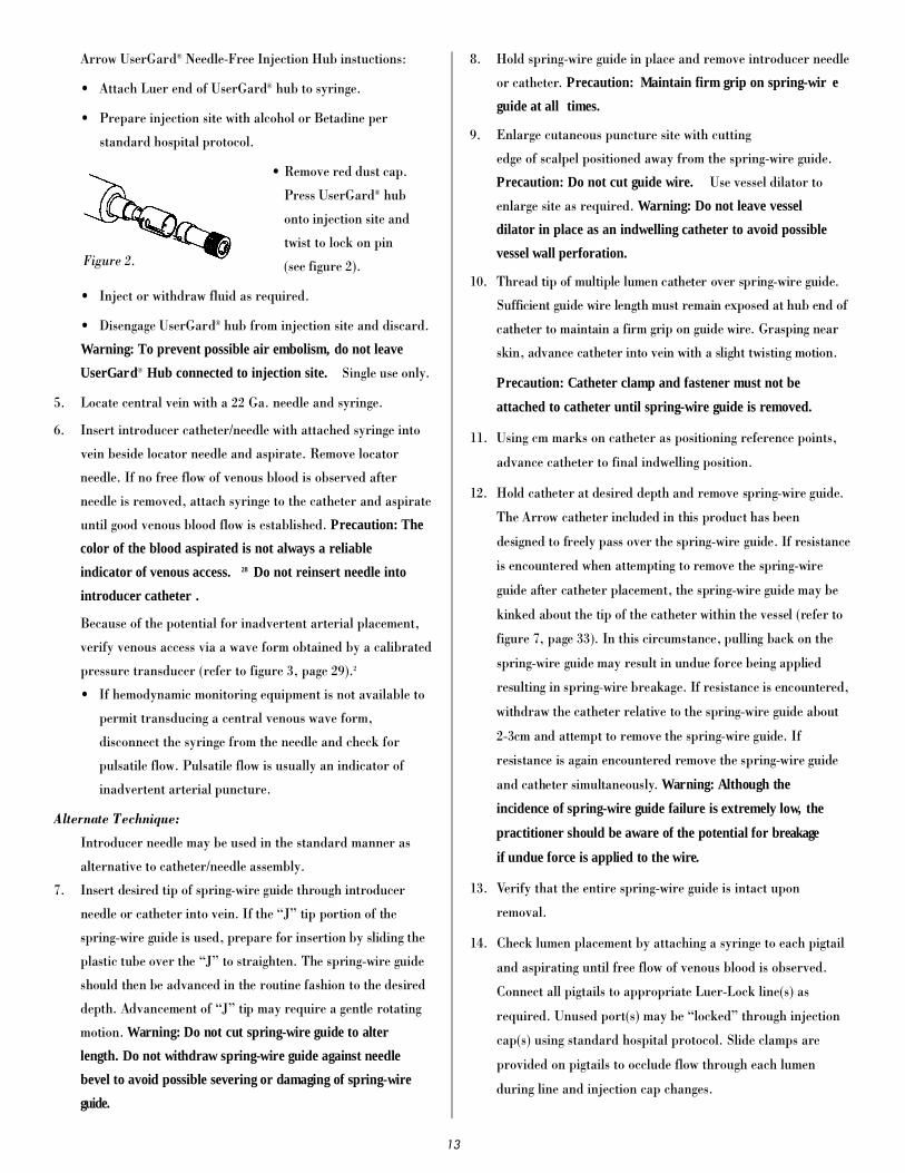
Arrow UserGard® Needle-Free Injection Hub instructions:
• Attach Luer end of UserGard® hub to syringe.
• Prepare injection site with alcohol or Betadine per
standard hospital protocol.
• Remove red dust cap. Press
UserGard® hub onto
injection site and twist to
lock on pin (see figure 2).
• Inject or withdraw fluid as
required.
• Disengage UserGard® hub from injection site and discard.
Warning: To prevent possible air embolism, do not
leave UserGar d® Hub connected to injection site. Single
use only.
5. Insert introducer needle with attached Arrow® Raulerson
Syringe into vein and aspirate. (If larger introducer needle is
used, vessel may be prelocated with 22 Ga. locator needle and
syringe). Remove locator needle.
Alternate Technique:
Catheter/needle may be used in the standard manner as
alternative to introducer needle. If catheter/needle is used,
Arrow® Raulerson Syringe will function as a standard
syringe, but will not pass a spring-wire guide. If no free flow
of venous blood is observed after needle is removed, attach
syringe to the catheter and aspirate until good venous blood
flow is established. Precaution: The color of the blood
aspirated is not always a reliable indicator of venous
access. 28 Do not reinsert needle into introducer catheter .
6. Because of the potential for inadvertent arterial placement,
one of the following techniques should be utilized to verify
venous access. Insert the fluid primed blunt tip transduction
probe into the rear of the plunger and
through the valves of the Raulerson
Syringe. Observe for central venous
placement via a wave form obtained by a
calibrated pressure transducer (refer to
figure 3). Remove transduction probe.
Alternate Technique:
If hemodynamic monitoring equipment is not available to
permit transducing a central venous wave form, check for
pulsatile flow by either using the transduction probe to open
the syringe valving system or by disconnecting the syringe
from the needle. Pulsatile flow is usually an indicator of
inadvertent arterial puncture.
10
Figure 1.
Figure 2.
Figure 3.

7 . Using the two-piece Arrow Advancer,™ advance spring-wire
guide through syringe into vein. Wa rning: Aspiration with
s p r i n g - w i r e guide in place will cause introduction of air into
syringe. Precaution: To avoid leakage of blood from syringe
cap do not reinfuse blood with spring-wire guide in place.
Two-Piece Arrow Advancer™ Instruction:
• Remove protective cap.
• Using your thumb, straighten the “J” by retracting the
spring-wire guide into the Arrow Advancer™
(refer to figure 4). When the tip is straightened, the
spring-wire guide is ready for insertion. Centimeter
marks are referenced
from “J” end. One band
indicates 10cm, two
bands 20cm, and three
bands 30cm.
Introducing the Spring-Wire Guide:
• Place the tip of the Arro w
Advancer™—with “J”
retracted—into the hole in
the rear of the Raulerson
syringe plunger (refer to
figure 5).
• Advance spring-wire guide into the syringe approximately
10cm until it passes through the valves.
• Lift your thumb and pull the Arrow Advancer™
approximately 4cm to 8cm
away from the syringe.
Lower thumb onto the
Arrow Advancer™ and
while maintaining a firm
grip on the spring-wire guide, push the assembly into the
syringe barrel to further advance the spring-wire guide.
Continue until spring-wire guide reaches desired depth
(refer to figure 6).
Alternate Technique:
If a simple straightening tube is preferred, the
straightening tube portion of the Arrow Advancer™ can
be disconnected from the unit and used separately.
Separate the Arrow Advancer™ tip or straightening tube
from the blue Arrow Advancer™ unit. If the “J” tip
portion of the spring-wire guide is used, prepare for
insertion by sliding the plastic tube over the “J” to
straighten. The spring-wire guide should then be advanced
in the routine fashion to the desired depth.
8. Advance guide wire until triple band mark reaches rear of
syringe plunger. Advancement of “J” tip may require a gentle
rotating motion. Warning: Do not cut spring-wire guide to
alter length. Do not withdraw spring-wire guide against
needle bevel to avoid possible severing or damaging of
spring-wire guide.
9. Hold spring-wire guide in place and remove introducer needle
and Raulerson syringe (or catheter). Precaution: Maintain
firm grip on spring-wire guide at all times. Use centimeter
markings on spring-wire guide to adjust indwelling length
according to desired depth of indwelling catheter placement.
10. Enlarge cutaneous puncture site with cutting edge of scalpel
positioned away from the spring-wire guide. Precaution: Do
not cut guide wire. Use vessel dilator to enlarge site as
required. Warning: Do not leave vessel dilator in place as
an indwelling catheter to avoid possible vessel
wall perforation.
11. Thread tip of multiple lumen catheter over spring-wire guide.
Sufficient guide wire length must remain exposed at hub end
of catheter to maintain a firm grip on guide wire. Grasping
near skin, advance catheter into vein with slight twisting
motion. Precaution: Catheter clamp and fastener must not
be attached to catheter until spring-wire guide is removed.
12. Using cm marks on catheter as positioning reference points,
advance catheter to final indwelling position.
1 3 . Hold catheter at desired depth and
remove spring-wire guide. The Arro w
catheter included in this product has
been designed to freely pass over the
s p r i n g - w i re guide. If resistance is
e n c o u n t e red when attempting to re m o v e
the spring-wire guide after catheter
placement, the spring-wire may be kinked about the tip of the
catheter within the vessel (refer to figure 7). In this circ u m-
stance, pulling back on the spring-wire guide may result in
undue force being applied resulting in spring-wire guide
b reakage. If resistance is encountered, withdraw the catheter
relative to the spring-wire guide about 2-3cm and attempt to
remove the spring-wire guide. If resistance is again
e n c o u n t e red remove the spring-wire guide and catheter
s i m u l t a n e o u s l y.
Warning: Although the incidence of spring-wire guide
failure is extremely low, the practitioner should be aware
of the potential for breakage if undue force is applied to
the wire.
11
Figure 4.
Figure 5.
Figure 6.
Figure 7.

14. Verify that the entire spring-wire guide is intact upon
removal.
15. Check lumen placement by attaching a syringe to each pigtail
and aspirate until free flow of venous blood is observed.
Connect all pigtails to appropriate Luer-Lock line(s) as
required. Unused port(s) may be “locked” through injection
cap(s) using standard hospital protocol. Slide clamps are
provided on pigtails to occlude flow through each lumen
during line and injection cap changes. Precaution: To avoid
damage to pigtails from excessive pressure, each clamp
must be opened prior to infusing through that lumen.
16. Secure and dress catheter temporarily.
17. Verify catheter tip position by chest x-ray immediately after
placement. Precaution: X-ray exam must show the catheter
located in the right side of the mediastinum in the SVC
with the distal end of the catheter parallel to the vena cava
wall and its distal tip positioned at a level above either the
azygos vein or the carina of the trachea, whichever is
better visualized. If the catheter tip is malpositioned,
reposition and reverify.
18. Secure the catheter to patient. Use triangular juncture hub
with integral suture ring and side wings as primary suture
site. In kits where provided, the catheter clamp and fastener
should be utilized as a secondary suture site as necessary.
Precaution: Do not suture directly to the out-side
diameter of the catheter to avoid cutting or damaging the
catheter or impeding catheter flow.
Catheter Clamp and Fastener Instructions:
• After spring-wire guide
has been removed and
the necessary lines
have been connected or
locked, spread wings of
rubber clamp and posi-
tion on catheter as
required to ensure
proper tip location
(refer to figure 8).
• Snap rigid fastener
onto catheter clamp
(refer to figure 9).
• Secure catheter to patient by
suturing the catheter clamp
and fastener together to the
skin, using side wings to
prevent catheter migration
(refer to figure 10).
19. Dress puncture site per hospital protocol. Precaution:
Maintain the insertion site with regular meticulous
redressing using aseptic technique.
20. Record on the patient’s chart the indwelling catheter length
as to centimeter markings on catheter where it enters the
skin. Frequent visual reassessment should be made to ensure
that the catheter has not moved.
Cent ra l Venous Ca t he teri za t ion must be
performed by trained personnel well versed in anatomical land-
marks, safe technique, and potential complications.
I n s e r t ion P roc edure
(w it hou t Rau l e rs on Syri nge)
Use Sterile Technique.
1. Precaution: Place patient in slight T rendelenburg position
as tolerated to reduce the risk of air embolism. If femoral
approach is used, place patient in supine position.
2. Prep and drape puncture site as required.
3. Perform skin wheal with desired needle (25 Ga. or 22 Ga.
needle).
4. Prepare catheter for insertion by flushing each lumen and
clamping or attaching the injection caps to the appropriate
pigtails. Leave the distal pigtail uncapped for guide wire
passage. Warning: Do not cut the catheter to alter length.
12
Figure 8.
CatheterClamp
Figure 9.
CatheterClamp
RigidFastener
Figure 10.
Arrow UserGard® Needle-Free Injection Hub instuctions:
• Attach Luer end of UserGard® hub to syringe.
• Prepare injection site with alcohol or Betadine per
standard hospital protocol.
• Remove red dust cap.
Press UserGard® hub
onto injection site and
twist to lock on pin
(see figure 2).
• Inject or withdraw fluid as required.
• Disengage UserGard® hub from injection site and discard.
Warning: To prevent possible air embolism, do not leave
UserGard® Hub connected to injection site. Single use only.
5. Locate central vein with a 22 Ga. needle and syringe.
6. Insert introducer catheter/needle with attached syringe into
vein beside locator needle and aspirate. Remove locator
needle. If no free flow of venous blood is observed after
needle is removed, attach syringe to the catheter and aspirate
until good venous blood flow is established. Precaution: The
color of the blood aspirated is not always a reliable
indicator of venous access. 28 Do not reinsert needle into
introducer catheter .
Because of the potential for inadvertent arterial placement,
verify venous access via a wave form obtained by a calibrated
pressure transducer (refer to figure 3, page 29).2
• If hemodynamic monitoring equipment is not available to
permit transducing a central venous wave form,
disconnect the syringe from the needle and check for
pulsatile flow. Pulsatile flow is usually an indicator of
inadvertent arterial puncture.
Alternate Technique:
Introducer needle may be used in the standard manner as
alternative to catheter/needle assembly.
7 . I n s e rt desired tip of spring-wire guide through intro d u c e r
needle or catheter into vein. If the “J” tip portion of the
s p r i n g - w i re guide is used, pre p a re for insertion by sliding the
plastic tube over the “J” to straighten. The spring-wire guide
should then be advanced in the routine fashion to the desire d
depth. Advancement of “J” tip may re q u i re a gentle ro t a t i n g
motion. Wa rning: Do not cut spring-wire guide to alter
length. Do not withdraw spring-wire guide against needle
bevel to avoid possible severing or damaging of spring-wire
g u i d e .
8. Hold spring-wire guide in place and remove introducer needle
or catheter. Precaution: Maintain firm grip on spring-wir e
guide at all times.
9. Enlarge cutaneous puncture site with cutting
edge of scalpel positioned away from the spring-wire guide.
Precaution: Do not cut guide wire. Use vessel dilator to
enlarge site as required. Warning: Do not leave vessel
dilator in place as an indwelling catheter to avoid possible
vessel wall perforation.
1 0 . T h read tip of multiple lumen catheter over spring-wire guide.
S u fficient guide wire length must remain exposed at hub end of
catheter to maintain a firm grip on guide wire. Grasping near
skin, advance catheter into vein with a slight twisting motion.
Precaution: Catheter clamp and fastener must not be
attached to catheter until spring-wire guide is removed.
11. Using cm marks on catheter as positioning reference points,
advance catheter to final indwelling position.
1 2 . Hold catheter at desired depth and remove spring-wire guide.
The Arrow catheter included in this product has been
designed to freely pass over the spring-wire guide. If re s i s t a n c e
is encountered when attempting to remove the spring-wire
guide after catheter placement, the spring-wire guide may be
kinked about the tip of the catheter within the vessel (refer to
f i g u re 7, page 33). In this circumstance, pulling back on the
s p r i n g - w i re guide may result in undue force being applied
resulting in spring-wire breakage. If resistance is encountere d ,
withdraw the catheter relative to the spring-wire guide about
2-3cm and attempt to remove the spring-wire guide. If
resistance is again encountered remove the spring-wire guide
and catheter simultaneously. Wa rning: Although the
incidence of spring-wire guide failure is extremely low, the
prac titioner s hould be aware of the potential for bre a k a g e
if undue force is applied to the w ire .
13. Verify that the entire spring-wire guide is intact upon
removal.
14. Check lumen placement by attaching a syringe to each pigtail
and aspirating until free flow of venous blood is observed.
Connect all pigtails to appropriate Luer-Lock line(s) as
required. Unused port(s) may be “locked” through injection
cap(s) using standard hospital protocol. Slide clamps are
provided on pigtails to occlude flow through each lumen
during line and injection cap changes.
13
Figure 2.
Precaution: To avoid damage to pigtails from excessive
pressure, each clamp must be opened prior to infusing
through that lumen.
15. Secure and dress catheter temporarily.
16. Verify catheter tip position by chest x-ray immediately after
placement. Precaution: X-ray exam must show the catheter
located in the right side of the mediastinum in the SVC
with the distal end of the catheter parallel to the vena cava
wall and its distal tip positioned at a level above either the
azygos vein or the carina of the trachea, whichever is
better visualized. If the catheter tip is malpositioned,
reposition and re-verify.
17. Secure catheter to patient. Use triangular juncture hub with
integral suture ring and side wings as primary suture site. In
kits where provided, the catheter clamp and fastener should
be utilized as a secondary suture site as necessary.
Precaution: Do not suture directly to the outside diameter
of the catheter to avoid cutting or damaging the catheter
or impeding catheter flow .
Catheter Clamp and Fastener Instructions:
• After spring-wire guide has been removed and the
necessary lines have been connected, spread wings of
rubber clamp and position on catheter as required to
ensure proper tip location (refer to figure 8, page 35).
• Snap rigid fastener onto catheter clamp (refer to figure 9,
page 35).
• Secure catheter to patient by suturing the catheter clamp
and fastener together to the skin, using side wings to
prevent catheter migration (refer to figure 10, page 36).
18. Dress puncture site per hospital protocol. Precaution:
Maintain the insertion site with regular meticulous
redressing using aseptic technique.
19. Record on the patient’s chart the indwelling catheter length
as to centimeter markings on the catheter where it enters the
skin. Frequent visual reassessment should be made to ensure
that the catheter has not moved.
p o rt d e s i g n a t i o nThe ports of a multiple lumen central venous catheter should be
labeled for designated use, and the information should be entered
into the chart and onto the patient’s Kardex or information sheet.
The reason for port designation is to ensure uniform use of the
catheter lumens by health care personnel who are providing
treatments through the catheter.
There is a lack of scientific data to support many of the
designation protocols currently in use. Most choices have been
made using deductive reasoning. For example, the distal port is
usually used for central venous pressure monitoring. The reasons
given for this choice are that the distal lumen is the largest lumen
and it is closest to the heart, but theoretically other lumens could
be used provided they exit within the central venous system, i.e.
the superior or inferior vena cava (See Monitoring section).
The proximal port is often designated for blood sampling. This
choice is made because the rapid flow of blood within the large
central vein quickly carries the infusates from the more distal
lumens, that might affect laboratory tests, away from the proximal
sampling port. As an additional safeguard against erroneous lab
results, it is recommended that all other infusions be turned off
prior to blood sampling. (See Blood Sampling in Catheter
Maintenance Section).
Another designation that has gained widespread acceptance is the
need to reserve one lumen exclusively for total parenteral
nutrition (TPN). The rationale for this designation is the
prevention of catheter-related infections. When using a triple
lumen catheter, the middle port is often chosen. The following
designations are examples for port usage and do not represent the
only way the lumens can be used:
Proximal: Blood Sampling
Medications
Blood Administration
Medial: Total Parenteral Nutrition
Medications (only if TPN use is not
anticipated)
Distal: CVP Monitoring
Blood Administration
High Volume or Viscous Fluids
Colloids
Medication
4th Lumen: Infusion
Medication
14
More scientific study is needed to further define the scope of port
designation. With the knowledge that is available, the most
important issue is that the designations must be uniform when
used by all persons involved in patient care.
c o m p l i c a t i o n sIt is estimated that approximately 10% of patients who have a
central line placed will experience a complication secondary to the
catheter insertion or use. 44 Whether or not complications occur
depends upon a number of factors including the experience of the
inserting physician,30,51 anatomical distortion at the potential
insertion site, and the patient’s condition. Each site that can be
used, e.g. subclavian vs. internal jugular vs. femoral, has certain
risks involved due to the normal body anatomy in that area.19,20
Other individual patient factors are also important, such as
underlying disease, tolerance of Trendelenburg position,
laboratory levels associated with bleeding, and the patient’s
mental or emotional status to mention a few.
The complications are generally divided into two groups,
immediate or delayed, dependent upon the time they appear in
relation to the catheter insertion. Immediate complications are
usually associated with catheter placement; however, some may
develop later under certain circumstances. Delayed complications
are manifested after the catheter has been indwelling for a period
of time. Only the most frequently encountered complications will
be included in this section.
Immediate Delayed
venous air embolism catheter-related infection
cardiac tamponade catheter-related thrombosis
catheter embolus/rupture hydrothorax/vessel erosion
arterial puncture
cardiac dysrhythmia
nerve injury
catheter malposition
pneumothorax, hemothorax
Immedia t e Compl i ca t ions
Venous Air Embolism –A bolus of air within the venous circ u l a t i o n .
P a t h o p h y s i o l o g y The pre s s u res within the central venous
c i rculation are in direct accord with the
p re s s u res involved with respiration. On
expiration the intrathoracic and
intravenous pre s s u res are greater than the
atmospheric pre s s u re making it less likely
that air would enter the venous system. On
inspiration the opposite is true and, in
a c c o rd with equalizing pre s s u res, air is
sucked through an opening into the venous
system. The air proceeds as a bolus into the
h e a rt where it usually lodges against the
pulmonic valve and blocks the pulmonary
blood flow. With the increasing force of the
pumping right ventricle, the air bolus may
b reak up into smaller bubbles that enter
the pulmonary circulation. This causes
m o re blood obstruction which leads to
localized tissue hypoxia, decreased card i a c
output, and a resultant generalized
d e c rease in tissue perfusion. Wi t h o u t
i n t e rvention the condition rapidly
p ro g resses to shock and death. The
m o rtality rate ranges from 29-50%.7 3
Factors that affect the severity of the
condition are the volume of air that enters
the circulation, the rate of the air entry,
patient hydration status, and the position
of the patient. If the air enters rapidly, it
will more likely form a bolus that blocks
blood circulation as opposed to the
dispersed pattern which results with slower
air entry. It is estimated that symptoms will
appear if air enters the venous system at
20ml/sec., and death can occur at 75-
150ml/sec.59,70 When looking at these
numbers, the possibility of this amount of
air influx seems unlikely; however, it has
been shown that 100ml/sec. flow of air can
occur through a 14 ga. needle with a
pressure gradient of 5cm H2O.59,70
Concerning volume, 70-300cc may be
fatal.72 The symptoms will also be more
severe when the patient is in an upright
position, is dehydrated or hypovolemic.
Occurrence/ • at insertion of CVC
Predisposing • with break in catheter connection
Factors • with open damage to hub or catheter body
• after catheter removal due to remaining
subcutaneous track58,62,70,73,74
• if IV fluids run dry
• if a percutaneous sheath remains in place
without catheter or obturator 60
• during neurosurgical and head/neck
surgical procedures in sitting position66
15

Signs, Symptoms • may be nonspecific
& Data • sudden unexplained hypoxia or
cardiovascular collapse
• pulmonary: sucking sound on inspiration,
pulmonary hypertension, respiratory
distress, tachypnea progressing to apnea
• cardiovascular: millwheel murmur over
precordium, increased pulmonary artery
pressure, jugular vein distention, decreased
cardiac output, chest pain, hypotension,
tachycardia, increased central venous
pressure.
• neurological: anxiety, hypoxic symptoms,
change in mental status, dizziness,
confusion, syncope, seizures, coma
Interventions • prompt recognition and immediate action
• access CVC line and correct any problems
• place patient on left side in steep
Trendelenburg
• administer 100% oxygen
• notify physician
• chest massage to displace air bolus from
pulmonic valve55,64,65,73
• catheter aspiration of air from right ventricle
• hyperbaric chamber
Prevention • place patient in Trendelenburg or flat
position for catheter insertion and removal
• have patient use Valsalva maneuver or hold
breath during injection hub or tubing
changes
• use Luer-Lock connection, tape
• apply occlusive dressing after CVC removal
(24-72 hours)58,62,73
• use Raulerson syringe or occlude open hub
with finger prior to guide wire insertion
• occlude all ports of MLC
• flush air out of all catheter ports
• anchor catheter securely
• use slide clamps for tubing change
• turn stopcocks to a position that closes the
line to air
• give sedation, if necessary
• patient teaching
• provide adequate hydration
• check all catheter connections and catheter
integrity regularly
Cardiac Tamponade A syndrome of circulatory abnormalities
caused by excess fluid in the pericardial
space.
Pathophysiology The pericardial sac normally holds 10-20ml
of fluid which serves to cushion and protect
the heart. When excess fluid is introduced
the pressure in the pericardial space
increases and impairs the filling of the
heart during diastole. As the pressure
builds, cardiac output is decreased with
detrimental effects on the systemic
circulation. If left unchecked, the condition
will lead to total circulatory collapse,
shock, and death.
Cardiac tamponade may be acute or long
term. Symptomatology depends on the rate
of fluid accumulation, the amount of fluid
build-up and the pericardial compliance. In
acute tamponade the fluid accumulation
occurs rapidly and the pericardium
remains inelastic and restraining. In this
setting 100-300ml of excess fluid is
potentially fatal.83,90
Tamponade that develops over a longer
period of time is due to a slow fluid
accumulation which provides time for
pericardial compliance. It is not unusual to
find an accumulation of 1000-2000ml of
fluid in this setting. 83,90
A majority of central venous catheter-
induced cases of tamponade reported in the
literature have been acute in origin and
represent a gravely emergent complication
with a mortality rate of 70-85% often due
to a delay in diagnosis.84 The fluid
accumulation is usually due to a catheter
perforation of the superior vena cava, right
atrium, or right ventricle.
Occurrence/ • catheter in right atrium or right ventricle
Predisposing • catheter migration with arm or neck
Factors motion, depends on the insertion site
• catheter not parallel to the vessel wall
• catheter made of “stiff” material or dilator
remains indwelling87
• left-sided insertion88
16

Signs, Symptoms •acutely ill appearance
& Data •may mimic other conditions
•all signs and symptoms may not be present
or detected
•acute presentation is a true emergency
•cardiovascular: tachycardia, hypotension,
distant or muffled heart sounds, chest or
epigastric pain, venous engorgement of neck
and face, thready or absent peripheral
pulses, dusky or pale nail beds, pericardial
friction rub, cyanosis, narrow arterial pulse
pressure, elevated CVP, ECG abnormalities
(low voltage, ST changes, electrical
alternans), decreased cardiac output,
cardiac arrest, Beck’s triad = increased CVP
+ decreased arterial pressure + quiet heart
•nausea and abdominal pain
•respiratory: dyspnea, tachypnea, short of
breath, pulsus paradoxus, hypoxemia,
respiratory alkalosis,Kussmaul’s sign
• n e u rological: restlessness, diaphore s i s ,
confusion decreased level of consciousness,
c o m a
• non-acute diagnostics:
echocardiogram - demonstrates echo-free
space separating pericardium from heart
wall
chest x-ray - demonstrates cardiomegaly,
large cardiac silhouette or “water bottle”
heart associated with massive pericardial
effusion
ECG - S-T changes, low voltages, electrical
alternans
CAT, MRI
Interventions • prompt recognition and immediate action
• notify physician
• lower infusion bag to facilitate gravity
drainage75,77,78
• physician aspiration through catheter
• pericardiocentesis
• thoracostomy
Prevention • be aware of tamponade potential
• avoid using “stiff” catheters
• do not use a beveled catheter
• use soft guide wire, J tip
• perform preinsertion anatomic measurement
• verify blood return from all ports after
insertion and recheck regularly
• secure catheter with sutures and tape
• document length of catheter inserted and
routinely check for catheter migration
• verify placement of catheter tip in superior
vena cava by chest x-ray immediately after
insertion and before infusion, including
catheters changed over a guide
wire31,32,34,38,40,41,85
• periodically reassess tip position by
chest x-ray
• remove catheter when no longer needed
Catheter Embolus/
Rupture
Pathophysiology At the time of insertion or sometime after the
multiple lumen catheter is indwelling,
physical damage to the catheter body can
occur. During insertion the catheter can be
damaged by exposure to the sharp edge of
the introducer needle. The catheter can be
nicked or a portion can be sheared off and
enter the bloodstream or adjacent structures
as an embolic fragment. The final resting
location of the fragment will determine the
severity of the patient’s symptoms. A portion
of the catheter can also be broken off if
extreme pressure is exerted and the catheter
is improperly secured.
“Softer” catheters can be damaged when
excessive force is applied at the time of
irrigation. The force can rupture the
catheter body and the resultant catheter
dysfunction will be related to the location of
the rupture. The rupture can also be a
function of the catheter insertion site if it is
“pinched” between the clavicle and the first
rib due to subclavian placement.
Occurrence/ • use of syringe smaller than 10cc to irrigate
Predisposing or declot a blocked catheter92
Factors • patient with altered mental status
• insertion technique - catheter or spring-
wire guide pulled back against the needle
bevel93,94
• use of scissors or other sharp instruments
during dressing change or reinsertion overa guide wire
• poorly secured catheter• forceful irrigation• “soft” catheter material
17
Signs, Symptoms • fluid leakage from the insertion siteor Data • cardiopulmonary signs and symptoms,
e.g., palpitations, shortness of breath• dysrhythmias on ECG• catheter malfunction
Interventions • catheter removal or guide wire exchange• catheter fragment retrieval (hooked
catheters, wire loops, stone baskets,endoscopic forceps, surgery)
Prevention • only advance the catheter forwardthrough the introducer needle
• if the insertion procedure is unsuccessful,withdraw the catheter and needle as oneunit
• secure catheter to the patient• take measures to prevent the patient from
forcefully removing the catheter• use a 10cc syringe or larger to irrigate or
declot an occluded catheter92
Arterial Puncture
Pathophysiology Due to the anatomical proximity of arteries
to veins at certain insertion sites, it is
possible that an artery can be entered,
transected or lacerated during the insertion
procedure. The sites where this complication
occurs most frequently are the subclavian,
the internal jugular and the femoral veins.
Many arterial punctures are uncomplicated;
however, a hematoma can develop that can
represent significant blood loss from
circulation and might impinge upon other
anatomical structures.
Occurrence/ • insertion site
Predisposing • patient dehydration
Factors • insertion technique
• inserter experience
Signs, Symptoms • bright red blood in syringe
& Data Note: the color of the blood may not be a
reliable indicator dependent upon the
patient’s conditions28
• pulsatile blood flow
• hematoma development
Intervention • observe for bright red, pulsatile blood
flow and if noted, remove needle
immediately, apply pressure
• direct pressure for 5-10 minutes
• pressure dressing
• monitor vital signs
• x-ray to determine extent of blood
collection
• comparative blood gases
Prevention • knowledge about potential for this
complication
• ECG confirmation of venous placement
using pressure transducer
• use of small locator needle to find vessel95
Dysrhythmias
Pathophysiology When the multiple lumen catheter or guide
wire is advanced too far and enters the
heart, the myocardium may be stimulated
and result in an abnormal cardiac rhythm.
The dysrhythmias may be atrial, ventricular
or in the form of a conduction abnormality,
i.e., a bundle branch block. Another
mechanism for producing a dysrhythmia is
the stimulation of the carotid sinus during an
internal jugular insertion attempt. In both
cases the change in cardiac rhythm may be
detrimental to the patient. The rhythm
abnormality will usually disappear when the
offending stimulation is discontinued. If, due
to guide wire or catheter-induced damage, a
conduction pathway is disturbed, the
conduction abnormality may be permanent.
An extremely difficult situation might arise if
the patient already has an existing left
bundle branch block. A right bundle branch
block induced by a catheter or guide wire
may lead to complete heart block and severe
patient compromise.96
Occurrence/ • a difficult internal jugular insertion
Predisposing (possible carotid sinus stimulation)
Factors • antecubital insertion (catheter migration
into the heart with arm movement)
• improper catheter tip placement on
insertion
• lack of chest x-ray confirmation of
catheter location
Signs, Symptoms • cardiopulmonary signs and symptoms
& Data dependent upon dysrhythmia
• ECG irregularities
• pulse irregularities
18
Interventions • withdraw the catheter and position the
tip correctly
• artificial pacemaker for complete heart
block
Prevention • use marked spring-wire guides
• estimate length of catheter and guide wire
needed by external anatomy
• secure catheter to prevent migration
• chest x-ray immediately after insertion
and periodically while catheter is
indwelling to verify correct tip location
• ECG monitoring during insertion
• have pacing equipment available for
catheterization of patient with existing
bundle branch block
Nerve Injury
Pathophysiology The injury of nerves occurs almost
exclusively during a central venous catheter
insertion into the subclavian and internal
jugular sites where there are many nerve
pathways in the surrounding area.47 The
damage is usually inflicted by probing during
the attempt to locate the targeted vessel. The
following deficits have been recorded in the
literature: sensory-motor loss to the upper
extremities, paralyzed diaphragm,
hoarseness, Horner’s syndrome and others.
If the nerve was only compressed, full
recovery from the nerve deficit is expected;
however, if the nerve was cut or damaged in
some way, the regeneration may take a long
period of time or may never occur.
Occurrence/ • subclavian or internal jugular
Predisposing insertion sites
Factors • difficult insertion
• inexperienced inserter
Signs, Symptoms • numbness, tingling of extremities
& Data • respiratory difficulties
• hoarse voice
• painful parasthesias
• muscular twitches
• contraction of pupil, partial ptosis of the
eyelid, enophthalmos (Horn e r’s syndro m e )
Interventions • symptomatic
• physical therapy
Prevention • knowledge about complication possibility
with subclavian or internal jugular
catheter insertions
• supervision of inexperienced inserters
Catheter
Malposition Incorrect placement of the catheter tip.
Pathophysiology The tip of an indwelling central venous
catheter should be located within the
superior vena cava (SVC) at a level 3-4cm
above the junction of the SVC and the right
atrium.35 This placement helps to prevent
complications such as vessel perforation or
thrombosis that can lead to cardiac
tamponade, hydrothorax or catheter
occlusion. Malposition of the catheter tip will
predispose a patient to complications and
can occur at the time of catheter insertion or
spontaneously at some time while the
catheter is indwelling. The probability of this
occurring is dependent upon the insertion
site, the individual patient’s venous anatomy
and the catheter material. Catheters made of
“soft” materials are the most prone to
malposition.
During insertion the catheter can be
advanced into the wrong vessel. Overall
rates of 1-6% have been noted for all types
of CVC insertion.102 In the literature the
incidence of malpositioned catheters inserted
into the subclavian vein has been found to
vary from 5.5 to 29%. For the antecubital
approach, the incidence varies from 21-
55%.101 A study conducted by Lum and Soski
that investigated malpositioned subclavian
and antecubital lines revealed that the most
common sites for aberrant placement were
the internal jugular vein (43.4%), the
axillary vein (19%), the contralateral
innominate vein (11.2%) and the right
atrium (9.8%).101 Other insertion sites, such
as the femoral, can be implicated with
malpositioned catheters as well, but the rates
are usually lower.
Central venous catheters that have been
indwelling can become malpositioned
spontaneously due to a change in
intrathoracic pressure, a rapid infusion or
19

random body movement. The change in
catheter position can occur at any time after
insertion and has even been reported in
long-term catheters. Coughing46,100 or
vomiting46 are two conditions that increase
the intrathoracic pressure and have been the
cause for a change in catheter position.
Depending upon the site of insertion,
random body movements can cause the
catheter tip to advance beyond its intended
resting point. The tips of catheters that have
been inserted into the arm have been shown
to advance several centimeters when the arm
is abducted making it advisable to insert the
catheter with the arm in that position to
avoid having the catheter tip advance into
the heart. Curelaru35 noted that catheters
dwelling within the internal jugular vein can
advance 1.5-3cm with maximum neck
flexion. This movement must be considered
during catheter insertion for final tip
placement.
When a catheter is malpositioned it can
empty into a smaller vessel that will be
adversely affected by an infusion of
hyperosmolar solution or extremes of pH.
Other problems that could occur in this
situation include a disturbance in the blood
flow pattern or partial occlusion. All of these
conditions predispose the patient to
thrombus formation.
Occurrence/ • insertion site
Predisposing • insertion technique
Factors • patient anatomy
• catheter material
• significant change in thoracic pressure
• rapid IV infusion
• body movements
Signs, Symptoms • might be asymptomatic
& Data • inadequate blood return
• pain upon infusion
• leaking at insertion site
• palpitations, dysrhythmias
• internal jugular - gurgling sound in
ipsilateral ear, hear fluids infusing, ear or
neck pain99
• abnormal central venous pressure
Interventions • if asymptomatic, intervention might not
be necessary
• change patient position
• attempt rapid fluid infusions
• guide wire or snare manipulation
• remove and replace catheter
Prevention • insertion by experienced personnel
• chest X-ray for tip location immediately
after insertion and periodically while
catheter is indwelling
• secure catheter to prevent migration
Pneumothorax
Hemothorax Pneumothorax: A collection of air within the
pleural cavity.
Hemothorax: A collection of bloody fluid
within the pleural cavity.
Pathophysiology During the insertion of a central venous
catheter it is possible that the guide wire or
catheter might puncture the vessel wall and
enter the pleural cavity. This is especially
true with a subclavian approach due to the
close proximity of the subclavian vein and
the parietal pleura. These two structures are
adjacent at the posterior/inferior side of the
subclavian vein at the point where it passes
from the first rib to join the innominate
vein.
A pneumothorax develops when the pleural
cavity is violated and air enters. If a blood
vessel, e.g. subclavian artery or vein, is
lacerated in the process and blood enters the
pleural cavity, a hemothorax develops.
Symptoms appear due to the pressure that is
exerted on the lung tissue and in more
extensive cases, the heart.
The extent of a pneumo/ hemothorax can
vary. Many cases can be managed
conservatively;8 however, fatal cases have
been documented. Likewise, the onset of
symptoms can vary from slow to rapid. Rare
cases of bilateral pneumothoraces have been
reported due to insertion attempts on both
the right and left sides without an x-ray
being taken to rule out a puncture from the
initial attempts made on the first side.103
20

The incidence of pneumothorax during a
subclavian catheter insertion is 0-6% 47,104 and
is dependent upon the experience of the
inserter. A person experienced in the
procedure usually demonstrates a low rate in
the range of 0-0.5%. 8
Due to the life-threatening nature of
pneumo/hemothorax it is important to
consider this diagnosis when dealing with
any patient who has a central line and
develops respiratory distress.
Occurrence/ • subclavian approach
Predisposing • inexperienced inserter
F a c t o r s • patients with COPD or bullous lung disease
• positive pressure ventilation
• obesity
Signs, Symptoms • onset may be sudden or gradual
& Data • pain on inspiration and expiration
• dyspnea
• apprehension
• decreased breath sounds over affected
side
• decreased chest wall movement
• tachycardia
• advanced:hypoxia, shock
• diagnostic chest x-ray
Interventions • O2 administration
• chest tube, needle or catheter drainage
• if respiratory distress during insertion,
stop and assess for
hemothorax/pneumothorax
• hemothorax: fluid replacement, blood
transfusion, as needed
• treatment according to extent of
pneumo/hemothorax
Prevention • supervise inexperienced personnel
• do not use subclavian approach on high
risk patients, e.g. COPD, positive
pressure ventilation
• chest x-ray after each procedure whether
or not successful
• verify non-visualized catheter tip location
by dye insertion
Delayed Comp l i c a ti ons
Catheter-Related The state or condition in which, due to an
Infection indwelling catheter, the body or a part of it
is invaded by a pathogenic agent that, given
favorable conditions, multiplies and
produces injurious effects.
Pathophysiology Indwelling intravenous catheters are prone
to infection. The catheters bypass the
cutaneous protective barriers and provide a
direct route of entry for microorganisms. In
addition, the catheters are foreign bodies
within the host and serve to alter the local
immune response.109
Within minutes of a catheter insertion a film
of proteinaceous material and blood
components begins to form on its surface.
This film encases the intravascular portion
of the catheter within a fibrin sheath. With
further progression a thrombus may form.
If bacteria are introduced into the area the
fibrin sheath and thrombus serve as a bed
for bacterial adherence and growth.122 Some
bacteria, such as certain strains of coagulase
negative staphylococci, produce a glycocalyx
or “slime” which serves as further protection
for the microorganisms.111
Once the organisms have taken up residence
on the catheter there are a number of
conditions that may follow, either localized
or systemic. The best way to determine
whether the catheter is the infection culprit
is to perform quantitative or semiquantita-
tive catheter cultures along with peripheral
blood cultures. The most popular method is
the semiquantitative method developed by
Maki116 in which portions of the catheter are
removed aseptically and rolled on blood agar
plates. If the culture grows <15 colonies the
catheter is probably contaminated, but
growth of ≥15 colonies has been associated
with local infection or systemic infection if
peripheral blood cultures match the plated
organism(s). Purulent drainage from the
catheter site is also a positive indicator of
localized infection.
21

Using a semiquantitative method for catheter
culture the following definitions are
applicable to the diagnosis of infection:
sterile - no growth
contamination - <15 CFU (colony-forming
units) from catheter tip or intracutaneous
catheter segment.
localized:
colonization - organisms present without
symptoms: ≥15 CFU from catheter tip and/or
intracutaneous catheter segment; present in
5-25% catheters removed.
i n s e rtion site/track infection - ≥15CFU on
c u l t u re from catheter tip and/or intracuta-
neous catheter segment; may have extern a l
signs and symptoms of localized infection.
tunnel infection - ≥15 CFU on culture from
catheter tip and/or segment from tunneled
portion of catheter; external signs and
symptoms along tunnel track.
systemic:
catheter-related or primary catheter
bacteremia - ≥15 CFU on culture from
catheter tip and/or intracutaneous catheter
segment and positive peripheral blood
cultures with the same organism; no other
site of infection identified.
Other conditions that can be associated with
indwelling IV catheters are suppurative
thrombophlebitis, in which a thrombus that
has developed at the catheter site becomes
infected, and systemic extension of the
catheter-related infection to other sites, e.g.
endocarditis.
T h e re are three mechanisms by which
indwelling IV catheters can become
colonized/infected with micro o rg a n i s m s.1 0 6 , 1 1 2 , 1 1 3
The most prevalent cause is the migration of
o rganisms down the external surface of the
catheter via the intracutaneous track to the
fibrin sheath. Usually the organisms are fro m
the patient’s skin flora. The catheter can also
be contaminated during handling, and the
causative organisms will be from the hands of
the person manipulating the catheter,
contaminated antiseptic solution, or some
other external source. Another mechanism
for the introduction of organisms into the
catheter is the infusion of contaminated IV
fluid. The contamination can be attributed to
the organisms growing within the fluid or to
the introduction of organisms into the
i n t e rnal lumen of the tubing or catheter
t h rough in-line sites such as stopcocks,
transducers, and hubs. This type of
contamination is usually related to the
amount of manipulation to which the line is
subjected. The third way that IV catheters
become colonized is by seeding from a re m o t e
infection site during transient or continuous
b a c t e re m i a .
The organisms that cause most catheter-
related infections are found on the skin, most
notably coagulase negative staphylococci.1 1 8 , 1 2 6
Staphylococcus aureus, Candida, and some
gram-negative bacilli have also been
implicated, but to a lesser degre e .
Occurrence/ • difficult insertion or inserter
Predisposing lack of experience
Factors • break in aseptic technique
• established remote infection
• prolonged placement
• increased frequency of catheter
manipulation
• type of device
• use of TPN
• patient status (e.g. age, immune
compromise, etc.)
• stiff or “rough” catheter material
• inadequate site care
• contaminated equipment or infusate
Signs, Symptoms local:
& Data • erythema, tenderness, induration,
may be purulent exudate, fever
• positive exudate culture and/or
Gram stain
systemic:
• fever without another source, chills,
hyperventilation, nausea, vomiting,
malaise, headache
• may progress to shock with certain
organisms (hypotension, increased heart
rate, nausea, vomiting, confusion,
seizures, death)
• positive peripheral blood cultures
22

• positive semiquantitative or quantitative
cultures of the tip of the catheter and the
intracutaneous segment
Interventions • determined on a case-by-case basis
• in questionable cases the catheter may be
changed over a guide wire and the tip and
intracutaneous segment cultured - if the
catheter cultures are negative the newly
placed catheter remains - if the catheter
cultures are positive the catheter is
removed and a new catheter is inserted
into another site.121
• may remove catheter
• may remove catheter and treat with anti-
biotics specific to the pathogen(s) involved
• may keep catheter in place and treat with
antibiotics specific to the pathogen(s)
involved
Prevention known:
• meticulous aseptic technique
• hand washing prior to insertion or any
manipulation
• experienced and knowledgeable personnel
to insert
• remove the catheter as soon as it is no
longer needed
• maintenance by IV team or highly
educated personnel
• occlusive dressings
• routine site care according to established
hospital protocol
• routine replacement of IV setup and
infusate
• antimicrobial subcutaneous cuff114
• antiseptic bonded catheters117
• mix solutions under laminar flow hood
• observe infusate containers for cracks or
cloudiness
• anchor catheter securely
• maximal sterile barrier precautions
(sterile gloves, drape, mask, gown, cap)272
controversial:
(see Catheter Maintenance section)
• type of dressing (gauze vs. transparent)
• frequency of dressing change
• use of antibiotic ointment
• type of antibiotic ointment
(PNB, iodophor, mupirocin)
• antiseptic for skin cleansing (iodophor vs.
chlorhexidine)
• routine catheter change over guide wire
Catheter-related
Thrombosis The formation, development or
existence of a blood clot or thrombus related
to the presence of an intravenous catheter
within the vascular system.
Pathophysiology Catheter-related thrombosis is a common
occurrence during central venous catheter
use. Although usually not significant, in a
certain percentage of cases progression of
the clot or fibrin sheath formation can cause
catheter malfunction or serious patient
problems. The incidence of thrombosis has
been shown to range from 0.3-71%.53
Symptomatic cases are usually found in less
than 5% of the catheters entered into
documented studies.133
The form of thrombosis that is found most
frequently is the fibrin sheath that develops
around the catheter body while it is
indwelling. The process begins as soon as the
catheter is inserted and is precipitated by
the body’s reaction to the catheter as a
foreign object. In the blood stream, platelets
and fibrin are deposited along the catheter
body. The speed at which this occurs is
related to the smoothness of the catheter
surface.35,131,132 As the process continues, a
“film” is formed that can encase the entire
intravenous portion of the catheter. When
the lumen openings become involved a
partial occlusion can result that mimics the
action of a ball valve, i.e., fluids can be
infused but not withdrawn.
Another type of thrombus involves the vessel
wall and can form at the site where the
catheter enters the blood vessel or where the
tip rubs against the vessel wall. The process
is initiated by the injury to the lining of the
vessel. Platelets gather and clump at the site
and the clot continues to grow.
23

This type of clot is called a mural thrombus.
If the growth continues unchecked, it may
result in occlusion of the blood vessel.
One of the worst complications that can
result from central venous occlusion is
superior vena cava syndrome in which the
superior vena cava is blocked. This
condition can lead to fatal cerebral and
vocal cord edema. Fortunately, it is an
uncommon occurrence.
A correlation has been made between the
formation of a fibrin sheath or a thrombus
and adherence of microorganisms to the
resultant rough surface. If the thrombus
becomes infected, a septic condition can
develop that usually requires removal of the
affected portion of the vein for resolution.
Thrombi must also be viewed as a possible
source for emboli that can migrate to other
locations, most frequently, the lungs.
Occurrence/ • duration of indwelling catheterization
Predisposing • rigidity of catheter material
Factors • hypertonic or irritating solutions
• coagulopathies
• sluggish flow through vessels
• repeated use of a vein
Signs, Symptoms • rare signs and symptoms
& Data • may be subtle and delayed
• poor catheter function
• withdrawal or partial occlusion
• insertion site edema
• occlusive: prominent superficial collateral
veins, neck pain, numbness or tingling in
ipsilateral extremity, change in skin
temperature or color, swelling in
ipsilateral arm, neck or face
• pain along vein
• fever, malaise
• tachycardia
Interventions • diagnostic study, i.e. venography,
ultrasonography
• remove catheter, if indicated
• anticoagulant therapy
• thrombolytic therapy
• warm compresses to affected area
Prevention • avoid using “stiff” catheters
• remove catheters when no longer needed
• secure catheters
• dilute irritating solutions
• use least thrombogenic catheters
• maintain IV flow
• maintain optimum patient hydration
• utilize routine flushing procedures
Hydrothorax
Vessel erosion A non-inflammatory collection of fluid in the
pleural cavity.
Pathophysiology Although a hydrothorax can develop soon
after a catheter is inserted due to vessel
perforation, it is more often the result of the
catheter eroding through the vessel wall. The
erosion is usually caused by the friction
created when the catheter tip rubs or presses
against a point on the wall. The process can
be compounded by chemical irritation of the
vessel intima due to the infusion of
hyperosmolar solutions. Over a period of
time, the vessel wall is damaged to the extent
that the vessel is perforated and the solution
infuses into the thorax, mediastinum or gut,
depending on the catheter insertion site.
With increasing fluid collection, pressure is
exerted on vital organs with resultant signs
and symptoms. The incidence at which vessel
erosion occurs is 0.4-1.0%.134
Occurrence/ • catheter tip against vessel wall
Predisposing • left-sided approach
Factors • hyperosmolar infusion
• stiff catheters
Signs, Symptoms • fast or slow onset
& Data • failure to aspirate blood
• increasing hypoxia
• chest pain
• cardio-respiratory compromise
• mediastinal widening
Interventions • discontinue fluid administration
• oxygen administration
• diagnostic chest x-ray
• remove catheter and evacuate fluid
• thoracentesis or mediastinal fluid
aspiration
24

Prevention • awareness of potential development
• use catheter of soft material with pliable tip
• right-sided approach when possible
• catheter of sufficient length for left-sided
approach to achieve proper catheter
placement so tip is parallel to vessel wall
• chest x-ray after insertion and periodically
to check catheter tip placement
catheter m a i n t e n a n c eCentral venous catheters are invasive devices that predispose the
patient to complications that are costly in financial terms as well
as in patient morbidity and mortality. Methods used in the care
and maintenance of the catheter during insertion and while
indwelling can have a significant effect upon the incidence of
catheter-related complications. It is vital that health care
personnel be knowledgeable and experienced in the care of central
venous catheters, and that policies, procedures and protocols be
based upon available scientific studies that support the methods
chosen for that care.
Due to a lack of study or conflicting available study results, there
are ongoing debates about the best methods for use in some
aspects of catheter care. For these issues, consideration should
be given to conducting research in an effort to develop the
scientific basis for standards of care.
D r e s s i n g s
The placement of a dressing over the catheter insertion site serves
a number of purposes. It must be occlusive and water-repellent to
protect the area from extrinsic contamination and to keep the site
clean of any secretions or drainage from surrounding anatomical
sites. The dressing also helps to stabilize the catheter and aids in
the protection of the catheter body close to the insertion site.
Sterile supplies must be used to prevent the introduction of
pathogenic microorganisms at the time the dressing is applied.
An ideal dressing would consist of materials with the following
attributes: is safe and comfortable for the patient, is easy to apply
and remove, provides an occlusive seal and clear observation of
the site, is waterproof and provides protection against infections.
In addition, the dressing should be inexpensive and readily
accessible. Unfortunately, the ideal dressing has not yet been
developed. Practitioners must attempt to provide optimal
protection by manipulating the elements involved in the
maintenance of the catheter dressing. The elements include (1)the
type of dressing material; (2)skin preparation; (3)redressing
interval; (4)aseptic technique; (5)use of antimicrobial ointment;
(6)hand washing and; (7)health care worker expertise.
Of the elements, those with the highest level of acceptance within
the medical community are the use of aseptic technique, hand
washing and the skill of the person redressing the site. The other
elements are surrounded by controversy and need to be discussed
individually.
Dress ing Mater ia l
The classic IV dressing has consisted of gauze secured with tape;
however, the introduction of transparent, semipermeable
dressings has initiated a controversy as to which dressing material
is superior. Each has its advantages and disadvantages.
Advantages Disadvantages
gauze • low cost& tape • adherence
Advantages Disadvantages
transparent
25
• frequent changesfor visual site assessment
• contaminated bysecretions/discharge/moisture
• patient discomfort, irritationand bulk
• increased microbialcolonizationof the site
• lack of adherence todiaphoretic skin
• skill to apply• increased cost
per unit
• continuousvisualassessment
• patient comfort• longer dressing
change interval• less
maintenance
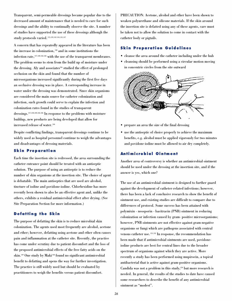
Transparent, semi-permeable dressings became popular due to the
decreased amount of maintenance that is needed to care for such
dressings and the ability to continually observe the site. A number
of studies have supported the use of these dressings although the
study protocols varied.115,139,142-144,145,147
A concern that has repeatedly appeared in the literature has been
the increase in colonization, 138 and in some institutions the
infection rate,137,140,142,146 with the use of the transparent membranes.
The problem seems to stem from the build up of moisture under
the dressing. Aly and associates136 studied the effect of prolonged
occlusion on the skin and found that the number of
microorganisms increased significantly during the first five days
an occlusive dressing was in place. A corresponding increase in
water under the dressing was demonstrated. Since skin organisms
are considered the main source for catheter colonization and
infection, such growth could serve to explain the infection and
colonization rates found in the studies of transparent
dressings.137,138,140,142,146 In response to the problems with moisture
buildup, new products are being developed that allow for
increased release of water.144
Despite conflicting findings, transparent dressings continue to be
widely used as hospital personnel continue to weigh the advantages
and disadvantages of dressing materials.
Sk in P reparat i on
Each time the insertion site is redressed, the area surrounding the
catheter entrance point should be treated with an antiseptic
solution. The purpose of using an antiseptic is to reduce the
number of skin organisms at the insertion site. The choice of agent
is debatable. The main antiseptics that are used are alcohol,
tincture of iodine and povidone-iodine. Chlorhexidine has more
recently been shown to also be an effective agent and, unlike the
others, exhibits a residual antimicrobial effect after drying. (See
Site Preparation Section for more information.)
Defat ti ng t he Sk in
The purpose of defatting the skin is to reduce microbial skin
colonization. The agents used most frequently are alcohol, acetone
and ether; however, defatting using acetone and ether often causes
pain and inflammation at the catheter site. Recently, the practice
has come under scrutiny due to patient discomfort and the loss of
the proposed antimicrobial effects of the free fatty acids on the
skin.149 One study by Maki 148 found no significant antimicrobial
benefit to defatting and opens the way for further investigation.
The practice is still widely used but should be evaluated by
practitioners to weigh the benefits versus patient discomfort.
PRECAUTION: Acetone, alcohol and ether have been shown to
weaken polyurethane and silicone materials. If the skin around
the insertion site is defatted using any of these agents, care must
be taken not to allow the solution to come in contact with the
catheter body or pigtails.
Sk in P repa rat i on Guide l ines
• cleanse the area around the catheter including under the hub
• cleansing should be performed using a circular motion moving
in concentric circles from the site outward
• prepare an area the size of the final dressing
• use the antiseptic of choice properly to achieve the maximum
benefits, e.g. alcohol must be applied vigorously for two minutes
and povidone-iodine must be allowed to air dry completely.
Anti mi c rob ia l Oin tment
Another area of controversy is whether an antimicrobial ointment
should be used under the dressing at the insertion site, and if the
answer is yes, which one?
The use of an antimicrobial ointment is designed to further guard
against the development of catheter-related infections; however,
there has been a lack of conclusive research to show the benefit of
ointment use, and existing studies are difficult to compare due to
differences of protocol. Some success has been attained with
polymixin - neosporin - bacitracin (PNB) ointment in reducing
colonization or infection caused by gram- positive microorganisms;
however, PNB ointments are not effective against gram-negative
organisms or fungi which are pathogens associated with central
venous catheter use.151,152 In response, the recommendation has
been made that if antimicrobial ointments are used, povidone-
iodine products are best for central lines due to the broader
spectrum of organisms against which they are active. More
recently a study has been performed using mupirocin, a topical
antibacterial that is active against gram-positive organisms.
Candida was not a problem in this study,150 but more research is
needed. In general, the results of the studies to date have caused
some researchers to describe the benefit of any antimicrobial
ointment as “modest”.
26

In addition to the questionable efficacy of using an antimicrobial
ointment, other problems must be considered. If used with a
transparent membrane dressing, an ointment covers the insertion
site and negates the advantage of site visibility. The amount of
ointment that is used is also important, since too much mayinterfere with the membrane adherence. A question can also be
asked about how long the ointment will be effective given the
longer periods of time between redressing that are part of some
hospitals’ protocols. One opinion held by some practitioners is
that the ointment provides a wetter environment under theocclusive dressing that may lead to organism growth resistant to
the antimicrobial.
The final decision for or against an antimicrobial ointment
should be based upon a review of the literatur e, consideration
for other dressing attributes that may be affected and thedocumented rate of infection within the hospital setting.
Consideration of the added costs might weigh heavily against a
treatment that has not been proven beneficial.
Redress ing In t er v a l
Hospital protocols vary with respect to the amount of time a
central venous catheter dressing is left in place. The situations for
redressing that are universally accepted are when a dressing is no
longer adherent or occlusive and when a gauze dressing becomes
soiled. Routine redressing protocols vary concerning the interval
between changes from daily up to the lifetime of the catheter or
dressing, whichever comes first. Some protocols provide for a
redressing when the IV tubing is changed at 48 to 72 hours
depending on the hospital policy. Longer intervals are often used
in correlation with the use of transparent dressings,3,115 and in
fewer cases have been extended to gauze redressings.115
Due to the lack of data, the choice must be made within individual
hospitals based on patient factors, infection rates and the type of
dressing desired.
Red res s i ng P roc edure Guide l ines
• hands must be washed thoroughly prior to the redressing
• Universal Precautions must be observed
• to prevent cutting the catheter when removing old dressings, do
not use scissors or sharp instruments
• use strict aseptic technique, sterile gloves, masks (gowns when
required)
• if possible, turn patient’s head away from dressing site or have
patient wear a mask
• visually examine insertion site at least daily or with each
dressing change for erythema, drainage, tenderness, suture
integrity and catheter position
• use circular motion to cleanse the area around the catheter,
working from the site out in concentric circles and under the
catheter hub
• document dressing change with the date and initials of person
redressing
D e l i v e r y Sy s tem
In an effort to keep the number of microorganisms within the IV
set-up at a minimum, the solution and tubing should be changed at
regular intervals. Although most catheter-related infections are
caused by skin organisms migrating down the subcutaneous
catheter track, it is possible to have an infection develop due to
contamination of the infusate. If the IV set or solution hangs for
prolonged periods of time, there is more risk of introducing a
significant number of organisms as a result of frequent
manipulations. Concerning the solution, if small numbers of
certain Gram-negative rods, e.g. Klebsiella species, Enterobacter
species are present in certain solutions as contaminants, a
prolonged hanging time would allow the organisms to multiply to
significant numbers.113 In both cases the result could be a catheter-
related bacteremia.
In the 1970’s, in response to epidemics of IV- related infections
caused by intrinsically contaminated IV fluids, the Centers for
Disease Control and Prevention (CDC) made the re c o m m e n d a t i o n
to change IV administration sets every 24 hours. Studies in the late
1 9 7 0 ’s and early 1980’s demonstrated that it was possible to extend
the interval to every 48 hours with the exception of TPN which was
not included in all of the studies.1 5 3 - 1 5 5 This information was
included in the CDC Guidelines for the Prevention and Control of
Nosocomial Infections.1 5 8 In 1987 re s e a rch was published that
demonstrated it was possible to extend the interval for IV tubing
change, including TPN to at least 72
h o u r s .1 5 6 , 1 5 7 , 1 5 9 The following infusion
sets were exempted from the 72 hour
change: blood, lipids, art e r i a l
p re s s u re monitoring and all
infusions during an IV- re l a t e d
epidemic. These IV sets should be
changed more fre q u e n t l y, i.e., after
each infusion (blood administration)
or every 24 to 48 hours.1 1 3
Since microorganisms can proliferate within solutions, the amount
of time that it takes for a bag of solution to infuse is important.
The frequency with which the bags must be changed is determined
by the capacity for bacterial growth and/or the breakdown of
components within the solution. For example, bags of IV fluids
that are used at a slow rate to keep the line patent should be
changed every 24 hours according to CDC.158 Protocols in some
27

hospitals have been changed to correspond with tubing changes at
48 to 72 hours as long as the system remains closed. Blood should
not infuse for longer than 4 hours.113 Hyperalimentation should
not hang for more than 24 hours, and infusions of lipid emulsions
have a 12 hour limit.158
S l i de C lamps
Slide clamps are provided on each pigtail to facilitate tubing or
injection cap changes. The clamps should not be considered a
guarantee against air entry while the catheter is indwelling due to
the shape and ability of the clamp to “slide” off given the right
circumstances, e.g. patient movement or tampering. Instead,
emphasis must be placed on the use of tightened luer lock
connections as a vital step in the prevention of an air embolus.
Tests have been performed to determine the effect of Arrow clamp
placement on the catheter pigtails.160 During one trial the clamp
was maintained in a closed position for a prolonged period of time
(1 week). When released the flow rates were temporarily reduced
for approximately one hour before returning to pre-test levels.
The clamp can also be applied repeatedly; however, an attempt
should be made to vary the clamp position along the pigtail.
The slide clamps are removable. In situations where the slide
clamps may be inadvertently removed and potentially aspirated by
children or confused adults, they can be taken off and reapplied
only when needed. Do not substitute other clamping devices, such
as hemostats, for the slide clamps. Serrated and sharp edges may
damage the pigtails.
P r i m a r y In j ec t ion S i tes
When continuous infusions are no longer needed, the pigtail hub is
occluded by using a Luer-Lock injection cap. The caps are utilized
as sites for intermittent infusions or injections. Because the caps
are handled repeatedly, it is possible that pathogenic
microorganisms can enter at the hub during a manipulation. To
reduce the chance of the caps becoming a source of catheter-
related infection, it has been recommended that they be changed
at regular intervals. Many protocols provide for the caps to be
changed every 72 hours in conjunction with tubing changes. A
factor that helps to determine how often the caps are changed is
the number of manipulations to which they are subjected. In some
situations the interval between changes has been extended in
hospital/agency protocols such as with a long term catheter
through which treatments are given infrequently.
Every manipulation must be performed using aseptic technique,
and prior to each puncture the injection site must be wiped with
an effective antiseptic, e.g. alcohol or iodine. Likewise, before
each cap change, the hub should be cleaned with an antiseptic
solution prior to disconnection.
The injection caps can withstand repeated punctures. The Arrow
cap (UG-14703) has been tested with a 22 Ga. dry needle and a
needle free hub each used 50 times. The hub did not leak after the
test completion.161
Mainta in i ng Patency
When a catheter lumen is no longer used for continuous infusions,
it can be capped and “locked” in preparation for intermittent or
future use. To “lock” a catheter lumen a solution must be instilled
to fill the entire space of the lumen and injection cap.
Theoretically this prevents a backflow of blood that would cause
clotting within the catheter. Some procedures call for using larger
amounts of solution to “flush” the catheter. A controversy exists
concerning which solution is most appropriate, a heparinized
saline solution or saline alone.
Heparinized saline has been used primarily due to the
a n t i t h rombolytic pro p e rties of heparin. Heparin inhibits clot
f o rmation by inactivating thrombin and other coagulation
f a c t o r s .1 9 2 T h e re are few adverse effects routinely attributed to
heparin. Hemorrhage (7-10%) and allergic reactions (2-5%) are
the most prevalent, but in the past decade another complication of
heparin administration, heparin-induced thrombocytopenia and
t h rombosis syndrome (HITTS), has received much attention.1 6 6 , 1 6 8 , 1 8 3 -
1 8 5 HITTS is associated most frequently with continuous IV heparin
t h e r a p y, but it has also occurred with intermittent subcutaneous
and flushing/locking administration1 7 1 , 1 8 1 , 1 8 8 , 1 9 1 and in one re p o rt was
associated with heparin-coated pulmonary art e ry catheters.1 8 6
HITTS occurs in approximately 5%-30% of persons re c e i v i n g
h e p a r i n .1 6 6 , 1 8 7 , 1 9 1 , 1 9 6
28
Two clinical presentations of heparin-induced thrombocytopenia
and thrombosis syndrome have been identified. One is a mild form
characterized by a transient decrease in platelets that usually
occurs around the second or third day of therapy. This is the most
frequent presentation. The other type is believed to be an immune
response to heparin and occurs 6 to 12 days after therapy is
started.185 It is characterized by a severe decrease in platelets that
may be accompanied by thrombosis, the result of platelet
aggregation. The clots may form in arteries or veins and affect any
portion of the body including organs. The clotting phase of the
disease has also been called “white clot syndrome” due to the color
of the clots which consist of platelets and fibrin. Although this
presentation is rare (0.6%), the incidence of limb amputation has
been reported as 21% and the mortality rate 29%.166
Consideration of the serious side effects of heparin, cost
containment and the systemic antithrombolytic effects that can
occur with heparin flush/lock have led researchers to investigate
other regimens for maintaining catheter patency. Most research
has been conducted using peripheral indwelling intermittent
infusion devices (heparin locks); however, studies can be found
that used arterial lines, peripherally inserted central catheters
(PICC) and central venous catheters. Generally, the information
from these devices has been applied to the maintenance of
multiple lumen catheters.
The current literature identifies the most widely used concentration
of heparin used in intermittent peripheral devices as 100 U/ml 0.9%
sodium chloride solution.1 6 3 , 1 6 9 , 1 7 0 , 1 7 4 Studies have also demonstrated
success with 10 U/ml1 6 4 , 1 8 0 and saline alone1 6 5 , 1 6 7 , 1 7 2 , 1 7 3 , 1 7 5 , 1 7 6 , 1 7 8 , 1 7 9 , 1 9 0 , 1 9 4 , 1 9 5
and have measured success by the number of IV re s t a rts that are
needed due to clotting or patient complications such as phlebitis.
Meta-analyses of similar re s e a rch projects have served to support
the use of saline in indwelling intermittent devices.1 7 7 , 1 8 9 A d v a n t a g e s
that have been attributed to saline use are as follows: decre a s e d
e x p o s u re of the patient to the effects of heparin, decreased risk of
medication errors, decreased nursing time, cost eff e c t i v e n e s s ,
avoidance of drug incompatibilities, and avoidance of heparin
hypersensitivity re a c t i o n s .
Various concentrations of heparin, 0-1000U/ml. normal saline
have successfully been used in Arrow central venous catheters.
The concentration needed to maintain patency is a clinical issue
and must be based on factors specific to the patient population
and treatment setting.
Another aspect of the procedure that is not standardized is the
frequency with which the IV device is flushed. A regimen used
frequently is to flush at the time of medication administration using
the S.A.S.H. method and at least every eight hours or once per shift.
The S.A.S.H. method consists of a flush of saline, followed by the
medication, another saline flush and finally, heparin to lock the
device. This prevents the interaction of heparin with other
medications. Without heparin an S.A.S. routine is used. The range
of intervals in published surveys of flushing protocols is mainly every
eight hours to 24 hours in hospital settings and extends to longer
periods in the home setting.1 7 4
The volume of solution to use is another factor that must be
considered. The range of volumes used extends from the exact
volume of the lumen to 6 ml in one study when saline alone was
instilled every 24 hours. 190 The volume is more critical if a
heparinized solution is used. If too much is given, the patient’s
coagulation status may be altered. For peripheral devices the
Revised IV Nursing Standards of Practice recommend the use of a
minimal concentration of heparinized saline of an amount that is
2x the size of the lumen.182 There is no standard for central venous
catheters.
Considering the elements involved in the flushing/ locking of IV
devices, solution c o n c e n t r a t i o n, flush f re q u e n c y and v o l u m e, there
a re many combinations that can be used.1 6 3 , 1 7 0 , 1 7 4 The trend seems to
be moving toward the use of the least of each of the elements that will
be successful given the patient condition and circumstances. That is,
use the least amount of heparin (down to 0) in the smallest volume
using the longest interval between doses that still keeps the lumen
patent. Curre n t l y, this approach seems applicable to most
intravenous devices.
In contrast, the recently published results of the Thunder Pro j e c t ,
a re s e a rch project conducted through the American Association of
Critical Care Nurses, found that heparinized saline solution was
definitely preferable to maintain the patency of arterial lines in
c e rtain patient populations.1 6 2 The large sample of patients
(n=5139) gave credence to the study that was conducted in 198 U.S.
hospitals. Other factors, in addition to heparin, that were
associated with arterial line patency were: additional anticoagulant
or thrombolytic therapy, catheters longer than 2 inches, femoral
i n s e rtion and male gender. Using these factors can help to
d e t e rmine the population for which heparinized saline should
definitely be used. Determination of the need should be made on a
patient-to-patient basis.
F lush i ng/Lock ing Guide l ines
• use aseptic technique
• cleanse injection hub
with approved antiseptic
solution
• monitor patient clotting
factors during heparin use
• use a 10 cc or greater syringe for flushing/locking to reduce the
risk of exceeding pressure capacity of the catheter92
29

• maintain gentle positive pressure on syringe plunger while
removing from injection hub
• observe Universal Precautions
Blood Samp l ing
While a catheter is indwelling, it is usually necessary for the
patient to have blood samples withdrawn for laboratory testing.
The ideal site for blood sampling is a peripheral vessel; however, a
central venous catheter is often inserted into patients who have no
available peripheral sites making blood withdrawals through the
catheter a necessity. Due to the patient’s condition, it may also be
beneficial to use the catheter for blood sampling to avoid added
stress or sleep disruption.
When a multiple lumen catheter is used for sampling the proximal
port is usually chosen for the procedure. The rationale for the
choice is that if solutions are infusing through the other ports of
the catheter, they will be carried away from the sampling port by
the flow of blood within the vessel, thus reducing the chance for
contamination of the specimen. 198 For additional insurance that the
laboratory results will not be altered, the distal infusions should
be turned off for at least one minute before the blood sample is
obtained.
Syringes or a vacuum setup can be used to withdraw the blood
from the catheter. If syringes are used, it is important to aspirate
slowly to prevent hemolysis of the specimen and/or collapse of the
catheter or vessel. Bubbles in the blood as it is being aspirated
signal that too much force is being applied. After the blood is
withdrawn, the syringe is used to transfer the blood into the
appropriate laboratory test tubes. A vacuum setup is appropriate
for use with pigtails or sampling ports that are secured with a
Luer-Lock injection cap and provide for flow of the blood into the
designated evacuated test tubes.
Several methods can be used to withdraw a blood specimen
through a central venous catheter. Withdrawal can be performed
1) through an injection port, 2) by opening the system or 3)
through one or two stopcocks depending upon whether the initial
amount of solution aspirated is to be reinfused. All sampling
attempts represent an opportunity for microbial contamination of
the systems so use of strict aseptic technique and meticulous hand
washing are of the utmost importance.
Of the three methods, the open technique is the most risky in
terms of microbial contamination. This technique requires the
removal of the injection cap or disruption of an infusion to
facilitate the attachment of various syringes to the Luer-Lock
connection of the catheter. The exposed hub is a well-documented
source of catheter-related infection, and the amount of
manipulation required during the procedure is a contributing
factor to catheter colonization that can lead to infection. The use
of stopcocks can be dangerous for the same reason: the amount of
manipulation needed for the procedure predisposes the system to
microbial contamination, up to 46% in a study by Dryden.204
The actual steps taken in the blood sampling pro c e d u re depend on
a number of variables, e.g. whether the line is used for an infusion
(including TPN), whether the catheter is locked with heparin vs.
n o rmal saline, whether the catheter is heparin-coated, and the
condition of the patient. If there are compounds within or on the
s u rface of the catheter that would alter the study results, attempts
must be made to remove them. Examples are electrolyte-or glucose-
containing solutions if the patient’s electrolytes or blood sugar are
to be measured, or the presence of heparin if coagulation studies or
potassium levels are ord e re d .2 0 2 , 2 0 9 To clear the offending solution, a
d i s c a rd sample must be withdrawn. Some hospital pro c e d u re s
p rovide for an initial saline flush as further assurance that the line
is cleared. A word of caution about flushing is necessary if the
catheter is filled with a concentrated heparin-lock solution such as
100 U/ml of saline solution because if blood samples are taken
f requently the flushing may provide the patient with a therapeutic
dose of heparin that could be detrimental.
The amount of blood that is needed for discard is a controversial
topic and many studies have been performed to determine the
amount of discard that is necessary to assure accuracy of the test
result. Most of the research has been conducted using arterial
catheters that are flushed with a dilute heparin solution to
maintain patency. Very little information is available for multiple
lumen catheters that may utilize more concentrated heparin
solutions. The studies that have been conducted using arterial
catheters are very difficult to compare due to differing study
designs and a lack of priming volume information for
interpretation of results.215
Recommendations for the discard volume in arterial and central
venous catheters have ranged from the priming volume (dead space)
to moderate amounts (3-6ml)2 0 1 , 2 0 3 , 2 0 7 , 2 1 0 , 2 1 2 to 10ml2 0 8 , 2 1 6 with the larg e s t
volumes being used for heparin-locked or flushed catheters when
drawing coagulation studies. Another way the recommendation has
been stated is in the number of priming volumes that are necessary
for clearing the lumen, a range of 2x to 6x its volume.2 1 1 , 2 1 3 A few
re f e rences continue to resolutely state that coagulation studies
should not be drawn from heparinized lines.1 9 7 , 2 1 6
The practice of taking blood cultures through a catheter is
discouraged due to the rate of catheter lumen colonization.199
Blood cultures through a line would only be applicable in an
attempt to diagnose a catheter-related bacteremia as previously
discussed in the “Complications” Section.
30

A concern that is frequently expressed is the amount of blood that
a patient, especially in a critical care setting, loses to laboratory
testing when the sample and discard volumes are combined.
Smoller and Kruskall214 looked at total amounts of blood drawn
from patients on general wards and in the intensive care unit. The
results of their record review revealed that the patients on general
wards had blood drawn on an average of 1.1 times each day for a
mean daily volume of 12.4 ml and a total amount of 175 ml for
their entire hospital stay. In contrast, intensive care patients had
blood drawn on an average of 3.4 times per day at a mean daily
volume of 41.5 ml and a total volume of 762.2 ml. If the patients
had arterial lines, the total increased to 944.0ml. Similar
results were shown in research by Eyster205 and Foulke.206 Some
patients require transfusions to treat the iatrogenic anemia that
results from such frequent blood sampling.
In response to the concern about iatrogenic anemia from blood
sampling, many procedures have been changed to provide for the
lowest discard and sampling volumes and least frequent testing
schedules that are possible considering the patient’s condition and
IV or arterial line regimen. Strategies that have proven useful to
accomplish this goal include the use of smaller test tubes in
accordance with laboratory instrumentation, batching of
laboratory samples, documentation of patient blood loss on the
chart or flow sheet, in line infusion set-ups that provide reservoirs
and allow for immediate reinfusion of the discard specimen, and
new technologies that allow for laboratory testing at the bedside
using minimal amounts of blood.200,214
Final decisions about blood sampling should be made in
conjunction with the laboratory and take
into consideration the solutions that are being infused, the
presence or absence of heparin within the catheter, the lumen
priming volume, the frequency of sampling, and the patient’s
welfare. More research is needed
in this area.
Blood Sampl i ng Guide l i nes
• use aseptic technique and meticulous hand washing
• use peripheral sites whenever possible
• cleanse hubs, injection ports and stopcocks with an approved
antiseptic
• avoid opening the system, whenever possible
• utilize strategies to minimize patient blood loss
• reduce manipulation of the system, whenever possible
• review laboratory results carefully to detect error related to
blood sampling techniques
• replace injection caps that become contaminated with blood
• turn off infusions for one minute prior to sampling, if possible
• do not apply too much pressure when aspirating through a
catheter using a syringe
• use the least amount of discard blood that is possible to achieve
an accurate laboratory test result
Home Ca re
In today’s health care environment, patients are living at home
with central venous catheters in place. The basic principles for
care and maintenance of the catheters apply to all settings but
must be tailored to meet the specific patient’s needs and conditions
found within the home.
I n f u s i o n sA major advantage to using multiple lumen central venous catheters
is the ability to deliver multiple infusions through one perc u t a n e o u s
IV site. Due to tip placement within a large central vein the scope of
t reatment is expanded to include solutions that could not be given
p e r i p h e r a l l y. Proper tip placement in a large vessel is of utmost
i m p o rtance to ensure rapid dilution when such fluids are infused. If
a hypertonic solution is infused into a smaller vessel, the risk of
t h rombophlebitis or vessel erosion is increased significantly.
Solutions that are infused through multiple lumen catheters may
include routine replacement fluids, e.g. 0.9% sodium chloride or
5% dextrose in water, medications, blood products and
hyperalimentation. Consideration must be given to the effects that
certain medications may have upon the catheter as well as other
simultaneous infusions. Examples are: the leaching of plasticizers
from polyvinyl chloride (PVC) material, the weakening of
polyurethane by alcohol, the absorption or adsorption of
medications to certain plastics such as PVC, and medication
incompatibilities.
In compat ib le Inf us ions
Certain medications/solutions will interact when they are in direct
contact. The results may be the formation of a precipitate, a
change in drug potency, total inactivation of a medication or the
formation of toxic or harmful
products.219,222 The simultaneous
infusion of incompatible
medications through a multiple
lumen catheter has raised concerns
about these possible results.
31

The infusion of incompatible medications is possible through a
multiple lumen catheter,220 but studies have shown that important
factors for success are a sufficient blood volume and the
configuration of the lumen exits at the distal portion of the
catheter body.217,218,221 The blood volume is determined by catheter
tip placement, patient hydration status and cardiac output.
Concerning the exit holes, when they are staggered and rotated
around the circumference and along the length of the catheter,
there is much less chance for interaction between incompatible
medications because the proximal infusions are diluted within the
blood flow by the time the next exit hole is approached. Central
venous catheters that are constructed with the exit holes side-by-
side have demonstrated more precipitate formation.218
In fu si on Guide l i nes
• infusions should be monitored frequently to ensure proper flow
rate and medication delivery
• the catheter should be flushed after medication administration
to deliver any residual portion of the dosage to the patient
and to prevent interaction of the medication/solution with
future infusions
• due to the potential for infection, one lumen should be
designated for TPN use only
• medications containing high concentrations of alcohol should
not be infused through polyurethane catheters
• medications/solutions such at Taxol or lipid infusions should
not be infused through catheters made of polymers that
contain plasticizers, e.g. PVC, (polyurethane does not
contain plasticizers)
• consideration must be given to dosages of medications, e.g.
nitroglycerin or insulin, that are absorbed or adsorbed by
certain polymers, e.g. PVC.
m o n i t o r i n gMultiple lumen central venous catheters can be used to monitor
cardiac function. The location of the catheter lumen exit holes
within the superior vena cava (SVC) make it possible to measure
the central venous pressure (CVP), a reflection of the pressure in
the right atrium. The CVP provides valuable information about
the efficiency with which venous blood is returned to the heart,
the intravascular blood volume and right ventricular function.
The normal range for CVP values is 0-7mm Hg with 3mm Hg
considered as a normal average.27 A low CVP is an indication of
fluid depletion, and an elevation signals right ventricular failure.
Measurements can be made using a manometer or calibrated
pressure transducer system.
The distal lumen of a multiple lumen catheter has usually been
employed for CVP measurement. The rationale for this choice is
that the distal lumen is larger and the lumen exit is closest to the
right atrium; however, theoretically a measurement could be taken
from anywhere within the central venous system which includes
the superior vena cava and inferior vena cava within the thoracic
cavity.224,225 That would indicate that any lumen exiting within
those vessels could be used for CVP’s.
The superior vena cava averages 7cm in length.223 Brandt found
the length of the SVC varied with a range of 3cm to 10cm in a
group of 50 patients he studied. He found the length of the SVC to
be proportional to the height of the patient and it could be
estimated by measuring the lower portion of the sternum.33 Taking
into consideration the spacing of the lumen exits on Arrow
multiple lumen catheters (2.2cm), the amount of the distal
catheter that must be within the SVC on a triple lumen catheter is
4.4cm. This length would fall within the SVC of most patients.
There is a lack of published data about using other lumens for
CVP’s. One small unpublished study that was performed using a
heterogeneous group of 38 critical care patients demonstrated no
significant difference between CVP’s measured through the three
lumens of a triple lumen catheter.226 The measurements were taken
from all three ports in succession with the patients supine at a 30°
angle. The pressure transducer set up was recalibrated between
port measurements. This study provides interesting results to
support the theory that there is no significant difference between
lumen CVP measurements, but further evaluation is needed.
p roblem i n t e rv e n t i o nManagement o f Oc c l us ions
Catheter occlusion is a frequently encountered problem that is
associated with most types of vascular access devices. An occlusion
can be caused by a mechanical obstruction, a blood clot, chemical
precipitate accumulation or lipid sludge build-up. In the past if
the occlusion in a single lumen catheter could not be aspirated or
the mechanical problem could not be corrected, the usual recourse
was to change or remove the catheter. With multiple lumen
catheters, when only one lumen became occluded, the same
options were available but there was also a tendency to mark that
lumen as blocked while the remaining lumen(s) continued to be
used. This practice increases the risk of catheter-related infections
and should be discouraged.
The first step in the evaluation of an occluded catheter is to
determine whether there is a mechanical cause for the lack of flow,
e.g. a kink in the tubing, a dry infusion bag or a positional
32

occlusion of the catheter. Measures should be taken to correct any
mechanical problem. If the catheter remains occluded or no
mechanical problem has been found, a thrombolytic agent or
chemical compound can be used to “dissolve” the occlusion.
With the advent of thrombolytic agents, i.e. urokinase and
streptokinase, and the discovery of the composition of chemical
and lipid occlusions, catheters are being salvaged through the use
of various declotting procedures. Urokinase,233,235,244,245,249,252,253
streptokinase243,247 and tissue plasminogen activator (t-PA)230 have
been successfully used to dissolve blood clots. Of these agents,
urokinase 5000 IU/ml is used most frequently because it is less
expensive than t-PA and does not cause allergic reactions as
streptokinase does. The most frequently used procedure calls for
the instillation of a volume of urokinase sufficient to fill the
occluded lumen followed by a dwell time and repeated efforts to
aspirate the clot at regular time intervals.235,236,244 If the initial
instillation does not open the lumen the process can be repeated
again. It is important to remember that the older the occlusion the
less responsive it will be to the therapy. If the lumen remains
blocked after the additional instillations, an infusion of urokinase
might be used231,240 or the cause of the occlusions should be
reevaluated to determine the benefit of using other agents.
As a prophylactic measure against central venous catheter
thrombosis, Bern and associates evaluated the use of warfarin in
low doses,232 and found their regimen to be successful. For long
term catheters the use of prophylactic doses of thrombolytic
agents has also been discussed academically, but no studies have
been published to document the efficacy of such a protocol.
When a thrombolytic agent fails to clear a catheter the reason for
the failure might be that the occlusion is chemical in nature.
Chemical occlusions can be caused by the infusion of poorly
soluble components or incompatible medications. Chemical
occlusions can often be solubized by introducing a compound that
will alter the pH at the blockage site. An evaluation of the
solutions that have been infused through the lumen will help to
determine the course of action.
The agent that is used to lower the pH is a dilute solution of
hydrochloric acid (HCl), 0.1N, in an amount necessary to fill the
occluded lumen.234,237,248,251 The HCl is allowed to dwell within the
lumen for 20 minutes after which the patency of the lumen is
checked. If the occlusion remains the process may be repeated
twice. The only side effect of HCl administration that has been
reported was fever that occurred as part of the Breaux study.234 A
possible explanation for the fever is that the HCl was infused
systematically in that study and not confined to the catheter
lumen.
The agent used to increase the pH within a lumen is sodium
bicarbonate (NaHCO3) in an 8.4% solution. As with the other
procedures an amount is instilled equal to the filling volume of the
catheter lumen and allowed a period of dwell time.239,251 Usually the
dissolution of a chemical occlusion occurs rapidly when the
correct agent is used, and in the two reported uses of NaHCO3 no
additional instillations were necessary.
When lipids are infused a build-up of “lipid sludge” can cause
catheter occlusion. The sludge consists of waxy lipid material that
accumulates gradually on the surface of the catheter lumen.
Catheters used for “all-in-one” solutions may be more prone to
sludge development.238 Incomplete occlusions of lipid sludge have
been treated with success using a 70% solution of ethanol.246 The
ethanol was allowed to dwell for one hour and the catheters were
then flushed and locked. In Pennington’s study the ethanol was
injected into silastic catheters without any effect on the catheter or
patient. This is not the case when polyurethane catheters are
used. 70% ethanol has been shown to reduce the tensile
strength of Ar row polyurethane catheters when the conditions
of clinical ethanol use have been simulated. 229 Sodium
hydroxide, 0.1N, has also been used for lipid occlusions,250
but no information is currently available about its effects
on polyurethane.
As an aid in determining what agent should be used to treat a
central venous catheter occlusion, the algorithm developed by
Holcombe, Forloines-Lynn and Garmhausen242 is very useful.
(See algorithm pages 124 and 125) The algorithm was developed
for silastic, long-term central venous access devices so it does
not caution about the use of ethanol in polyurethane catheters.
Declo t t ing Gu ide l ine s:
• Prior to declotting verify catheter tip placement by chest x-ray.
• Rule out mechanical occlusion first.
• Evaluate medications infused through the line for possible clues
about the nature of the occlusion.
• Use a 10cc syringe or larger to avoid excessive pressure.92
• Do not use excessive force during the declotting procedure.
• Use gentle aspiration to dislodge any occlusion.
• Do not exceed catheter lumen filling volume.
• Make sure the catheter is completely flushed out prior to trying
another declotting agent.
33

Cathet er Repair
Damage to the catheter within the body cannot be repaired.
Intraluminal damage in a multiple lumen catheter is also
irreparable. In most cases the catheter must be removed because
change over a guide wire would increase the risk for further
catheter damage. The main focus of attention must be on
prevention through proper catheter placement and the avoidance
of excess intraluminal pressures and external stresses.
The internal (within the patient’s bloodstream) or external
(outside of the patient’s body) portion of a central venous catheter
can become damaged during catheter use. Internal damage can be
related to catheter position. If the catheter has been placed in the
subclavian vein and is pinched between the clavicle and the first
rib the continued pressure (pinch-off syndrome) and shearing
forces exerted on the catheter can cause partial or complete
fracture of the catheter.254 The catheter can also be damaged
internally if too much force is exerted during a flushing
procedure. If a catheter becomes partially fractured, the function
of the catheter may be compromised, but a worse problem occurs
if the catheter breaks completely and the broken segment
embolizes within the heart or pulmonary artery. Patient symptoms
will be dependent upon where the segment lodges.
The external portion of a catheter can be damaged when it is cut
by a sharp instrument, such as scissors or a scalpel, clamped with
a serrated edge clamp or when it is subjected to excess external
force when snagged or pulled. If a break does occur, it is vital to
clamp below the break to avoid having air enter the catheter.
Measures should be taken to avoid situations that would lead to
catheter breakage; however, if a break occurs there are
commercial kits available to repair central venous catheters.
Temporary repair of a catheter does involve an amount of risk,
and that risk must be carefully weighed when repair is considered
an alternative to catheter exchange or removal. Arrow does not
support this procedure or provide a repair kit for their products.
34
Algorithm for Assessment and Management of Occluded Long-Term Central Venous Access Devices
Printed with permission of authors B. Holcombe Pharm D, L. Garmhousen RN, S. Forloines - Lynne RN. UNCHospitals 1991

infection c o n t ro lHeal th Care Wo r k e r s
Due to the threat of exposure to blood during central venous
catheter insertion and certain aspects of catheter care, all
involved personnel must comply with the safety measures provided
by using Universal Precautions. Protective garb and needlestick
prevention measures should be utilized based on the task and
amount of blood exposure that is anticipated. Needle free systems
and needle containment devices have been developed to further
reduce personnel exposure to needles during the admixture and
administration of injectible medications. Systems for use with
central venous catheters are available.
P a t i e n t
Routine measures as discussed throughout the booklet can be
taken to reduce the risk of local and systemic catheter-related
infections. They include hand washing, site preparation, catheter
stabilization, occlusive dressings, site evaluation, minimal
handling, set-up inspection and exchange, admixture precautions
and inserter expertise.
In addition, advances designed to provide additional protection
against catheter-related infection have been made in IV catheter
technology, e.g. antiseptic-impregnated catheter materials, silver-
impregnated collagen cuffs and antibiotic coated catheters. The
action of these products is directed toward preventing the
migration of skin micro-organisms down the catheter track.
ARROWg+ard™ is an antiseptic surface that has been shown in a
large randomized clinical study by Maki to reduce the risk of
catheter-related infection by 80%.117 The antiseptic surface
consists of a combination of silver-sulfadiazine and chlorhexidine
that is impregnated into the polyurethane catheter surface. The
agents act synergistically to prevent replication of microorganisms.
The process involves alteration of the cell wall of the organism by
chlorhexidine which then allows the entry of the silver ions into
the cell. The silver ions bind to the DNA helix and prevent the cell
from replicating.256 Both agents are active against gram- positive
and gram-negative bacteria as well as yeast. The agents are
released slowly over a period of time, up to 15 days, after which
the release is reduced significantly.255 By using a multiple lumen
catheter made with the antiseptic surface, the benefits will include
a longer catheter dwell time, decreased patient morbidity and
mortality and cost effectiveness.
A similar principle was evaluated using the idea of antibiotic-coated
central venous catheters. However, the use of antibiotics, usually
ampicillin or a cephalosporin, raises the following concerns: there is
a possibility that resistant micro o rganisms will develop with
repeated antibiotic use, time must be spent to pre p a re the catheter
for insertion and the agents used are not effective against yeasts.
A silver impregnated collagen cuff has also been shown to be
effective in reducing the incidence of catheter-related infection.
The cuff is attached to a catheter prior to insertion, and when the
catheter is in position the cuff is inserted beneath the skin into the
subcutaneous tissue. The cuff will become anchored in place by
tissue growth into the collagen material, and silver ions are
released from the cuff over time. This provides a physical barrier
(collagen cuff) as well as antimicrobial action (silver ions) to
prevent microorganism migration down the catheter.114
Advantages Disadvantages
Antiseptic surface • Routine catheter
(silver sulfadiazine/ • insertion
chlorhexidine) technique
• Synergistic
activity
of agents
• Surface
impregnation
effective against
bacteria and
Candida
Antibiotic coating • Allows choice • Preparation of
antibiotic time prior to
insertion
• Possible
emergence of
resistant strains
• Lack of action
against Candida
Silver-impregnated • Additional • Insertion
collagen cuff mechanical technique
barrier
35

Combined with routine infection control measures, new IV
technology such as ARROWg+ard™ can work to prevent the
development of catheter-related infections in patients with central
venous access. Measures such as these are important in all
patients with multiple lumen catheters since they represent a
compromised population in terms of morbidity and mortality.
catheter e x c h a n g eThe amount of time a catheter remains in one site is a
controversial issue. In hospitals a variety of protocols are followed
including routine changes over a guide wire, routine changes to a
new site and leaving the catheter in until complications occur or
the catheter is no longer needed. There are pros and cons to all of
these methods. Unfortunately, the studies that have been done to
determine which protocol is best are difficult to compare due to
their differences in target populations, change intervals, catheter
types and other variables that are studied simultaneously, e.g.,
use of collagen cuffs.
The practice of routinely changing catheters, especially in the
critical care setting, is the product of studies that have shown a
direct relationship between increased catheter
colonization/infection and the length of catheter indwelling
time.261,263,265,266,270 In response, many protocols have incorporated
within them a catheter change every three to seven days either
over a guide wire or into a new site to reduce the infection risk
associated with prolonged catheterization.257,259,264,267 However, the
routine changing of catheters is not without risk.
Placing catheters in new sites has drawbacks that include the small
number of sites available for rotation and the possibility of
complications associated with each catheter insertion, such as air
embolism malpositioning, pneumothorax and cardiac tamponade to
name a few. A complication that can be associated with guide wire
exchanges is colonization or infection. If the site is already colonized
when a new catheter is inserted, colonization and future infection of
the newly inserted catheter is almost ensure d .2 6 8 When the two
methods for replacing catheters are compared, the pro b l e m s
associated with new insertions have caused some re s e a rchers to
recommend guide wire exchanges instead of rotation to new sites.2 6 9
Recent studies have addressed the issue of routine replacement of
catheters to assess the benefits of the procedure. In a study by
Bonawitz, et al.,258 three day catheter exchanges were compared to
seven day exchanges. No statistically significant differences were
noted between the patient groups in this prospective, randomized
trial of critically ill subjects. Another study, by Cobb, et al.,260
concluded that replacing catheters every three days does not
prevent infections in adult patients in ICU but does expose
patients to complications associated with the changes. A third
study by Eyer, et al.262 that compared changes every seven days in
surgical ICU patients also found no benefit to the procedure. The
Cobb and Eyer studies led to conclusions on the part of the
researchers that central venous catheters should be maintained
using strict aseptic technique and should be allowed to remain in
the same site unless removal is clinically indicated.
If the catheters are allowed to remain in one site, newer
technologies such as an antiseptic impregnated catheter can off e r
f u rther infection protection in addition to meticulous catheter care .
catheter re m o v a lWhen a catheter is removed, precautions must be taken to pre v e n t
associated complications. During removal Universal Pre c a u t i o n s
must be employed to protect the health care worker from potential
e x p o s u re to bloodborne pathogens. Aseptic technique must be used
at the insertion site. To increase intra-thoracic pre s s u re, the patient
should be placed in Tre n d e l e n b u rg position, if tolerated, or flat in
bed and should be instructed to hold his/her breath or perf o rm the
Valsalva maneuver. If the patient cannot cooperate with the
i n s t ructions, the catheter should be removed during expiration.
After the catheter is removed pressure must be maintained with
sterile gauze at the site until hemostasis is achieved, approximately
five minutes. A totally air occlusive dressing must be placed over
the insertion site to prevent an air embolism caused by air
entering the body through the residual subcutaneous catheter
track.58,59,70,73,271 The dressing must remain in place for 24-72 hours
depending on the length of time the catheter was indwelling.
Dressing materials that have been suggested are Vaseline
gauze70,72,271 and telfa gauze with antimicrobial ointment.72 The use
of semipermeable membrane dressings has not been published in
the literature, although they are used in some hospitals. Plain
gauze with ointment is not acceptable as demonstrated in a case
presented by Hanley.58 In addition to the dressing, as further
security the wound can be sutured closed.
During the time following catheter removal the patient should be
observed closely for any signs and symptoms of complications,
especially bleeding, air embolism or infection at the insertion site.
Removal Guide l ines
• turn off all infusions
• wash hands
• don protective apparel according to Universal Precautions
guidelines
36

• place patient in Trendelenburg or supine position
• instruct patient to perform Valsalva maneuver or hold breath
on command
• if catheter cultures are collected have sterile field, sterile
scissors and specimen containers available
• to collect catheter cultures the skin around the insertion site
must be cleansed with an acceptable antiseptic, the catheter
must be removed at a 90° angle from the skin and the desired
segments (tip, subcutaneous) are cut with the sterile scissors
and placed into the specimen containers
• as the catheter is removed the patient should perform the
Valsalva maneuver or hold his/her breath
• immediately cover the insertion site with sterile gauze to apply
pressure to the area
• cover the site with an occlusive dressing such as Vaseline gauze
while the patient is performing the Valsalva maneuver or
holding his/her breath
• leave occlusive dressing in place 24-72 hours depending upon
the length of time the catheter was indwelling
• observe patient for complication signs and symptoms.
d o c u m e n t a t i o nThe following information should be included within the patient’s
chart:
• product name (refrain from misnomers, e.g. calling all
thermodilution catheters a “Swan-Ganz”, all tunneled catheters
as “Hickman’s”)
• date of insertion, inserter
• anatomical location
• catheter depth according to catheter reference markings
• x-ray confirmation of catheter tip location
• port designation for infusions/measurements, e.g. TPN, CVP,
medications
• amount, type and frequency of flush solution
• dressing and tubing changes
• site assessment, patient condition
• catheter guide wire exchanges
• complications
• catheter removal and application of air-impermeable dressing.
b i b l i o g r a p h yGeneral Information1 Baranowski L. Central venous access devices. J Intravenous Nurs. 1993;16:167-194.2 Farber BF. The multi-lumen catheter: proposed guidelines for its use. Infect Control Hosp Epidemiol. 1988;9:206-208.3 Hadaway LC. Evaluation and use of advanced I.V. technology part 1: central venous access devices. J Intravenous
Nurs. 1989;12:73-82.4 Holder C, Alexander J. A new and improved guide to IV therapy. February 1990:43-47.5 Jones S. New IV catheters that can do it all. RN. February 1985:2-5. Reprint.6 Kaufman JL, Rodriguez JL, McFadden JA, Brolin RE. Clinical experience with the multiple lumen central venous
catheter. J Parenteral and Enteral Nutrition. 1986;10:487-489.7 Recker DH, Metzler DJ. Use of the multi-lumen catheter. Crit Care Nurse. May/June 1984:1-3.8 Seneff M. Central venous catheters. In: Rippe JM, Irwin RS, Alpert JS, Fink MP, eds. Intensive Care Medicine. 2nd ed.
Boston, Mass: Little Brown and Co; 1991:17-37.
Types of Catheters9 Viall CD. Your complete guide to central venous catheters. Nursing ‘90. February 1990:34-41.
Catheter Materials and Proper ties10 Crocker KS, Devereaux GB, Ashmore DL, Coker MH. Clinical evaluation of elastomeric hydrogel peripheral catheters
during home infusion therapy. J Intravenous Nurs. 1990;13:89-97.11 Dieckmann D. Plasticizers. In: Greene R, ed. Modern Plastics Encyclopedia ‘92. Hightstown, NJ: McGraw Hill; Mid-
October 1991:184-187.12 Frye RB. Silicones. In: Greene R, ed. Modern Plastics Encyclopedia ‘92. Hightstown, NJ: McGraw Hill; Mid-October
1991:138-141.13 Gabbett JF. Introduction to PVC. In: Greene R, ed. Modern Plastics Encyclopedia ‘92. Hightstown, NJ: McGraw Hill;
Mid-October 1991:122-123.14 Gillis HR. Polyurethanes. In: Greene R, ed. Modern Plastics Encyclopedia ‘92. Hightstown, NJ: McGraw Hill; Mid-
October 1991:135-138.15 Schatten KL. Cardiovascular catheter polymer materials. JVAN. 1991;1:20-21.16 Szycher M. Biocompatible Polymers, Metals and Composites. Lancaster, Pa: Technomic Publishing Co., Inc.; 1983.17 Szycher M, Robinson, WJ. Synthetic Biomedical Polymers: Concepts and Applications. Lancaster, Pa: Technomic
Publishing Co., Inc.; 1980.
Insertion Sites1 8 M a l l o ry D, Sapienza R, McGee WT, et al. “State-of-the-art” vascular cannulation in the ICU. R e s M e d i c a. 1988;4:5-14.1 9 S e n e ff MG. Central venous catheterization: A comprehensive review, Part I. J Intensive Care Medicine. 1 9 8 7 ; 2 : 1 6 3 - 1 7 5 .2 0 S e n e ff MG. Central venous catheterization: A comprehensive review, Part II. J Intensive Care Medicine. 1 9 8 7 ; 2 : 2 1 8 - 2 3 2 .
Site Preparation21 Alexander JW, Fischer JE, Boyajian M, Palmquist J, Morris MJ. The influence of hair-removal methods on wound
infections. Arch Surg. 1983;118:347.22 Larson E. Guideline for use of topical antimicrobial agents. Am J Infection Control. December 1988;16:253-266.23 LeVeen HH, LeVeen RF, LeVeen EG. The mythology of povidone-iodine and the development of self-sterilizing plastics.
Surg Gynecol Obstet. 1993;176:183-190.24 Maki D, Ringer M, Alvarado CJ. Prospective randomized trial of povidone-iodine, alcohol, and chlorhexidine for
prevention of infection associated with central venous and arterial catheters. Lancet. August 1991; 338:339-343.25 Strand CL, Wajsbort RR, Sturmann K. Effect of iodopher vs. iodine tincture skin preparation on blood culture
contamination rate. JAMA. 1993;269:1004-1006.
Catheter Insertion26 Conahan TJ III, Schwartz AJ, Geer RT. Percutaneous catheter introduction: the Seldinger technique. JAMA.
1977;237:446-447. Letter.2 7 Daily EK, Schroeder JS. Techniques in Bedside Hemodynamic Monitoring. 4th ed. St. Louis, Mo: Mosby; 1989:59-62.28 Jobes DR, Schwartz AJ, Greenhow DE, Stephenson LW, Ellison N. Safer jugular vein cannulation: recognition of
arterial puncture and preferential use of the external jugular route. Anesthesiology. 1983;59:353-355.2 9 Seldinger SI. Catheter replacement of the needle in percutaneous art e r i o g r a p h y. A C TA Radiologica. 1 9 5 3 ; 3 9 : 3 6 8 - 3 7 6 .30 Sznajder J, Zveibil, F, Bitterman H, Weiner P, Bursztein S. Central vein catheterization. Failure and complication rates
by three percutaneous approaches. Arch Intern Med. February 1986;146:259-261.
Catheter Tip Placement31 Bar-Joseph G, Galvis AG. Perforation of the heart by central venous catheters in infants: guidelines to diagnosis and
management. J Pediatr Surg. 1983;18:284-287.32 Blitt CD, ed. Monitoring in Anesthesia and Critical Care Medicine. Central venous pressure monitoring. New York,
NY: Churchill Livingstone; 1985:121-165.33 Brandt RL, Foley WJ, Fink GH, Regan WJ. Mechanism of perforation of the heart with production of hydropericardium
by a venous catheter and its prevention. Am J Surg. 1970;119:311-316.34 Collier PE, Ryan JJ, Diamond DL. Cardiac tamponade from central venous catheters - report of a case and review of
the English literature. Angiology. September 1984;35:595-600.35 Curelaru J, Linder LE, Gustavsson B. Displacement of catheters inserted through internal jugular veins with neck flexion
and extension. Intens Care Med. 1980;6:179-183.36 Iberti TJ, Katz LB, Reiner MA, Brownie T, Kwun KB. Hydrothorax as a late complication of central venous indwelling
catheters. Surgery. November 1983:842-84637 Johans TG. Multiorificed catheter placement with an intravascular electrocardiographic technique . Anesthesiology.
1986;64:411-413. Letter.38 Maschke SP, Rogove HJ. Cardiac tamponade associated with a multilumen central venous catheter . Crit Care Med.
1984;12:611-612.39 McGee WT, Ackerman BL, Roubin LR, Prasad VM, Bandi V, Mallory DL. Accurate placement of central venous
catheters: a prospective, randomized, multicenter trial. Critical Care Medicine. 1993;21:1118-1123.40 Peters JL , ed. A Manual of Central Venous Catheterization and Parenteral Nutrition. Boston, Mass: John Wright PSG;
1983:58-61,155-157.41 Sigurdsson J, Riba P, Sigurdsson S. The wandering central venous catheter. Intensive Care Med. 1985;11:263-264.42 Tocino IM, Watanabe A. Impending catheter perforation of superior vena cava: radiographic recognition. Am J
Roentgenology. March 1986;146:487-490.
Complications(General)43 Beam TR, Goodman EL, Maki DG, Farr BM, Mayhall CG. Preventing central venous catheter-related complications, a
roundtable discussion. Infections in Surgery. October 1990:1-13.44 Collins JL. Central venous catheter complications. Oncology Nurs.Forum. 1991;18:819-820. Letter.45 Dawood MM, Trebbin WM. Complications associated with central venous cannulation. Hosp Pract. June 15,
1991;26:211-214,218-219.46 Klass K. Troubleshooting central line complications. Nursing 87. November 1987:58-61.47 Mihm FG, Rosenthal MH. Central venous catheterization. In: Benumof JL, ed. Clinical Procedures in Anesthesia and
Intensive Care. Philadelphia, Pa: J B Lippincott Co; 1992:365-370.
37
48 Parsa MH, Freeman HP. Precautions for long-term maintenance of central venous catheters: part I. Surg Rounds.November 1992:967-974.
49 Richardson D, Bruso P. Vascular access devices - management of common complications. J Intravenous Nurs.1993;16:44-49.
5 0 Ryan JA, Gough J. Complication of central venous catheterization for total parenteral nutrition. N I TA . 1 9 8 4 ; 7 : 2 9 - 3 5 .51 Scott WL. Complications associated with central venous catheters. Chest. 1988;94:1221-1224.52 Wickham R, Purl S, Welker D. Long-term central venous catheters: issues for care. Seminars in Oncology Nurs.
1992;8:133-147.53 Wilson GL, McGregor PJ, Thompson GR. Long-term complications of intravascular cannulation. In: Venus B, Mallory
DL, eds. Problems in Critical Care. Philadelphia, Pa: J.B. Lippincott Company; April-June 1988;2:296-313.54 Yerdel MA, Karayalcin K, Aras N, Bozatli L, Yildirim E, Anadol E. Mechanical complications of subclavian vein
catheterization. A prospective study. International Surgery. 1991;76:18-22.
Air Embolism55 Alvaran SB, Toung JK, Graff TE, Benson DW. Venous air embolism: comparative merits of external cardiac massage,
intracardiac aspiration, and left lateral decubitus position. Anesth Analg. 1978;57:166-170.56 Bedford RF. Venous air embolism: a historical perspective. Seminars in Anesth. September 1983;3:169-176.57 Coppa GF, Gouge TH, Hofstetter SR. Air embolism: a lethal but preventable complication of subclavian vein
catheterization. J Parenteral and Enteral Nutrition. March/April 1981;5:166-168.58 Hanley PC, Click RL, Tancredi RG. Delayed air embolism after removal of venous catheters. Ann Intern Med.
1984;101:401-402. Letter.59 Kashuk JL, Penn I. Air embolism with venous catheterization. Surg Gynecol Obstet. 1984;159:249-252.60 Kondo K, O’Reily LP, Chiota J Jr. Air embolism associated with an introducer for pulmonary arterial catheters. Anesth
Analg. 1984;63:871-872.6 1 McConnell EA. Preventing air embolism in patients with central venous catheters. Nurs Life. March/April 1986:47-49.62 Mendieta JM, Roy TM, Denny DM, Ossorio M. Venous air embolism associated with removal of central venous
catheters. J Kentucky Med Assoc. April 1988; 86:169-173.63 Michel L, Poskanzer DC, McKusick KA, Nardi GL, Malt RA. Fatal paradoxical air embolism to the brain: complication
of central venous catheterization. J Parenteral and Enteral Nutrition. January/February 1982;6:68-70.64 O’Quin RJ, Lakshminarayan S. Venous air embolism. Arch Intern Med. 1982;142:2173-2176.65 Ordway CB. Air embolus via CVP catheter without positive pressure: presentation of case and review. Ann Surg. April
1974;179:479-481.6 6 Orebaugh SL. Venous air embolism: clinical and experimental considerations. Crit Care Med. 1 9 9 2 ; 2 0 : 1 1 6 9 - 1 1 7 7 .67 O’Rourke ME. Reducing the risk of venous air embolism. Am J Nurs. June 1988:886-890.68 Ostrow LS. Air embolism and central venous lines. Am J Nurs. 1981;81:2036-2038.69 Peters JL, Armstrong R, Bradford R, Gelister JK. Air embolism: a serious hazard of central venous catheter systems.
Intensive Care Med. 1984;10:261-262.70 Phifer TJ, Bridges M, Conrad SA. The residual central venous catheter track - an occult source of lethal air embolism:
case report. J Trauma. 1991;31:1558-1560.71 Seidelin PH, Stolarek IH, Thompson AM. Central venous catheterization and fatal air embolism. Br J Hosp Med.
November 1987;438-439.7 2 Thielen JB. Air emboli: A potentially lethal complication of central venous lines. Focus on Crit Care. 1 9 9 0 ; 1 7 : 3 7 4 - 3 8 3 .73 Thielen JB, Nyquist J. Subclavian catheter removal. J Intravenous Nurs. 1991;14:114-118.74 Turnage WS, Harper JV. Venous air embolism occurring after removal of a central venous catheter. Anesth Analg.
1991;72:559-560.
Cardiac Tamponade75 Chamsi-Pasha H, Waldek S. Cardiac tamponade complicating central venous catheter. Postgrad Med J.
1988;64:290-291.76 Csanky-Treels JC. Hazards of central venous pressure monitoring. Anaesthesia. 1978;33:172-177.77 Defalque RJ, Campbell C. Cardiac tamponade from central venous catheters. Anesthesiology. 1979;50:249-252.78 Edwards H, King TC. Cardiac tamponade from central venous catheters. Arch Surg. 1982;117:965-967.79 Eide J, Odegaard E. Cardiac tamponade as a result of infusion therapy. A potentially amenable complication of
central venous catheters. Acta Anaesthesiol Scand. 1983;27:181-184.80 Estes MEZ. Management of the cardiac tamponade patient: a nursing framework. Crit Care Nurse. 1985;5:17-26.81 Fotre TV. Cardiac tamponade: early recognition and treatment. Emerg Care Q. 1988;4:19-28.82 Gordon HS. Action STAT - cardiac tamponade. Nursing 86. August 1986;33.83 Hancock EW. Cardiac tamponade. Med Clin North Am. January 1979;63:223-237.84 Hunt R, Hunter TB. Cardiac tamponade and death from perforation of the right atrium by a central venous catheter.
Amer J Roentgenology. 1988;151:1250. Letter.85 Kapadia CB, Heard SO, Yeston NS. Delayed recognition of vascular complications caused by central venous
catheters. J Clin Monitoring. 1988; 4:267-271.86 Long R, Kassum D, Donen N, DePape A, Taylor J, Warrian K. Cardiac tamponade complicating central venous
catheterization for total parenteral nutrition: a review. J Crit Care. March 1987;2:39-44.87 Mitchell MR, Wood DG, Naraghi M, Riopelle JM. Fatal cardiac perforation caused by the dilator of a central venous
catheterization kit. J Clin Monitoring. September 1993;9:288-291.88 Sheep RE, Guiney WB Jr. Fatal cardiac tamponade. JAMA. 1982;248:1632-1635.89 Suddleson EA. Cardiac tamponade: a complication of central venous hyperalimentation. J Parenteral and Enteral
Nutrition. 1986;10:528-529.90 Timperman W, Rath R. Cardiac tamponade. Contemporary Surg. 1989;34:53-58.9 1 Thomas CS, Carter JW, Lowder SC. Pericardial tamponade from central venous catheters. Arch Surg. 1 9 6 9 ; 9 8 : 2 1 7 -218.
Catheter Embolus/Rupture92 Conn C. The importance of syringe size when using implanted vascular access devices. J Vasc Access Nurs. Winter
1993;3:11-18.93 Cope C. Intravascular breakage of Seldinger spring wire guides. JAMA. June 23, 1962;180:1061-1063.94 Richardson JD, Grover FL, Trinkle JK. Intravenous catheter emboli. Experience with twenty cases and collective review.
Am J Surg. 1974;128:722-727.
Arterial Puncture95 Todd MR, Barone JE. Recognition of accidental arterial cannulation after attempted central venipuncture. Crit Care
Med. 1991;19:1081-1083.
Dysrhythmias96 Eissa NT, Kvetan V. Guide wire as a cause of complete heart block in patients with preexisting left bundle branch
block. Anesthesiology. 1990;73:772-774.97 Kasten GW, Owens E, Kennedy D. Ventricular tachycardia resulting from central venous catheter tip migration due to
arm position changes: report of two cases. Anesthesiology. 1985;62:185-187.
Nerve Injury98 Defalque RJ, Fletcher MV. Neurological complications of central venous cannulation. J Parenteral and Enteral
Nutrition. 1988;12:406-409.
Catheter Malposition99 Gilner LI. The “ear-gurgling” sign. NEJM. 1977;296:1301. Letter.100 Jacobs WR, Zaroukian MH. Coughing and central venous catheter dislodgement. J Parenteral and Enteral Nutrition.
August 1991;15:491-493.101 Lum PS, Soski M. Management of malpositioned central venous catheters. J Intravenous Nursing. December
1989;12:356-365.
102 Rosa UW, Foreman M, Willsie-Ediger S. Intermittent back pain after central venous catheter placement. J Parenteraland Enteral Nutrition. 1993;17:91-93.
Pneumothorax, Hemothorax103 Maggs PR, Schwaber JR. Fatal bilateral pneumothoraces complicating subclavian vein catheterization. Chest.
1977;71:552-553.104 McGoon MD, Benedetto PW, Greene BM. Complications of percutaneous central venous catheterization: a report of
two cases and review of the literature. Johns Hopkins Med J. July 1979;145:1-6.
Catheter-related Infection105 Beam TR, Goodman EL, Maki DG, Farr BM, Mayhall CG. Preventing central venous catheter-related complications. A
roundtable discussion. Infections in Surgery. October 1990:1-13.106 Bjornsen HS. Pathogenesis, prevention and management of catheter-associated infections. New Horizons. May
1993;1:271-278.107 Bozzetti F. Central venous catheter sepsis. Surg Gynecol Obstet. September 1985;161:293-301.108 Corona ML Peters SG, Narr BJ, Thompson RL. Infections related to central venous catheters. Mayo Clin Proc.
1990;65:979-986.109 Dickinson GM, Bisno AL. Infections associated with indwelling devices: concepts of pathogenesis; infections associated
with intravascular devices. Antimicrobial Agents Chemother. May 1989;33:597-601.110 Gil RT, Kruse JA, Thill-Baharozian MC, Carlson RW. Triple-vs single-lumen central venous catheters. Arch Intern Med.
1989;149:1139-1143.111 Goldmann DA. Coagulase-negative staphylococci: interplay of epidemiology and bench research. Am J Infection
Control. June 1990;18:211-221.112 Hampton AA, Sherertz RJ. Vascular-access infections in hospitalized patients. Surg Clin North Am. 1988;68:57-71.113 Maki DG. Infections due to infusion therapy. In: Bennett JV, Brachman PS, eds. Hospital Infections. Boston, Mass: Little,
Brown and Co; 1992:849-898.114 Maki DG, Cobb L, Garman JK, Shapiro JM, Ringer M. Helgerson RB. An attachable silver-impregnated cuff for
prevention of infection with central venous catheters: a prospective randomized multicenter trial. AJM. September1988;85:307-314.
1 1 5 Maki DG, Ringer M. Evaluation of dressing regimens for prevention of infection with peripheral intravenous catheters:gauze, a transparent polyurethane dressing, and an iodophor-transparent dressing. J A M A . 1 9 8 7 ; 2 5 8 : 2 3 9 6 - 2 4 0 3 .
116 Maki DG, Weise CE, Sarafin HW. A semiquantitative culture method for identifying intravenous-catheter-relatedinfection. NEJM. 1977;296:1305-1309.
117 Maki DG, Wheeler SJ, Stolz SM, Mermel LA. Clinical trial of a novel antiseptic-coated central venous catheter.Presented at 31st Interscience Conference on Antimicrobial Agents and Chemotherapy, Chicago, Ill, October 1, 1991.Abstract.
118 Mermel LA, McCormick RD, Springman SR, Maki DG. The pathogenesis and epidemiology of catheter-relatedinfection with pulmonary artery Swan-Ganz catheters: a prospective study utilizing molecular subtyping. AJM.September 1991;91 (Suppl. 3B):3B-197S-3B-205S.
119 Miller JJ, Venus B, Mathru M. Comparison of the sterility of long-term central venous catheterization using singlelumen, triple lumen, and pulmonary artery catheters. Crit Care Med. 1984;12:634-637.
120 Norwood S, Hajjar G, Jenkins L. The influence of an attachable subcutaneous cuff for preventing triple lumen catheterinfections in critically ill surgical and trauma patients. Surg Gynecol Obstet. July 1992;175:33-40.
121 Norwood S, Ruby A, Civetta J, Cortes V. Catheter-related infections and associated septicemia. Chest. 1991;99:968-975.
122 Passerini L, Lam K, Costerton JW, King EG. Biofilms on indwelling vascular catheters. Crit Care Med. 1992;20: 665-673.
123 Powell C, Fabri PJ, Kudsk KA. Risk of infection accompanying the use of single-lumen vs double-lumen subclaviancatheters: a prospective randomized study. J Parenteral and Enteral Nutrition. March/April 1988;12:127-129.
124 Recker D. Catheter-related sepsis: an analysis of the research. Dimensions of Crit Care Nurs. Sept-Oct 1992;11:249-262.
125 Rose SG, Pitsch RJ, Karrer W, Moor BJ. Subclavian catheter infections. J Parenteral and Enteral Nutrition.1988;12:511-512.
126 Snydman DR, Pober BR, Murray SA, Gorbea HF, Majka JA, Perry LK. Predictive value of surveillance skin cultures intotal-parenteral-nutrition-related infection. Lancet. 1982;2:1385-1388.
127 Toltzis P, Goldmann DA. Central venous catheter infections. Infections in the Immunocompromised Host (BurroughsWellcome). 1986;4:2-11.
128 Yeung C, May J. Hughes R. Infection rate for single lumen v triple lumen subclavian catheters. Infect Control HospEpidemiol. 1988;9:154-158.
Catheter-Related Thrombosis129 Curelaru I, Gustavsson B, Hultman E, et al. Material thrombogenicity in central venous catheterization III. A
comparison between soft polyvinyl/chloride and soft polyurethane elastomer, long, antebrachial catheters. ActaAnaesthesiol Scand. 1984;28:204-208.
130 Davis CL. Upper extremity venous thrombosis and central venous catheters. Critical Care Nurse. September1991;11:16-22.
131 Linder L-E, Curelaru I, Gustavsson B, Hansson H-A, Stenquist O, Wojciechowski J. Material thrombogenicity in central venous catheterization: a comparison between soft, antebrachial catheters of silicone elastomers andpolyurethane. J Parenteral and Enteral Nutrition. August 1984;8:399-406.
132 Stenqvist O, Curelaru I, Linder L-E. Gustavsson B. Stiffness of central venous catheters. Acta Anaesthesiol Scand.1983;27;153-157.
133 Williams EC. Catheter-related thrombosis. Clin Cardiol. 1990;13(suppl VI):VI34-36.
Hydrothorax/Vessel Erosion134 Duntley P, Siever J, Korwes ML, Harpel K, Heffner JE. Vascular erosion by central venous catheters. Chest.
1992;101:1633-1638.135 Ellis LM, Vogel SB, Copeland EM III. Central venous catheter vascular erosions. Ann Surg. 1989;209:475-478.
CATHETER MAINTENANCE
Dressings136 Aly R, Shirley C, Cunico B, Maibach HI. Effect of prolonged occlusion on the microbial flora, pH, carbon dioxide and
transepidermal water loss on human skin. J Invest Dermatol. 1978;71:378-381.137 Conly JM, Grieves K, Peters B. A prospective, randomized study comparing transparent and dry gauze dressings for
central venous catheters. J Infectious Diseases. February 1989;159:310-319.138 Craven DE, Lichtenberg DA, Kunches LM, et al. A randomized study comparing a transparent polyurethane dressing
to a dry gauze dressing for peripheral intravenous catheter sites. Infection Control. 1985;6:361-366.139 Eisenberg PG, Howard MP, Gianino MS. Improved long-term maintenance of central venous catheters with a new
dressing technique. J Intravenous Nurs. 1990;13:279-284.140 Gantz NM, Presswood GM, Goldberg R. Doern G. Effects of dressing type and change interval on intravenous
therapy complication rates. Diagn Microbiol Infect Dis. 1984;2:325-332.141 Hoffmann KK, Weber DJ, Samsa GP, Rutala WA. Transparent polyurethane film as an intravenous catheter dressing.
JAMA. 1992;267:2072-2076.142 Hoffmann KK, Western SA, Kaiser DL, Wenzel RP, Groschel DHM. Bacterial colonization and phlebitis-associated risk
with transparent polyurethane film for peripheral intravenous site dressings. Am J Infection Control. June1988;16:101-106.
143 Lawson M, Kavanagh T, McCredie K, Marts K, Barbour N, Chandler W. Comparison of transparent dressing to papertape dressing over central venous catheter sites. NITA. January/February 1986;9:40-43.
144 Maki DG, Stolz S, Wheeler S. A prospective, randomized, three-way clinical comparison of a novel, highlypermeable, polyurethane dressing with 206 Swan-Ganz pulmonary artery catheters: OpSite IV 3000 vs. Tegaderm vs.gauze and tape. In: Maki DG, ed. Improving Catheter Site Care. Royal Society of Medicine Services InternationalCongress and Symposium Series No. 179, Royal Society of Medicine Services Limited. London;1991:61-66.
38
145 Palidar PJ, Simonowitz DA, Oreskovich MR, et al. Use of OpSite as an occlusive dressing for total parenteral nutritioncatheters. J Parenteral and Enteral Nutrition. 1982;6:150-151.
146 Powell C, Regan C, Fabri PJ, Ruberg RL. Evaluation of OpSite catheter dressings for parenteral nutrition: aprospective, randomized study. J Parenteral and Enteral Nutrition. 1982;6:43-46.
147 Shivnan JC, McGuire D, Freedman S, et al. A comparison of transparent adherent and dry sterile gauze dressings forlong-term central catheters in patients undergoing bone marrow transplant. Oncology Nurs Forum. 1991;18:1349-1356.
Defatting the Skin148 Maki D, McCormack KN. Defatting catheter insertion sites in total parenteral nutrition is of no value as an infection
control measure. Am J Med. 1987;83:833-840.149 Rothman S, Lorincz AL. Defense mechanisms of the skin. Annual Rev Med. 1963;14:215-242.
Antimicrobial Ointment150 Hill RLR, Fisher AP, Ware RJ, Wilson S, Casewell MW. Mupirocin for the reduction of colonization of internal jugular
cannulae - a randomized controlled trial. J Hospital Infection. 1990;15:311-321.151 Levy RS, Goldstein J, Pressman RS. Value of a topical antibiotic ointment in reducing bacterial colonization of
percutaneous venous catheters. J Albert Einstein Med Center. Summer, 1970;18:67-70.152 Maki DG, Band JD. A comparative study of polyantibiotic and iodophor ointments in prevention of vascular catheter-
related infection. Am J Med. 1981;70:739-744.
Delivery System153 Band JD, Maki DG. Safety of changing intravenous delivery systems at longer than 24-hour intervals. Ann Intern Med.
1979;91:173-178.154 Buxton AE, Highsmith AK, Garner JS, et al. Contamination of intravenous infusion fluid: effects of changing
administration sets. Ann Intern Med. 1979;90:764-768.155 Gorbea HF, Snydman DR, Delaney A, Stockman J, Martin WJ. Intravenous tubing with burettes can be safely changed
at 48-hour intervals. JAMA. 1984;251:2112-2115.156 Josephson A, Gombert ME, Sierra MF, Karanfil LV, Tansino GF. The relationship between intravenous fluid
contamination and the frequency of tubing replacement. Infection Control. 1985;6:367-370.157 Maki DG, Botticelli JT, LeRoy ML, Thielke TS. Prospective study of replacing administration sets for intravenous therapy
at 48- vs. 72-hour intervals. JAMA. 1987;258:1777-1781.158 Simmons BP. Guideline for prevention of intravenous therapy-related infections. In: Guideline for Prevention of
Intravascular Infections. Centers for Disease Control, Atlanta, Ga: October 1981.159 Snydman DR, Donnelly-Reidy M, Perry LK, Martin WJ. Intravenous tubing containing burettes can be safely changed
at 72 hour intervals. Infection Control. 1987;8:113-116.
Slide Clamps160 Arrow International, data on file.Primary Injection Sites161 Arrow International, data on file.
Maintaining Patency1 6 2 American Association of Critical Care Nurses. Evaluation of the effects of heparinized and nonheparinized flush solutions
on the patency of arterial pressure monitoring lines: the AACN Thunder Project. Amer J Crit Care. 1 9 9 3 ; 2 : 3 - 1 5 .163 American Association of Critical Care Nurses. Nationwide practice survey results announced. AACN News. August
1990:3.164 Anderson KM, Holland JS. Maintaining the patency of peripherally inserted central catheters with 10 Units/cc
heparin. J Intravenous Nurs. March/April 1992;15:84-88.165 Ashton J, Gibson V, Summers S. Effects of heparin versus saline solution on intermittent infusion device irrigation.
Heart Lung. 1990;19:608-612.166 Baldwin DR. Heparin-induced thrombocytopenia. J Intravenous Nurs. 1989;12:378-382.167 Barrett PJ, Lester RL. Heparin versus saline flushing solutions in a small community hospital. Hosp Pharmacy. February
1990;25:115-118.168 Chang JC. White clot syndrome associated with heparin-induced thrombocytopenia: a review of 23 cases. Heart
Lung. July 1987;16:403-407.169 Cyganski JM, Donahue JM, Heaton JS. The case for the heparin flush. Am J Nurs. June 1987:796-797. 170 Deeb EN, DiMattia PE. The key question: how much heparin in the lock? Amer J IV Therapy. December/January
1976:22-26.171 Doty JR, Alving BM, McDonnell DE, Ondra SL. Heparin-associated thrombocytopenia in the neurosurgical patient.
Neurosurgery. July 1986;19:69-72.172 Dunn DL, Lenihan SF. The case for saline flush. Am J Nurs. June 1987:798-799.173 Epperson EL. Efficacy of 0.9% sodium chloride injection with and without heparin for maintaining indwelling
intermittent sites. Clin Pharmacy. November/December 1984;3:626-629.174 Fry B. Intermittent heparin flushing protocols. J Intravenous Nurs. 1992;15:160-163. 175 Garrelts JC, LaRocca J, Ast D, Smith DF, Sweet DE. Comparison of heparin and 0.9% sodium chloride injection in the
maintenance of indwelling intermittent i.v. devices. Clin Pharmacy. 1989;8:34-39.176 Geritz, MA. Saline versus heparin in intermittent infuser patency maintenance. West J Nurs Research. 1992;14:
131-141.177 Goode CJ, Titler M, Rakel B, et al. A meta-analysis of effects of heparin flush and saline flush: quality and cost
implications. Nurs Research. November/December 1991;40:324-330.178 Hamilton RA, Plis JM, Clay C, Sylvan L. Heparin sodium versus 0.9% sodium chloride injection for maintaining
patency of indwelling intermittent infusion devices. Clin Pharmacy. 1988;7:439-443.179 Harrigan CA. Intermittent I.V. therapy without heparin: a study. NITA. November/December 1985;8:519-520.180 Hanson RL, Grant AM, Majors KR. Heparin-lock maintenance with ten units of sodium heparin in one milliliter of
normal saline solutin. Surg Gynecol Obstet. 1976;142:373-376. 181 Heeger PS, Backstrom JT. Heparin flushes and thrombocytopenia. Ann Intern Med. 1986;105:143.182 Intravenous Nurses Society. Intravenous nursing standards of practice. J Intravenous Nurs. 1990; 13 (suppl):S1-S98.183 Irvin S. White clot syndrome: a life-threatening complication of heparin therapy. Focus on Crit Care. April
1990;17:107-110.184 King DJ, Kelton JG. Heparin-associated thrombocytopenia. Ann Intern Med. 1984;100:535-540.185 Kuc JA. When heparin causes clots. RN. March 1993;34-37.186 Laster JL, Nichols K, Silver D. Thrombocytopenia associated with heparin-coated catheters in patients with heparin-
associated antiplatelet antibodies. Arch Intern Med. 1989;149:2285-2287.187 Miller ML. Heparin-induced thrombocytopenia. Cleveland Clinic J Med. July/August 1989;56:483-490.188 Passanante A, Macik BG. Case report: the heparin flush syndrome: a cause of iatrogenic hemorrhage. Am J Med
Sciences. July 1988;296:71-73.189 Peterson FY, Kirchhoff KT. Analysis of the research about heparinized versus nonheparinized intravascular lines. Heart
Lung. 1991;20:631-642.190 Ridley PA, Dean TW. The use of unpreserved 0.9% sodium chloride injection for the maintenance of multilumen
subclavian catheters. CINA. January/March 1990;6:5,8-9.191 Rizzoni WE, Miller K, Rick M, Lotze MT. Heparin-induced thrombocytopenia and thromboembolism in the
postoperative period. Surgery. April 1988;103:470-476.192 Schwartz BS. Heparin: what is it? how does it work? Clin Cardiol. 1990;13:VI-12-15.193 Shearer J. Normal saline flush versus dilute heparin flush. NITA. November/December 1987:425-427.194 Taylor N. Hutchison E, Milliken W, Larson E. Comparison of normal versus heparinized saline for flushing infusion
devices. J Nurs Qual Assur. 1989;3:49-55.195 Tuten SH, Gueldner SH. Efficacy of sodium chloride versus dilute heparin for maintenance of peripheral intermittent
intravenous devices. Applied Nurs Research. May 1991;4:63-71.196 Warkentin TE, Kelton JG. Heparin-induced thrombocytopenia. Ann Rev Med. 1989;40:31-44.
Blood Sampling197 Almadrones L, Godbold J, Raaf J, Ennis J. Accuracy of activated partial thromboplastin time drawn through central
venous catheters. Oncol Nurs Forum. 1987;14:15-18.198 Bodai BI, Holcroft JW. Reply to letter re: Use of the pulmonary arterial catheter in the critically ill patient. Heart Lung.
1983;12:329.199 Bryant JK, Strand CL. Reliability of blood cultures collected from intravascular catheter versus venipuncture. Am J Clin
Pathol. 1987;88:113-116.200 Chernow B, Salem M, Stacey J. Blood conservation - a critical care imperative. Crit Care Med. 1991;19:313-314.
Editorial.201 Clapham MCC, Willis N, Mapleson WW. Minimum volume of discard for valid blood sampling from indwelling
arterial cannulae. Br J Anaesth. 1987;59:232-235.202 Cook JD, Koch TR, Knoblock EC. Erroneous electrolyte results caused by catheters. Clin Chem. 1988;34:211. Letter.203 Crocker, KS. Multilumen Central Venous Catheters. Nursing Care Policies and Guidelines. Reading, PA; Arrow
International, Inc. Educational Pamphlet204 Dryden GE, Brickler J. Stopcock contamination. Anesth Analg. March/April 1979;58:141-142.205 Eyster E, Bernene J. Nosocomial anemia. JAMA. 1973;223:73-74.206 Foulke GE, Harlow DJ. Effective measures for reducing blood loss from diagnostic laboratory tests in intensive care
unit patients. Crit Care Med. 1989;17:1143-1145.207 Gregerson RA, Underhill SL, Detter JC, Schmer G, Lax K. Accurate coagulation studies from heparinized radial artery
catheters. Heart Lung. November 1987;16:686-693.208 Jackson EF. The reliability of blood tests drawn from intravenous lines. Ohio State Med J. January 1972:32-35.209 Johnston JB, Messina M. Erroneous laboratory values obtained from central catheters. J Intravenous Nurs.
January/February 1991;14:13-15.210 Lew JKL, Hutchinson R, Lin ES. Intra-arterial blood sampling for clotting studies. Anaesthesia. 1991;46:719-721.211 Molyneaux RD Jr, Papciak B, Rorem DA. Coagulation studies and the indwelling heparinized catheter. Heart Lung.
1987;16:20-23.212 Palermo LM, Andrews RW, Ellison N. Avoidance of heparin contamination in coagulation studies drawn from
indwelling lines. Anesth Analg. 1980;59:222-224.213 Reinhardt ACR, Tonneson AS, Bracey A, Goodnough SKC. Minimum discard volume from arterial catheters to obtain
coagulation studies free of heparin effect. Heart Lung. 1987;16:699-705.214 Smoller BR, Kruskall MS. Phlebotomy for diagnostic laboratory tests in adults. N Engl J Med. 1986;314:1233-1235.215 Templin K, Shively M, Riley J. Accuracy of drawing coagulation samples from heparinized arterial lines. Am J Crit
Care. 1993;2:88-95.216 Weinstein SM. Plumer’s Principles and Practice of Intravenous Therapy. 5th ed. Philadelphia, Pa: JB Lippincott
Co;1993:233-239.
INFUSIONS
Incompatible Infusions217 Bell RM, Record K. Clinical Evaluation of the Arrow-Howes Multi-lumen Central Venous Catheter. Arrow International,
data on file.218 Collins JL, Lutz RJ. In vitro study of simultaneous infusion of incompatible drugs in multilumen catheters. Heart Lung.
1991;20:271-277.219 Davie IT. Specific drug interactions in anaesthesia. Anaesthesia. 1977;32:1000-1008.220 Klein DG. The multilumen central venous catheter. J Burn Care Rehabil. May/June 1984;5:236-238.221 Peterson FB, Clift R, Buckner CD, Sanders JE, Hickman R, Meyers J. Design and use of double lumen right atrial
catheters in bone marrow transplant recipients. Acta Anaesth Scand. 1985;81(suppl):16-19.222 Trissel LA. Handbook on Injectable Drugs. 6th ed. Bethesda, Md: American Society of Hospital Pharmacists; 1990.
Monitoring223 Clemente CD, ed. Gray’s Anatomy. 30th ed. Philadelphia, Pa: Lea & Febiger;1985:799.224 Johnson MS. Nurses’ guide to central venous pressure monitoring. Military Medicine. February 1970:100-106.225 Maier WP, Goldman LI, Rosemund GP. Central venous pressure monitoring. Pennsylvania Medicine. February
1969:58-61.226 Russo PR. Comparison of Central Venous Pressures through the Three Ports of the Triple Lumen Catheter. Houston, Tex:
The University of Texas Health Science Center at Houston; 1991. Thesis.227 Schwenzer KJ. Venous and pulmonary pressures. In: Lake CL. Clinical Monitoring. Philadelphia, Pa: WB Saunders;
1990:147-152.228 Yacone LA. Monitoring central venous pressure. In: Darovic GO. Hemodynamic Monitoring. Philadelphia, Pa: WB
Saunders Co; 1987:115-135.
Management of Occlusions229 Arrow International, data on file.230 Atkinson JB, Bagnall HA, Gomperts E. Investigational use of tissue plasminogen activator (t-PA) for occluded central
venous catheters. J Parenteral and Enteral Nutrition. May/June 1990;14:310-311.231 Bagnall HA, Gomperts E, Atkinson JB. Continuous infusion of low-dose urokinase in the treatment of central venous
catheter thrombosis in infants and children. Pediatrics. 1989;83:963-966.232 Bern MM, Lokich JJ, Wallach SR, et al. Very low doses of warfarin can prevent thrombosis in central venous catheters.
Ann Intern Med. 1990;112:423-428.233 Bjeletich J. Declotting central venous catheters with urokinase in the home by nurse clinicians. NITA.
November/December 1987:428-430.234 Breaux CW Jr, Duke D, Georgeson KE, Mestre JR. Calcium phosphate crystal occlusion of central venous catheters
used for total parenteral nutrition in infants and children: prevention and treatment. J Pediatr Surg. 1987;22:829-832.235 Brown LH, Wantroba I, Simonson G. Reestablishing patency in an occluded central venous access device. Crit Care
Nurs. 1989;9:114-121.236 Cunliffe MT, Polomano RC. How to clear catheter clots with urokinase. Nursing 86. December, 1986. Reprint.237 Duffy LF, Kerzner B, Gebus V, Dice J. Treatment of central venous catheter occlusions with hydrochloric acid. J Pediatr.
1989;114:1002-1004.238 Freund HR, Rimon B, Muggia-Sullam M, Gimmon Z. The “all in one” system for TPN causes increased rates of
catheter blockade. J Parenteral and Enteral Nutrition. September/October 1986;543.239 Goodwin ML. Using sodium bicarbonate to clear a medication precipitate from a central venous catheter. JVAN.
Winter 1991;1:23.240 Haire WD, Lieberman RP, Lund GB, Edney J. Wieczorek BM. Obstructed central venous catheters. Cancer.
1990;66:2279-2285.241 Hickman J, Danek G, Smith D. Use of intravenous sodium bicarbonate to clear occluded central venous catheters.
Presented at the 15th ASPEN Clinical Congress; January 27-31, 1991; San Francisco, Calif. Abstract.242 Holcombe BJ, Forloines-Lynn S, Garmhausen LW. Restoring patency of long-term central venous access devices. J
Intravenous Nurs. January/February 1992;15:36-41.243 Hurtubise MR, Bottino JC, Lawson M, McCredie KB. Restoring patency of occluded central venous catheters. Arch
Surg. February 1980;115:212-213.2 4 4 Lawson M. Partial occlusion of indwelling central venous catheters. J Intravenous Nurs. May/June 1991;14:157-159.245 Monturo CA, Dickerson RN, Mullen JL. Efficacy of thrombolytic therapy for occlusion of long-term catheters.
J Parenteral and Enteral Nutrition. 1990;14:312-314.246 Pennington R, Pithie AD. Ethanol lock in the management of catheter occlusion. J Parenteral and Enteral Nutrition.
1987;11:507-508.247 Schneider TC, Krzywda E, Andris D, Quebbeman EJ. The malfunctioning silastic catheter - radiologic assessment and
treatment. J Parenteral and Enteral Nutration. January/February 1986;10:70-73.248 Shulman RJ, Reed T, Pitre D, Laine L. Use of hydrochloric acid to clear obstructed central venous catheters.
J Parenteral and Enteral Nutrition. 1988;12:509-510.249 Suarez CR, Ow EP, Lambert GH, Anderson CL, Purewal NS. Amer J Hematology. 1989;31:269-272.250 ter Borg F, Timmer J, de Kam SS, Sauerwein HP. Use of sodium hydroxide solution to clear partially occluded vascular
access ports. J Parenteral and Enteral Nutrition. 1993;17:289-291.
39
251 Testerman EJ. Restoring patency of central venous catheters obstructed by mineral precipitation using hydrochloricacid. JVAN. Winter 1991;1:22-23.
252 Wachs T. Urokinase administration in pediatric patients with occluded central venous catheters. J Intravenous Nurs.1990;13:100-102.
253 Winthrop AL, Wesson DE. Urokinase in the treatment of occluded central venous catheters in children. J Ped Surg.October 1984;19:536-538.
Catheter Repair254 Hinke DH, Zandt-Stastny DA, Goodman LR, Quebbeman EJ, Krzywda EA, Andris DA. Pinch-off syndrome: a
complication of implantable subclavian venous access devices. Radiology. November 1990;177:353-356.
Infection Control255 Farber T. ARROWgard Antiseptic Surface - Toxicology Review. Monograph. Reading, PA: Arrow International, Inc.
1992.256 Modak SM, Sampath L. Development and evaluation of new polyurethane central venous antiseptic catheter: reducing
central venous catheter infections. Infections in Medicine. June 1992:23-29.
Catheter Exchange257 Bonadimani B, Sperti C, Stevanin A, et al. Central venous catheter guidewire replacement according to the Seldinger
technique: usefulness in the management of patients in total parenteral nutrition. J Parenteral and Enteral Nutrition.1987;11:267-270.
258 Bonawitz SC, Hammell EJ, Kirkpatrick JR. Prevention of central venous catheter sepsis: a prospective randomized trial.Am Surgeon. October 1991;57:618-623.
259 Bozzetti F, Terno G, Bonfanti G, et al. Prevention and treatment of central venous catheter sepsis by exchange via aguidewire. Ann Surg. July 1983;48-52.
260 Cobb DK, High KP, Sawyer RG, et al. A controlled trial of scheduled replacement of central venous and pulmonary-artery catheters. N Engl J Med. 1992;327:1062-1068.
261 Cronin WA, Germanson TP, Donowitz LG. Intravascular catheter colonization and related bloodstream infection incritically ill neonates. Infect Control Hosp Epidemiol. 1990;11:301-308.
262 Eyer S, Brummitt C, Crossley K. Siegel R, Cerra F. Catheter-related sepsis: prospective, randomized study of threemethods of long-term catheter maintenance. Crit Care Med. 1990;18:1073-1079.
263 Gil RT, Kruse JA, Thill-Baharozian MC, Carlson RW. Triple-vs single-lumen central venous catheters. Arch Intern Med.1989;149:1139-1143.
264 Hagley MT, Martin B, Gast P, Traeger SM. Infectious and mechanical complications of central venous catheters placedby percutaneous venipuncture and over guidewires. Crit Care Med. 1992;20:1426-1430.
265 Maki DG, Will L. Risk factors for central venous catheter-related infection within the ICU. A prospective study of 345catheters. Presented at the 3rd International Conference on Nosocomial Infections; 1990; Atlanta, Ga. Abstract.
266 Michel L, Marsh HM, McMichan JC, Southorn PA, Brewer NS. Infection of pulmonary artery catheters in critically illpatients. JAMA. 1981;245:1032-1036.
267 Norwood S, Jenkins G. An evaluation of triple-lumen catheter infections using a guidewire exchange technique. J Trauma. 1990;30:706-712.
268 Olson ME, Lam K, Bodey GP, King EG, Costerton JW. Evaluation of strategies for central venous catheter replacement.Crit Care Med. 1992;20:797-804.
269 Snyder RH, Archer FJ, Endy T, et al. Catheter infection. Ann Surg. 1988;208:651-653.270 Ullman RF, Gurevich I, Schoch PE, Cunha BA. Colonization and bacteremia related to duration of triple-lumen
intravascular catheter placement. Am J Infection Control. 1990;80:201-207.
Catheter Removal271 Paskin DL, Hoffman WS, Tuddenham WJ. A new complication of subclavian vein catheterization. Ann Surg. March
1974;179:266-268.272 Raad II, Hohn DC, Gilbreath BJ, et al. Prevention of central venous catheter-related infections by using maximal sterile
barrier precautions during insertion. Infect Control Hosp. Epidemiol. 1994;15:231-238.
M i s u n d e r s t a n d i n g
Morning r o u n d s , t h e i n t ern's order
Discontinue TLC a word and three letters puzzling to me,
d i s pl a y e d s o n e a r a pp r o a c h i n g de a t h a n d t hi s c ol d
r o b o t o f v o l u me a n d f l o w q u a nt i t a t i n g e v e r y b re a t h .
H o w i n a p p r op r i a t e , i t s e e m s , t o d i s c o n t i n u e t e n d e r
loving care for this spare woman scored with tears–
strange enough for me,
e m b a rr a s s e d t o i n q u i r e o f l o v e w i t h a s t e t ho s c op e
tickling my ears,
t o a s k h e r n u r s e t h e m e a n i n g o f th i s d o c t o r ’s o rd e r .
O h , s h e re p o rt e d ,
t h a t ’s Tr i p l e - L u m e n C a t h e t e r
in this ICU.
S o, n ow I u nd e r s t a nd t he m e a n i n g o f T L C i n ro o m
262, where we are headed this morning, what a
d i ff e re n c e t h r e e l e t t e r s c a n m a k e ,
how much the language of this work has changed.
E r i c L . D y e r, M . D . , N a s h v i l l e , Te n n e s s e e
as printed in the April 15, 1993 issue of Annals of Internal Medicine
Printed with permission of author.
40