Embed Size (px)
Citation preview
VOLUME XVII SUMMER 1981
A quarterly journal of the Gemological Institute of America
SUMMER 1981 Volume 17 Number 2
TABLE OF CONTENTS
EDITORIAL
FEATURE ARTICLES
NOTES AND NEW TECHNIQUES
REGULAR FEATURES
In Tribute to Lewis Kuhn
60 Color in Gems: The New Technologies George R. Rossman
72 Scanning Electron Microscopy in Gemology Carol M. Stockton and D. Vincent Manson
80 Emeralds of Colombia Peter C. Keller
93 A Cubic Zirconia Refractometer C.S. Hurlbut, Jr.
96 Hornbill Ivory Robert E. Kane
98 The Chromium Content of Lechleitner Synthetic Emerald Overgrowth K. Schmetzer, H. Bank, and V. Stable
101 GemTrade Lab Notes 107 Editorial Forum 109 Gemological Abstracts 117 Gem News 119 Suggestions for Authors
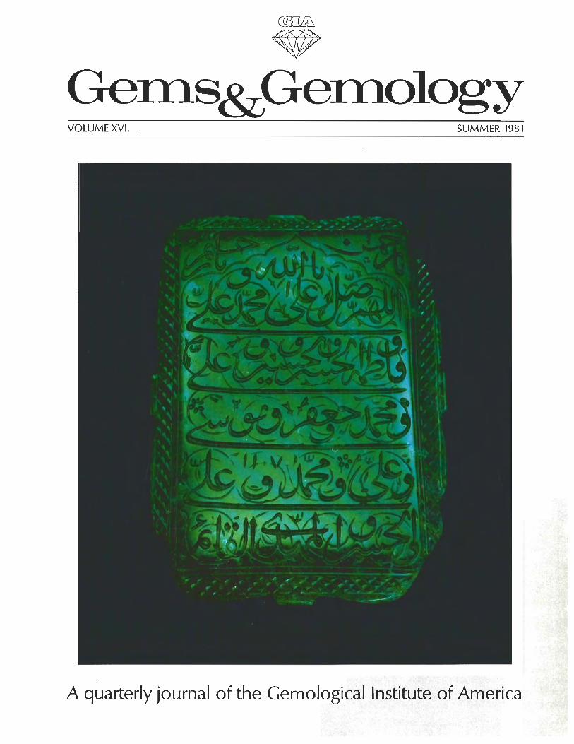
ABOUT THE COVER: This 217.8-ci. Mogul emerald is a fine example of the early Colombian stones that were treasured b y the Mogul nobility i n India. The Islamic prayer in this dramatic representation includes the date 1695 A.D. This and other famous Colombian emeralds, together with a detailed description of mining and marketing activities in Colombia, are discussed in Dr. Keller's article in this issue. The Mogul emerald is the property of Allan Caplan, New York, NY. Photograph '" 1981 Harold and Erica Van Pelt.
Composition for Gems & Gemology i s by Printed Page Graphics, Fullerton, CA. The color separations are by Effective Graphics, Conipton, CA. Printing is by Waverly Press, Easton, MD.
x37 98 7 Gemological Institute o f America. All rights reserved. ISSN 0016-62X
COLOR IN GEMS: THE NEW TECHNOLOGIES By George R. Rossman
Advances in technology have brought great advances i n our ability to impart color to natural gemstones as well as to create synthetics. Simultaneously, technologies are also being developed to distinguish natural from artificial colors as well as natural from synthetic materials. This article discusses some of the work being done to enhance color i n gemstones today and some of the techniques used b y the laboratories at the California Institute of Technology to determine whether a color originates naturally or in the laboratory. Dyed jade, synthetic turquoise, heat-treated beryl and zircon, irradiated spodumene, and topaz that has been irradiated and heat treated to produce a pleasing blue color are some of the specific examples included.
ABOUT THE AUTHOR
Dr. Rossman is associate professor of mineralogy at the California Institute of Technology, Pasadena, CA.
Acknowledgments: Photographs 4, 20, 23, and 24 were taken by G. P. Meeker of the California Institute of Technology, Pasadena, CA. The balance of the photos, except as noted in the legends, were provided by the author or by the Gem Media division of the Gemological Institute of America.
The author's research on the effects of radiation in minerals is funded in part by the National Science Foundation.
^I981 Gemological Institute of America
0 ne of the most important attributes of a gem ma- terial is its color. For centuries, man has used a va-
riety of technologies to enhance the color of natural gems and to produce materials that imitate natural stones. To- day, these technologies have grown in sophistication to the point that the gemologist faces formidable problems in identifying some of the products of these technologies. Concurrently, much scientific activity has been directed toward understanding the origin of color in minerals. So- phisticated instruments are being used in a number of laboratories to investigate color and other physical and chemical properties of minerals almost to the level of atomic resolution.
This article will consider a number of case studies, drawn largely from the experience of the laboratories at the California Institute of Technology, which illustrate methods used to study color in gem minerals. The ex- amples presented have been chosen to show how the scientific study of the basic properties of minerals can provide useful gemological information for determining whether the color in a particular gem originates naturally or in the laboratory. Included in the discussion are jade, turquoise, beryl, zircon, spodumene, and topaz, as well as a variety of other materials that have also been the sub- jects of color experimentation.
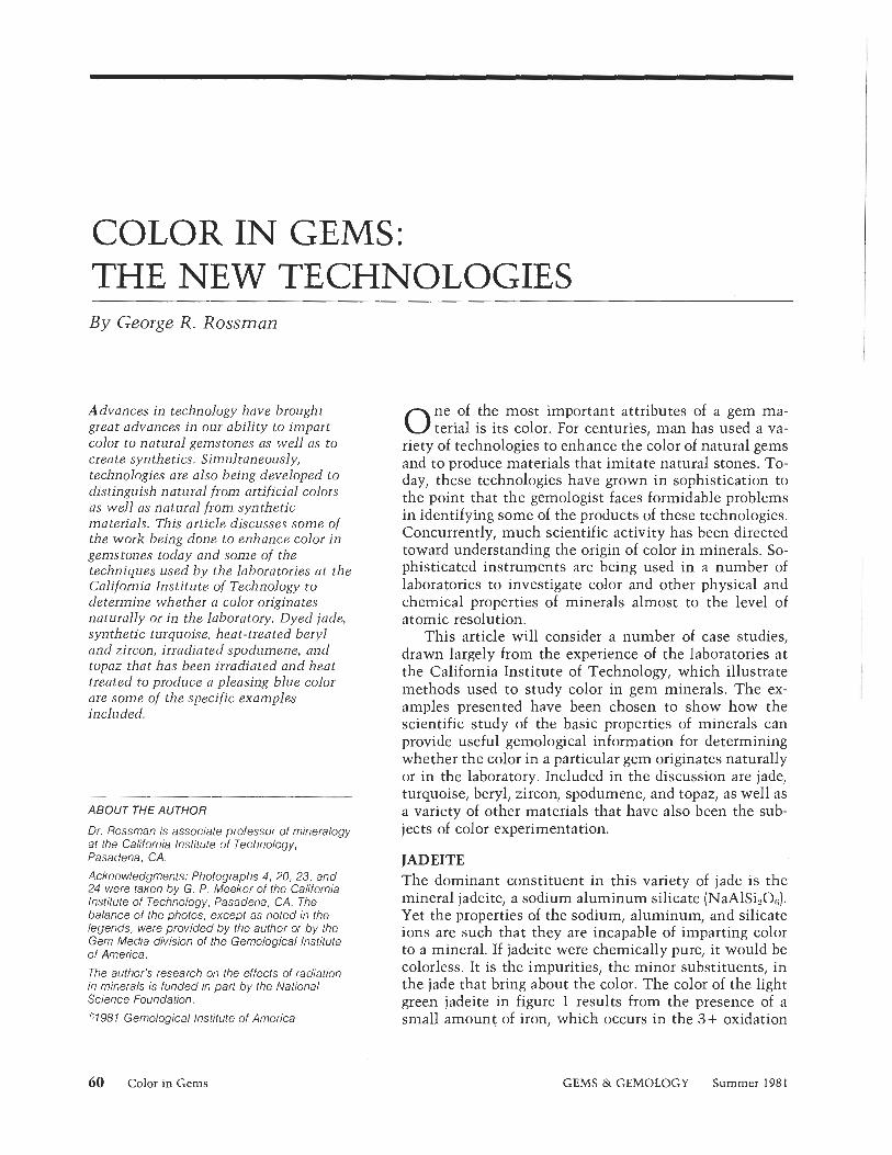
JADEITE The dominant constituent in this variety of jade is the mineral jadeite, a sodium aluminum silicate (NaAISizOg). Yet the properties of the sodium, aluminum, and silicate ions are such that they are incapable of imparting color to a mineral. If jadeite were chemically pure, it would be colorless. It is the impurities, the minor substituents, in the jade that bring about the color. The color of the light green jadeite in figure 1 results from the presence of a small amount of iron, which occurs in the 3+ oxidation
60 Color in Gems GEMS & GEMOLOGY Summer 1981
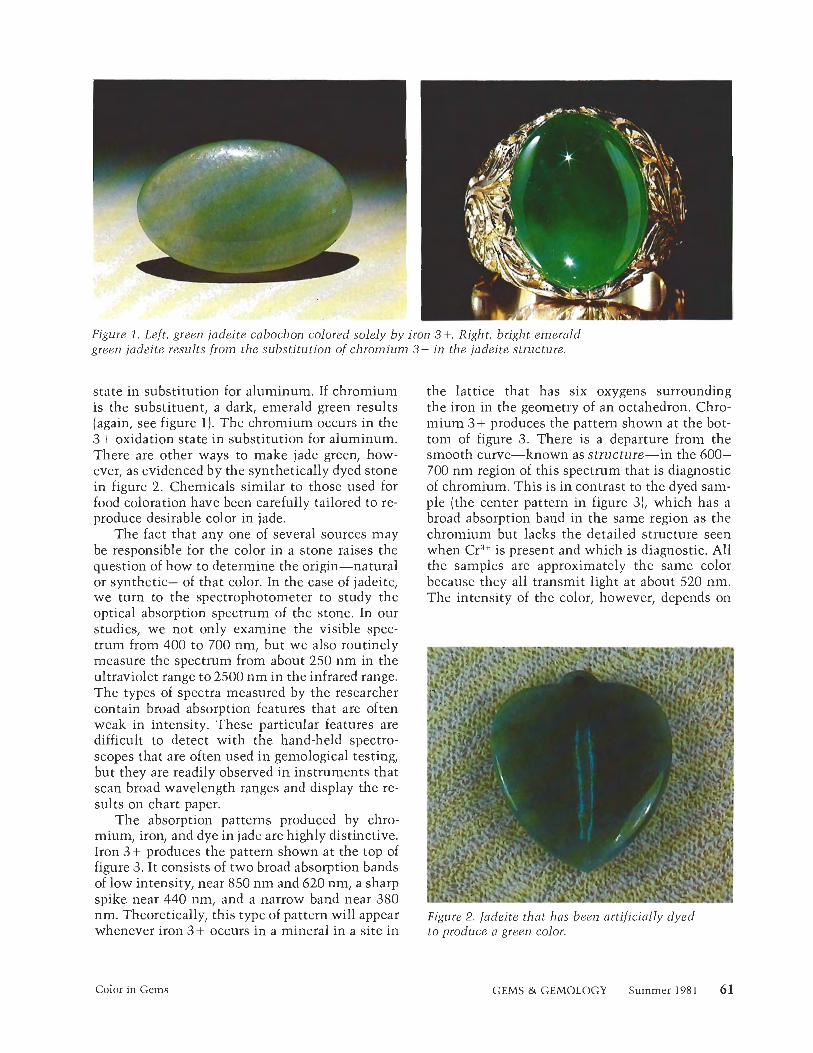
Figure 1 . Left, green jadeite cabochon colored solely by iron 3+. Right, bright emerald green jadeite results from the substitution of chromium 3+ in the jadeite structure.
state in substitution for aluminum. If chromium is the substituent, a dark, emerald green results (again, see figure 1). The chromium occurs in the 3 + oxidation state in substitution for aluminum. There are other ways to make jade green, how- ever, as evidenced by the synthetically dyed stone in figure 2. Chemicals similar to those used for food coloration have been carefully tailored to re- produce desirable color in jade.
The fact that any one of several sources may be responsible for the color in a stone raises the question of how to determine the origin-natural or synthetic-of that color. In the case of jadeite, we turn to the spectrophotometer to study the optical absorption spectrum of the stone. In our studies, we not only examine the visible spec- trum from 400 to 700 nm, but we also routinely measure the spectrum from about 250 nm in the ultraviolet range to 2500 nm in the infrared range. The types of spectra measured by the researcher contain broad absorption features that are often weak in intensity. These particular features are difficult to detect with the hand-held spectro- scopes that are often used in gemological testing, but they are readily observed in instruments that scan broad wavelength ranges and display the re- sults on chart paper.
The absorption patterns produced by chro- mium, iron, and dye in jade are highly distinctive. Iron 3 + produces the pattern shown at the top of figure 3. It consists of two broad absorption bands of low intensity, near 850 nm and 620 nm, a sharp spike near 440 nm, and a narrow band near 380
the lattice that has six oxygens surrounding the iron in the geometry of an octahedron. Chro- mium 3 + produces the pattern shown at the bot- tom of figure 3. There is a departure from the smooth curve-known as structure-in the 600- 700 nm region of this spectrum that is diagnostic of chromium. This is in contrast to the dyed sam- ple (the center pattern in figure 3), which has a broad absorption band in the same region as the chromium but lacks the detailed structure seen when CrW is present and which is diagnostic. All the samples are approximately the same color because they all transmit light at about 520 nm. The intensity of the color, however, depends on
nm. Theoretically, this type of pattern will appear Figure 2. Jadeite that has been artificially dyed whenever iron 3 + occurs in a mineral in a site in to produce a green color.
Color in Gems GEMS & GEMOLOGY Summer 1981 61
--
-̂I--
, , , , JADE I TE ~ e ~ ' '1
4 0 0 6 0 0 000 1000 WAVELENGTH (nm)
Figure 3. Comparison of the absorption spectra of jadeite colored by: top, iron; center, organic dye; bottom, chromium. The origin of color i n jadeite can be readily determined from these spectra. Note that the sharp line near 440 nm i n the dyed sample indicates that some iron is naturally present i n this sample.
the extent of absorption on either side of the transmission band at 520 nm.
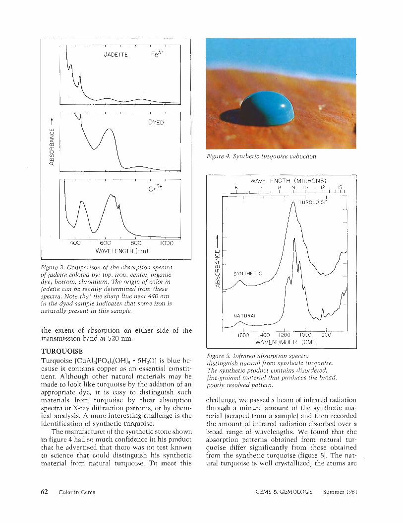
TURQUOISE Turquoise [ C U A ~ ~ ( P O J ~ ( O H ) ~ 5HgO] is blue be- cause it contains copper as an essential constit- uent. Although other natural materials may be made to look like turquoise by the addition of an appropriate dye, i t is easy to distinguish such materials from turquoise by their absorption spectra or X-ray diffraction patterns, or by chem- ical analysis. A more interesting challenge is the identification of synthetic turquoise.
The manufacturer of the synthetic stone shown in figure 4 had so much confidence in his product that he advertised that there was no test known to science that could distinguish his synthetic material from natural turquoise. To meet this
Figure 4 . Synthetic turquoise cabochon.
WAVELENGTH (MICRONS)
NATURAL j
WAVENUMBER (cM")
Figure 5. Infrared absorption spectra distinguish natural from synthetic turquoise. The synthetic product contains disordered, fine-grained material that produces the broad, poorly resolved pattern.
challenge, we passed a beam of infrared radiation through a minute amount of the synthetic ma- terial (scraped from a sample) and then recorded the amount of infrared radiation absorbed over a broad range of wavelengths. We found that the absorption patterns obtained from natural tur- quoise differ significantly from those obtained from the synthetic turquoise (figure 5). The nat- ural turquoise is well crystallized; the atoms are
62 Color in Gems GEMS & GEMOLOGY Summer 198 1
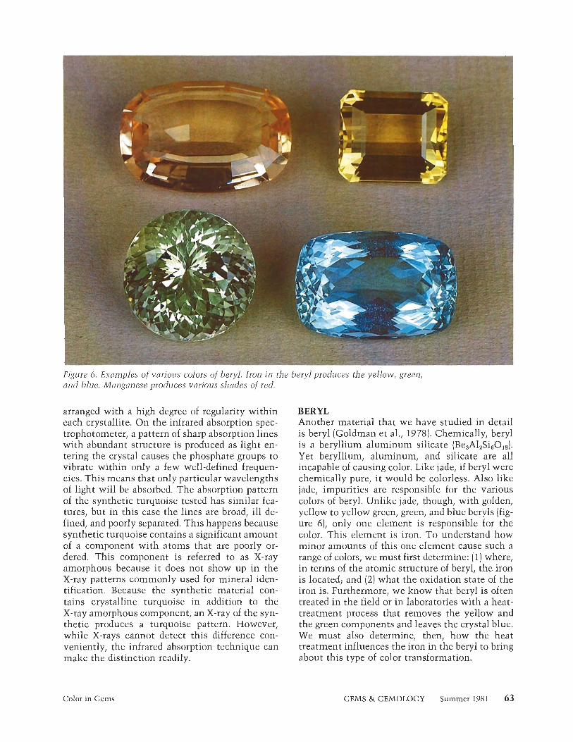
Figure 6. Examples of various colors of beryl. Iron in the beryl produces the yellow, green, and blue. Manganese produces various shades of red.
arranged with a high degree of regularity within each crystallite. On the infrared absorption spec- trophotometer, a pattern of sharp absorption lines with abundant structure is produced as light en- tering the crystal causes the phosphate groups to vibrate within only a few well-defined frequen- cies. This means that only particular wavelengths of light will be absorbed. The absorption pattern of the synthetic turquoise tested has similar fea- tures, but in this case the lines are broad, ill de- fined, and poorly separated. This happens because synthetic turquoise contains a significant amount of a component with atoms that are poorly or- dered. This component is referred to as X-ray amorphous because it does not show up in the X-ray patterns commonly used for mineral iden- tification. Because the synthetic material con- tains crystalline turquoise in addition to the X-ray amorphous component, an X-ray of the syn- thetic produces a turquoise pattern. However, while X-rays cannot detect this difference con- veniently, the infrared absorption technique can make the distinction readily.
Color in Gems
BERYL Another material that we have studied in detail is beryl (Goldman et al., 1978). Chemically, beryl is a beryllium aluminum silicate (Be&SieOi8). Yet beryllium, aluminum, and silicate are all incapable of causing color. Like jade, if beryl were chemically pure, it would be colorless. Also like jade, impurities are responsible for the various colors of beryl. Unlike jade, though, with golden, yellow to yellow green, green, and blue beryls (fig- ure 6), only one element is responsible for the color. This element is iron. To understand how minor amounts of this one element cause such a range of colors, we must first determine: (1) where, in terms of the atomic structure of beryl, the iron is located; and (2) what the oxidation state of the iron is. Furthermore, we know that beryl is often treated in the field or in laboratories with a heat- treatment process that removes the yellow and the green components and leaves the crystal blue. We must also determine, then, how the heat treatment influences the iron in the beryl to bring about this type of color transformation.
GEMS & GEMOLOGY Summer 1981
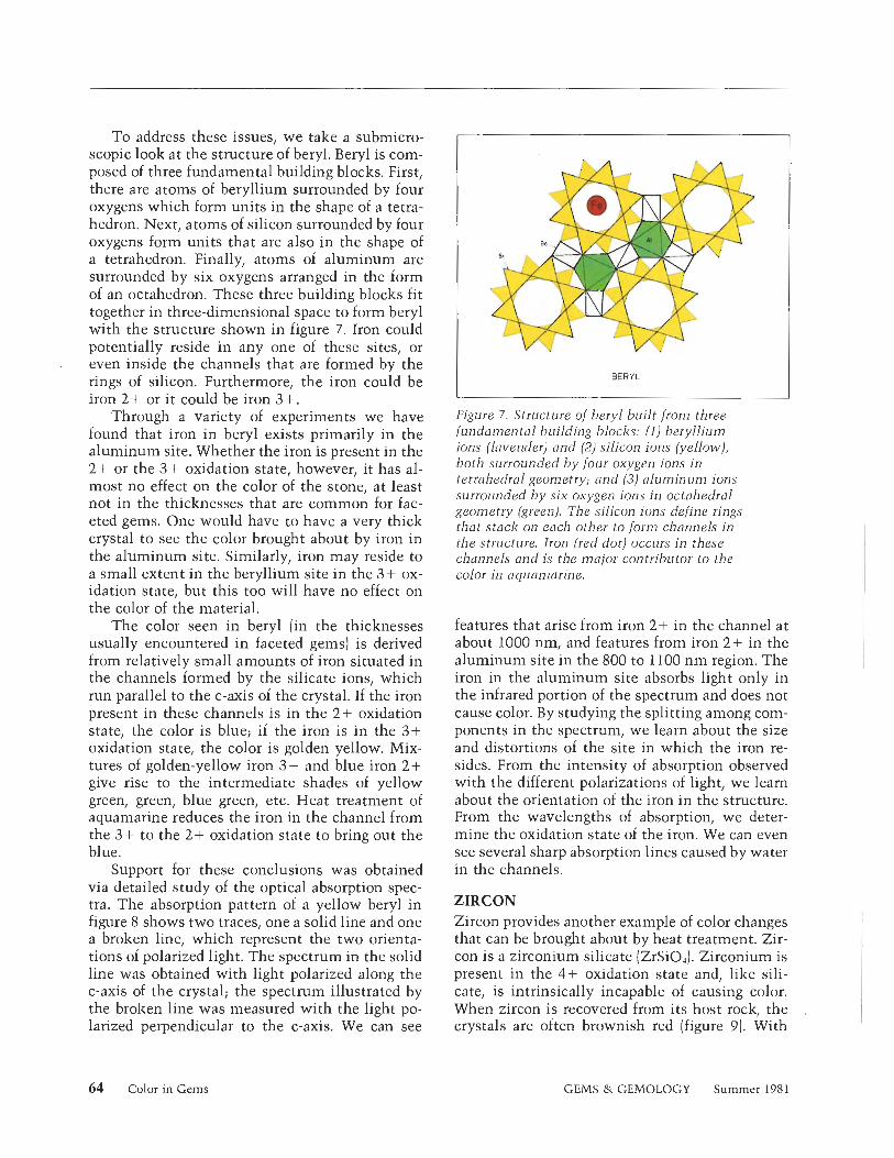
To address these issues, we take a submicro- scopic look at the structure of beryl. Beryl is com- posed of three fundamental building blocks. First, there are atoms of beryllium surrounded by four oxygens which form units in the shape of a tetra- hedron. Next, atoms of silicon surrounded by four oxygens form units that are also in the shape of a tetrahedron. Finally, atoms of aluminum are surrounded by six oxygens arranged in the form of an octahedron. These three building blocks fit together in three-dimensional space to form beryl with the structure shown in figure 7. Iron could potentially reside in any one of these sites, or even inside the channels that are formed by the rings of silicon. Furthermore, the iron could be iron 2+ or it could be iron 3 +.
Through a variety of experiments we have found that iron in beryl exists primarily in the aluminum site. Whether the iron is present in the 2+ or the 3+ oxidation state, however, it has al- most no effect on the color of the stone, at least not in the thicknesses that are common for fac- eted gems. One would have to have a very thick crystal to see the color brought about by iron in the aluminum site. Similarly, iron may reside to a small extent in the beryllium site in the 3+ ox- idation state, but this too will have no effect on the color of the material.
The color seen in beryl (in the thicknesses usually encountered in faceted gems) is derived from relatively small amounts of iron situated in the channels formed by the silicate ions, which run parallel to the c-axis of the crystal. If the iron present in these channels is in the 2+ oxidation state, the color is blue; if the iron is in the 3+ oxidation state, the color is golden yellow. Mix- tures of golden-yellow iron 3 + and blue iron 2+ give rise to the intermediate shades of yellow green, green, blue green, etc. Heat treatment of aquamarine reduces the iron in the channel from the 3+ to the 2+ oxidation state to bring out the blue.
Support for these conclusions was obtained via detailed study of the optical absorption spec- tra. The absorption pattern of a yellow beryl in figure 8 shows two traces, one a solid line and one a broken line, which represent the two orienta- tions of polarized light. The spectrum in the solid line was obtained with light polarized along the c-axis of the crystal; the spectrum illustrated by the broken line was measured with the light po- larized perpendicular to the c-axis. We can see
-
BERYL
I I Figure 7. Structure of beryl built from three fundamental building blocks: (1) beryllium ions (lavender) and (2) silicon ions (yellow), both surrounded by four oxygen ions in tetrahedral geometry; and (3) aluminum ions surrounded by six oxygen ions in octahedral geometry (green). The silicon ions define rings thai stack on each other to form channels in the structure. Iron (red dot) occurs in these channels and is the major contributor to the color in aquaniurine.
features that arise from iron 2+ in the channel at about 1000 nm, and features from iron 2+ in the aluminum site in the 800 to 1100 nm region. The iron in the aluminum site absorbs light only in the infrared portion of the spectrum and does not cause color. By studying the splitting among com- ponents in the spectrum, we learn about the size and distortions of the site in which the iron re- sides. From the intensity of absorption observed with the different polarizations of light, we learn about the orientation of the iron in the structure. From the wavelengths of absorption, we deter- mine the oxidation state of the iron. We can even see several sharp absorption lines caused by water in the channels.
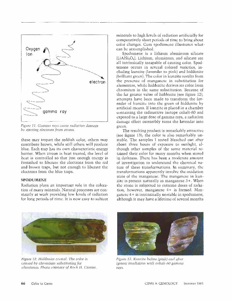
ZIRCON Zircon provides another example of color changes that can be brought about by heat treatment. Zir- con is a zirconium silicate (ZrSiOJ. Zirconium is present in the 4+ oxidation state and, like sili- cate, is intrinsically incapable of causing color. When zircon is recovered from its host rock, the crystals are often brownish red (figure 9). With
64 Color in Gems GEMS & GEMOLOGY Summer 1981
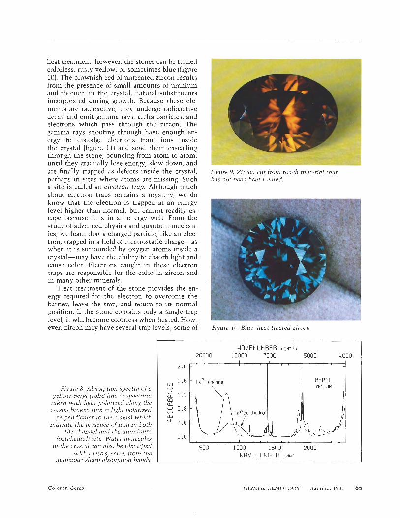
heat treatment, however, the stones can be turned colorless, rusty yellow, or sometimes blue (figure 10). The brownish red of untreated zircon results from the presence of small amounts of uranium and thorium in the crystal, natural substituents incorporated during growth. Because these ele- ments are radioactive, they undergo radioactive decay and emit gamma rays, alpha particles, and electrons which pass through the zircon. The gamma rays shooting through have enough en- ergy to dislodge electrons from ions inside the crystal (figure 11) and send them cascading through the stone, bouncing from atom to atom, until they gradually lose energy, slow down, and are finally trapped as defects inside the crystal, perhaps in sites where atoms are missing. Such a site is called an electron trap. Although much about electron traps remains a mystery, we do know that the electron is trapped at an energy level higher than normal, but cannot readily es- cape because it is in an energy well. From the study of advanced physics and quantum mechan- ics, we learn that a charged particle, like an elec- tron, trapped in a field of electrostatic charge-as when it is surrounded by oxygen atoms inside a crystal-may have the ability to absorb light and cause color. Electrons caught in these electron traps are responsible for the color in zircon and in many other minerals.
Heat treatment of the stone provides the en- ergy required for the electron to overcome the barrier, leave the trap, and return to its normal position. If the stone contains only a single trap level, it will become colorless when heated. How- ever, zircon may have several trap levels; some of
Figure 8. Absorption spectra of a yellow beryl (solid line = spectrum
taken wi th light polarized along the c-axis; broken line = light polarized
perpendicular to the c-axis) which indicate t-he presence of iron i n both
the channel and the a luminum (octahedral) site. Water molecules
i n the crystal can also be identified wi th these spectra, from the
numerous sharp absorption bands.
Figure 9. Zircon cut from rough material that has not been heat treated,
Figure 10. Blue, heat-treated zircon.
WRVENUMBER (CM-1 )
10000 7000 5000 4000
500 1000 1500 2000 WRVELENGTH ( N M )
Color in Gems GEMS & GEMOLOGY Summer 1981 65
Ion Oxygen @! 3 -1 gamma ray
electron
Figure 11. Gamma rays cause radiation damage by ejecting electrons from atoms.
these may impart the reddish color, others may contribute brown, while still others will produce blue. Each trap has its own characteristic energy barrier. When zircon is heat treated, the level of heat is controlled so that just enough energy is furnished to liberate the electrons from the red and brown traps, but not enough to liberate the electrons from the blue traps.
SPODUMENE Radiation plays an important role in the colora- tion of many minerals. Natural processes are con- stantly at work providing low levels of radiation for long periods of time. It is now easy to subject
minerals to high levels of radiation artificially for comparatively short periods of time to bring about color changes. Gem spodumene illustrates what - can be accomplished.
Spodumene is a lithium aluminum silicate (LiAISizOe). Lithium, aluminun~, and silicate are all intrinsically incapable of causing color. Spod- umene occurs in several colored varieties, in- cluding lzunzite (lavendar to pink) and hiddenite (brilliant green). The color in lzunzite results from the presence of manganese in substitution for aluminum, while hiddenite derives its color from chromium in the same substitution. Because of the far greater value of hiddenite (see figure 121, attempts have been made to transform the lav- endar of kunzite into the green of hiddenite by artificial means. If lzunzite is placed in a chamber containing the radioactive isotope cobalt-60 and exposed to a large dose of gamma rays, a radiation damage effect ostensibly turns the lavendar into green.
The resulting product is remarlzably attractive (see figure 13); the color is also remarlzably un- stable. The samples I tested bleached out after about three hours of exposure to sunlight, al- though other samples of the same material re- tained their color for many months when stored in darkness. There has been a moderate amount of investigation to understand the chemical na- ture of these transformations. In summary, the transformations apparently involve the oxidation state of the manganese. The manganese in lzun- zite is present naturally as manganese 3+. When the stone is subjected to extreme doses of radia- tion, however, manganese 4+ is formed. Man- ganese 4+ is intrinsically unstable in spodumene, although it may have a lifetime of several months
. ',$
Figure 12. Hiddenite crystal. The color is caused b y chromium substituting for aluminum. Photo courtesy of Rock H. Currier.
Figure 13. K~lnzite before (pink) and after (green) irradiation with cobalt-60 gamma rays.
66 Color in Gems GEMS & GEMOLOGY Summer 198 1
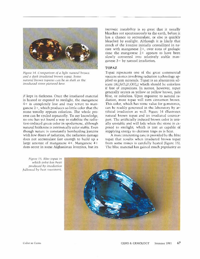
Figure 14. Comparison of a light natural brown and a dark irradiated brown topaz. Some natural brown topazes can be as dark as the irradiated stone pictured here.
if kept in darkness. Once the irradiated material is heated or exposed to sunlight, the manganese 4+ is completely lost and may revert to man- ganese 2+, which produces so little color that the stone usually appears colorless. The whole pro- cess can be cycled repeatedly. To my knowledge, no one has yet found a way to stabilize the radia- tion-induced green color in spodumene, although natural hiddenite is intrinsically color stable. Even though nature is constantly bombarding kunzite with low doses of radiation, the radiation damage does not accumulate fast enough to build up a large amount of manganese 4+. Manganese 4+ does occur in some Afghanistan kunzites, but its
intrinsic instability is so great that it usually bleaches out spontaneously in the earth, before i t has a chance to accumulate, or else is quickly bleached by sunlight. Although it is likely that much of the kunzite initially crystallized in na- ture with manganese 2+, over eons of geologic time the manganese 2+ appears to have been slowly converted into relatively stable man- ganese 3+ by natural irradiation.
TOPAZ Topaz represents one of the great commercial success stories involving radiation technology ap- plied to gem minerals. Topaz is an aluminum sil- icate [AlzSi04[F,0H]a] which should be colorless if free of impurities. In nature, however, topaz generally occurs as yellow or yellow brown, pale blue, or colorless. Upon exposure to natural ra- diation, most topaz will turn cinnamon brown. This color, which has some value for gemstones, can be readily generated in the laboratory by ar- tificial irradiation as well. Figure 14 illustrates natural brown topaz and its irradiated counter- part. The artificially induced brown color is usu- ally unstable and will fade when the stone is ex- posed to sunlight, which is just as capable of supplying energy to electron traps as is heat.
A more interesting case is provided by the blue topaz that results when irradiated brown topaz from some mines is carefully heated (figure 15). The blue material has gained much popularity as
Figure 15. Blue topaz i which color has bee
produced by irradiation followed by heat treatment.
Color in Gems GEMS & GEMOLOGY Summer 198 1 67
a gemstone because of its pleasing color and the stability of the color. Even though blue topaz has received a great amount of study, we do not yet know the ultimate cause of its color. The fact that most properties of irradiated blue topaz are essen- tially the same as those of natural blue topaz has suggested the possibility that the natural color is itself a product of natural irradiation.
Because the properties-chemistry, color, in- dices of refraction, etc.-of natural and irradiated blue topaz are almost identical, the gemologist is faced with the formidable task of distinguishing the natural from the irradiated material. After considerable investigation, we have developed one method that has been effective in the laboratory for making this distinction. It involves thermo- luminescence, that is, light caused by heat.
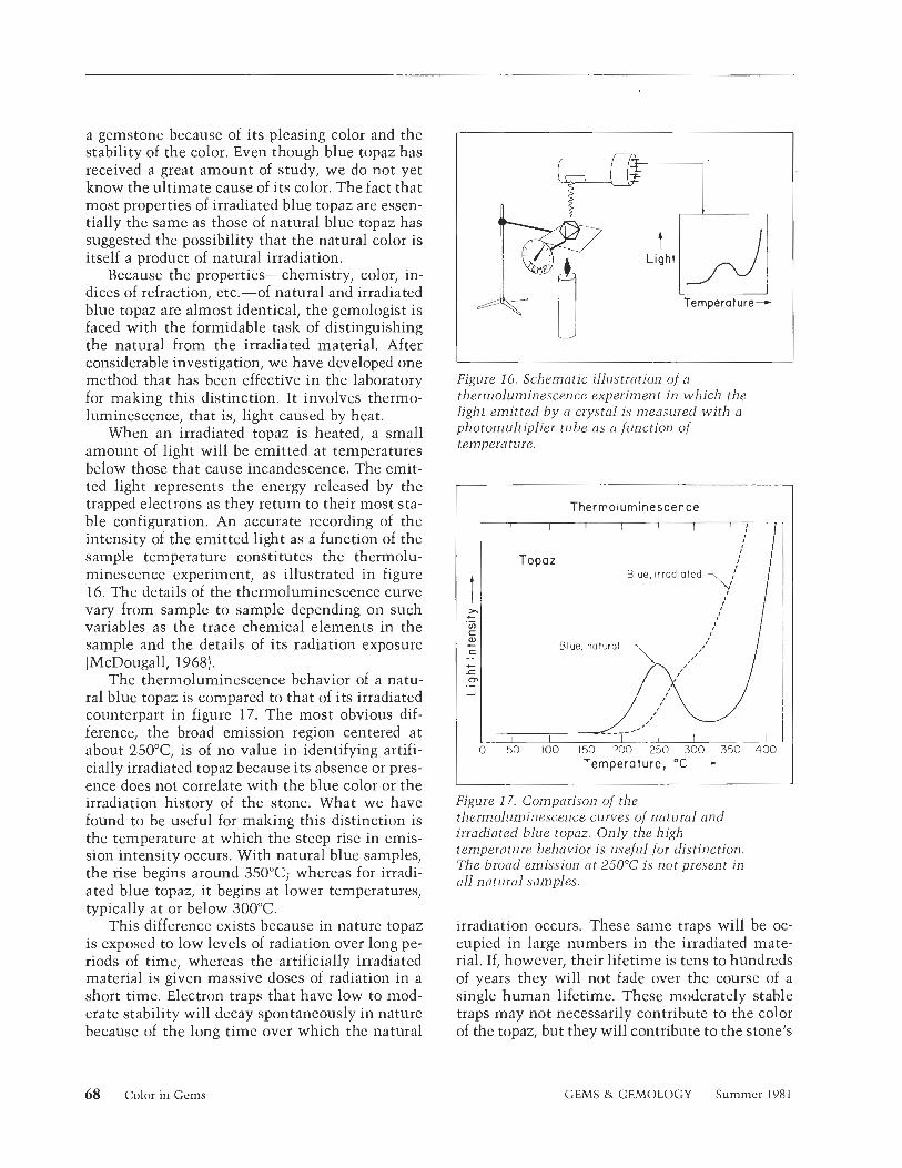
When an irradiated topaz is heated, a small amount of light will be emitted at temperatures below those that cause incandescence. The emit- ted light represents the energy released by the trapped electrons as they return to their most sta- ble configuration. An accurate recording of the intensity of the emitted light as a function of the sample temperature constitutes the thermolu- minescence experiment, as illustrated in figure 16. The details of the thermoluminescence curve vary from sample to sample depending 011 such variables as the trace chemical elements in the sample and the details of its radiation exposure (McDougall, 1968).
The thermoluminescence behavior of a natu- ral blue topaz is compared to that of its irradiated counterpart in figure 17. The most obvious dif- ference, the broad emission region centered at about 250°C is of no value in identifying artifi- cially irradiated topaz because its absence or pres- ence does not correlate with the blue color or the irradiation history of the stone. What we have found to be useful for making this distinction is the temperature at which the steep rise in emis- sion intensity occurs. With natural blue samples, the rise begins around 350°C whereas for irradi- ated blue topaz, it begins at lower temperatures, typically at or below 300¡C
This difference exists because in nature topaz is exposed to low levels of radiation over long pe- riods of time, whereas the artificially irradiated material is given massive doses of radiation in a short time. Electron traps that have low to mod- erate stability will decay spontaneously in nature because of the long time over which the natural
I
Fjgure 16. Schematic illustration of a thermoluminescencc experiment in which the light emitted by a crystal is measured with a photomultiplier tube as a function of temperature.
Thermoluminescence
Blue, natural 7
I Temperature, OC - - Figure 17. Comparison of the t l~ern~olumi~~escence curves of natural and irradiated blue topaz. Only the high- temperature behavior is useful for distinction. The broad emission at 250° is not present in all natural samples.
irradiation occurs. These same traps will be oc- cupied in large numbers in the irradiated mate- rial. If, however, their lifetime is tens to hundreds of years they will not fade over the course of a single human lifetime. These moderately stable traps may not necessarily contribute to the color of the topaz, but they will contribute to the stone's
68 Color in Gems GEMS & GEMOLOGY Summer 1981
thermoluminescence behavior at high temper- atures.
At this time, the thermoluminescence method is not suitable for routine testing in the jeweler's or gemologist's lab because the extreme heat re- quired for the measurement bleaches the blue color. It is possible to scrape a small quantity of material from the edge of the stone to make the measurement; about 500 micrograms is ideal. Although a small amount of repolishing where the stone was sampled would remove the scrape marks, even this method is not suitable for rou- tine testing because of the time involved in sam- ple preparation.
ADDITIONAL EXAMPLES OF RADIATION-INDUCED COLOR IN MINERALS Radiation plays a role in the coloration of many other minerals as well. For example, common salt, the mineral halite (NaCl), is one of the sub- stances most often used in studies of radiation damage. An amber color develops when salt is exposed to gamma rays, and with a high enough dose the salt can be turned black. It is important to remember that salt, like all minerals, does not become radioactive when irradiated by X-rays or by the more energetic gamma rays.
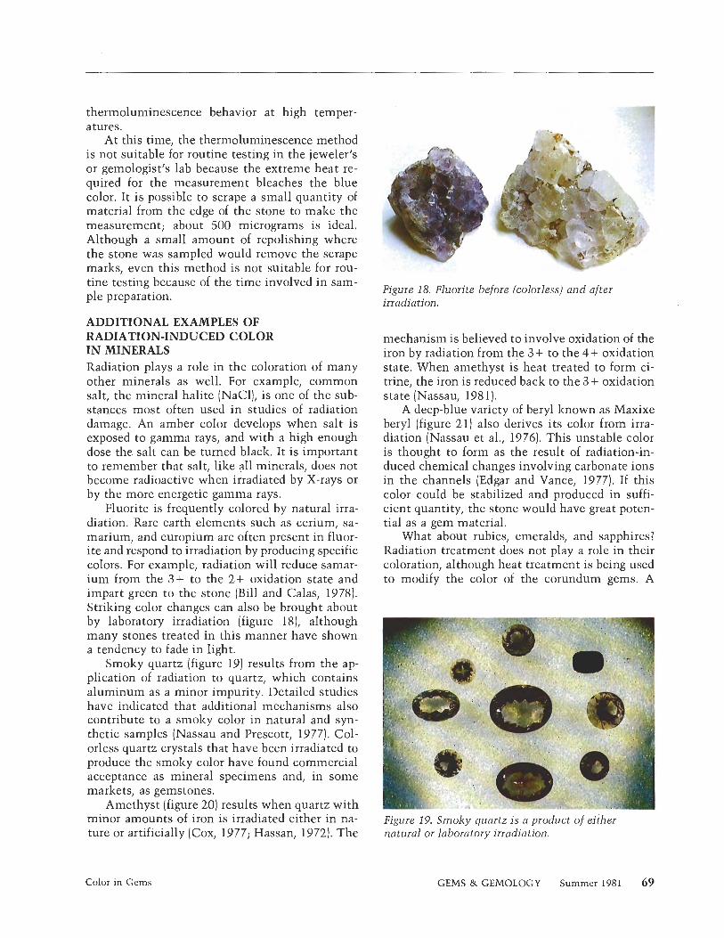
Fluorite is frequently colored by natural irra- diation. Rare earth elements such as cerium, sa- marium, and europium are often present in fluor- ite and respond to irradiation by producing specific colors. For example, radiation will reduce samar- ium from the 3+ to the 2+ oxidation state and impart green to the stone (Bill and Calas, 1978). Striking color changes can also be brought about by laboratory irradiation (figure 181, although many stones treated in this manner have shown a tendency to fade in light.
Smoky quartz (figure 19) results from the ap- plication of radiation to quartz, which contains aluminum as a minor impurity. Detailed studies have indicated that additional mechanisms also contribute to a smoky color in natural and syn- thetic samples (Nassau and Prescott, 1977). Col- orless quartz crystals that have been irradiated to produce the smoky color have found commercial acceptance as mineral specimens and, in some markets, as gemstones.
Amethyst (figure 20) results when quartz with minor amounts of iron is irradiated either in na- ture or artificially (Cox, 1977; Hassan, 1972). The
"'"I
Figure 18. Fluorite before (colorless) and after irradiation.
mechanism is believed to involve oxidation of the iron by radiation from the 3+ to the 4+ oxidation state. When amethyst is heat treated to form ci- trine, the iron is reduced back to the 3 + oxidation state (Nassau, 1981).
A deep-blue variety of beryl known as Maxixe beryl (figure 21) also derives its color from irra- diation (Nassau et al., 1976). This unstable color is thought to form as the result of radiation-in- duced chemical changes involving carbonate ions in the channels (Edgar and Vance, 1977). If this color could be stabilized and produced in suffi- cient quantity, the stone would have great poten- tial as a gem material.
What about rubies, emeralds, and sapphires? Radiation treatment does not play a role in their coloration, although heat treatment is being used to modify the color of the corundum gems. A
Figure 19. Smoky quartz is a product of either natural or laboratory irradiation.
Color in Gems GEMS & GEMOLOGY Summer 1981 69
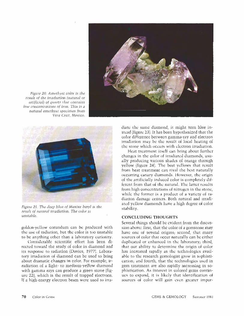
Figure 20. Amethyst color is the result of the irradiation (natural or
artificial) of quartz that contains low concentrations of iron. This is a
natural amethyst specimen from . - Vera Cruz, Mexico.
J . ! .
Figure 21. The deep blue of Maxixe beryl is the result of natural irradiation. The color is unstable.
golden-yellow corundum can be produced with the use of radiation, but the color is too unstable to be anything other than a laboratory curiosity.
Considerable scientific effort has been di- rected toward the study of color in diamond and its response to radiation (Davies, 1977). Labora- tory irradiation of diamond can be used to bring about dramatic changes in color. For example, ir- radiation of a light- to medium-yellow diamond with gamma rays can produce a green stone (fig- ure 22), which is the result of trapped electrons. If a high-energy electron beam were used to irra-
diate the same diamond, i t might turn blue in- stead (figure 23). It has been hypothesized that the color difference between gamma-ray and electron irradiation may be the result of local heating of the stone which occurs with electron irradiation.
Heat treatment itself can bring about further changes in the color of irradiated diamonds, usu- ally producing various shades of orange through yellow (figure 24). The best yellows that result from heat treatment can rival the best naturally occurring canary diamonds. However, the origin of the artificially induced color is completely dif- ferent from that of the natural. The latter results from high concentrations of nitrogen in the stone, while the former is a product of a variety of ra- diation damage centers. Both natural and irradi- ated yellow diamonds have a high degree of color stability.
CONCLUDING THOUGHTS Several things should be evident from the discus- sion above: first, that the color of a gemstone may have one of several origins; second, that many sources of color that occur naturally can be either duplicated or enhanced in the laboratory; third, that our ability to determine the origin of color has increased rapidly as the technologies avail- able to the research gemologist grow in sophisti- cation; and fourth, that the technologies used in gem treatment are also rapidly increasing in so- phistication. As interest in colored gems contin- ues to expand, it is likely that identification of sources of color will gain even greater impor-
70 Color in Gems GEMS & GEMOLOGY Summer 1981
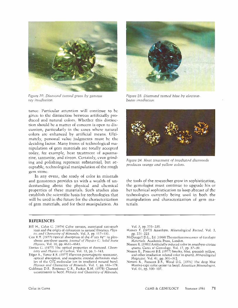
Figure 22. Diamond turned green b y gamma- ray irradiation.
Figure 23. Diamond turned blue b y electron- beam irradiation.
tance. Particular attention will continue to be given to the distinction between artificially pro- duced and natural colors. Whether this distinc- tion should be a matter of concern is open to dis- cussion, particularly in the cases where natural colors are enhanced by artificial means. Ulti- mately, personal value judgments must be the deciding factor. Many forms of technological ma- nipulation of gem materials are totally accepted today, for example, heat treatment of aquama- rine, tanzanite, and zircon. Certainly, even grind- ing and polishing represent substantial, but ac- ceptable, technological manipulation of the rough gem stone.
In any event, the study of color in minerals and gemstones provides us with a wealth of un- derstanding about the physical and chemical properties of these materials. Such studies also establish the scientific basis for technologies that will be used in the future for the characterization of gem materials, and for their manipulation. As
REFERENCES Bill H., Calas G. (1978) Color centers, associated rare-earth
ions and the origin of coloration in natural fluorites. Phys- ics and Chemistry of Minerals, Vol. 3, pp. 1 1 7- 13 1 .
Cox R.T. (1977) Optical absorption of the d' ion Fe"+ in pleo- chroic amethyst quartz. /ournal of Physics C: Solid State Physics, Vol. 10, pp. 463 1-4643.
Davies G. (1977) The optical properties of diamond. Chem- istry and Physics of Carbon, Vol. 13, pp. 1- 143.
Edgar A., Vance E.R. (1977) Electron paramagnetic resonance, optical absorption, and magnetic circular dichroism stud- ies of the COY molecular ion in irradiated natural beryl. Physics and Chemistry of Minerals, Vol. 1, pp. 165-178.
Goldman D.S., Rossman G.R., Parkin K.M. (1978) Channel constituents in beryl. Physics and Chemistry of Minerals,
Figure 24. Heat treatment o f irradiated diamonds produces orange and yellow colors.
the tools of the researcher grow in sophistication, the gemologist must continue to upgrade his or her technical sophistication to keep abreast of the technologies currently being used in both the manipulation and characterization of gem ma- terials.
Vol. 3, pp. 225-235. Hassan F. (19721 Amethyst. Mineralogical Record, Vol. 3,
pp. 221-225. McDougall D.L., Ed. (19681 Thermoluminescence of Geologic
Materials. Academic Press, London. Nassau K. (1981) Artificially induced color in amethyst-citrine
quartz. Gems 6) Gemology, Vol. 17, pp. 37-39. Nassau K., Prescott B.E. (1977) Smoky, blue, greenish yellow,
and other irradiation-related color in quartz. Mineralogical Magazine, Vol. 4 1, pp. 301-312.
Nassau K., Prescott B.E., Wood D.L. (1976) The deep blue Maxixe-type color center in beryl. American Mineralogist, VOI. 61, pp. 100-107.
Color in Gems GEMS & GEMOLOGY Summer 1981 71
SCANNING ELECTRON MICROSCOPY IN GEMOLOGY By Carol M. Stockton and D. Vincent Manson
T h i s article examines both the principles behind the functioning of the scanning electron microscope-energy dispersive spectrometer system and its application to the chemical and structural analyses of gems. The examples presented describe the distinction of natural from treated opals, the relationships observed between a diamond and its kimberlite matrix, the identification of inclusions in ruby and taaffeite, the elemental mapping and chemical analysis of maw-sit-sit, and the study of chemical interrelationships between various types of garnets.
ABOUT THE AUTHORS
Carol M. Stockton is research gemologist and D. Vincent Manson is director of research at the Gemological Institute of America, Santa Monica, CA.
"1981 Gemological Institute of America
A n important function of the Gemological Institute of America's Department of Research is the appli-
cation of modem, advanced methods of analysis to gem- ological questions that cannot be readily answered using the usual gemological tests. One of the principal tools in our search for more knowledge about gem materials is the scanning electron microscope-energy dispersive spec- trometer system (SEM-EDS; fig. 1). Additional electronic components and a computer provide for automation within the SEM-EDS system and rapid execution of com- plex calculations with results displayed for interpretation by the research gemologist.
The following discussion seeks to explain the basic principles on which the SEM-EDS system operates, ex- amines the advantages it holds for gemology, and reviews a few of the gemological problems to which it is currently being applied.
THE SEM-EDS: HOW IT WORKS To understand the principles behind the SEM-EDS, a basic knowledge of atomic structure is helpful. Atoms are 'the smallest divisions of matter that retain the charac- teristics of the elements, but they are actually composed of yet smaller particles. A contemporary model of the atom portrays a central nucleus consisting of protons and neutrons (except hydrogen, which has only one proton in its nucleus), around which electrons are distributed in orbitals (fig. 2). An atom is usually referred to by its atomic number. This is simply the number of protons in the atom's nucleus, which, unlike the number of neu- trons, is constant for each element.
The scanning electron microscope works on the prin- ciple that a beam of electrons striking a sample results in the return of electrons from the sample. The varying intensity and distribution of these returning electrons
72 Scanning Electron Microscopy GEMS & GEMOLOGY Summer 198 1
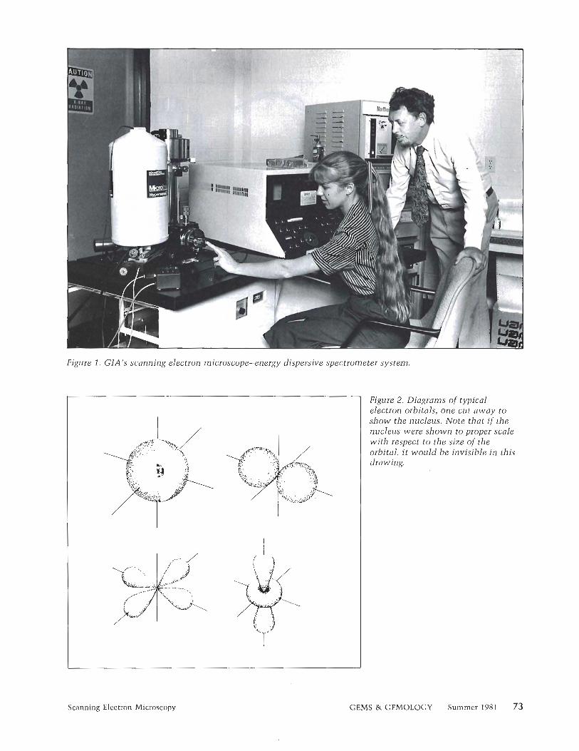
Figure 1. GIA's scanning electron microscope-energy dispersive spectrometer system.
Figure 2. Diagrams of typical electron orbitals, one cut away to show the nucleus. Note that if the nucleus were shown to proper scale with respect to the size of the orbital, i t would be invisible in this drawing.
Scanning Electron Microscopy GEMS & GEMOLOGY Summer 198 1 73
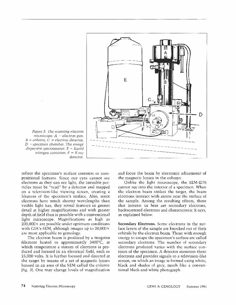
Figure 3. The scanning electron microscope: A = electron gun,
B = column, C = electron detector, D = specimen chamber. The energy dispersive spectrometer: E = liquid
nitrogen container, F = X-ray detector.
reflect the specimen's surface contours or com- positional features. Since our eyes cannot see electrons as they can see light, the invisible par- ticles must be "read" by a detector and mapped on a television-like viewing screen, creating a likeness of the specimen's surface. Also, since electrons have much shorter wavelengths than visible light has, they reveal features in greater detail at higher magnifications and with greater depth of field than is possible with a conventional light microscope. Magnifications as high as 200,000 x are possible under optimum conditions with GIA's SEMI although images up to 20,000x are most applicable to gemology.
The electron beam is produced by a tungsten filament heated to approximately 3400°C at which temperature a stream of electrons is pro- duced and focused in an electrical field, such as 25,000 volts. It is further focused and directed at the target by means of a set of magnetic lenses housed in an area of the SEM called the column (fig. 3). One may change levels of magnification
and focus the beam by electronic adjustment of the magnetic lenses in the column.
Unlike the light microscope, the SEM-EDS cannot see into the interior of a specimen. When the electron beam strikes the target, the beam electrons interact with atoms near the surface of the sample. Among the resulting effects, those that interest us here are secondary electrons, backscattered electrons and characteristic X-rays, as explained below.
Secondary Electrons. Some electrons in the sur- face layers of the sample are knocked out of their orbitals by the electron beam. Those with enough energy to escape the specimen's surface are called secondary electrons. The number of secondary electrons produced varies with the surface con- tours of the specimen. A detector monitors these electrons and provides signals to a television-like screen, on which an image is formed using white, black and shades of grey, much like a conven- tional black-and-white photograph.
74 Scanning Electron Microscopy GEMS & GEMOLOGY Summer 1981
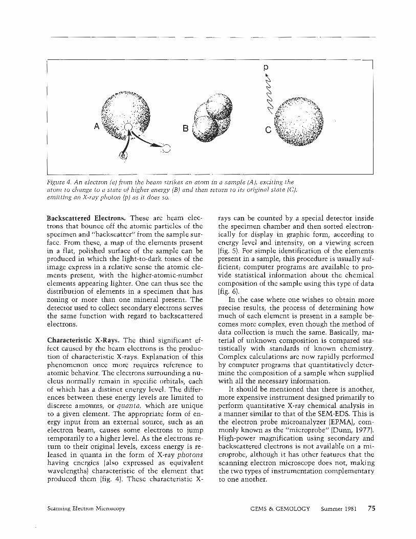
Figure 4. An electron (e) from the beam strikes an atom in a sample (A), exciting the atom to change to a state of higher energy (B) and then return to its original state (C), emitting an X-ray photon (p) as it does so.
Backscattered Electrons. These are beam elec- trons that bounce off the atomic particles of the specimen and "backscatter" from the sample sur- face. From these, a map of the elements present in a flat, polished surface of the sample can be produced in which the light-to-dark tones of the image express in a relative sense the atomic ele- ments present, with the higher-atomic-number elements appearing lighter. One can thus see the distribution of elements in a specimen that has zoning or more than one mineral present. The detector used to collect secondary electrons serves the same function with regard to backscattered electrons.
Characteristic X-Rays. The third significant ef- fect caused by the beam electrons is the produc- tion of characteristic X-rays. Explanation of this phenomenon once more requires reference to atomic behavior. The electrons surrounding a nu- cleus normally remain in specific orbitals, each of which has a distinct energy level. The differ- ences between these energy levels are limited to discrete amounts, or quanta, which are unique to a given element. The appropriate form of en- ergy input from an external source, such as an electron beam, causes some electrons to jump temporarily to a higher level. As the electrons re- turn to their original levels, excess energy is re- leased in quanta i n the form of X-ray photons having energies (also expressed as equivalent wavelengths) characteristic of the element that produced them (fig. 4). These characteristic X-
rays can be counted by a special detector inside the specimen chamber and then sorted electron- ically for display in graphic form, according to energy level and intensity, on a viewing screen (fig. 5). For simple identification of the elements present in a sample, this procedure is usually suf- ficient; computer programs are available to pro- vide statistical information about the chemical composition of the sample using this type of data (fig. 6).
In the case where one wishes to obtain more precise results, the process of determining how much of each element is present in a sample be- comes more complex, even though the method of data collection is much the same. Basically, ma- terial of unknown composition is compared sta- tistically with standards of known chemistry. Complex calculations are now rapidly performed by computer programs that quantitatively deter- mine the composition of a sample when supplied with all the necessary information.
It should be mentioned that there is another, more expensive instrument designed primarily to perform quantitative X-ray chemical analysis in a manner similar to that of the SEM-EDS. This is the electron probe microanalyzer (EPMA), com- monly known as the "microprobe" (Dunn, 1977). High-power magnification using secondary and backscattered electrons is not available on a mi- croprobe, although it has other features that the scanning electron microscope does not, making the two types of instrumentation complementary to one another.
Scanning Electron Microscopy GEMS & GEMOLOGY Summer 1981 75
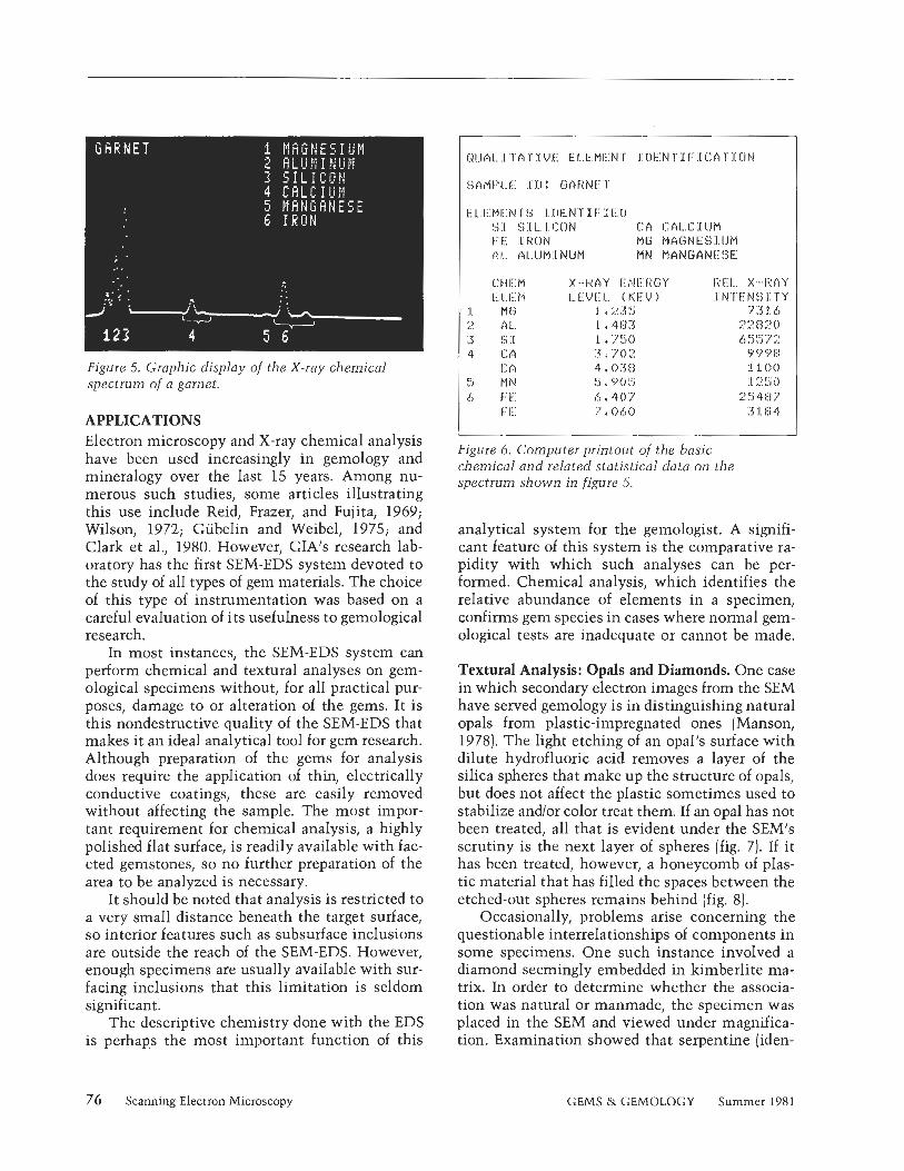
Figure 5. Graphic display of the X-ray chemical spectrum 0f.a garnet.
APPLICATIONS Electron microscopy and X-ray chemical analysis have been used increasingly in gemology and mineralogy over the last 15 years. Among nu- merous such studies, some articles illustrating this use include Reid, Frazer, and Fujita, 1969; Wilson, 1972; Gubelin and Weibel, 1975; and Clark et al., 1980. However, GIA's research lab- oratory has the first SEM-EDS system devoted to the study of all types of gem materials. The choice of this type of instrumentation was based on a careful evaluation of its usefulness to gemological research.
In most instances, the SEM-EDS system can perform chemical and textural analyses on gem- ological specimens without, for all practical pur- poses, damage to or alteration of the gems. It is this nondestructive quality of the SEM-EDS that makes it an ideal analytical tool for gem research. Although preparation of the gems for analysis does require the application of thin, electrically conductive coatings, these are easily removed without affecting the sample. The most impor- tant requirement for chemical analysis, a highly polished flat surface, is readily available with fac- eted gemstones, so no further preparation of the area to be analyzed is necessary.
It should be noted that analysis is restricted to a very small distance beneath the target surface, so interior features such as subsurface inclusions are outside the reach of the SEM-EDS. However, enough specimens are usually available with sur- facing inclusions that this limitation is seldom significant.
The descriptive chemistry done with the EDS is per hap,^ the most important function of this
Figure 6. Computer printout of the basic chemical and related statistical data on the spectrum shown in figure 5 .
analytical system for the gemologist. A signifi- cant feature of this system is the comparative ra- pidity with which such analyses can be per- formed. Chemical analysis, which identifies the relative abundance of elements in a specimen, confirms gem species in cases where normal gem- ological tests are inadequate or cannot be made.
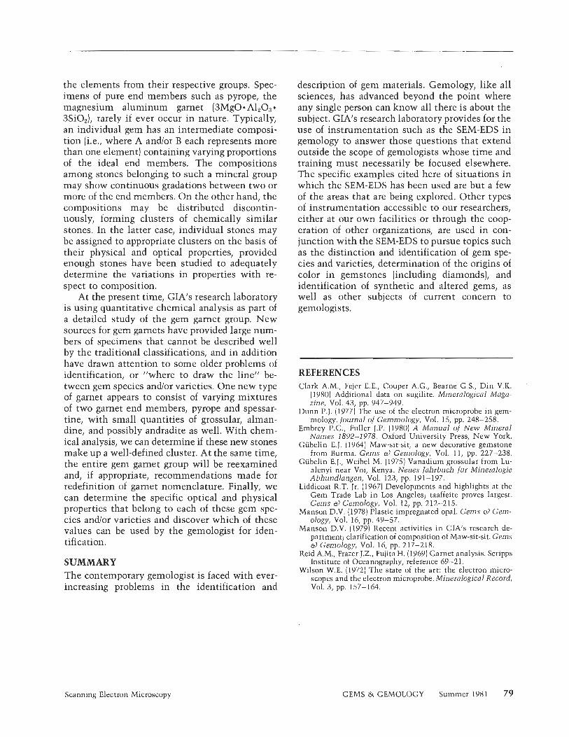
Textural Analysis: Opals and Diamonds. One case in which secondary electron images from the SEM have served gemology is in distinguishing natural opals from plastic-impregnated ones (Manson, 1978). The light etching of an opal's surface with dilute hydrofluoric acid removes a layer of the silica spheres that make up the structure of opals, but does not affect the plastic sometimes used to stabilize andlor color treat them. If an opal has not been treated, all that is evident under the SEM's scrutiny is the next layer of spheres (fig. 7). If it has been treated, however, a honeycomb of plas- tic material that has filled the spaces between the etched-out spheres remains behind (fig. 8).
Occasionally, problems arise concerning the questionable interrelationships of components in some specimens. One such instance involved a diamond seemingly embedded in kimberlite ma- trix. In order to determine whether the associa- tion was natural or manmade, the specimen was placed in the SEM and viewed under magnifica- tion. Examination showed that serpentine (iden-
76 Scanning Electron Microscopy GEMS & GEMOLOGY Summer 198 1
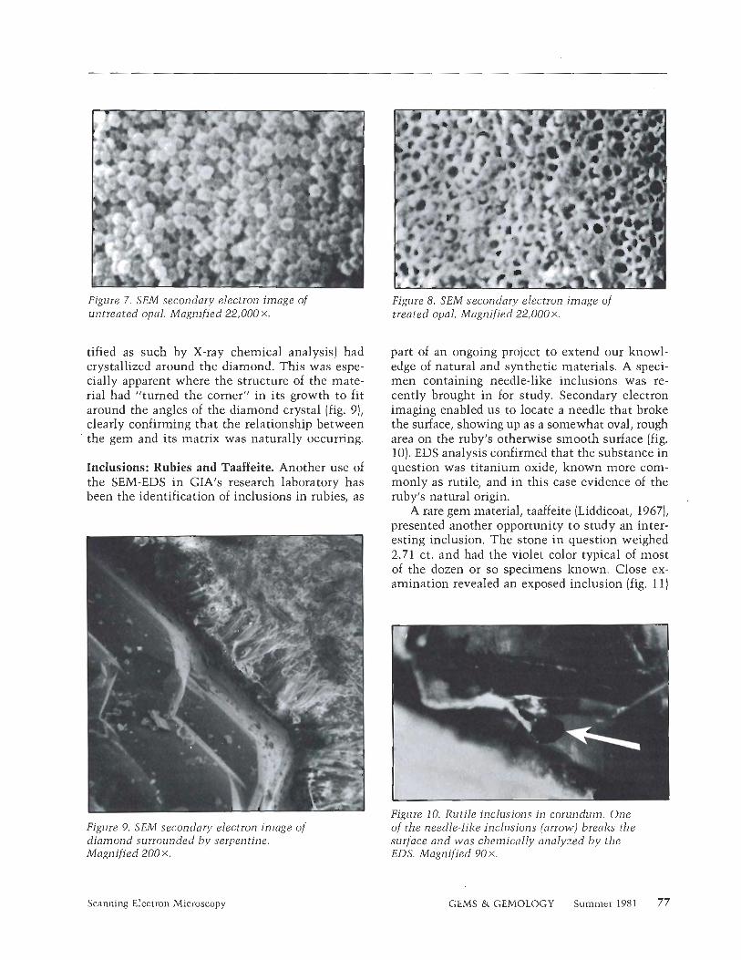
Figure 7. SEM secondary electron image o f untreated opal. Magnified 22,000 X.
tified as such by X-ray chemical analysis] had crystallized around the diamond. This was espe- cially apparent where the structure of the mate- rial had "turned the corner" in its growth to fit around the angles of the diamond crystal (fig. 9), clearly confirming that the relationship between
' the gem and its matrix was naturally occurring.
Inclusions: Rubies and Taaffeite. Another use of the SEM-EDS in GIA1s research laboratory has been the identification of inclusions in rubies, as
Figure 9. SEM secondary electron image o f diamond surrounded by serpentine. Magnified 200 x.,
Figure 8. SEM secondary electron image o f treated opal. Magnified 22,000 X.
part of an ongoing project to extend our knowl- edge of natural and synthetic materials. A speci- men containing needle-like inclusions was re- cently brought in for study. Secondary electron imaging enabled us to locate a needle that broke the surface, showing up as a somewhat oval, rough area on the ruby's otherwise smooth surface (fig. 10). EDS analysis confirmed that the substance in question was titanium oxide, known more com- monly as rutile, and in this case evidence of the ruby's natural origin.
A rare gem material, taaffeite (Liddicoat, 1967), presented another opportunity to study an inter- esting inclusion. The stone in question weighed 2.71 ct. and had the violet color typical of most of the dozen or so specimens known. Close ex- amination revealed an exposed inclusion (fig. 11)
Figure 10. Rutjle inclusions i n corundum. One o f the needle-like inclusions (arrow) breaks the surface and was chemically analyzed b y the EDS. Magnified 90 X.
Scanning Electron Microscopy GEMS & GEMOLOGY Summer 1981 77
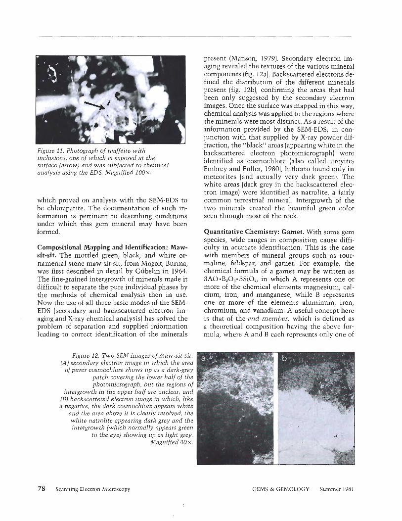
Figure 11. Photograph of taaffeite with inclusions, one of which is exposed at the surface (arrow) and was subjected to chemical analysis using the EDS. Magnified 1 0 0 ~ .
which proved on analysis with the SEM-EDS to be chlorapatite. The documentation of such in- formation is pertinent to describing conditions under which this gem mineral may have been formed.
Compositional Mapping and Identification: Maw- sit-sit. The mottled green, black, and white or- namental stone maw-sit-sit, from Mogok, Burma, was first described in detail by Gubelin in 1964. The fine-grained intergrowth of minerals made it difficult to separate the pure individual phases by the methods of chemical analysis then in use. Now the use of all three basic modes of the SEM- EDS (secondary and backscattered electron im- aging and X-ray chemical analysis) has solved the problem of separation and supplied information leading to correct identification of the minerals
present (Manson, 1979). Secondary electron im- aging revealed the textures of the various mineral components (fig. 12a). Backscattered electrons de- fined the distribution of the different minerals present (fig. 12b), confirming the areas that had been only suggested by the secondary electron images. Once the surface was mapped in this way, chemical analysis was applied to the regions where the minerals were most distinct. As a result of the information provided by the SEM-EDS, in con- junction with that supplied by X-ray powder dif- fraction, the "black" areas (appearing white in the backscattered electron photomicrograph) were identified as cosmochlore (also called ureyite; Embrey and Fuller, 1980), hitherto found only in meteorites (and actually very dark green). The white areas (dark grey in the backscattered elec- tron image) were identified as natrolite, a fairly common terrestrial mineral. Intergrowth of the two minerals created the beautiful green color seen through most of the rock.
Quantitative Chemistry: Garnet. With some gem species, wide ranges in composition cause diffi- culty in accurate identification. This is the case with members of mineral groups such as tour- maline, feldspar, and garnet. For example, the chemical formula of a garnet may be written as 3AO*B203*3Si02, in which A represents one or more of the chemical elements magnesium, cal- cium, iron, and manganese, while B represents one or more of the elements aluminum, iron, chromium, and vanadium. A useful concept here is that of the end member, which is defined as a theoretical composition having the above for- mula, where A and B each represents only one of
Figure 12. Two SEM images of maw-sit-sit: (A) secondary electron image in which the area
of purer cosmochlore shows up as a dark-grey patch covering the lower half of the photomicrograph, but the regions o f
intergrowth in the upper half are unclear; and (B) backscattered election image in which, liki a negative, the dark cosmochlore appears whitt
and the area above it is clearly resolved, tht, white natrolite appearing dark grey and the 1 in tergrow th (which normally appears greer.
to the eye) showing up as light grey. Magnified 40 X .
78 Scanning Electron Microscopy GEMS & GEMOLOGY Summer 1981
the elements from their respective groups. Spec- imens of pure end members such as pyrope, the magnesium aluminum garnet (3MgO- AlzOg 3SiOz), rarely if ever occur in nature. Typically, an individual gem has an intermediate composi- tion (i.e., where A and/or B each represents more than one element) containing varying proportions of the ideal end members. The compositions among stones belonging to such a mineral group may show continuous gradations between two or more of the end members. On the other hand, the compositions may be distributed discontin- uously, forming clusters of chemically similar stones. In the latter case, individual stones may be assigned to appropriate clusters on the basis of their physical and optical properties, provided enough stones have been studied to adequately determine the variations in properties with re- spect to composition.
At the present time, GIA1s research laboratory is using quantitative chemical analysis as part of a detailed study of the gem garnet group. New sources for gem garnets have provided large num- bers of specimens that cannot be described well by the traditional classifications, and in addition have drawn attention to some older problems of identification, or "where to draw the line" be- tween gem species andlor varieties. One new type of garnet appears to consist of varying mixtures of two garnet end members, pyrope and spessar- tine, with small quantities of grossular, alman- dine, and possibly andradite as well. With chem- ical analysis, we can determine if these new stones make up a well-defined cluster. At the same time, the entire gem garnet group will be reexamined and, if appropriate, recommendations made for redefinition of garnet nomenclature. Finally, we can determine the specific optical and physical properties that belong to each of these gem spe- cies and/or varieties and discover which of these values can be used by the gemologist for iden- tification.
SUMMARY The contemporary gemologist is faced with ever- increasing problems in the identification and
description of gem materials. Gemology, like all sciences, has advanced beyond the point where any single person can know all there is about the subject. GIA1s research laboratory provides for the use of instrumentation such as the SEM-EDS in gemology to answer those questions that extend outside the scope of gemologists whose time and training must necessarily be focused elsewhere. The specific examples cited here of situations in which the SEM-EDS has been used are but a few of the areas that are being explored. Other types of instrumentation accessible to our researchers, either at our own facilities or through the coop- eration of other organizations, are used in con- junction with the SEM-EDS to pursue topics such as the distinction and identification of gem spe- cies and varieties, determination of the origins of color in gemstones (including diamonds), and identification of synthetic and altered gems, as well as other subjects of current concern to gemologists.
REFERENCES d a r k A.M., Fejer E.E., Couper A.G., Bearne G.S., Din V.K. '
(1980) Additional data on sugilite. Mineralogical Maga- zine, Vol. 43, pp. 947-949.
Dunn P.J. (1977) The use of the electron microprobe in gem- mology. fournal of Gemmology, Vol. 15, pp. 248-258.
Embrey P.G., Fuller J.P. (1980) A Manual of New Mineral Names 1892-1978. Oxford University Press, New York.
Gubelin E.J. (1964) Maw-sit-sit, a new decorative gemstone from Burma. Gems a) Gemology, Vol. 11, pp. 227-238.
Gubelin E.J., Weibel M. (19751 Vanadium-grossular from Lu- alenyi near Voi, Kenya. Neues Jahrbuch fur Mineralogie Abhundlangen, Vol. 123, pp. 19 1- 197.
Liddicoat R.T. Jr. (1967) Developments and highlights at the Gem Trade Lab i n Los Angeles; taaffeite proves largest. Gems a) Gemology, Vol. 12, pp. 212-215.
Manson D.V. (1978) Plastic impregnated opal. Gems a) Gem- ology, Vol. 16, pp. 49-57.
Manson D.V. (1979) Recent activities in GIA's research de- partment; clarification of composition of Maw-sit-sit. Gems a) Gemology, Vol. 16, pp. 21 7-21 8.
Reid A.M., Frazer J.Z., Fujita H. (1969) Garnet analysis. Scripps Institute of Oceanography, reference 69-21.
Wilson W.E. (1972) The state of the art: the electron micro- scopes and the electron microprobe. Mineralogical Record, Vol. 3, pp. 157- 164.
Scanning Electron Microscopy GEMS & GEMOLOGY Summer 198 1 79
EMERALDS OF COLOMBIA By Peter C. Keller
There has been more activity in emerald mining in Colombia in the last few years than at any other time in the four centuries since the Spanish first discovered the deposits. This article combines a comprehensive review of the literature on the history and geology of Colombian emeralds with the author's own insighis into the current niarkeiing and mining of these stones. O f particular concern are the harsh nieihods now being used to extract the emeralds and the impact these methods will have on future production in Colombia. The past, however, has yielded many fabulous gems, the most famous of which are also described here and placed i n historical perspective.
ABOUT THE AUTHOR
Dr. Keller is director of education of the Gemolog~cal Institute of America, Santa Monica, CA.
Much 01 the research for this article was undertaken in preparation lor a special exhibit 01 Colombian emeralds and pre-Columbian gold artifacts held at the Natural History Museum of Los Angeles County July 4-September 6, 1981.
"-1 981 Gemological Institute of America
F ew gemstones on the world market today provide the aura of adventure and intrigue, wealth and beauty that
is so intrinsic to emerald. The first records of emeralds worn as jewelry date from the early days of the Roman Empire, in reference to stones recovered from now long- lost Egyptian mines. They have been treasured as gems ever since. Although the Egyptian mines may have intro- duced emeralds to the Western world, historically the fin- est emeralds have come from deposits in Colombia. The checkered history of these mines began cruelly with the Spanish conquerors' enslavement of local Indians to work the deposits, leading to the decimation of the native pop- ulation in just a few years. And as recently as the early 1970s, Muzo-the major mine-was forced to close because of over 900 emerald-related murders in one year.
Over the past half century, there have been numerous papers describing the status of the Colombian emerald mines, usually by recent visitors to the mining areas (MacFadden, 1934; Switzer, ,1948; Copeland, 1950; An- derton, 1950 and 1965; Bancroft, 1971; and Tenhagen, 1972). However, few, if any, of these papers have com- bined their observations (which were usually on a single mine) with a comprehensive review of the literature to provide a detailed account of the history and geology of the emerald deposits. Furthermore, in the past five years we hav) seen more activity in the Colombian mines than there has been since the Spanish first exploited them in the 16th century. The purpose of this paper is to combine a comprehensive review of the literature with an update on current mining and marketing activity, based on sev- eral recent trips to the area by the author. In addition, major emeralds originally mined in Colombia and now
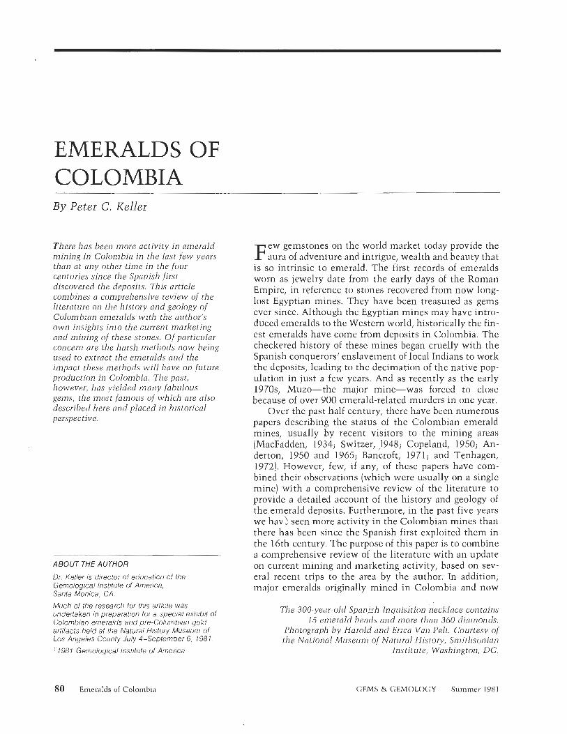
The 300-year-old S p m v h Inquisition necklace contains 15 emerald beads find more than 360 cliamonds.
Photograph by Harold and Erica Van Pelt. Courtesy of the National Museum of Natural History, Smithsonian
Institute, Washington, DC.
80 Emeralds of Colombia GEMS & GEMOLOGY Summer 198 1
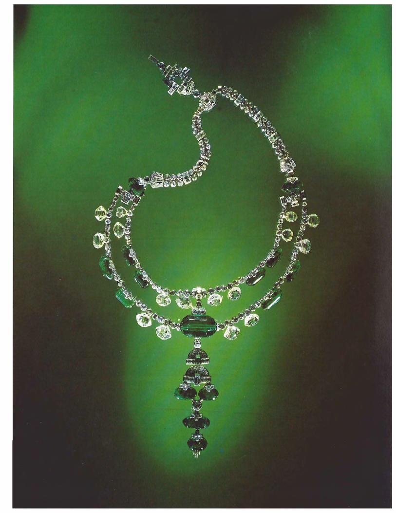
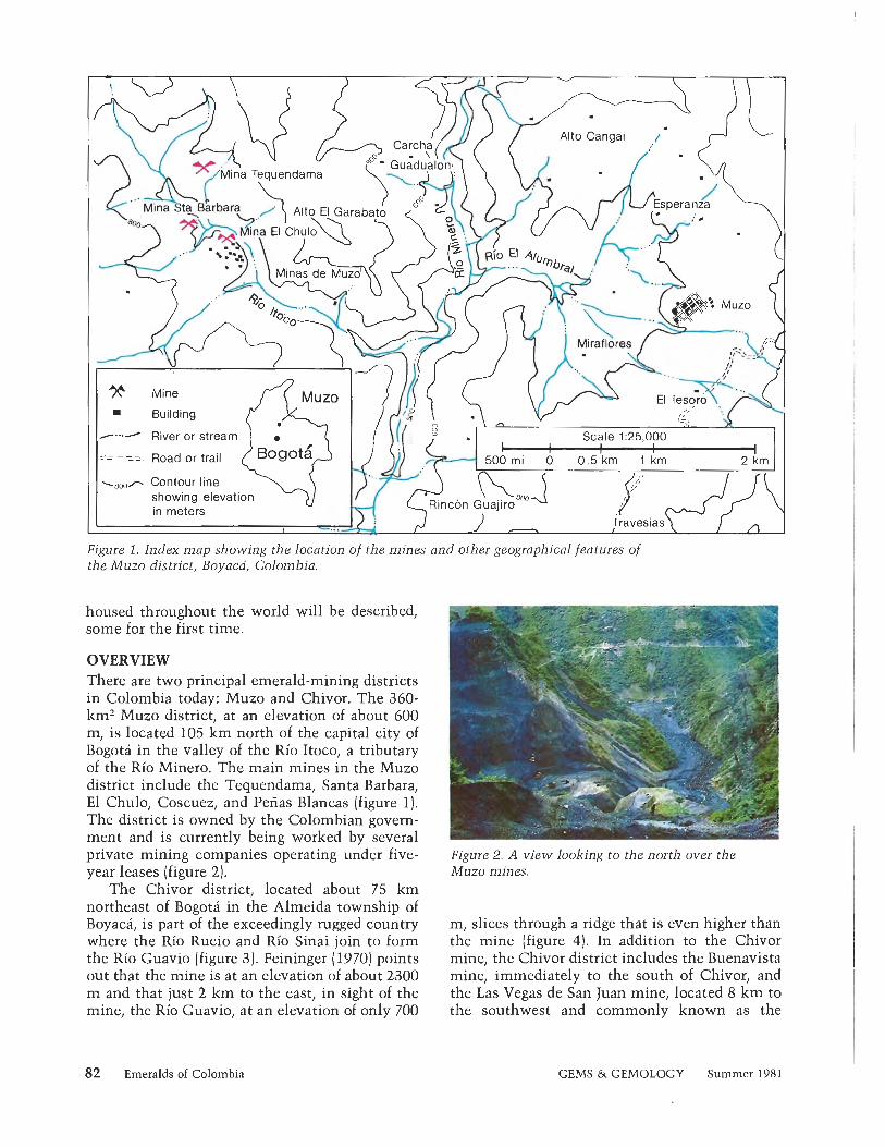
Figure 1. Index map showing the location of the mines and other geographical features of the Muzo district, Boyacd, Colombia.
housed throughout the world will be described, some for the first time.
OVERVIEW There are two principal emerald-mining districts in Colombia today: Muzo and Chivor. The 360- km2 Muzo district, at an elevation of about 600 m, is located 105 km north of the capital city of Bogota in the valley of the Rio Itoco, a tributary of the Rio Minero. The main mines in the Muzo district include the Tequendama, Santa Barbara, El Chulo, Coscuez, and Penas Blancas (figure 1). The district is owned by the Colombian govern- ment and is currently being worked by several private mining companies operating under five- year leases (figure 2).
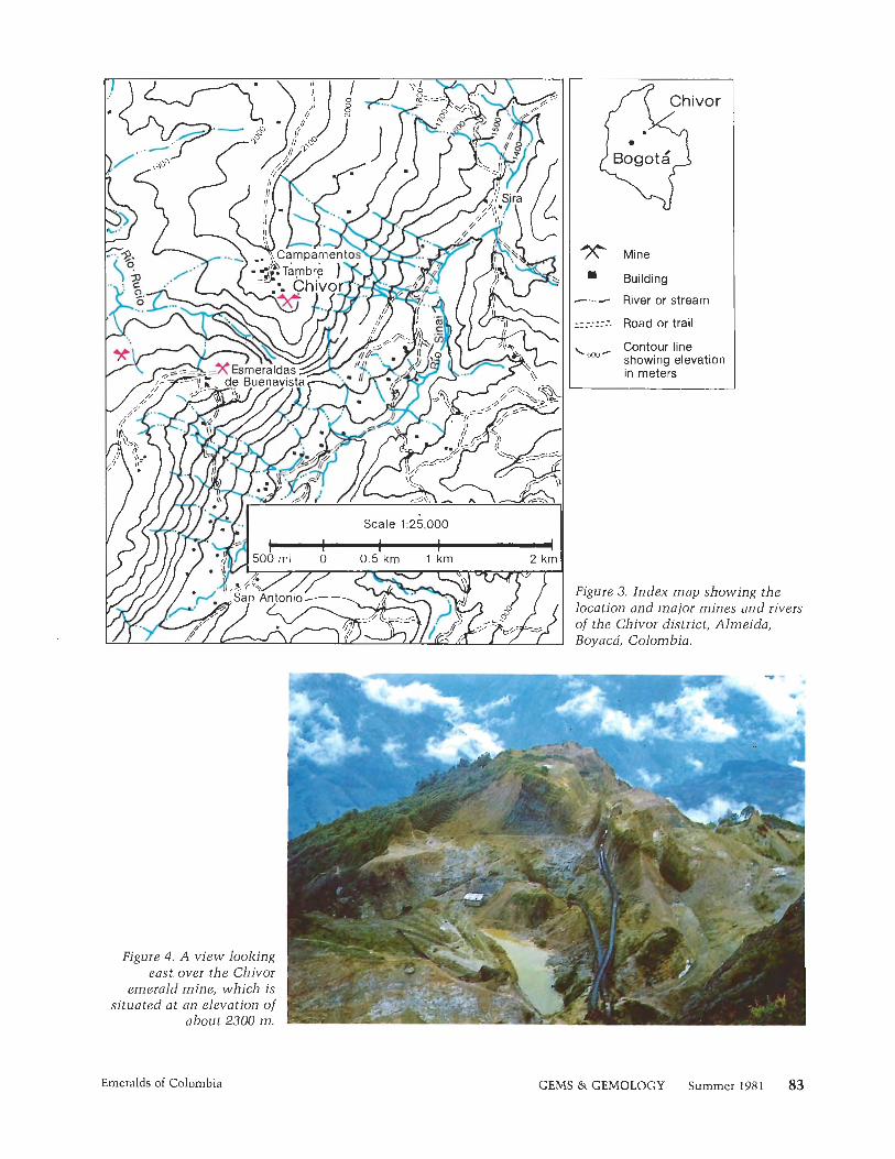
The Chivor district, located about 75 km northeast of Bogota in the Almeida township of Boyaca, is part of the exceedingly rugged country where the Rio Rucio and Rio Sinai join to form the Rio Guavio (figure 3). Feininger (1970) points out that the mine is at an elevation of about 2300 m and that just 2 km to the east, in sight of the mine, the Rio Guavio, at an elevation of only 700
82 Emeralds of Colombia
Figure 2. A view looking to the north over the Muzo mines.
m, slices through a ridge that is even higher than the mine (figure 4). In addition to the Chivor mine, the Chivor district includes the Buenavista mine, immediately to the south of Chivor, and the Las Vegas de San Juan mine, located 8 km to the southwest and commonly known as the
GEMS & GEMOLOGY Summer 1981
Bogota ovO /\ Mine
Building - River or stream
: - - Road or trail
^ew^ Contour line showing elevation in meters
Figure 3. Index map showing the location and major mines and rivers of the Chivor district, Almeida, Boyacd, Colombia.
Emeralds of Colombia GEMS & GEMOLOGY Summer 1981
G a c h a l i m i n e (Anderton, 1955). T h e Chivor m i n e is t h e on ly privately owned emerald m i n e i n Co- lombia. The Buenavista and G a c h a l i m i n e s op- era te a s concessions under a 1959 law, a n d their owners pay a 25% royalty t o t h e Co lombian gov- e r n m e n t (Colombian American Business, 1 979).
T h e r e have been a n u m b e r of s tudies o n t h e geology of M u z o and, t o a lesser extent, Chivor [see box). O u r cu r ren t knowledge of t h e geology tells us how t h e emeralds occur and where t o d i - rect fu tu re exploration. Thus far, however, a de- tailed s t u d y of t h e Eastern Cordillera h a s been in- hibi ted by rugged terrain a n d th ick vegetation, w h i c h h a s a lso restr icted exploration.
EARLY HISTORY Emeralds m i n e d i n Colombia have been used for t rade a n d personal adornment th roughou t much of Middle a n d Sou th America s ince pre-Colum- bian t imes . W h e n t h e first Spaniards arrived i n t h e N e w World in t h e ear ly 1 6 t h century, emer - a lds were being worshiped, were used i n jewelry, a n d played a n impor tan t role a s sacrificial offer- ings i n ceremonies s u c h a s t h e f a m o u s El Dorado ce remony o n Lake Guatavi ta , located just no r th - eas t of Bogota [Bray, 1978). Emeralds were being traded a s far s o u t h a s Peru a n d Chile a n d a s far no r th a s Mexico. According to Morello (1957], w h e n Spanish conqueror Cor tes m e t t h e Aztec
GEOLOGY OF MUZO AND CHIVOR The lower member at Muzo, the Cambiado, consists of highly folded, faulted, and fractured carbonaceous shales and thinly bedded limestones. The Cambiado is discordantly overlain by the Emerald Beds, the latter consisting of thinly bedded, weathered, yellowish-gray shales that have also been intensely folded and invaded by thin calcite veins. The Emerald Beds, as their name implies, contain localized concentrations of emeralds associated with calcite, dolomite, pyrite, quartz, and parisite. Locally, the Cambiado and Emerald Beds are separated by the Cama and the overlying Cenicero. The major difference between these two layers appears to be textural. The Cama consists of an agglomerate of relatively large calcite crystals, whereas the Cenicero, which is usually about a meter thick, consists of small calcite crystals along with pyrite in a carbonate ground mass. Barite has also been reported in the Cenicero (Oppenheim, 1948).
The Colon~bian Andes consists of three subparallel ranges: the Western, or Cordillera Occidental; the Central, or Cordillera Central; and the Eastern, or Cordillera Oriental. According to Clements (19411, the Western and Central ranges consist primarily of granites and are best known for their gold deposits (gold production). The Eastern range, however, consists mostly of sedimentary units, principally limestones and shales with minor igneous and metamorphic rocks exposed only locally. Clements places a Paleozoic age on these crystalline rocks.
The major emerald deposits are limited to the eastern (Chivor) and western (Muzo) margins of the Cordillera Oriental where Cretaceous sediments are well exposed. The geology of the Muzo district has been described by several authors, five of whom are outstanding: Pogue (1916), Lleras (1929), Scheibe (19331, Clements (1941), and Oppenheim (1948). These studies agree that emeralds are restricted to the Villeta formation of Lower Cretaceous age. The Villeta formation consists of a great thickness of intensely folded and fractured black carbonaceous shale and minor limestones. The black shale is so rich in carbon that it is impossible to handle without soiling one's hands. The Lower Cretaceous age of the Villeta has been determined from the presence of fossil ammonites. The highly fractured shales have been invaded by numerous white calcite fracture- filling veins, and it is in these veins that the emeralds occur. Dolomite, quartz, pyrite, and the uncommon rare-earth calcium carbonate, parisite, are accessory minerals con~monly found with emerald in these veins. Lleras (19291, Scheibe (1933), and Oppenheim (1948) noted albite as a common vein mineral at Muzo, but Clements (1941) did not observe albite, nor did this author.
The Villeta formation was divided into two members by Lleras (19291, namely, the Cambiado and the overlying Emerald Beds. Locally, these two members are separated by two thin agglomeratic layers of calcite crystals designated the Canla and the Cenicero (Oppenheiin, 1948).
84 Emeralds of Colombia GEMS & GEMOLOGY Summer 1981
emperor Montezuma in Mexico i n 1519, the lat- ter was bedecked with fine emeralds. Reportedly, Spanish conqueror Pizarro sent four chests of em- eralds from Peru to the King of Spain i n 1533. These were all undoubtedly of Colombian origin (Ball, 193 1) .
Chivor was the first operating emerald mine discovered by the Spaniards in their conquest of the New World. Gonzalo Jimenez de Quesada saw the first sign of emeralds in Colombia a t Turq- mequ6, Boyaca, i n 1537 (Colombian American Business, 1979). Quesada sent Captain Pedro F. de Valenzuela t o find the source. Tha t same year, h e located the well-developed Chibcha Indian mine
of Somondoco, later t o be named Chivor after a nearby valley. Soon thereafter, the Spaniards were vigorously working the Chivor mine using local Indians as slave labor.
Five years after the founding of Santi'sima Trinidad de 10s Muzos in 1559 (Wokittel, 19601, the Muzo and Caijma Indians' mine was located some 7 km to the west on the Itoco Hill. Actual mining of the Muzo area by t he Spaniards began i n 1567, and initial production is said t o have overshadowed production a t Chivor (Feininger, 1970). By the end of the 16th century, both Chivor and Muzo were being vigorously worked using Indian slave labor. In 1592, the first recorded grant
The sedimentary rock units at Chivor are somewhat different from those described for Muzo. At Chivor, such units are almost entirely shales and argillites, with minor limestone and sandstone (Rainier, 1929). The general geology and stratigraphy of this area are not as well known as at Muzo. The stratigraphic section at Chivor appears toconsist primarily of at least 1000 m of conformable sediments. A good description of these sediments and the geology is found in Johnson (1961). Johnson indicates that the emerald zone at Chivor runs about 10 km east-west and about 5 km north-south. Fossil ammonites, bivalves, and ferns indicate that the sediments of the district are of Cretaceous age. As at Muzo, these sediments are heavily faulted and folded. They are mostly shales and argillites with some blocks or floaters of carbonaceous limestone present near the top of the stratigraphic section. The most prominent unit is a poorly cemented yellowish shale which overlies a thick sequence of gray-blue shales and argillites. No emeralds have been reported from the yellowish shale cap. Most emeralds occur in a blue-gray argillite about midway through the section. Johnson (1961) suggests that the occurrence of emeralds at Chivor is structurally related, as if the emerald veins were concentrated along the axes of tight folds in the argillites. He states that, "If a vein is discovered traveling in the trough of a syncline, the production of stones may be immense."
Chivor emeralds are found mostly in veins, but in rare instances they may occur in cavities, as was the case with the famous Patricia emerald discussed elsewhere in this paper. The cavities, when present, are always associated with the veins. The veins run parallel to the bedding of the sediments, which suggests that separations between bedding laminae provided the avenues of least resistance for fissure-filling hydrothermal solutions that crystallized to form veins. Such veins occur up to 15 cm in thickness but rarely exceed 65 m in length. Emeralds commonly are found where two veins intersect. Johnson (1961) divided the veins into three mineralogical types: (1) pyrite, (2) albite, and (3) pyrite with albite. The mineralization is in sharp contrast to the emerald- bearing veins of Muzo, where the gem material occurs in white calcite or dolomite or both. It is stressed that there have been no reports of emeralds occurring in calcite or dolomite at Chivor; both quartz and pyrite are very common there.
Three more-or-less parallel "iron bands," consisting of pyrite and limonite, appear about 50 m apart from one another, interlaminated in the stratigraphic section at Chivor. These bands appear to control the distribution of emeralds to some extent. Emeralds are most prevalent below the lowest of the three iron bands or between the lowest and middle bands. Very few emeralds have ever been reported above the middle or upper iron bands. This suggests that these bands may have acted as impervious "dams" for the rising emerald-bearing solutions.
Emeralds of Colombia GEMS & GEMOLOGY Summer 1981 85
of Chivor was given to Francisco Maldonado de Mendoza by Antonio Gonzalez, president of the New Kingdom of Granada. By this time, however, the treatment of the Indian slaves was so inhu- mane that on September 22, 1593, President Gon- zalez issued a 39-article decree protecting the In- dians (Johnson, 1961). This decree was soon followed in 1602 by several royal orders from Phillip 111 of Spain to enforce the law. By this time, however, the Indian population had already been decimated. As a consequence of this loss of cheap labor and the litigation that followed the royal orders, production of Colombian emeralds declined drastically. In 1650, the Muzo mines were declared royal property, and production fur- ther declined. By 1675, the Chivor mine had been abandoned; its location eventually became a mys- tery that endured for over 200 years. Muzo con- tinued to be worked sporadically throughout the 17th, 18th) and 19th centuries (Barriga and Bar- riga, 1973) until the government declared it the National Emerald Domain in 1871 (Colombian American Business, 1979). When the mines at Muzo came under government control, produc- tion all but ceased and lawless disorder came to characterize the area. This situation has changed only very recently.
Soon after Muzo was placed under govern- ment control, the Chivor mine was rediscovered on the basis of a description written almost 300 years earlier. In 1888, Colombian mining engi- neer Don Francisco Restrepo found a manuscript dating back to the early 17th century in a Do- minican convent in Quito, Equador. This manu- script, written by Fray Martin de Aguado, de- scribed the location of the Chivor mine as the only place in the Andes where one could see through a pass in the mountains to the plains of the Orinoco. Restrepo's search for the legendary mine ended successfully in 1896. Although legal problems with the government hampered Res- trepo's early mining activities, his early 20th cen- tury partnership with German mining engineer Fritz Klein coincided with the lifting of some of these restrictions and promising production at the mine. When World War I broke out, however, Klein returned to Germany for military service. Restrepo died at Chivor, and, with Germany's loss of the war, Klein lost all rights to the mine as a result of alien property legislation. In 1919, Chivor was purchased by the Colombian Emerald Syndicate, Ltd., an American company. Since then
it has changed hands many times with varying degrees of success, and has been managed by such notable mining engineers as Peter W. Rainier (au- thor of Green Fire, 1942) and Willis Bronkie. The Chivor mines are currently in the hands of the Quintero family.
In 1953, a new mine was discovered 8 km southwest of Chivor at Gachala, reportedly when a woodcutter's mule uncovered an emerald-bear- ing rock (Anderton, 1955). Although the mine has produced only sporadically since 1953, in 1967 an 858-ct. crystal, generally considered one of the finest in existence, was found there. The 5- cm hexagonal prism is known simply as the Gachali emerald and is housed in the Smithson- ian Institution (Trapp, 1969).
RECENT HISTORY AND PRODUCTION Emerald mining under government control has been a questionable business proposition. In 1946, the government entrusted the management of the Muzo mines, including the power to regulate all mining and marketing of emeralds from that dis- trict, to the Banco de la Republics (Feininger, 1970). However, illicit emerald mining and deal- ing continued to be widespread, with an esti- mated loss to the government of more than 28 million pesos during the period 1946 to 1969, when the Bank relinquished control of the Muzo emeralds to the Empresa Colombiana de Minas (ECOMINAS), the government's mining agency. Still, the situation at Muzo worsened, to the point that in 1973 over 900 people were killed in feuds and the mines were forced to close. The mines stayed officially closed until 1977, when the gov-
Figure 5. Active mining of emeralds at Muzo. Note the white calcite veins.
86 Emeralds of Colombia GEMS & GEMOLOGY Summer 1981

Figure 6. Looking eastward at guaqueros working in the Rio Itoco below the main mining operations at Muzo.
ernment solicited bids for five-year leases on the Muzo mines, the Coscuez mine, and the Peiias Blancas mine. After extensive negotiations, the Muzo lease was awarded to the Sociedad Minera Boyacense Ltda., the Coscuez lease went to Es- meraldas y Minas de Colombia, S.A. (ESMERA- COL), and the Perias Blancas lease went to the Quintero brothers, who also control the Chivor mine [Colombian American Business, 1979).
Unfortunately, f ive-year leases encourage the lessees to mine the area as rapidly as possible, and their methods are not as conservative of resources as they should be (figure 5). When the author vis- ited Muzo in 1979 and again in 1980, the main area was being worked harshly, using bulldozers and dynamite, a method not adopted in the past because of the fragility of the emeralds. After an area has been blasted, bulldozers scrape off the overburden until the white calcite veins are ex- posed. Then, teams are brought in to work each vein with pick and shovel. When emeralds are found, they are placed in a canvas bag for sorting by the mine lessees each evening. These sorted parcels are then sealed and taken to Bogota for further grading and subsequent marketing.
Because the main mine area is being stripped away so rapidly, a significant portion of the po- tential emerald production is lost to the gravels of the Rio Itoco, with the result that an estimated 15,000 guaqueros (independent miners, directly translated as "treasure hunters") mine the riv- erbed each day (figure 61. Theseguaqueros usually work alone or in small groups, and they live en- tirely on what they find. They subsist in nearby rancheros on the flanks of the Itoco Valley, eating pigs, chickens, vegetables, and fruits that they raise or grow locally. Their lifestyle closely re- sembles that of the miners who joined the mid- 19th century California Gold Rush.
The terms of the government leases are very demanding. For example, over five years, ESMER- ACOL will pay ECOMINAS a total rent of 260 million pesos (about US$6.53 million) plus 5% of the gross production at Coscuez. ESMERACOL started working Coscuez in March of 1978; by 1979, five bulldozers were working the mine, and a force of 60 National Police patrolled the area to prevent poaching (Colombian American Busi- ness, 1975). The status at Muzo is very similar.
Emerald production statistics for Colombia
Emeralds of Colombia GEMS & GEMOLOGY Summer 1981 87
during the 1970s, as reported by Baskin in the Minerals Yearbook (19791, provide some indica- tion of what has happened to emerald production since the leases were awarded. In 1973, total ex- ports of emeralds from Colombia were reported to be US$2 million. In 1978, these exports rose to US$40 million; and in the first seven months of 1979, exports rose to US$75 million. If this trend continues, Colombia should derive far more rev- enue from emerald mining than ever in the past.
MARKETING THE EMERALDS Presently, there are two distinct systems in Co- lombia for marlzeting emeralds: (1) the official marketing required by law of the private mining companies under leases from the government, and (2) the illicit marlzeting done by the gua- queros in the Rio Itoco and, to a lesser extent, at other mines. In the case of the private mining companies, the value of their production is deter- mined by the Controloria General de la Republica, representatives of ECOMINAS, and by the own- ers of the mining companies. The private mining companies then market their emeralds through whichever channels they choose. The amount of emerald mined and subsequent marlzeting by the guaqueros working in the Rio Itoco below the main mines at Muzo is much more secretive. When a guaquero finds an emerald, eager buyers are waiting on the dirt road next to the river. These esmeralderos, independent emerald dealers from Bogota, gather to buy whatever is found in the river, then take their goods to Bogota to be cut and subsequently traded on the sidewalks off Bogota's Avenida Jimenez. On any given day, one can see emeralds of any quality and quantity being traded on Avenida Jimenez, ironically, within sight of the Banco de la Republica.
It is, however, buyer beware when purchasing emeralds on the Avenida Jimenez. Recently, a number of very cleverly enhanced emeralds and 'imitations" have been sold to the unwary. These include quartz crystals that have been cored, filled with green plastic, polished, and then glued into a plausible shale matrix; the discreet use of green ink to make moralla, or pale green emerald, ap- pear to be of a fine green color; as well as green glass and plastic imitations.
IMPORTANT COLOMBIAN EMERALDS Even though emeralds have been mined in Co- lombia since pre-Columbian times, relatively few
very fine large examples are known today. Because of the extremely high value of faceted stones, fine large emerald crystals are particularly rare. The following is a brief discussion of what many would agree are some of the most important emeralds, both cut and uncut, produced by the Colombian mines.
During the 17th century, large Colombian em- eralds were eagerly sought by the Mogul nobility of India. By way of Spain, a great many of the early Colombian stones found a ready marlzet in India through well-established trade routes. One partic- ularly fine example of these emeralds is the "Mo- gul" [see cover], which is currently owned by private collector Allan Caplan. The Mogul em- erald measures 5 cm x 3.8 cm x 3.5 cm and weighs 217.8 cts. (Caplan, 1968). The front of the stone is carved with the floral motif typical of the Mogul carvers; the back contains an Islamic prayer and includes the date 1695 A.D. The drilled stone was probably worn on an article of clothing, per- haps a turban.
Many of these Colombian emeralds were taken from India in 1739 during the sacking of Delhi by the Persians, and now are part of the Crown Jew- els of Iran in Tehran. Meen (1969) examined over one thousand of these emeralds and reported that most were over 10 cts. and some exceeded 300 cts. (Meen and Tushingham, 1968). Certainly this collection and the collection in the Topkapi Mu- seum in Istanbul, Turkey, must be considered the two largest collections of Colombian emeralds in the world.
One of the most spectacular pieces of emerald jewelry on public display in the United States is the 300-year-old Spanish Inquisition neclzlace (see frontispiece), which is in the Smithsonian In- stitution's collection. This neclzlace was report- edly worn in the Spanish and, later, French courts. The drilled hexagonal and cylindrical beads that make up the neclzlace may very well have originally belonged to articles of pre-Columbian jewelry and were simply reset into this magnifi- cent piece. A 24 mm x 15 mm drilled emerald bead forms the focal point of the necklace; in addition, there are 14 smaller emerald beads av- eraging 16 mm x 7 mm. Dunn (1975) reports that the neclzlace also contains over 360 mine-cut dia- monds, 16 of which must be considered major gems themselves. It is interesting to note that these 16 large diamonds also have been drilled.
Another spectacular Colombian emerald in
88 Emeralds of Colombia GEMS & GEMOLOGY Summer 1981
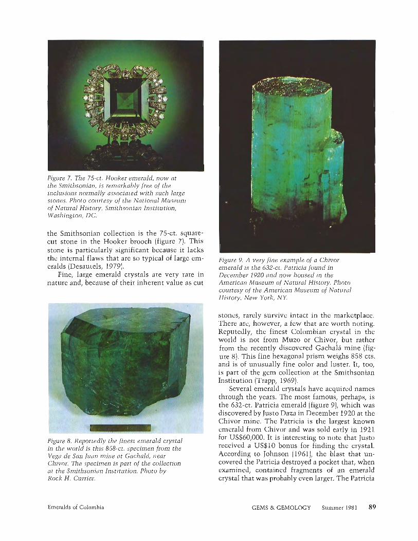
Figure 7, The 75-ct. Hooker emerald, now at the Smithsonian, is remarkably free of the inclusions normally associated with such large stones. Photo courtesy of the National Museum of Natural History, Smithsonian Institution, Washington, DC.
the Smithsonian collection is the 75-ct. square- cut stone in the Hooker brooch (figure 7). This stone is particularly significant because it lacks the internal flaws that are so typical of large em- eralds (Desautels, 1979).
Fine, large emerald crystals are very rare in nature and, because of their inherent value as cut
rGuze O . Reportedly the finest emerald crystal in the world is this 858-ct. specimen from the Vega de San Juan mine at Gachala, near Chivor. The specimen is part of the collection at the Smithsonian Institution. Photo by Rock H. Currier,
Emeralds of Colombia
Figure 9. A very fine example of a Chivor emerald is the 632-ct. Patricia found in December 1920 and now housed in the American Museum of Natural History. Photo courtesy of the American Museum of Natural History, New York, NY.
stones, rarely survive intact in the marketplace. There are, however, a few that are worth noting. Reputedly, the finest Colombian crystal in the world is not from Muzo or Chivor, but rather from the recently discovered Gachala mine (fig- ure 8). This fine hexagonal prism weighs 858 cts. and is of unusually fine color and luster. It, too, is part of the gem collection at the Smithsonian Institution (Trapp, 1969).
Several emerald crystals have acquired names through the years. The most famous, perhaps, is the 632-ct. Patricia emerald (figure 9)) which was discovered by Justo Daza in December 1920 at the Chivor mine. The Patricia is the largest known emerald from Chivor and was sold early in 1921 for US$60,000. It is interesting to note that Justo received a US$10 bonus for finding the crystal. According to Johnson (1961)) the blast that un- covered the Patricia destroyed a pocket that, when examined, contained fragments of an emerald crystal that was probably even larger. The Patricia
GEMS & GEMOLOGY Summer 1981
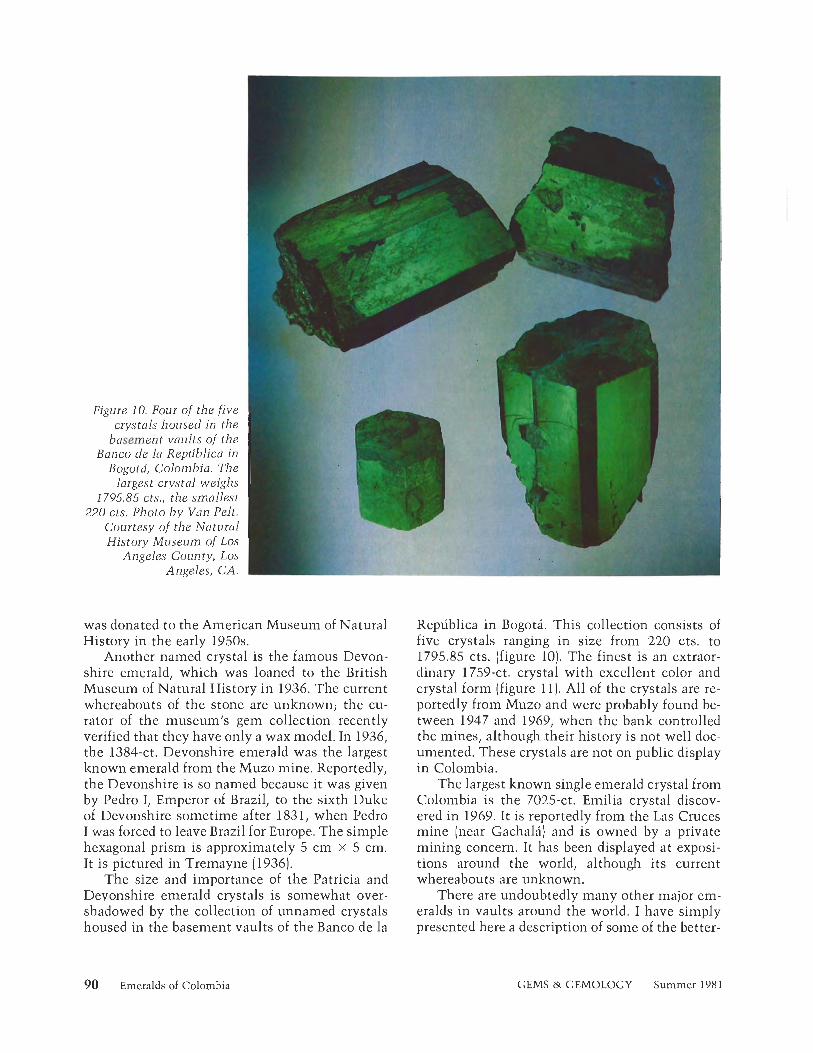
Figure 10. Four of the five crystals housed in the
basement vaults of the Banco de la Republica in
Bogota, Colombia. The largest crystal weighs I
1795.85 cts., the smallest 220 cts. Photo by Van Pelt.
Courtesy of the Natural History Museum of Los
Angeles County, Los Angeles, CA.
was donated to the American Museum of Natural History in the early 1950s.
Another named crystal is the famous Devon- shire emerald, which was loaned to the British Museum of Natural History in 1936. The current whereabouts of the stone are unknown; the cu- rator of the museum's gem collection recently verified that they have only a wax model. In 1936, the 1384-ct. Devonshire emerald was the largest known emerald from the Muzo mine. Reportedly, the Devonshire is so named because it was given by Pedro I, Emperor of Brazil, to the sixth Duke of Devonshire sometime after 1831, when Pedro I was forced to leave Brazil for Europe. The simple hexagonal prism is approximately 5 cm x 5 cm. It is pictured in Tremayne (1936).
The size and importance of the Patricia and Devonshire emerald crystals is somewhat over- shadowed by the collection of unnamed crystals housed in the basement vaults of the Banco de la
90 Emeralds of Colombia
Republica in Bogota. This collection consists of five crystals ranging in size from 220 cts. to 1795.85 cts. (figure 10). The finest is an extraor- dinary 1759-ct. crystal with excellent color and crystal form (figure 11). All of the crystals are re- portedly from Muzo and were probably found be- tween 1947 and 1969, when the bank controlled the mines, although their history is not well doc- umented. These crystals are not on public display in Colombia.
The largest known single emerald crystal from Colombia is the 7025-ct. Emilia crystal discov- ered in 1969. It is reportedly from the Las Cruces mine (near Gachala) and is owned by a private mining concern. It has been displayed at exposi- tions around the world, although its current whereabouts are unknown.
There are undoubtedly many other major em- eralds in vaults around the world. I have simply presented here a description of some of the better-
GEMS & GEMOLOGY Summer 198 1
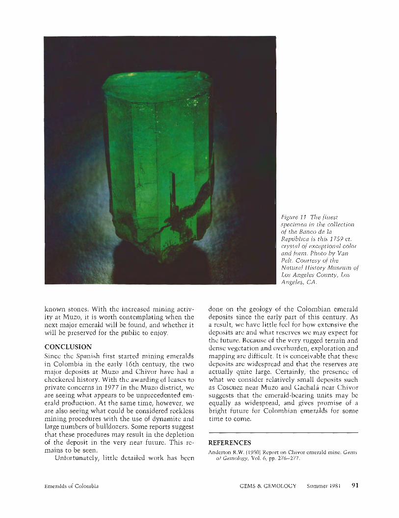
known stones. With the increased mining activ- ity at Muzo, it is worth contemplating when the next major emerald will be found, and whether it will be preserved for the public to enjoy.
CONCLUSION Since the Spanish first started mining emeralds in Colombia in the early 16th century, the two major deposits at Muzo and Chivor have had a checkered history. With the awarding of leases to private concerns in 1977 in the Muzo district, we are seeing what appears to be unprecedented em- erald production. At the same time, however, we are also seeing what could be considered reclzless mining procedures with the use of dynamite and large numbers of bulldozers. Some reports suggest that these procedures may result in the depletion of the deposit in the very near future. This re- mains to be seen.
Unfortunately, little detailed work has been
Emeralds of Colombia
Figure 11, The finest specimen i n the collection of the Banco de la Republics is this 1759-ct. crystal of exceptional color and form. Photo b y Van Pelt. Courtesy of the Natural History Museum o f Los Angeles County, Los Angeles, CA.
done on the geology of the Colombian emerald deposits since the early part of this century. As a result, we have little feel for how extensive the deposits are and what reserves we may expect for the future. Because of the very rugged terrain and dense vegetation and overburden, exploration and mapping are difficult. It is conceivable that these deposits are widespread and that the reserves are actually quite large. Certainly, the presence of what we consider relatively small deposits such as Coscuez near Muzo and Gachala near Chivor suggests that the emerald-bearing units may be equally as widespread, and gives promise of a bright future for Colombian emeralds for some time to come.
REFERENCES Anderton R.W. (1950) Report on Chivor emerald mine. Gems
a) Gemology, Vol. 6, pp. 276-277.
GEMS & GEMOLOGY Summer 1981
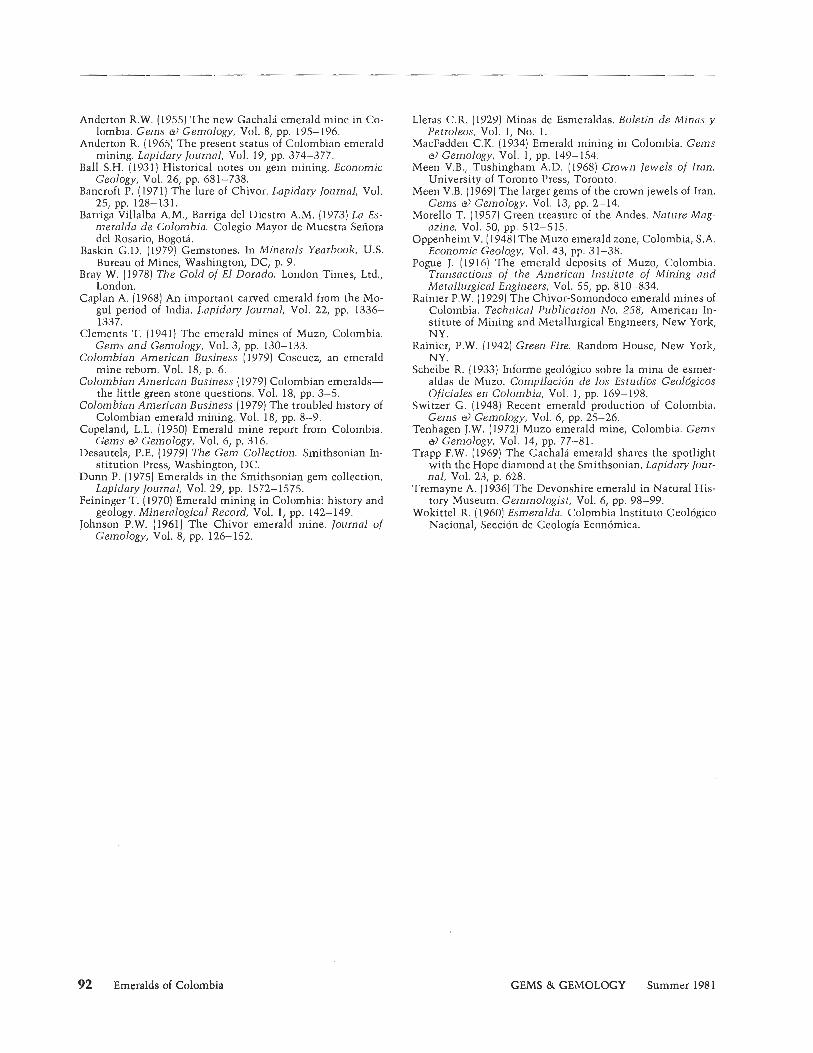
Anderton R.W. (1955) The new Gachala emerald mine in Co- lombia. Gems a) Gemology, Vol. 8, pp. 195-196.
Anderton R. (1965) The present status of Colombian emerald mining. Lapidary Journal, Vol. 19, pp. 374-377.
Ball S.H. (1931) Historical notes on gem mining. Economic Geology, Vol. 26, pp. 681-738.
Bancroft P. (1971) The lure of Chivor. Lapidary Journal, Vol. 25, pp. 128-131.
Barriga Villalba A.M., Barriga del Diestro A.M. (1973) La Es- meralda de Colombia. Colegio Mayor de Muestra Senora del Rosario, Bogota.
Baskin G.D. (1979) Gemstones. In Minerals Yearbook, U.S. Bureau of Mines, Washington, DC, p. 9.
Bray W. (1978) The Gold of El Dorado. London Times, Ltd., London.
Caplan A. (1968) An important carved emerald from the Mo- gul period of India. Lapidary Journal, Vol. 22, pp. 1336- 1337.
Clements T. (1941) The emerald mines of Muzo, Colombia. Gems and Gemology, Vol. 3, pp. 130-133.
Colombian American Business ( 1979) Coscuez, an emerald mine reborn. Vol. 18, p. 6.
Colombian American Business (1979) Colombian emeralds- the little green stone questions. Vol. 18, pp. 3-5.
Colombian American Business (1979) The troubled history of Colombian emerald mining. Vol. 18, pp. 8-9.
Copeland, L.L. (1950) Emerald mine report from Colombia. Gems St) Gemology, Vol. 6, p. 316.
Desautels, P.E. (1979) The Gem Collection. Smithsonian In- stitution Press, Washington, DC.
Dunn P. (1975) Emeralds in the Smithsonian gem collection. Lapidary Journal, Vol. 29, pp. 1572-1575.
Feininger T. (1970) Emerald mining in Colombia: history and geology. Mineralogical Record, Vol. 1, pp. 142-149.
Johnson P.W. (1961) The Chivor emerald mine. Journal of Gemology, Vol. 8, pp. 126- 152.
Lleras C.R. (1929) Minas de Esmeraldas. Boletin de Minas y Petroleos, Vol. 1, No. 1.
MacFadden C.K. (1934) Emerald mining in Colombia. Gems a) Gemology, Vol. 1, pp. 149- 154.
Meen V.B., Tushingham A.D. (1968) Crown Jewels of Iran. University of Toronto Press, Toronto.
Meen V.B. (1969) The larger gems of the crown jewels of Iran. Gems a) Gemology, Vol. 13, pp. 2-14.
Morello T. (19571 Green treasure of the Andes. Nature Mag- azine, Vol. 50, pp. 512-515.
Oppenheim V. (1948) The Muzo emerald zone, Colombia, S.A. Economic Geology, Vol. 43, pp. 31-38.
Pogue J. (1916) The emerald deposits of Muzo, Colombia. Transactions of the American Institute of Mining and Metallurgical Engineers, Vol. 55, pp. 810-834.
Rainier P.W. (1929) The Chivor-Somondoco emerald mines of Colombia. Technical Publication No. 258, American In- stitute of Mining and Metallurgical Engineers, New York, NY.
~aik ie r , P.W. (1942) Green Fire. Random House, New York, NY.
Scheibe R. (1933) Informe geologic0 sobre la mina de esrner- aldas de Muzo. Compilacion de 10s Estudios Geologicos Oficiales en Colombia, Vol. 1, pp. 169- 198.
Switzer G. (1948) Recent emerald production of Colombia. Gems a) Gemology, Vol. 6, pp. 25-26.
Tenhagen J.W. (1972) Muzo emerald mine, Colombia. Gems St) Gemology, Vol. 14, pp. 77-81.
Trapp F.W. (1969) The Gachala emerald shares the spotlight with the Hope diamond at the Smithsonian. Lapidary lour- nal, Vol. 23, p. 628.
Tremayne A. (1936) The Devonshire emerald in Natural His- tory Museum. Gemmologist, Vol. 6, pp. 98-99.
Wokittel R. (1960) Esmeralda. Colombia Institute Geoldgico National, Seccion de Geologia Economica.
92 Emeralds of Colombia GEMS & GEMOLOGY Summer 198 1
A N D
N E W T E C H N I Q U E S
A CUBIC ZIRCONIA REFRACTOMETER By C. S. Hurlbut , Jr.
With the development by C. D. West o f practical high- refractive-index liquids, the major barrier to extending the range and durability of the refractometer has been the need for an isotropic substance with an index of refraction higher than that o f the glass commonly used. On the basis of the author's experiments with a cubic zirconia refractometer, CZ-with its relatively l o w cost, high refractive index, and hardness-shows great promise for meeting this need.
Since the development of a direct-reading jew- eler's refractometer by G. F. Herbert Smith (1907)) numerous changes have been incorporated into the design of such instruments. However, the basic principle on which they operate remains the same: that is, the refractive index (R.I.) of the gemstone is determined by means of the stone's total reflection and critical angle. Light entering the instrument passes through a dense glass, im- pinges on the gemstone, and, at the critical angle, is totally reflected back through the glass. The dense, highly refractive glass may be in the form of a hemisphere, as in early models, or a hemi- cylinder or prism.The glass and the liquid that is interposed between it and the gemstone must both have a refractive index higher than that of the gemstone. Thus, these two items (but usually the liquid) set the upper limit to R.I. deter- minations.
The liquid most commonly used today has a refractive index of about 1.8 1. While glass contin- ues to be the most common isotropic substance
used, some refractometers have been constructed using diamond, sphalerite, and, most recently, strontium titanate and cubic zirconia (CZ) in place of glass. The ready availability, high refractive in- dex, and hardness of cubic zirconia, together with other properties, suggest that it may be particu- larly well suited for use in the refractometer.
REFRACTOMETERS WITH HIGH REFRACTIVE INDEX READINGS The two basic requirements for increasing the range of a refractometer are: (1) an isotropic sub- stance with an index of refraction higher than that of available glass, and (2) a liquid with a refractive index above that of commonly encoun- tered gemstones. When C. D. West (1936) devel- oped practical liquids in the refractive index range of 1.78-2.06, one of these problems was over- come. But what highly refractive, transparent, isotropic mineral could be used in place of glass? B. W. Anderson and C. J. Payne (1939) found two answers to this question: diamond and sphalerite (blende).
ABOUT THE AUTHOR
Dr. Hurlbut is professor emeritus in the Department of Geological Sciences, Harvard University, Cambridge, MA
Acknowledgments: The author is greatly indebted to the Ceres Corporation, Waltham, MA, for supplying the cubic zirconia from which the hemicylinders were cut; and to the Gemological Institute of America for its continuing interest in this project and for furnishing the Duplex I1 refractometers for modification.
'198 1 Gemological Institute of America
Notes and New Techniques GEMS & GEMOLOGY Summer 1981 93
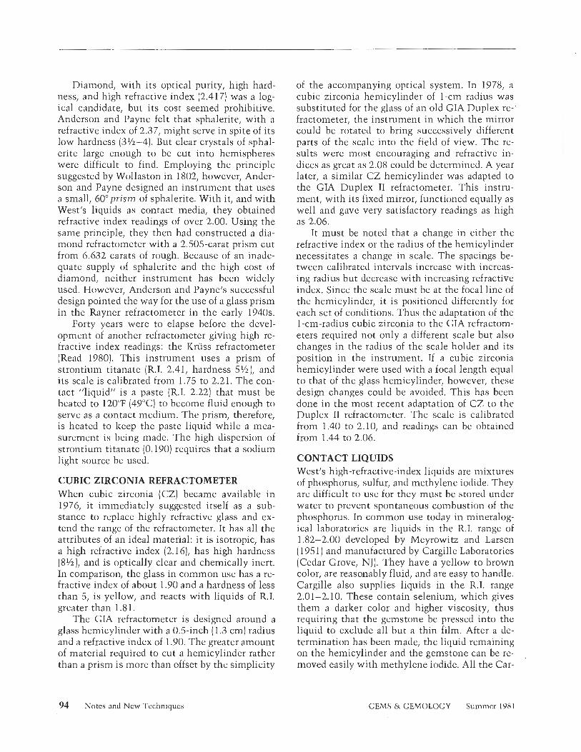
Diamond, with its optical purity, high hard- ness, and high refractive index (2.417) was a log- ical candidate, but its cost seemed prohibitive. Anderson and Payne felt that sphalerite, with a refractive index of 2.37, might serve in spite of its low hardness (3%-4). But clear crystals of sphal- erite large enough to be cut into hemispheres were difficult to find. Employing the principle suggested by Wollaston in 1802, however, Ander- son and Payne designed an instrument that uses a small, 60°pris of sphalerite. With it, and with West's liquids as contact media, they obtained refractive index readings of over 2.00. Using the same principle, they then had constructed a dia- mond refractometer with a 2.505-carat prism cut from 6.632 carats of rough. Because of an inade- quate supply of sphalerite and the high cost of diamond, neither instrument has been widely used. However, Anderson and Payne's successful design pointed the way for the use of a glass prism in the Rayner refractoinetcr in the early 1940s.
Forty years were to elapse before the devel- opment of another refractometer giving high re- fractive index readings: the Kriiss refractometer (Read 1980). This instrument uses a prism of strontium titanate (R.I. 2.41, hardness SYs), and its scale is calibrated from 1.75 to 2.21. The con- tact "liquid" is a paste (R.I. 2.22) that must be heated to 120° (49OC) to become fluid enough to serve as a contact medium. The prism, therefore, is heated to keep the paste liquid while a mea- surement is being made. The high dispersion of strontium titanate (0.190) requires that a sodium light source be used.
CUBIC ZIRCONIA REFRACTOMETER When cubic zirconia (CZ) became available in 1976, i t immediately suggested itself as a sub- stance to replace highly refractive glass and ex- tend the range of the refractometer. It has all the attributes of an ideal material: i t is isotropic, has a high refractive index (2.161, has high hardness (8V2), and is optically clear and chemically inert. In comparison, the glass in common use has a re- fractive index of about 1.90 and a hardness of less than 5, is yellow, and reacts with liquids of R.I. greater than 1.8 1.
The GIA refractometer is designed around a glass hemicylinder with a 0.5-inch (1.3 cm) radius and a refractive index of 1.90. The greater amount of material required to cut a hemicylinder rather than a prism is more than offset by the simplicity
of the accompanying optical system. In 1978, a cubic zirconia hemicylinder of 1-cin radius was substituted for the glass of an old GIA Duplex re-' fractometer, the instrument in which the mirror could be rotated to bring successively different parts of the scale into the field of view. The re- sults were most encouraging and refractive in- dices as great as 2.08 could be detern~ined. A year later, a similar CZ hemicylinder was adapted to the GIA Duplex I1 refractometer. This instru- ment, with its fixed mirror, functioned equally as well and gave very satisfactory readings as high as 2.06.
It must be noted that a change in either the refractive index or the radius of the hemicylinder necessitates a change in scale. The spacings be- tween calibrated intervals increase with increas- ing radius but decrease with increasing refractive index. Since the scale must be at the focal line of the hemicylinder, i t is positioned differently for each set of conditions. Thus the adaptation of the 1-cm-radius cubic zirconia to the GIA refractom- eters required not only a different scale but also changes in the radius of the scale holder and its position in the instrument. If a cubic zirconia hemicylinder were used with a focal length equal to that of the glass hemicylinder, however, these design changes could be avoided. This has been done in the most recent adaptation of CZ to the Duplex I1 refractometer. The scale is calibrated from 1.40 to 2.10, and readings can be obtained from 1.44 to 2.06.
CONTACT LIQUIDS West's high-refractive-index liquids are mixtures of phosphorus, sulfur, and methylene iodide. They are difficult to use for they must be stored under water to prevent spontaneous combustion of the phosphorus. In common use today in mineralog- ical laboratories are liquids in the R.I. range of 1.82-2.00 developed by Meyrowitz and Larsen (1951) and manufactured by Cargille Laboratories (Cedar Grove, NJ). They have a yellow to brown color, are reasonably fluid, and are easy to handle. Cargille also supplies liquids in the R.I. range 2.01-2.10. These contain selenium, which gives them a darker color and higher viscosity, thus requiring that the gemstone be pressed into the liquid to exclude all but a thin film. After a de- termination has been made, the liquid remaining on the hemicylinder and the gemstone can be re-
,
moved easily with methylene iodide. All the Car-
94 Notes and New Techniques GEMS & GEMOLOGY Summer 198 1
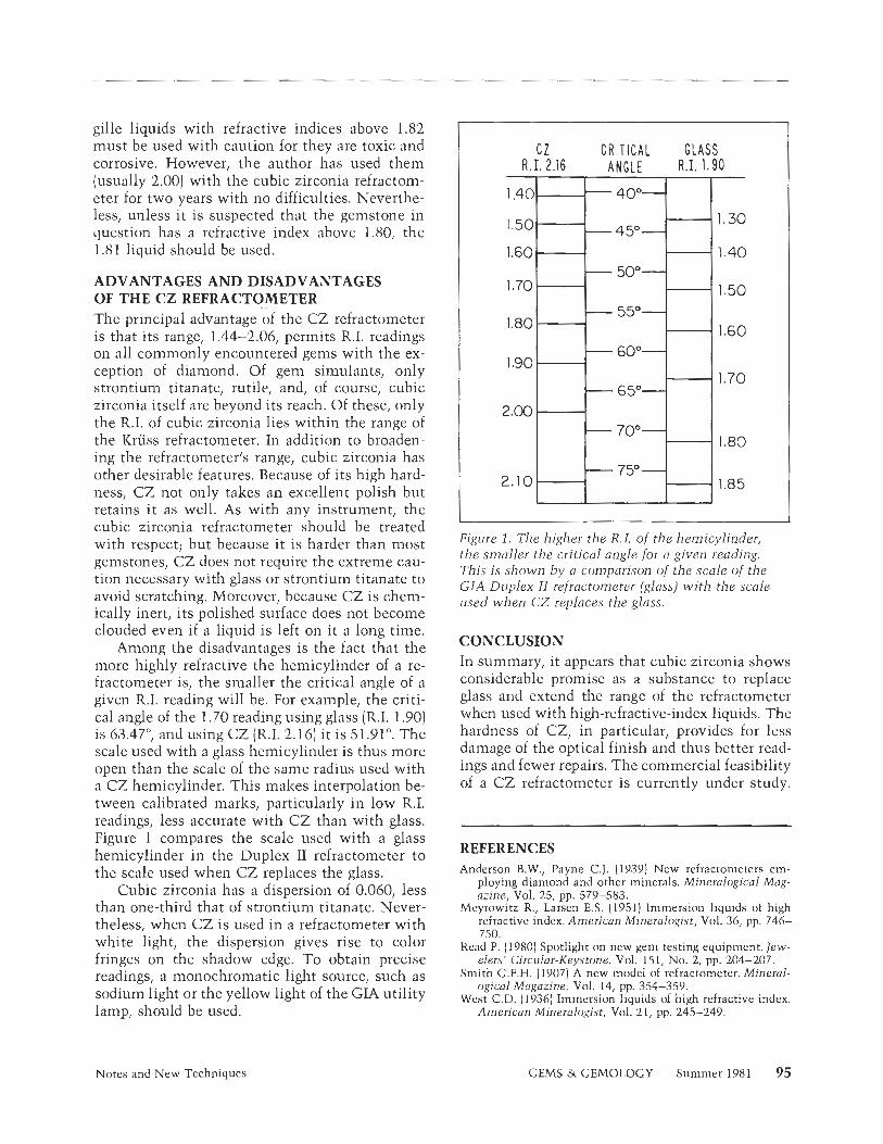
gille liquids with refractive indices above 1.82 must be used with caution for they are toxic and corrosive. However, the author has used them (usually 2.00) with the cubic zirconia refractom- eter for two years with no difficulties. Neverthe- less, unless i t is suspected that the gemstone in question has a refractive index above 1.80, the 1.81 liquid should be used.
ADVANTAGES AND DISADVANTAGES OF THE CZ REFRACTOMETER The principal advantage of the CZ refractometer is that its range, 1.44-2.06, permits R.I. readings on all commonly encountered gems with the ex- ception of diamond. Of gem siinulants, only strontium titanate, rutile, and, of course, cubic zirconia itself are beyond its reach. Of these, only the R.I. of cubic zirconia lies within the range of the Kriiss refractometer. In addition to broaden- ing the refractometerfs range, cubic zirconia has other desirable features. Because of its high hard- ness, CZ not only takes an excellent polish but retains i t as well. As with any instrument, the cubic zirconia refractometer should be treated with respect; but because it is harder than most gemstones, CZ does not require the extreme cau- tion necessary with glass or strontium titanate to avoid scratching. Moreover, because CZ is chem- ically inert, its polished surface does not become clouded even if a liquid is left on it a long time.
Among the disadvantages is the fact that the more highly refractive the hemicylinder of a re- fractoineter is, the smaller the critical angle of a given R.I. reading will be. For example, the criti- cal angle of the 1.70 reading using glass (R.I. 1.90) is 63.47' and using CZ (R.I. 2.16) it is 51.91'. The scale used with a glass hemicylinder is thus more open than the scale of the same radius used with a CZ hemicylinder. This makes interpolation be- tween calibrated marks, particularly in low R.I. readings, less accurate with CZ than with glass. Figure 1 compares the scale used with a glass hemicylinder in the Duplex I1 refractometer to the scale used when CZ replaces the glass.
Cubic zirconia has a dispersion of 0.060, less than one-third that of strontium titanate. Never- theless, when CZ is used in a refractometer with white light, the dispersion gives rise to color fringes on the shadow edge. To obtain precise readings, a monochromatic light source, such as sodium light or the yellow light of the GLA utility lamp, should be used.
C 2 C R I T I C A L G L A S S R . I . 2.16 ANGLE R . I . 1.90
Figure 1 . The higher the R.I. o f the hemicylinder, the smaller the critical angle for u given reading. This is shown b y a comparison of the scale of the GIA Duplex I1 refractometer (glass) with the scale used when CZ replaces the glass.
CONCLUSION In summary, it appears that cubic zirconia shows considerable promise as a substance to replace glass and extend the range of the refractometer when used with high-refractive-index liquids. The hardness of CZ, in particular, provides for less damage of the optical finish and thus better read- ings and fewer repairs. The commercial feasibility of a CZ refractometer is currently under study.
REFERENCES Anderson B.W., Payne C.J. (1939) New refractometers em-
ploying diamond and other minerals. Mineruloglcal Mag- azine, Vol. 25, pp. 579-583.
Meyrowitz R , Larsen E.S. (1951) Immersion liquids of high refractive index. American Mineralogist, Vol. 36, pp. 746- 750.
Read P. (1980) Spotlight on new gem testing equipment. few- elers' Circular-Keystone, Vol. 151, No. 2, pp. 204-207.
Smith G.F.H. (1907) A new model of refractometer. Mineral- ogical Magazine, Vol. 14, pp. 354-359.
West C.D. (1936) Immersion liquids of high refractive index. American Mineralogist, Vol. 21, pp. 245-249.
Notes and New Techniques GEMS & GEMOLOGY Summer 1981 95
HORNBILL IVORY B y Robert E. Kane
A rare gem material is found 111 the skull of the hel- meted hornbill, an exotic bird native to Southeast Asia. This yellowish-white material with its unique, vibrant, brownish-red coating has been valued for carvings by the Chinese and other Asian societies for many centuries and has recently captivated Western collectors as well. The author reviews the nature of this material and some of the forms into which it has
, been fashioned, as well as means of identifying horn- bill ivory from other ivories and from imitations.
Figure 1. An exquisitely carved hornbill ivory snuff bottle measuring approximately 70 m m high by 44 m m wide by 15 m m thick. This bottle is reportedly over 300 years old and is of Chinese origin. The Chinese call this highly regarded material ho-ting, the Chinese version of the Malaysian word gading, which means ivory.
T h e world of gems includes numerous minerals and some organic materials. Among the organic materials, one of the rarest and most unusual is a substance known as hornbill ivory. Figure 1 shows an exquisitely carved hornbill ivory snuff bottle that was recently brought into the Los An- geles office of the GIA Gem Trade Laboratory.
Hornbill ivory is not a true ivory. Ivory-or, more correctly, dentine - is a substance that comprises teeth or teeth modifications such as the tusks of mammals (Webster, 1975). The ma- terial used for hornbill ivory carvings is cut from the casque (the large protuberance covering a por- tion of the head and bill) of the helmeted hornbill. These unusual birds grow to about five feet in length and have large heads and bills. They live in tall trees in the jungles of Southeast Asia and range from Tenasserim southward over the Malay Peninsula to Borneo and Sumatra (Lint, 1 972).
The hornbill family of birds consists of many species; the helmeted hornbill, Rhinoplax vigil, is the only species that possesses ivory suitable for ornamental carving. The helmeted hornbill is unique in that it is the only species that has a solid casque; all other large hornbill casques are light and spongy inside (Lint, 1972). The casque of the helmeted hornbill is primarily a solid yel- lowish-white to whitish-yellow material with a very thin outer covering that is an intense brown- ish red.
Hornbill casques have been carved for many centuries. Carvings over 2,000 years old have been found in Borneo, and Chinese carvings have been traced back to the Tang Dynasty (618-907 A.D.) The Chinese had a high regard for this material and have called it "golded jade" (Munsuri, 1973). In addition to snuff bottles, hornbill ivory has been carved into such items as rings, bracelets, spoons, and belt buckles. In some cases, the en- tire skull has been kept intact and portions of the
ABOUT THE AUTHOR
Mr. Kane is gem identification supervisor in the Gem Trade Laboratory, Inc., Los Angeles, CA.
"1981 Gemological Institute of America
96 Notes and New Techniques GEMS & GEMOLOGY Summer 1981
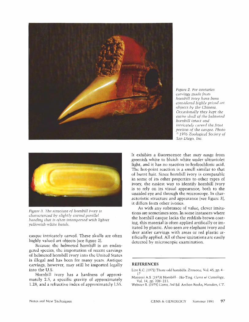
Figure 3. The structure of hornbill ivory is characterized by slightly curved parallel banding that is often interspersed with lighter yellowish-white bands,
casque intricately carved. These skulls are often
Figure 2. For centuries carvings made from hornbill ivory have been considered highly prized art objects by the Chinese. Occasionally they kept the entire skull of the helmeted hornbill intact and intricately carved the front portion of the casque. Photo ' 1976 Zoological Society of Son Diego, Inc.
It exhibits a fluorescence that may range from greenish white to bluish white under ultraviolet light, and it has no reaction to hydrochloric acid. The hot-point reaction is a smell similar to that of burnt hair. Since hombill ivory is comparable in some of its other properties to other types of ivory, the easiest way to identify hombill ivory is to rely on its visual appearance, both to the unaided eye and through the microscope. In char- acteristic structure and appearance (see figure 3), it differs from other ivories.
As with any substance of value, clever imita- tions are sometimes seen. In some instances where the hornbill casque lacks the reddish-brown coat- ing, this material is often applied artifically or im- itated by plastic. Also seen are elephant ivory and deer antler carvings with areas or red plastic ar- tificially applied. All of these imitations are easily detected by microscopic examination. highly valued art objects (see figure 2).
Because the helmeted hombill is an endan- gered species, the importation of recent carvings of helmeted hombill ivory into the United States is illegal and has been for many years. Antique carvings, however, may still be imported legally REFERENCES into the U.S. Lint K.C. (19721 Those odd hornbills. Zoonooz, Vol. 45, pp. 4-
Hornbill ivory has a hardness of approxi- mately 2.5, a specific gravity of approximately 1.28, and a refractive index of approximately 1.55.
. ..
17. Munsuri A.S. (1973) Hornbill-Ho-Ting. Gems &> Gemology,
Vol. 14, pp. 208-21 1. Webster R. (1975) Gems, 3rd Ed. Archon Books, Hamden, CT.
Notes and New Techniques GEMS & GEMOLOGY Summer 1981 97
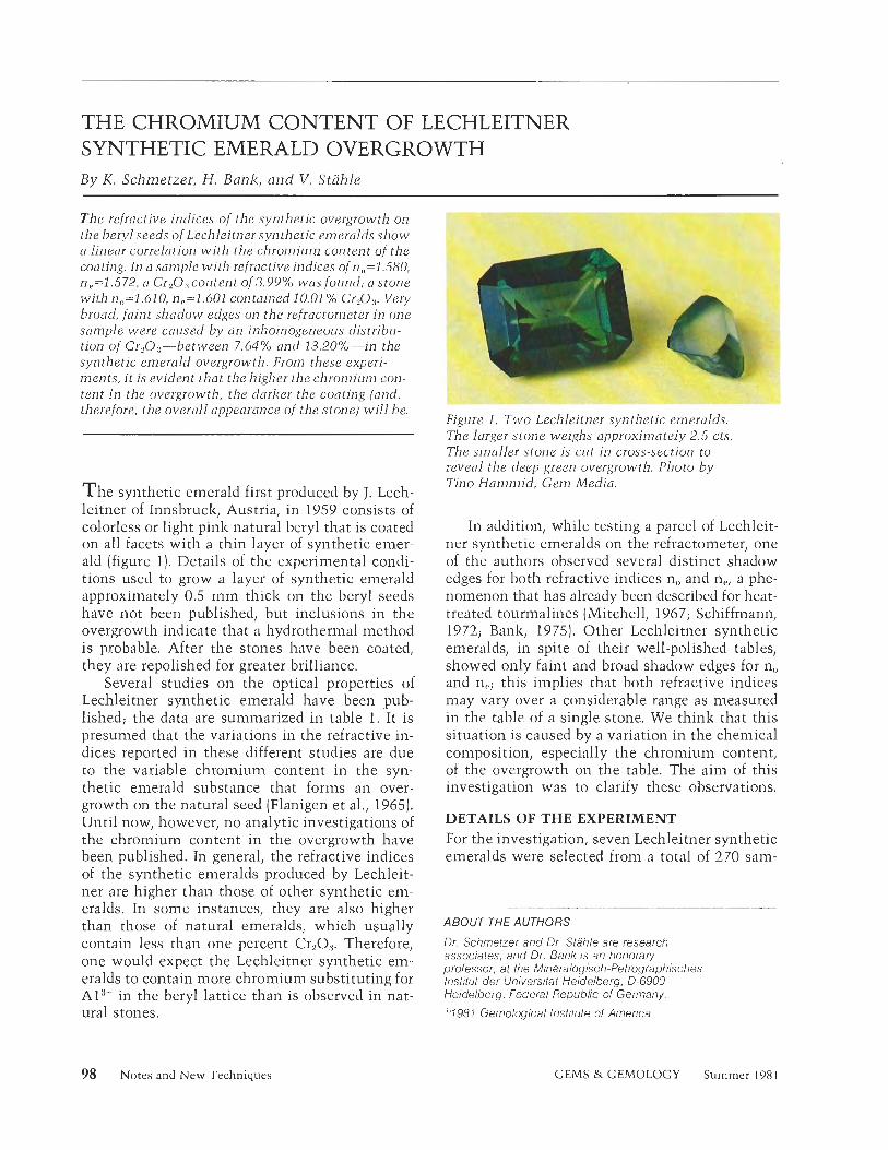
THE CHROMIUM CONTENT OF LECHLEITNER SYNTHETIC EMERALD OVERGROWTH B y K. Schmetzer, H. B a n k , and V . Stahle
The refractive indices of the synthetic overgrowth on the beryl seeds of Lechleitner synthetic emeralds show a linear correlation with the chromium content of the coating. In a san7ple with refractive indices of n1,=1.580, nC1=1.572, a Cr20:,content of 3.99% was found; a stone with n1=1.610. n.,=1.601 contained 10.01 % Cr,O..,. Very broad, faint shadow edges on the refractometer in one sample were caused by an inhomogeneous distribu- tion of Cr,O;,-between 7.64% and 13.20%-in the synthetic emerald overgrowth. From these experi- ments, it is evident that the higher the chromium con- tent in the overgrowth, the darker the coaling (and, therefore, the overall appearance of the stone) will be.
T h e synthetic emerald first produced by J. Lech- leitner of Innsbruck, Austria, in 1959 consists of colorless or light pink natural beryl that is coated on all facets with a thin layer of synthetic emer- ald (figure 1). Details of the experimental condi- tions used to grow a layer of synthetic emerald approximately 0.5 m m thick on the beryl seeds have not been published, but inclusions in the overgrowth indicate that a hydrothermal method is probable. After the stones have been coated, they are repolished for greater brilliance.
Several studies on the optical properties of Lechleitner synthetic emerald have been pub- lished; the data are summarized in table 1. It is presumed that the variations in the refractive in- dices reported in these different studies are due to the variable chromium content in the syn- thetic emerald substance that forms an over- growth on the natural seed (Flanigen et al., 1965). Until now, however, no analytic investigations of the chromium content in the overgrowth have been published. In general, the refractive indices of the synthetic emeralds produced by Lechleit- ner are higher than those of other synthetic em- eralds. In some instances, they are also higher than those of natural emeralds, which usually contain less than one percent Cr&. Therefore, one would expect the Lechleitner synthetic em- eralds to contain more chromium substituting for A13+ in the beryl lattice than is observed in nat- ural stones.
Figure 1. Two Lechleitner synthetic emeral~.,. The larger stone weighs approximately 2.5 cts. The smaller stone is cut in cross-section to reveal the deep green overgrowth. Photo by Tino Hammid, Gem Media.
In addition, while testing a parcel of Lechleit- ner synthetic emeralds on the refractometer, one of the authors observed several distinct shadow edges for both refractive indices no and ne, a phe- nomenon that has already been described for heat- treated tourmalines (Mitchell, 1967; Schiffmann, 1972; Bank, 1975). Other Lechleitner synthetic emeralds, in spite of their well-polished tables, showed only faint and broad shadow edges for n;, and np; this implies that both refractive indices may vary over a considerable range as measured in the table of a single stone. We think that this situation is caused by a variation in the chemical composition, especially the chromium content, of the overgrowth on the table. The aim of this investigation was to clarify these observations.
DETAILS OF THE EXPERIMENT For the investigation, seven Lechleitner synthetic emeralds were selected from a total of 270 sam-
ABOUT THE AUTHORS
Dr. Schmetzer and Dr. Stahle are research associates, and Dr. Bank is an honorary professor, at the Mineralogisch-Pelrographisches Inslilul der Universital Heidelberg, 0-6900 Heidelberg, Federal Republic of Germany.
"1981 Gemological Instilute of America
98 Notes and New Techniques GEMS & GEMOLOGY Summer 198 1
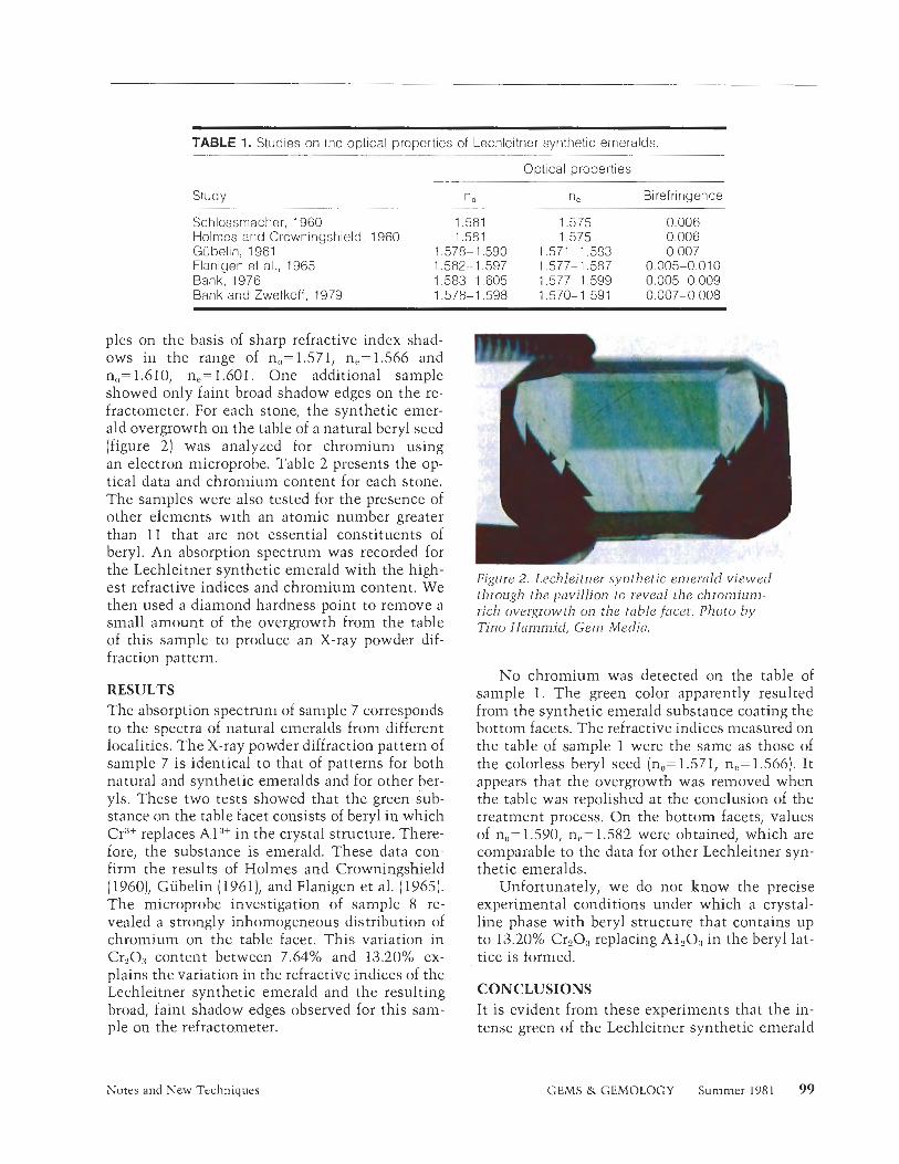
TABLE 1. Studies on the optical properties of Lechleitner synthetic emeralds.
Optical properties
Study .. . . . .- - .
n o - --
Schlossrnacher, 1960 1.581 Holmes and Crowningshield, 1960 1.581 Gubelin, 1961 1.578- 1.590 Flanigen et al,, 1965 1.582- 1.597 Bank, 1976 1.583-1.605 Bank and Zwetkoff, 1979 1.578-1.598
ne Birefringence
1.575 0.006 1.575 0.006
1.571 -1.583 0.007 1.577-1.587 0.005-0,010 1.577-1.599 0.005--0.009 1.570-1.591 0.007-0.008
pies on the basis of sharp refractive index shad- ows in the range of nu=1.571, 11~~1.566 and no= 1.610, ne= 1.601. One additional sample showed only faint broad shadow edges on the re- fractometer. For each stone, the synthetic emer- ald overgrowth on the table of a natural beryl seed (figure 21 was analyzed for chromium using an electron microprobe. Table 2 presents the op- tical data and chromium content for each stone. The samples were also tested for the presence of other elements with an atomic nunlber greater than 11 that are not essential constituents of beryl. An absorption spectrum was recorded for the Lechleitner synthetic emerald with the high- est refractive indices and chromium content. We then used a diamond hardness point to remove a small amount of the overgrowth from the table of this sample to produce an X-ray powder dif- fraction pattern.
RESULTS The absorption spectrum of sample 7 corresponds to the spectra of natural emeralds from different localities. The X-ray powder diffraction pattern of sample 7 is identical to that of patterns for both natural and synthetic emeralds and for other ber- yls. These two tests showed that the green sub- stance on the table facet consists of beryl in which CrC1+ replaces Al-"+ in the crystal structure. There- fore, the substance is emerald. These data con- firm the results of Holmes and Crowningshield (1960)) Gubelin (1961), and Flanigen et al. (1965). The microprobe investigation of sample 8 re- vealed a strongly inhomogeneous distribution of chromium on the table facet. This variation in Cr203 content between 7.64% and 13.20% ex- plains the variation in the refractive indices of the Lechleitner synthetic emerald and the resulting broad, faint shadow edges observed for this Sam- ple on the refractometer.
Figure 2. Lechleitner synthetic emerald viewed through the pavillion to reveal the chromium- rich overgrowth on the table facet. Photo by Tino Hammid, Gem Media.
No chromium was detected on the table of sample 1. The green color apparently resulted from the synthetic emerald substance coating the bottom facets. The refractive indices measured on the table of sample 1 were the same as those of the colorless beryl seed (no= 1.57 1, ne= 1.566). It appears that the overgrowth was removed when the table was repolished at the conclusion of the treatment process. On the bottom facets, values of no= 1.590, ne= 1.582 were obtained, which are comparable to the data for other Lechleitner syn- thetic emeralds.
Unfortunately, we do not know the precise experimental conditions under which a crystal- line phase with beryl structure that contains up to 13.20% CraO:) replacing A1203 in the beryl lat- tice is formed.
CONCLUSIONS It is evident from these experiments that the in- tense green of the Lechleitner synthetic emerald
Notes and New Techniques GEMS & GEMOLOGY Summer 1981 99
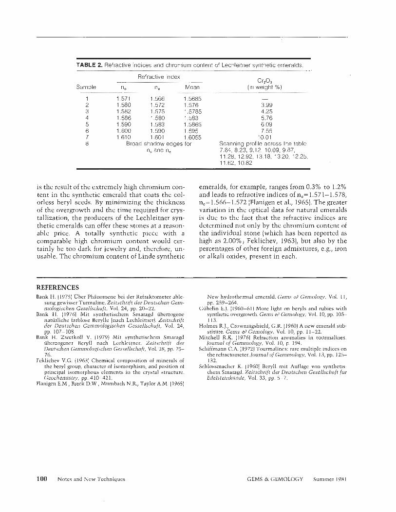
TABLE 2. Refractive indices and chromium content of Lechleitner synthetic emeralds.
Refractive index
Sample no ne Mean Cr203
(in weight %) - -
1 1.571 1.566 1.5685 -
2 1.580 1.572 1.576 3.99 3 1.582 1.575 1.5785 4.25 4 1.586 1.580 1.583 5.76 5 1.590 1.583 1 5865 6.09 6 1.600 1.590 1.595 7.55 7 1.610 1.601 1.6055 10.01 8 Broad shadow edges for Scanning profile across the table:
no and n, 7.64, 8.23, 9.12, 10.09, 9.87, 11.28, 12.92, 13.18, 13.20, 12.25, 11.62, 10.82
is the result of the extremely high chromium con- tent in the synthetic emerald that coats the col- orless beryl seeds. By minimizing the thickness of the overgrowth and the time required for crys- tallization, the producers of the Lechleitner syn- thetic emeralds can offer these stones at a reason- able price. A totally synthetic piece with a comparable high chromium content would cer- tainly be too dark for jewelry and, therefore, un- usable. The chromium content of Linde synthetic
emeralds, for example, ranges from 0.3% to 1.2% and leads to refractive indices of no= 1.571-1.578, ne= 1.566- 1.572 (Flanigen et al., 1965). The greater variation in the optical data for natural emeralds is due to the fact that the refractive indices are determined not only by the chromium content of the individual stone (which has been reported as high as 2.00%; Felzlichev, 1963), but also by the percentages of other foreign admixtures, e.g., iron or alkali oxides, present in each.
REFERENCES Bank H. (19751 Uber Phsnomenc bei der Refraktometer able-
sung gewisser Turmaline. Zeitschrift der Dentschen Cem- mologischen Gesellschaft, Vol. 24, pp. 20-22.
Bank H. (1976) Mit synthetischem Smaragd uberzogene naturliche farblose Berylle (nach Lechleitner]. Zeitschrift der Deutschen Gemmologischen Gessellschaft, Vol. 24, pp. 107-108.
Bank H. Zwetkoff V. (1979) Mit synthetischcm Smaragd uberzogener Beryll nach Lechleitner. Zeitschrift der Deutschen Gemmo1ogisc11en Gessellschaft, Vol. 28, pp. 75- 76
~eki ichev V.G. (1963) Chemical composition of minerals of the beryl group, character of ison~orphism, and position of principal isornorphous elements in the crystal structure. Geochemistry, pp. 4 10-42 1.
Flanigen E.M., Breck D.W., Mumbach N.R., Taylor A.M. (19651
New hydrothermal emerald. Gems ei) Gemology, Vol. 11, pp. 259-264,
Giibelin E.J. (1960-61) More light on beryls and rubies with synthetic overgrowth. Gems a) Gemology, Vol. 10, pp. 105- 113
Holmes R.J., Crowningshield, G.R. (19601 A new emerald sub- stitute. Gems oJ Gemology, Vol. 10, pp. 1 1-22.
Mitchell R.K. (1976) Refraction anomalies in tourmalines. Journal of Gemmology, Vol. 10, p. 194.
Schiffmann C.A. (1972) Tourmalines: rare multiple indices on the refractometer. fournal of Gemmology, Vol. 13, pp. 125- 132.
Schlossmacher K. (1960) Beryll mit Auflage von synthetis- chem Smaragd. Zeitschrift der Devtschen Gesellschaft fur Edelsteinkunde, Vol. 33, pp. 5-7.
100 Notes and New Techniques GEMS & GEMOLOGY Summer 1981
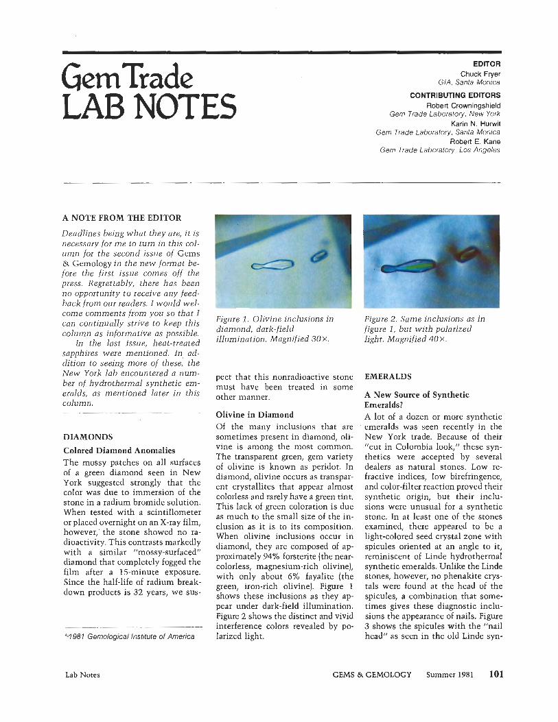
LAB NOTES EDITOR
Chuck Fryer GIA, Santa Monica
CONTRIBUTING EDITORS Robert Crowningshield
Gem Trade Laboratory, New York Karin N. Hurwit
Gem Trade Laboratory, Santa Monica Robert E. Kane
Gem Trade Laboratory, Los Angeles
A NOTE FROM THE EDITOR
Deadlines being what they are, i t is necessary for m e to turn in this col- u m n for the second issue of Gems & Gemology i n the n e w format be- fore the first issue comes off the press. Regrettably, there has been n o opportunity t o receive any fee back from our readers. I would w e come comments from you so that I can continually strive t o keep this column as informative as possible.
In the last issue, heat-treated sapphires were mentioned. In, ad- dition to seeing more of these, the New York lab encountered a num- ber of hydrothermal synthetic em- eralds, as mentioned later i n this column.
DIAMONDS
Colored Diamond Anomalies The mossy patches on all surfaces of a green diamond seen in New York suggested strongly that the color was due to immersion of the stone in a radium bromide solution. When tested with a scintillometer or placed overnight on an X-ray film, however,' the stone showed no ra- dioactivity. This contrasts markedly with a similar "mossy-surfaced" diamond that completely fogged the film after a 15-minute exposure. Since the half-life of radium break- down products is 32 years, we sus-
'"1981 Gemological Institute of America
Figure 1. Olivine inclusions in diamond, dark-field illumination. Magnified 3 0 x .
pect that this nonradioactive stone must have been treated in some other manner.
Olivine in Diamond Of the many inclusions that are sometimes present in diamond, oli- vine is among the most common. The transparent green, gem variety of olivine is known as peridot. In diamond, olivine occurs as transpar- ent crystallites that appear almost colorless and rarely have a green tint. This lack of green coloration is due as much to the small size of the in- clusion as it is to its composition. When olivine inclusions occur in diamond, they are composed of ap- proximately 94% forsterite (the near- colorless, magnesium-rich olivine), with only about 6% fayalite (the green, iron-rich olivine). Figure 1 shows these inclusions as they ap- pear under dark-field illumination. Figure 2 shows the distinct and vivid interference colors revealed by po- larized light.
Figure 2, Same inclusions as i n figure 1, but wi th polarized light. Magnified 4 0 ~ -
EMERALDS
A New Source of Synthetic Emeralds? A lot of a dozen or more synthetic emeralds was seen recently in the New York trade. Because of their 'cut in Colombia look," these syn- thetics were accepted by several dealers as natural stones. Low re- fractive indices, low birefringence, and color-filter reaction proved their synthetic origin, but their inclu- sions were unusual for a synthetic stone. In at least one of the stones examined, there appeared to be a light-colored seed crystal zone with spicules oriented at an angle to it, reminiscent of Linde hydrothermal synthetic emeralds. Unlike the Linde stones, however, no phenakite crys- tals were found at the head of the spicules, a combination that some- times gives these diagnostic inclu- sions the appearance of nails. Figure 3 shows the spicules with the "nail head" as seen in the old Linde syn-
Lab Notes GEMS & GEMOLOGY Summer 198 1 101
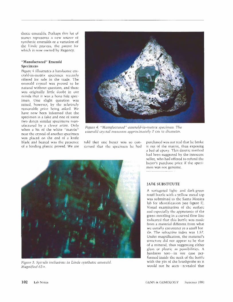
thetic emeralds. Perhaps this lot of stones represents a new source of synthetic en~eralds or a variation of the Linde process, the patent for which is now owned by Regency.
"Manufactured" Emerald Specimens Figure 4 illustrates a handsome em- erald-in-matrix specimen recently offered for sale in the trade. The emerald crystal was proved to be natural without question, and there was originally little doubt in our minds that it was a bona fide spec- imen. One slight question was raised, however, by the relatively reasonable price being asked. We have now been informed that the specimen is a fake and one of some two dozen similar specimens man-
-- - - - - -
ufactured by a 'lever artist ' Only Fig~ue 4, "Manu/ac~~,red" emerald-in-n~atrix specinlen. The when a bit of the white "matrix" emerald crystal measures approxin~ately 2 cn7 in diameter. near the crystal of another specimen was placed on the end o f a knife blade and heated was the presence told that one buyer was so con- purchased was not real that he broke of a binding plastic proved. We are cerned that the specimen he had it out of the matrix, thus exposing
a bed of epoxy. This drastic method had been suggested by the innocent seller, who had offered to refund the buyer's purchase price if the speci-
'1'. Â , men was not genuine.
JADE SUBSTITUTE
A variegated light- and dark-green snuff bottle with a yellow metal top was submitted to the Santa Monica lab for identification (see figure 5). Visual examination of the surface and especially the appearance of the green mottling in a curved flow line indicated that this bottle was made from a material different from what we usually encounter as a snuff bot-
I tle. The refractive index was 1.57. Under magnification, the material's structure did not appear to be that of a mineral, thus suggesting either glass or plastic as possibilities. A hardness test-in our case per- formed inside the neck of the bottle with the pin of the brushprobe so it would not be seen-revealed that
102 Lab Notes GEMS & GEMOLOGY Summer 1981
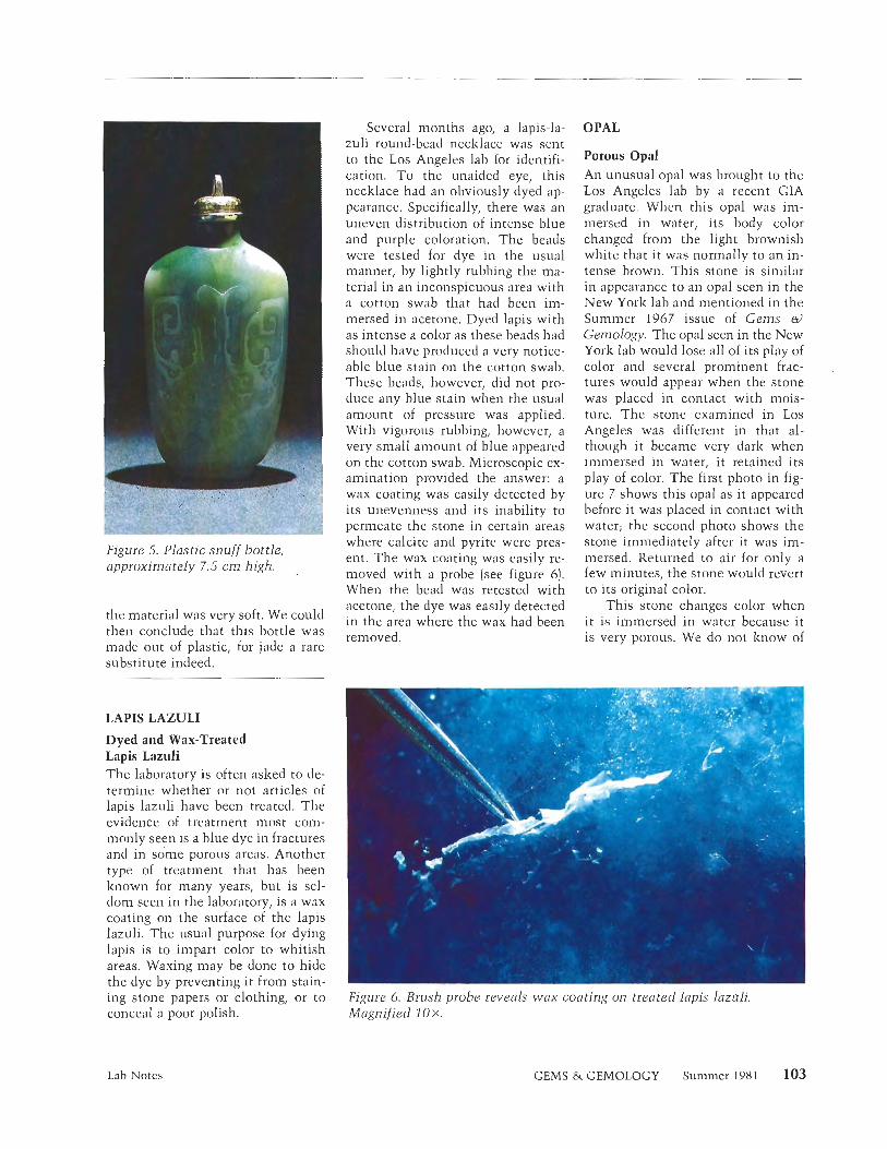
Figure 5. Plastic snuff bottle, approximately 7.5 cm high.
the material was very soft. We could then conclude that this bottle was made out of plastic, for jade a rare substitute indeed.
LAPIS LAZULI
Dyed and Wax-Treated Lapis Lazuli The laboratory is often asked to de- termine whether or not articles of lapis lazuli have been treated. The evidence of treatment most com- monly seen is a blue dye in fractures and in some porous areas. Another type of treatment that has been known for many years, but is sel- dom seen in the laboratory, is a wax coating on the surface of the lapis lazuli. The usual purpose for dying lapis is to impart color to whitish areas. Waxing may be done to hide the dye by preventing it from stain- ing stone papers or clothing, or to conceal a poor polish.
Lab Notes
Several months ago, a lapis-la- zuli round-bead necklace was sent to the Los Angeles lab for identifi- cation. T o the unaided eye, this necklace had an obviously dyed ap- pearance. Specifically, there was an uneven distribution of intense blue and purple coloration. The beads were tested for dye in the usual manner, by lightly rubbing the ma- terial in an inconspicuous area with a cotton swab that had been iin- mersed in acetone. Dyed lapis with as intense a color as these beads had should have produced a very notice- able blue stain on the cotton swab. These beads, however, did not pro- duce any blue stain when the usual amount of pressure was applied. With vigorous rubbing, however, a very small amount of blue appeared on the cotton swab. Microscopic ex- amination provided the answer: a wax coating was easily detected by its unevenness and its inability to permeate the stone in certain areas where calcite and pyrite were pres- ent. The wax coating was easily re- moved with a probe (see figure 61. When the bead was retested with acetone, the dye was easily detected in the area where the wax had been removed.
OPAL
Porous Opal An unusual opal was brought to the Los Angeles lab by a recent GIA graduate. When this opal was inl- mersed in water, its body color changed from the light brownish white that it was normally to :in in- tense brown. This stone is similar in appearance to an opal seen in the New York lab and mentioned in the Summer 1967 issue of Gems a)
Gemology. The opal seen in the New York lab would lose all of its play of color and several prominent frac- tures would appear when the stone was placed in contact with mois- ture. The stone examined in Los Angeles was different in that al- though i t became very dark when immersed in water, it retained its play of color. The first photo in fig- ure 7 shows this opal as it appeared before it was placed in contact with water; the second photo shows the stone immediately after it was im- mersed. Returned to air for only a few minutes, the stone would revert to its original color.
This stone changes color when it is immersed in water because it is very porous. We do not know of
GEMS & GEMOLOGY Summer 198 1
Figure 6. Brush probe reveals wax coating on treated lapis lazuli. Magnified 10 X.
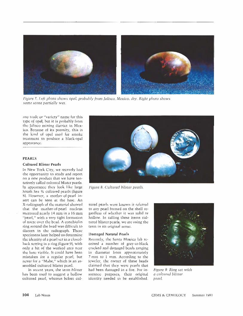
Figure 7. Left photo shows opal, probably from lalisco, Mexico, dry. Right photo shows same stone partially wet.
any trade or "variety" name for this type of opal, but i t is probably from the Jalisco mining district in Mex- ico. Because of its porosity, this is the kind of opal used for smoke treatment to produce a black-opal appearance.
PEARLS
Cultured Blister Pearls In New York City, we recently had the opportunity to study and report on a new product that we have ten- tatively called cultured blister pearls. In appearance they look like large South Sea ¥ cultured pearls (figure 8). However, a mother-of-pearl in- sert can be seen at the base. An X-radiograph of the material showed that the mother-of-pearl nucleus measured %-early 14 m m in a 16 m m "pearl," with a very tight formation of nacre over the bead. A conchiolin ring around the bead was difficult to discern in the radiograph. These specimens later helped u s determine the identity of a pearl set in a closed- back setting in a ring (figure 9)) with only a bit of the worked area near the base visible. It could have been mistaken for a regular pearl, but never for a "Mabe," which is an as- sembled cultured blister pearl.
In recent years, the term blister has been used to suggest a hollow cultured pearl, whereas before cul-
Figure 8. Cultured blister pearls.
tured pearls were known it referred to any pearl formed on the shell re- gardless of .whether i t was solid or hollow. In calling these items cul- tured blister pearls, we are using the term in its original sense.
Damaged Natural Pearls Recently, the Santa Monica lab re- ceived a number of gray-to-black cracked and damaged beads ranging in diameter from approxin~ately 7 m m to 1 mm. According to the jeweler, the owner of these beads claimed that they were pearls that had been damaged in a fire. For in- surance purposes, their original identity needed to be established.
Figure 9. Ring set with a cultured blister pearl.
104 LabNotes GEMS & GEMOLOGY Summer 1981
Sampling through the assortment, we found evidence of orient on some of the least damaged beads. This led us to believe that the beads indeed were pearls. An X-radiograph was taken to reveal their internal struc- ture, which proved that the beads were natural pearls.
--
RUBY
A natural ruby that was recently sent to our Los Angeles lab for identifi- cation contained a rather strikingly beautiful inclusion. While we were examining this ruby under the mi- croscope using dark-field illumina- tion, we noted the inclusion shown in figure 10. Knowing the nature of this type of "thin-film" inclusion, we switched off the dark-field illu- mination and employed vertical il- lumination. The result was the mul- ticolored inclusion shown in figure 11. Thin-film inclusions are not un- common in many minerals; they ap- pear in the form of ultrathin films fcommonlv 100 n m or less in thiclz- ness) composed of solids, liquids, gases, or any combination thereof.
A gemologist who knows how to recognize a thin-film inclusion and how to correctly illuminate i t will not only discover a pleasing pattern, but may also learn something more about the stone that would nor- mally have been overloolzecl.
SUGILITE
A small, thin slab of a new material from Africa was sent to Santa Mon- ica for identification. Figure 12 shows the beautiful purple color and delicate bandings of this stone. In its relatively brief history, this material has been tentatively identified by several different sources as sogdi- anite, sugilite, and even leuco- phoenicite. Extensive testing on the sample we received, including X-ray diffraction and chemical analysis, proved it to be manganoan sugilite.
Lab Notes
Figure 10. Thin-film inclusion in natural ruby, dark-field illumination. Magnified 25 X,
Figure 11. Same inclusion as in figure 10, but with vertical illumination. Magnified 50 X,
Figure 12. Banding in sugilite. Magnified 15 X.
Table 1 compares the refractive in- sample we received with those of dices and specific gravities of the the three minerals named. Table 2
TABLE 1. Refractive indices and specific gravities of sugilite, sogdianite, leucophoenicite, and the GTL sample.
Material Refractive index Specific gravity -.
Sugilite 1.607-1.610 2.74 Sogdianite 1,606-1.608 2.82-2.90 Leucophoenicite 1.75-1.78 3.85 Sample 1 .60a 2.75
"Hazy due to poor polish.
GEMS & GEMOLOGY Summer 1981
compares the eight strongest lines in the diffraction patterns for the four n~inerals.
Analysis by the energy disper- sive system of the electron micro- scope in the CIA research depart- ment confirmed the presence of manganese in addition to potas- sium, sodiun~, iron, and silicon, all essential elements in sugilite. Zir- conium is an essential element in sogdianite, and no zirconium was present in the sample tested. Leu- cophoenicite is a relatively simple manganese silicate hydroxide and would not have all these other ele- ments.
The color of sugilite does change with the type of lighting. It is a
bluish purple in daylight and a pleasing reddish purple under incan- descent light. The diagnostic ab- sorption spectrum as seen through a thin edge is shown in figure 13.
UNCLASSIFIED ODDITIES
A rather attractive polished slab came into the Santa Monica Lab for identification. The material con- sisted of small [ 1 to 2 mm) trans- parent to transluscent pink and green granules interspersed with larger areas of blue (see figure 14). The re- fractorneter revealed that the pinlz- to-red material was corundum, the green was tourmaline, and the blue,
TABLE 2. d spacing and relative intensity (I) of the eight strongest lines in the diffraction patterns for the GTL sample, sugilite, sogdianite, and leucophoenicite.
Sample Sugilite Sogdianite Leucophoenicite
Figure 13. Drawing of absorption spectrum of sugjfite.
figure 14. Slab containing a mixture of pink corundum, green tourmaline, and blue lzyanite. Magnified 20 x.
kyanite. The hexagonal zoning in the corundum, together with the constant nlaximuin birefringence readings of both the corundum and the tourmaline, indicates that these two constituents had grown so that their respective c-axes were parallel to each other and perpendicular to the polished face of the slab. The last time we encountered a similar material in our lab was in 1975. However, that particular sample did not have the lzyanite present.
ACKNOWLEDGMENTS
The photographs used in figures 1 , 2 , 5 , 10, and 11 were taken by Robert E . Kane of the Los Angeles lab. Figures 4, 7, and 8 were supplied by Rene Moore in New York. In Santa Monica, Tino Hammid is re- sponsible for figure 6, Mike Havstad for figure 9, and Chuck Fryer lor figures 3, 12, and 14. Chuck Fryer also prepared the absorption spectrum diagram in figure 13.
106 Lab Notes GEMS & GEMOLOGY Summer 198 1
Editorial Forum OUR READERS REVIEW THE NEW G&G
What a magnificent issue the G&G turned out to be! I cannot think of any aspect which can be improved. The color reproduction is superb, the articles are ex- cellent, and the whole lay-out conveys just that image of the one and only CIA.
Truly a "Gem"!
Kurt Nassau, P11.D. Bell Laboratories Murray Hill, NJ
I think the new Gems o) Gemology is simply beautiful! Congratulations on a magnificent job.
I would like to enter two subscriptions. . . . Donald E. Petersen Ford Motor Company Dearborn, MI
I was very pleased to receive the first issue of your "New Look" journal. It certainly is first class with ex- cellent colour reproductions and I wish you every suc- cess with this publication in the future.
H. Wheeler, Secretary Gemmological Association of
Great Britain London, England
The traditional issues of this quarterly publication were very educational, but I feel that so much more can be learned by jewelers with the aid of the color photo- graphs. The old saying is so relevant here, "a picture is worth a thousand words." Thank you for a very cur- rent, interesting, and educational publication.
Thomas A. Cleves Cleves & Lonneman, Registered
Jewelers Bellevue, KY
Congratulations on the really superb professional ap- proach in every one of the articles published in this is-
sue. As a reader of all gemological publications, I must honestly say that Gems o) Gemology is the first that I have read in years that gives such readable, in-depth study on the subjects covered. Keep up the good work.
Henry T. Levi, C.]. Levi Jewlers Nanticolze, PA
I felt I must write you a line to congratulate you and your fine organization on the magnificent first issue of the new format Gems a) Gemology. I must admit that when I first heard that this was coming I felt regret at the passing of the old familiar form of the journal, which has been a good friend for years, but improve- merit was due.
After reading this first issue, I feel despair in ever keeping pace with our galloping science. Who would have expected, after all those years, that major finds of diamonds would be made in Australia and in China. And how supermen like Edward [Giibelinj and K m Nassau find time and energy to pour out such riches of information acconlpanied by fine colour pictures passes my comprehension.
Basil W. Anderson Devon, England
The new Gems el Gemology is truly n~agnificent. It is certainly the "National Geo" of gemology.
E. A. "Ted" Brockie, C.G. Fort Frances, Ontario, Canada
Bravo!!
Wayne and Dona Leicht Kristalle Laguna Beach, CA
We are grateful for the time so many of our readers have taken to comment on the new Gems & Cemol- ogy, and regret that we could not include all o f your letters in our limited space. Thank you for your con- fidence and support. -Editor
Editorial Forum GEMS &. GEMOLOGY Summer 1981 107
DIAMOND PROBEQ3
"A Simple Approach to Detecting Diamond Simu- lants" (Spring 1981), by Jill Hobbs, is a very informative article. However, I am most disturbed by the flagrant use of the term "diamond probe." Please be advised that Diamond Probe'* is a registered trademark (see Registration No. 1,150,283 dated April 7, 1981) which refers specifically to the thermal conductivity gem identification instrument manufactured by Ceres Elec- tronics Corporation. '
While we are flattered that the author feels that our tradename has entered the realm of common usage (like Xerox, kleenex, and formica), I would expect that the Editors and Editorial Board should have known better.
Joseph F. Wenckus, President Ceres Electronics Corporation Waltham, MA
Our thanks to Mr. Wenckus for bringing this i tem to our attention. In defense of the journal's editorial staff and those members of the editorial board who reviewed this article, w e wish to point out that this issue of the journal was at the printer on the day the Diamond Probe trademark was issued; the article (in manuscript form) went through the review process some three months earlier. We are, however, sensitive to Mr, Wenclius's concern and will certainly honor the Ceres trademark on all future issues of Gems & Gem- ology. -Editor
in the hope of developing standards for the trade. The development of these standards is not a simple matter. The requirements of the public must be weighed against the properties of real materials, and the standards and tests must be severe enough to protect the public while not condemning suitable materials.
While there has been some controversy about the color stability of blue topaz, actual cases of stones being returned to the seller because of fading have not been reported by the trade. Treated blue topaz is not an especially new material, it has been on the market for over twelve years, and we do have a good sample to draw experience from.
Experts now consider blue topaz to be a completely satisfactory gem material, with color stability better than most well-known gem materials. For example, the G1A considers treated blue topaz to be suitable for use as a reference color sample included with their color grading instrument. Treated blue topaz makes an un- usually good color standard because all unstable colors are removed in the process of developing the blue color. The material is first irradiated, which develops a brownish color; the brownish color is then changed to blue with a heat treatment. The heat treatment re- moves all unstable colors. For this reason we can be sure that every treated blue topaz is color stable under all normal conditions for gemstone usage.
Joe Borden San Diego, CA
THE COLOR STABILITY OF BLUE TOPAZ
Recently there have been some misstatements in the press regarding the color stability of blue topaz. Since any solid can be destroyed under extreme conditions, we need to realize that all durability standards are rel- ative. In the jewelery trade we need standards based on the success and failure of materials in the hands of the public and the trade, yet at this time there are no sys- tematic standards for the color stability of gemstones. Preliminary studies have been made and published by Kurt Nassau, and the GIA is conducting experiments
We are pleased to report that the first issue of G d G in the new format has created an overwhelming influx of new subscriptions. We are delighted at the response and wish to give credit to some of the people who have helped process the vast amounts of paperwork in- volved: William P. Bender, Randee L. Bender, Carol S. Kay, Diane 1. Jett, and ludic L. Ervin, as well as the many others who have pitched in along the way. Special thanks go to our new subscriptions manager, Janet M. Fryer. -Editor
108 Editorial Forum GEMS & GEMOLOGY Summer 1981
GEMOLOGICAL ABSTRACTS
Dona M. Dirlam, Editor
REVIEW BOARD
Stephanie L. Dillon GIA Santa Monica Bob F. Effler GIA, Santa Monica Caroline K. Goldberg GIA, Santa Monica Joseph P. Graf Gem Trade Lab, Inc., Santa Monica Gary S. Hill GIA, Santa Monica Jill M. Hobbs GIA, Santa Monica
Karin N. Hurwit Gem Trade Lab, Inc., Santa Monica John I . Koivula GIA, Santa Monica Noel P. Krieger GIA, Santa Monica Shane F. McClure Gem Trade Lab, Inc,, Los Angeles Elise B. Misiorowski Gem Trade Lab, Inc., Santa Monica Gary A. Roskin Gem Trade Lab, Inc., Los Angeles Michael L. Ross GIA, Santa Monica
Richard J. Saindon GIA, New York Andrea L. Saito GIA, Santa Monica Peter C. Schneirla GIA, New York Frances Smith Gem Trade Lab, Inc., Los Angeles R. Stephen Smith GIA, Santa Monica Carol M. Stockton GIA, Santa Monica Evelyn Tucker GIA, Santa Monica
COLORED STONES AND ORGANIC MATERIALS Additional data on sugilite. A. M. Clark, E. E. Fejer,
A. G. Couper, C. S. Bearne, and V. K. Din, Miner- alogical Magazine, Vol. 43, 1980, pp. 947-949.
The authors record a third occurrence of manganoan sugilite, in India. Chemical and X-ray data are com- pared for the material from this locality with the Af- rican material and the Japanese sugilite originally cle- scribed. The refractive indices determined for the Indian material were (ii 1.595, e 1.590. This compared with (0
This section is designed to provide as complete a record as possible of the recent literature on gems and gemology. Articles are selected lor abstracting solely at the discretion of the section editor and her reviewers, and space limitations may require that we include only those articles that will be of greatest interest to our readership.
Inquiries for reprints of articles abstracted must be addressed to the author or publisher 01 the original material.
The reviewer of each article is identified by his or her initials at the end ot each abstract. Guest reviewers are identified by their full names.
981 Gemological Institute ot America
1.610 and e 1.606 for the African sugilite. Dichroisn~, if present at all, is very weak.
Chemical analyses were determined by electron probe, supplemented by atomic absorption determina- tion for lithium and wet chemical analysis for iron and manganese. The similarities in the results for the three localities are easily seen in the table given. Further- more, the absence of zirconium and titanium confirms that the material is not sogdianite, with which it has been confused in the past. X-ray powder patterns of the three specimens match very closely. Unit-cell dimen- sions are also in close agreement. Chuck Fryer
Additional information on charoite. A. Kraeff, R. P: E. Poorter, R. D. Schuiling, Neues Jahrbuch fur Mi - neralogie Monat she f t e , No. l l , 1980, pp. 498-500.
The authors have compiled data on charoite, the fibrous purple mineral that is used for ornamental pur- poses. This material occurs in a metasomatically al- tered zone in carbonate rocks with accessory minerals of quartz, aegirine, microcline, and tinalzsite. The re- fractive indices are quoted as a 1.550, p 1.553, and y 1.554; the stone has a 2V angle of 30° With the micro- probe, the following possible formula was derived: (Ca,Na), (KISrlBa)., Sig0.a(OH,F]2 xHzO.
The major X-ray lines and their intensities were re- ported to be 12.35 (61, 3.35 (5)) 3.20 ( lo) , 3.12 (8)) 3.08 (6), and 1.79 (10). The X-ray lines were obtained using
Gemological Abstracts GEMS & GEMOLOGY Summer 1981 109
CuK alpha radiation; the intensities were determined by means of visual estimates. GSH
The colour of natural corundun~. K. Schmetzer and H. Bank, Neues Jahrbuch fur Mineralogie Monat- shefte. No. 2, 1981, pp. 59-68.
Schmetzer and Bank sumn~arize the results of several investigations into the causes of color in corundum. Almost all the colors described result from metallic ion impurities. The overlapping of absorption bands caused by several trace elements results in combinations of hues such as bluish green and reddish violet. A sche- matic diagram, in conjunction with a table, illustrates the relations between the colors and their alleged causes. Some of the terminology, such as spin-allowed and spin-forbidden transitions, assumes some familiarity with the jargon of quan tun~ mechanics.
The authors repudiate a previous interpretation by Harder concerning the influence of vanadium on the color of violet sapphires. In Schmetzer and Bank's in- terpretation, vanadium acts to intensify the red in some rubies. A combination of lattice defects and chromium impurities is identified as the coloring agent of the Sri Lankan padparadscha sapphires. The schematic dia- gram, which is labeled a general survey of corundum colors, shows Ti+:' (titanium] to be responsible for the so-called rose-colored sapphires. Since this assignment is based on the analysis of only one specimen, however, the reader should be hesitant to view this article as the final word on the cause of color in rose-colored sap- phires specifically, or corundum in general. BFE
The fluorescence of benitoite. R. K. Mitchell, Journal of Gemmology, Vol. 17, No. 3, 1980, p. 149.
Mr. Mitchell describes the fluorescence possible for benitoite. When he examined a quantity of benitoite rough ranging from colorless to blue, he found that the strength of the fluorescence was proportional to the depth of color. The colorless am1 nearly colorless ma- terial fluoresced a dull red to long-wave ultraviolet (UV) light. The deeper blue the material was, the weaker the fluorescence was. When he tested dark-blue, fac- eted stones, there was no fluorescence to long-wave UV light. All the stones, however, fluoresced light blue under short-wave UV light. E T
Idocrase from the Kozaui area (northern Greece). E. S. Sapountzis and C. Katagis, Neues fahrbuch fur Mineralogie Monatshefte, No. 10, 1980, pp. 461- 468.
This article reports the results of a study of idocrase crystals from Rodiani village in northern Greece. These free-growing euhedral crystals may form up to 3 m m in length and are green to greenish yellow. The specific gravity is 3.38 and the hardness is 6-6%.
The main body of the article is divided into two sec- tions: optical and chemical data, and X-ray data. In ad- dition to its discussion of the refractive indices, bire- fringence, and other optical properties of idocrase, the
section on optical and chemical data contains a chart summarizing the chemical composition of the stone based on electron-probe analysis of crystals from Ro- diani village. Interestingly, idocrase is such a complex silicate that its chemical formula has not been fully established. The authors summarize the different pos- sibilities as they present their results. In addition, X-ray data obtained with a single-crystal diffractometer confirm known similarities between the lattice struc- tures of idocrase and grossularite.
This well-researched technical article also discusses other idocrase studies and provides a useful list of references. RSS
Jade (part 1). R. A. Ball, Australian Journal of Gem- mology, Vol. 1, No. 4, 1980, pp. 4-9.
This is a small, informal presentation of facts, hy- potheses, and speculations gleaned from various pres- ent-day authors. It contains no new inforn~ation, but the bibliography is useful. The five black-and-white photographs of carved birds are uncaptioned and are only marginally related to the text.
Bell begins with a few broad comments about the use of jade in various cultures. Areas covered include China, North and Central America, New Zealand, and Japan. He also provides tables of the Chinese dynasties and color terms for Chinese jade, which may be helpful reference tools.
In his discussion, he alludes to several persistent questions, such as native sources for jade in China and the possible connection between Chinese and Central American cultures. MLR
Natural and radiation-induced colouration of smoky quartz. R. A. Powell, Australian Journal of Gem- mology, Vol. 1, No. 3, 1980, pp. 12- 15.
This paper is a summary of past and present theories on the causes of color in smoky quartz. The early the- ories of color induced by the presence of a hydrocarbon material or organic matter in the quartz are given. The research done in the early 1900s using radium salts to change rock crystal quartz into smoky quartz is also cited. The modern technique of electron spin resonance measurement is noted with regard to its application to the problem of smoky quartz coloration. These theories are further documented in the 28 references at the end of the article. Unfortunately, several typographical er- rors make this section of the text somewhat difficult to understand. The term impaired electron is used in- stead of unpaired, hi* is given as a chemical syn~bol for lithium ion (Li+) and It+ is used to indicate the hydro- gen ion (H+].
The author's research is discussed complete with a detailed chemical analysis of the trace impurities found in three san~ples of rock crystal. These san~ples were then subjected to a source of gamma radiation and mea- sured by a spectrometer. Because the resulting absorp- tion bands agree with those found in natural smoky
110 Gemological Abstracts GEMS & GEMOLOGY Summer 198 1
quartz, Powell concludes that the color is related to a luminun~ substituting for silicon. Finally, he suggests that for much of the smoky quartz the color was pro- duced by underground radioactivity acting on rock- crystal deposits. // /<
Note sur andalusite gemme de la Chapada Diamantina (Note on the gem andalusite of Chapada Diaman- tine). J. P. Cassedanne and J. 0. Cassedanne, Revue de Gemmologie a.f.g, No. 63, 1980, pp. 15-17.
This article focuses on the geology, geography, and mineralogy of two localities in the state of Bahia, Bra- zil, where small waterworn pebbles of green andalusite are recovered from alluvium. The authors suggest that the andalusite has weathered from the rhyolite por- phyries.
The material itself varies from yellow green to per- idot green and has strong yellowish-green to rosy-orange pleochroism. Its indices of refraction are 1.638-1.649 [birefringence, 0.01 11 and the principal inclusions ob- served are small hematite flakes. It is inert to ultravi- olet light and does not react under the chelsea filter. I11 conclusion, the indices, spectrum, and X-ray diffrac- tion pattern of this material are consistent with classic properties of andalusite. CKG
Royal lavulite update. B. Jones, Rock d Gem, Vol. 11, No. 2, 1981, pp. 24 and 51.
Bob Jones has updated his previous article on "royal lavulite," which was published in the January 1981 is- sue of .Rock &> Gem (and abstracted in the Spring 1981 issue of Gems o) Geniology). He states that the mate- rial now on the market was first discovered about 1973 at the Wessels mine in the Kalahari manganese field of the Republic of South Africa. The material was exposed when an incline collapsed. Since this is the only ma- terial found to date, and it was not within the actual ore body, the chances of obtaining more material dur- ing future mining operations are not good.
Mineral dealer Prosper Willianls obtained some material on a trip to South Africa and sent a sample to Peter Dunn of the Smithsonian Institute. Dunn, Joseph Brumn~er, and Howard Belsky determined that this material was sugilite and published their findings in the Canadian Mineralogist, Vol. 18, 1980, pp. 31- 39. GSH
That's only garnet? ,1. H. Cornwall, Lapidary journal, VOI. 34, NO. 10, 1981, pp. 2114-2163.
This 16-page article, which is accompanied by 44 plio- tographs of rough and faceted material, inclusions, and mining, provides a general review of the garnet group as well as specific information on the garnets of the Adirondack region. Theories of garnet forn~ation, based on the Adirondack deposits, are set forth with specific examples and locations. The major gem garnet species and varieties are touched on, including tsavorite and "inalaya," with meniion of their physical and optical properties as well as some localities. As is appropriate
for the journal of publication, methods of cutting and polishing especially applicable to garnets are covered. Most uniquely, Mr. Cornwall draws on his personal experiences and knowledge of garnets and mining in the Adirondack area to provide interesting accounts of the lore and history of the region's garnet deposits.
CMS
DIAMONDS Connoisseurship in coloured diamonds. B. Zucker,
Connoisseur, Vol. 205, No. 826, 1980, pp. 252-259. Reflecting on his many years as a colored-diamond con- noisseur, the author provides an updated account of one of nature's rare and beautiful oddities. This appre- ciation for colored diamonds is skillfully explained with detailed historical accounts from Jean Baptiste Tavernier's travels to India in 1661. At that time, the Golconda mines of India produced the finest blues and pinks, among which the most notable include the Hope and Darya-i-Nur diamonds.
The variety of color in diamonds is discussed using subjective color descriptions and supported by some excellent photographs. Assessment of the slight nu- ances of color aid the connoisseur in better ~~nderstt ind- ing color appearance in diamond.
A 7-ct. pink diamond reportedly sold for $123,000, and a 3-ct. blue diamond for $100,000, in recent auc- tions. Mention is also made of the color appearance of a "chameleon diamond." Distinctions between natural . and treated diamonds are also discussed.
While there are some inaccuracies in the author's conclusions about the causes of color, this article pro- vides the reader with a good historical and descriptive account of colored dian~onds. Stephen C. Hofer
Connoisseurship in white diamonds. B. Zucker, Con- noisseur, Vol. 206, No. 830, 1981, pp. 300-305.
This article examines the development of the white diamond as an important gemstone throughout history. From a description of the perfect diamond written in sixth-century India, to present-day quality analysis and certification according to standards established by the C.I.A., Zucker shows how perceptions of diamond have developed and changed as cultures advanced techno- - logically and new sources of rough appeared.
In India, diamonds were appreciated for their clarity, purity, and dispersion. Because these properties coin- cided with religious symbolism, only the Brahmins, or members of the highest caste, were allowed to possess pure, colorless diamonds. Yellow stones and bort were - ,
relegated to the lower castes. Romans admired d k x o n d s for the hardness that
could "break iron," and the Chinese en~ployed them chiefly as engraving tools. In medieval times, diamonds were used primarily as accents for other gems, until it was discovered in the 14th century that diamonds could be faceted by grinding down the points of octahedral
Gemological Abstracts GEMS & GEMOLOGY Summer 1981 111
rough. With this development revealing the inherent beauty of the stone, diamond rose in popularity and value. Ultimately, in the 1900s, Tolkowskyrs mathe- matical formula for perfect proportions in a round bril- liant cut transformed diamond from a found object of mystic symbolism to a product of precise, scientific technology.
The author goes on to show how each new source of rough brought diamond further into the limelight. With diamond's popularity and demand ever increas- ing, a precise standard for the analysis of diamonds be- came logical and necessary. Zucker then outlines in detail the method of color and clarity grading developed by the G.I.A., and discusses the impact of computerized reports on future connoisseurship of diamonds. His hope is that people will continue to appreciate dia- monds for their beauty and rarity and will not value them solely for the piece of paper that certifies their quality. EBM
Determining the amount of nitrogen in natural dia- inond. G. Davies, Industrial Diamond Review, December, 1980, pp. 466-469.
This article begins with a very informative summary of the effects of nitrogen on the properties of diamond. Nitrogen is the dominant impurity found in natural dianlonds, readily entering the lattice during growth. High temperatures may cause some nitrogen atoms to move around in the diamond, altering the configura- tion of the nitrogen forms and thus having a profound influence on the properties of the stone. The dominant forms "A" (a pair of nitrogen atoms) and "B" (unknown structure] are believed to contain most of the nitrogen. Detailed absorption plots are recorded to show the rel- ative strength of these features.
Methods of calculating the concentration of nitro- gen in a natural diamond by means of infrared absorp- tion analysis are discussed in detail and illustrated with graphs based on data from 44 diamonds studied. Once the forms and amount of nitrogen in a diamond have been determined, a number of conclusions may be drawn about such items as the geological history of the original deposit and any subsequent natural or man- induced temperature treatment of the stone.
Stephen C. Hofer
Diamonds from the Kalahari. R. Hoppe, Engineering and Mining Journal, Vol. 18 1, No. 5, 1980, pp. 64- 69.
The diamond pipe at Orapa, also known as AKI, in the Kalahari Desert is the second largest in the world. Orapa currently produces 15,000 carats per day, of which about 2,300 carats, or 15%) are gem quality. About 40% of the stones produced at the nearby Letlhakane mine also are gem quality. These two mines are among DeBeersls most important diamond operations. The re- covered gems are sorted and valued in Gaborone by the Botswana Diamond Valuing Company before sale.
Hoppe also discusses the discovery of the pipe, the developnlent of the comnlunity of Orapa, and the min- ing procedures employed. An extensive and interesting discussion of recovery methods is illustrated by four detailed flowchart diagrams. ERL
Spectroscopic investigation of a canary yellow dia- mond. A. T. Collins, Journal of Gemmology, Vol. 17, NO. 4, 1980, pp. 2 13-222.
Diamond is unique among all gemstones with regard to the detailed inquiries that have been made into its origin, properties, and significance to the industry. Sur- prisingly, however, one of the principal results of all this research has been to identify how much remains to be discovered. In reporting some detailed studies on the absorption and fluorescence spectra of a single ca- nary yellow diamond, Collins epitomizes the kind of basic and somewhat esoteric research being carried on in a few universities and research laboratories. It is this kind of research, though, that provides fresh under- standing of phenomena periodically encountered by jeweler-gemologists. Here he reports specifically on numerous sharp lines observed in the luminescence spectra of a canary yellow dianlond cooled to 77'K. He also describes how the color of such a stone is ac- counted for by a broad absorption in the blue portion of the visible spectrum. This is typical of the absorp- tion associated with the presence of isolated substitu- tional nitrogen atoms in the diamond lattice and is characteristic of type 1B diamonds.
D. Vincent Munson
GEM INSTRUMENTS AND TECHNIQUES Analysis of a simple gem-testing phenomenon. T. C.
Wong' /oiirnal of CemmoIogy, Vol. 17, No. 5, 198 1, pp. 324-333.
Mr. Wong offers an interesting approach to distinguish- ing diamond from cubic zirconia. The analysis is based on a "dot ring" technique. When certain round-bril- liant-cut stones are placed table down over a small hole that has been cut into a white piece of paper, several dots or a ring of dots corresponding to the reflection of the hole will be seen provided that there is adequate overhead lighting. If properly proportioned, CZ will show the "dot ring," while diamond will not.
Wong also suggests that. the technique could be use- ful in the separation of other singly refractive diamond simulants that are over the limits of the conventional refractometer. If cut as properly proportioned round brilliants, strontium titanate will not exhibit a "dot ring" while YAG will. [Note: This technique is similar in theory to the read-through effect of many diamond simulants; that is, when some round-brilliant-cut stones are placed table-down over a colored background, the background is visible to different degrees for each type of stone depending on the cut and critical angle of the material.]
112 Gemological Abstracts GEMS & GEMOLOGY Summer 198 1
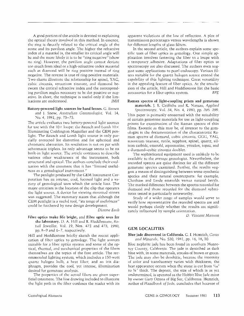
A good portion of the article is devoted to explaining the optical theory involved in this method. In essence, the ring is directly related to the critical angle of the stone and its pavilion angle. The higher the refractive index of a material is, the smaller its critical angle will be and the more likely it is to be "ring negative" (show no ring). However, the pavilion angle cannot deviate too much from ideal or a high-refractive-index material such as diamond will be ring positive instead of ring negative. The reverse is true of ring-positive materials. Two charts illustrate the relationship for spinel, YAG, cubic zirconia, strontium titanate, and diamond be- tween the critical refractive index and the correspond- ing pavilion angles necessary to be dot positive or neg- ative. In short, the technique is useful only if the lim- itations are understood. IMH
Battery-powered light sources for hand lenses. G. Brown and J . Snow, Australian Gemmologist, Vol. 14, NO. 4, 1981, pp. 73-75.
The article evaluates two battery-powered light sources for use with the l o x loupe: the Bausch and Loinb l o x Illuminating Coddington Magnifier and the GEM pen- light. The Bausch and Lomb light source is only par- tially corrected for distortion and for spherical and chromatic aberration. Its resolution is not on par with achromatic triplets. Its only advantage seems to be its built-in light source. The article continues to describe various other weaknesses of the instrument, both structural and optical. The authors conclude their eval- uation with the comment that it has "limited useful- ness as a gemological instrument."
The penlight produced by the GEM Instrument Cor- poration has an intense, cool, focused light and a va- riety of gemological uses which the article lists. The major criticism is the location of the clip that operates the light source, A device for viewing mounted stones was suggested. The summary states that although the GEM penlight is a useful tool, "its range of usefulness" could be furthered by new design developn~ent.
Dianne Eash
Fibre optics make life bright, and Fibre optic uses for the laboratory. D. A. Hill and R. Huddlestone, Re- tail Jeweller, Vol. 19, Nos. 472 and 473, 1980, pp. 8-9 and 6-7, respectively.
Hill and Huddlestone briefly sketch the recent appli- cation of fiber optics to gemology. The light sources suitable for a fiber optics system and some of the op- tical, thermal, and mechanical properties of the fibers themselves are the topics of the first article. The rec- ommended lighting system, which includes a 150-watt quartz halogen bulb, a heat filter, and an iris dia- phragm, provides the cool, yet intense, illumination desired for gemstone analysis.
The properties of the actual fibers are given super- ficial treatment. The line drawing included to illustrate the light path in the fiber confuses the reader with i ts
apparent violations of the law of reflection. A plot of transmission percentages versus wavelengths is shown for different lengths of glass filters.
In the second article, the authors explain some spe- cific uses of fiber optics in gemology. One simple ap- plication involves fastening the fiber to a loupe with a temporary adhesive. Adaptations of fiber optics to spectroscopy are also discussed. The authors even sug- gest some applications to pearl endoscopy. Various fil- ters suitable for the quartz halogen source extend the capability of this lighting technique. Great versatility is the appealing feature of fiber optics. At the conclu- sion of the article, Hill and Huddlestone list the basic accessories for a fiber optics system. B FE
Raman spectra of light-coupling prism and gemstone materials. J. E. Griffiths and K. Nassau, Applied Spectroscopy, Vol. 34, No. 4, 1980, pp. 395-399.
This paper is primarily concerned with the suitability of certain gemstone materials for use as light-coupling prisms for examination of the Raman spectra of thin films. Esoteric as this may be, of interest to the gem- ologist is the documentation of the characteristic Ra- man spectra of diamond, cubic zirconia, GGG, YAG, strontium titanate, rutile, sapphire, spinel, quartz, sil- icon carbide, emerald, aquamarine, yttralox, topaz, and a diamond-cubic zirconia doublet.
The sophisticated equipment used is unlikely to be available to the average gemologist. Nevertheless, the recorded spectra are quite distinct for all the different gemstone species examined. Further, the results sug- gest a means of distinguishing between some synthetic species and their natural counterparts: for example, Chatham and Linde emeralds versus natural beryls. The marked difference between the spectra recorded for diamond and those recorded for the diamond substi- tutes tested is particularly noteworthy.
Study of a wider range of samples would serve to verify how representative the recorded spectra are and would perhaps clarify whether the results are signifi- cantly influenced by sample orientation.
D. Vincent Manson
GEM LOCALITIES Blue jade discovered in California. G. I. Hemrich, Gems
and Minerals, No. 520, 1981, pp. 16, 18, 20. Blue nephrite jade has been found in southern Monte- rey County, California. The jade is described as dark blue with, in some materials, streaks of brown or green. The jade may also be dendritic, because the intensity of color and translucency varies with thickness; the best appearance occurs when the stone is cut from 1/16"
to '/4" thick. The deposit, the size of which is as yet undetermined, is operated as the Hobbit Blue Jade mine by owner Gary Ozuna of Big Sur, California. Hemrich, author of Handbook of lade, concludes that because of
Gemological Abstracts GEMS & GEMOLOGY Summer 1981 113
the rarity of "blue" jadeite or nephrite, this may be one of the major jade discoveries in California. GSH
Minerals of the Wausau Pluton. A. Falster, Mineral- ogical Record, Vol. 12, No. 2, 1981, pp. 93-97.
Mr. Falster describes a pegmatite area near Rib Moun- tain in Marathon County, Wisconsin, that has yielded at least 44 different mineral species. A brief history and a description of the geology of the area are given, fol- lowed by a list of the species that have been identified thus far. Although gem-quality material is apparently rare, the list includes beryl, found as gemmy aquama- rine crystals reaching 1.2 cm in length; phenakite, found as colorless, yellow, purple, or pale pink crystals reaching 3 cm; and ainazonite, found rarely as fine crystals that average 2 cm but have been encountered as large as 29 cm.
Falster indicates that this area, the Wausau Pluton, has good potential for producing more minerals, but adds that the dense forests and swamps covering the region make prospecting difficult. S FM
Notes on New Hampshire gem pockets. B. M. Shaub, Gems and Minerals, No. 520, 1981, pp. 44-47, 58-59.
Mr. Shaub theorizes that the gem crystals found in the pegmatites near Conway, New Hampshire, were bro- ken and tumbled by a natural, mechanical type of weathering. This is in contrast to other explanations that have been offered for the state of these crystals when found, such as explosions caused by steam build- up in the gem pocket or a natural nuclear explosion.
According to Mr. Shaub's theory, after the final crystallization of the gems in pegmatite, water vapor above the critical temperature would occupy the pores between mineral grains and in the gem pockets. As var- ious sections of the pegmatite cooled at different rates, part of the vapor would be super cooled and would cre- ate an unstable system. As this vapor condensed, the sudden release of thermal energy would break the gem crystals within the chamber.
I11 substantiating his theory, the author compares and contrasts New Hampshire pegmatites with the Dunton, Black Mountain, and Mount Mica (Paris) peg- m a t i t e ~ in Maine. He concludes that this type of re- action does not happen in pegmatites where hydrous minerals such as mica occur. GS H
The Urubu pegmatite and vicinity. J. and J. Casse- danne, Mineralogical Record, Vol. 12, No. 2, 198 1, pp. 73-77.
This area in the northeastern corner of Minas Gerais, Brazil, has been known for many years as a large pro- ducer of commercial minerals as well as gemstones. This article gives a good description of the geology of the area and its location and access. Petalite, occurring as flawless transparent fragments up to 10 cm long, is listed among the important minerals of the area, as is spodumene, which is abundant but very rarely trans-
parent. The Urubu pegmatite itself has produced light green elbaite crystals near the entrance to the quarry. These are described as very characteristic: "bottle- green" to "apple-green" with a rubellite core, showing a darker color toward the termination. Some green and, very occasionally, pink gem elbaites have been found in the northwest end of the quarry. Beryl, petalite, and amblygonite are also mentioned, but only as industrial- quality material. S FM
JEWELRY ARTS Antiques: old Chinese ornamental carvings. E. Doini-
nik, Architectural Digest, Vol. 38, No. 2, 198 1, pp. 64-69.
The Chinese have always placed jade above any other natural material. As eloquently explained by Mr. Dom- inik, jade, called yu in Chinese, is intimately entwined with this very old culture. As far back as 6 B.C. Con- fucius, and later the Taoist, ascribed qualities to this stone that transcended jade's physical properties- which are admirable in their own right.
Two minerals are generally designated as jade: nephrite, the traditional variety; and jadeite, the newer, more jewel-like material. Neither is actually carved, but both are ground and polished by abrasives using centuries-old methods. The craftsman studies the rough material with great respect to determine the design best suited for each individual piece. Chalcedony and lapis lazuli are also mentioned as often-carved materials.
T o match the short but passionate text a series of fine photographs of excellent carvings, obviously cho- sen with care, are included. Mr. Dominik has suc- ceeded in producing an enticing introduction for the beginning student or collector. Fred Gray
Immortal jade and the Brundage bequest. T . Albright, Museum, Vol. 2, No. 1, 1981, pp. 74-77.
Mr. Albright begins with an introduction to San Frail- cisco's Asian Art Museum, foIIows with a short biog- raphy of Avery Brundage, and then proceeds to provide a broad outline of the formation of the Brundage col- lection and the development of the museum to house that collection. Museum director Rene-Yvon Lefebvre d1Argence's recollections about organizing the collec- tion and working with Brundage give some interesting glimpses into the exotic world of the big-time collector,
The actual discussion of the largest jade collection in the U.S. is short. Twelve thousand pieces "span the entire history of Chinese jade carving," with heavy em- phasis on the Ming and Ching periods. A special feature of the collection is its inclusion of animal sculpture from every period of Chinese art. Mr. Albright also mentions other groups in the Brundage collection- bronzes, ceramics, and paintings from a number of Asian countries-and then concludes with brief com- ments about the museum's current problems and pros- pects. Three color photographs of jade objects illustrate the article. MLR
114 Gemological Abstracts GEMS & GEMOLOGY Summer 1981
The imperial Ju-I sceptres. L. Liang-yu (translated by M. Man-li Loh), Arts of Asia, Vol. 10, No, 5, 1980, pp. 124-127.
The history and development of the Ju-I sccptre are briefly described in this article. Unlike other art ob- jects, the shapes of which were determined by function, the JLI-I sceptre was adapted to meet symbolic purposes.
The author divides the sceptre's history into three phases. The first stage begins in the dawn of Chinese history, when authority was synlbolized by the stone axe. During the second stage, the stone axe gradually fell into disuse. It remained a symbol of royalty, how- ever, and stone tablets carved in the shape of axes were used as pledges when titles were bestowed on officials. Gradually, it became an established custom for officials to carry jade tablets while in audience with the em- peror. In the third and final stage, the Ju-I developed into its present form. The author suggests that it was gradually adapted to imitate the shape of the lingchih, the sacred fungus of immortality. Since these sceptres were given as gifts on happy occasions, the decorative patterns on them carry good wishes and auspicious meanings.
The article is well illustrated with two full pages of color photographs showing the variety of materials from which these sceptres are fashioned. These include nephrite and jadeite as well as the colored stones and pearls with which the sceptres arc often inlaid. ET
"La donna idolo" (The woman idol). 18 Karati, Vol. 10, NO. 49, 1980-81, pp. 73-77.
As the author says, "An Indian woman . . . can no more be separated from them (jewels) than . . . from the spirit of India." Indian women wear garlands of flowers threaded on fine chains of gold or silver, they crown their middle teeth with gold, they wear gems on their foreheads as well as rings (on fingers and toes), brace- lets, anklets, neclzlaces, tiaras, navel ornaments, and nose studs. Jewelry, taking its design from architectural as well as natural forms, signals a woman's caste and marital status, in addition to her financial standing.
The article describes some of the traditional types of jewelry worn in India and provides six excellent color photos of women wearing jewelry in the styles of some Indian states. SLD
Master jewelers: Twentieth century artists. R. Slivlza, Architectural Digest, Vol. 38, 198 1, pp. 104- 109.
The founder of Quarterly Craft International, Rose SLi.vlza, introduces the reader to the jewelry of notable artists of the 20th century. The author first reviews the function of jewelry from prehistoric to modern times, arguing that jewelry represents a "quest for self visi- bility: ourselves seeing ourselves." She also refers to the long tradition of artists of other media making jew- elry, including Renaissance masters Ghiberti, Botti- celli, and Brunelleschi.
Slivlza then turns to the 20th century, describing it
as a revolutionary and exciting period in the history of jewelry. Eight color photographs of jewelry by Alex- ander Calder, Giorgio de Chirico, Louise Nevelson, Sal- vador Dali, Roy Lichtenstein, Georges Braque, Pablo Picasso, and Erte complement the text. She briefly de- scribes the style of each artist and how this style is re- flected in his or her jewelry. Slivka concludes that the ancient power of jewelry as adornment and protection has a new dimension in today's economic and social context. DMD
SYNTHETICS AND SIMULANTS Characteristics of synthetic hydrothermal emeralds
from USA and USSR (in Japanese). H. Talzubo, Journal of the Gemmological Society of Japan, Vol. 6, NO. 4, 1979, pp. 3-18.
Complete with an abstract in English detailing the properties of the stones tested and the conclusions of the study, this article presents a comprehensive com- parison of the synthetic hydrothermal emeralds pro- duced in the U.S.A. with those produced in the U.S.S.R. The author also includes frequent references to the flux-grown synthetics and the natural emerald coun- terparts. Talzubo's well-organized, highly technical, and scientifically documented presentation includes charts that record the exact size and properties of the stones examined, diagrams of internal and external character- istics, and 32 photographs. The sequence of these pho- tographs permits a visual comparison of the character- istic differences between the rough and cut material produced in the U.S.A. (Linde and Regency] and their Russian counterparts. Emphasis is placed on the types of optical strain, n~icrotextures, and cracks. X-ray anal- ysis did not produce any definitive results with regard to identifying the stones..
Talzubo maintains that many of the differences be- tween the U.S.A. and U.S.S.R. synthetic hydrothermal emeralds result from the conditions of formation. He speculates on the temperatures and pressures used and on the influence that these factors have on the crystal faces and inclusions.
The author concludes that the synthetic hydro- thermal emeralds produced in the U.S.S.R. most closely resemble the natural stone and that only internal char- acteristics distinguish the two. These characteristics include the texture, cracks, zonal structure, and two- phase inclusions. He states that there is a 90% simi- larity between the Russian hydrothermal product and the natural counterpart, an 80% similarity between the American hydrothermal emerald and the natural, and a 40%-60% similarity between the flux-grown emer- ald and the natural stone. The chart at the conclusion of Talzubo's article facilitates comparison by summa- rizing all the characteristics of the U.S.S.R. hydro- thermal, U.S.A. hydrothermal, flux-grown, and natural stones. A LS
Gemological Abstracts GEMS & GEMOLOGY Summer 1981 115
Czochralski growth of single-crystal fayalite under controlled oxygen fugacity conditions. C. B. Finch, G. W. Clark, and 0. C. Kopp, American Mineral- ogist, Vol. 65, No. 3 and 4, 1980, pp. 38 1-389.
The production of synthetic fayalite should be of in- terest to all gemologists, inasmuch as fayalite is the iron-rich end member of the olivine series to which the gem peridot also belongs. The single-crystal boules are up to 1 cm in diameter and 4 cm in length. They are nearly opaque and of a brownish-black color. Magnetite crystals and clusters formed by the reaction of excess iron with oxygen are the most common inclusions ob- served. The synthetic fayalites are grown in inductively heated platinum crucibles which contain the iron sil- icate melt; the Czochralski pulling method is used to form the boules. U K
Internal textures and growth conditions of flux-grown emeralds from USSR (in Japanese). H. Takubo, Y. Kitamura, Y. Nakazumi, and M. Koizumi, Journal o f the Gemmological Society o f Japan, Vol. 6, No. 1 and 2, 1979, pp. 22-28.
The authors analyze flux-grown emeralds from the U.S.S.R. and speculate on the conditions of formation. The many inclusions and flaws in the Russian stones can be understood only by an examination of the crit- ical periods of growth, namely the initial and advanced stages. Compared to the 30%-40% usage rate for syn- thetic emeralds grown elsewhere, only 20%-25% of the U.S.S.R.-created crystals can be considered gem material.
All the internal characteristics of the Soviet mate- rial indicate rapid growth under high temperatures and high pressure. Sixteen photographs, which magnify the internal characteristics, emphasize the weak mosaic structure, the negative crystals, and the flux inclusions. In the late stage of development, veil-like secondary inclusions penetrate the mosaic structure, which was formed by rapid growth at the bottom of the crucible. Unusual silk-like inclusions, similar to silk inclusions found in some star rubies, suggest the possible creation of chatoyant emerald. Less clear than their Chatham counterparts, the Russian stones resemble more closely the product of the Japanese K Company. The heavy flux, composed of V f O s (vanadium oxide), darkens the stones,
The Soviet flux stones are probably grown in a cru- cible approximately 7-10 cm wide and 10-15 cm high, with initial temperatures of over 1 100°C The many cracks and external blemishes are due to the high tem- peratures used in the advanced stage when cooling oc- curs, probably around 90WC as opposed to the usual temperatures of 750°-800° Included crystals indicate that in the course of the slow cooling process, the cru- cible is turned and stirring techniques are used to ac- celerate growth, A LS
MISCELLANEOUS Gold Mines and Gold Mining. Mineralogy, Inc., Gold-
smith, Vol. 158, No. 6, 1981, pp. 83-84, 88. This article contains some basic information on gold: its specific gravity, hardness, melting point, etc. Gold production is discussed, along with information on types of gold deposits and how each type is mined.
Also included in this issue of Goldsmith are nine other articles on gold, covering such topics as the Lon- don fixing of gold prices, gold bullion vs. coins, and other investment-related subjects. FSS
Interview: Allan Caplan, Mineralogical Record, Vol. 11, No. 6, 1980, pp. 35 1-360.
This is an interesting and informative interview with Alan Caplan, a gem dealer who was the first to import museum-quality mineral specimens to the United States from Brazil. Included in the specimens that Mr. Caplan brought to the United States are some of the largest and finest gem topaz, aquamarine, and chrysoberyl crystals on display in our most important museums. Some of these are pictured in the 16 photographs accompanying the article. The interview gives the reader a rare insight into the world of Brazilian gem mining in the 1930s and '40s through the eyes of a pioneer in the Brazilian gem and mineral market. Peter Keller
Metamorphic processes in gemstone formation. A. F. Wilson, Australian Gemmologist, Vol. 14, No. 4, 1981, pp. 57-63.
Allan F. Wilson, of the Department of Geology and Mineralogy at the University of Queensland, reviews the conditions under which various metamorphic gem- stones formed and then illustrates his discussion with chemical, compositional, and pressure-temperature diagrams. Exsolution and the diagnostic features it pro- duces (such as chatoyancy, asterism, and adulares- cence) are related to the metamorphic process. In ad- dition to citing numerous Australian gem localities, the author describes the Barton garnet deposit at Gore Mountain in New York State in considerable detail to illustrate the growth of large crystals in metamorphic rocks. Dr. Wilson concludes this interesting article with a discussion of metasomatism, i.e., the intro- duction of new elements into the metamorphic process. GSH
The quest for quartz. S. B. N. Cooper, Journal of Gem- mology, Vol. 17, No, 3, 1980, pp. 150- 152,
Nikon Cooper discusses the origin of the word quartz. He traces a number of possible origins and in the pro- cess introduces the reader to several of the earliest sci- entific works on minerals, He documents the hypoth- esis that the word quartz is derived from quarzum, which is a latinized version of kwardy. Kwardy is a west Slav word originally meaning hard. It was used by miners in 14th-century Bohemia to describe massive quartz found in hard-rock mining. ET
116 Gemological Abstracts GEMS & GEMOLOGY Summer 1981
Stephanie Dillon, Editor
AGS CONCLAVE '81 The American Gem Society's 47th annual conclave was held at Chicago's Marriott Hotel April 24-28. Its theme, "Gemology: The Total Concept," was intro- duced in a speech by Michael Allbritton, GIA1s super- visor of one-week gemology classes.
At this year's meeting, Arnold Bockstruck, C.G., was elected president of the jewelers' organization for a tenure of two years.
The convention featured many laboratory work- shops, organized by GIA staff and AGS members, which. afforded opportunities for the practical application of gemology. Highlighted were several chief concerns of jewelers: appraisals, the detection of synthetics and im- itations, grading mounted diamonds, handling repairs, and the place of gem knowledge in selling.
The conclave was attended by approximately 1300 AGS members and their families. Of this total nearly 700 are registered jewelers.
An announcement from the AGS board of directors that next year's conclave may be declared open to non- members was a source of discussion amone attendees. The directors have yet to agree on guidelines for such a move.
ARGENTINE RHODOCHROSITE - A joint Japanese-Argentinian mining venture is cur- rently rehabilitating the Capillitas rhodochrosite mines of Catamarca, Argentina. The intention is to use elec- trolytic methods to process the ore, which is rich in gold, silver, and copper, and then to continue to mine the rhodochrosite.
Capillitas was mined first by the Incas for gold and later by the Spaniards for copper. In the early 1 9 0 0 ~ ~ both English and French concerns operated the mine. Although the Incas discovered the rhodochrosite and adopted it into use and legend, it was not until 1937 that the mine was worked exclusively for rhodochro- site, by Dr. Franz Mansfeld. In 1942, the military gov- ernment assumed control over the mine and rhodo- chrosite production was halted; it was resumed in 1951.
The deposit lies in arid, mountainous terrain at an
altitude of 3,000 m above sea level. The mine has ap- proximately 66 km of tunnels running on several levels with numerous entrances.
From estimates based on earlier production, the mine is expected to yield 200 to 300 tons of rhodo- chrosite annually.
AUSTRALIAN DEVELOPMENTS - View toward Gem Cutting. A diamond-mining con- cern, with the cooperation of the Australian govern- ment, is studying the possibility of establishing a gem-cutting industry in Perth, the capital of Western Australia. Presently, all gemstones are taken out of the country for cutting in Germany or Thailand.
Diamond. Diamond mining in the Kimberly region of Western Australia recently produced an 11-ct. crystal. Fancies have also been discovered in the Ashton Joint- Venture fields, notably some attractive pink stones.
Emerald. The Aga Khan mine, in the state of Western Australia, has been worked intermittently since 1906, yielding emeralds as a by-product of ores. The quantity of emeralds taken out has never been revealed.
Currently, an examination of the emerald-bearing rock is under way. Host rock is known to extend at least 1.6 km beyond the present workings. Both open- cut and underground methods are being used to remove the ore and the emeralds.
Most of the emeralds are small, the largest crystal yet found being 9.6 cts. The material is comparatively clear. In the past it has been sent to Germany for cut- ting, but current production is being stockpiled, along with the scheelite also produced from the mine, await- ing a decision on the establishment of local cutting facilities.
Sapphire. Up to 40 separate companies are currently mining sapphires in the 30 square miles of known re- serves in Central Queensland. The chief product is yel- low sapphires, generally found in 3- to 10-ct. sizes. The largest piece of translucent rough discovered thus far is 2,000 cts.
Gem News GEMS & GEMOLOGY Summer 1981 117
U.S. PERIDOT One of the most important sources of jewelry-sized per-, idot in the world today is the San Carlos Apache res- ervation, east of Phoenix, Arizona.
The source is a mesa, measuring approximately 2.5 knl x 4 km, which resulted when a volcanic eruption produced a basalt flow. It is estimated that peridot may comprise 40% to 50% of the entire structure. The ma- terial has formed in pods of basalt, coating the basalt with stones that may be as small as grains of sand or as large as good-sized pebbles. The Indians separate and sell the peridot, which is exported throughout the world and frequently marketed as the product of other regions.
A comprehensive report on this locality is slated to appear in an upcoming issue of Gems d Gemology.
ZAIRE BREAKS WITH DEBEERS - With the sale this spring of 620,000 cts. of diamonds to three independent European dealers, the government of Zaire marked the end of i ts 14-year exclusive mar- keting arrangement with DeBeers Consolidated Mines Ltd.
The sale, announced by Soza'com, Zaire's mineral- marketing agency, accounts for about 10% of this year's anticipated production at Miba (Societe Miniere de Bakwanga), the country's main diamond mine.
The change is not expected to have a major effect 011 the world diamond market. Over 95% of Zaire's production is industrial quality; this output represents approxin~ately 30% of the world supply of industrial diamonds.
Los Angeles County Museum of Natural History-900 Exposition Blvd., Los Angeles, CA 90007. Tel- ephone: (213) 744-3328. "Sweat of the Sun, Tears of the Moon" is the largest exhibit of Co- lombian gold artifacts and rough and cut emeralds ever to be exhibited outside of the Republic of Colom- bia. Opened July 4 and continues through September 6, 198 1.
Smithsonian InstitutionINational Museum of Man-1000 Jefferson Dr., SW, Washington, DC 20560. Telephone: (2021 357-1304. The ex- hibit "5000 Years of Korean Art" comprises 345 treasures from Ko- rean national museums, universi- ties, and private collections. Over 30 gold artifacts from the Three Kingdom Period (1st c. B.C.-7th c, A.D.) exemplify advanced skills in metalwork. The pieces, including three spectacular crowns as well as pendants and earrings, are set with jade and other gemstones. The show inaugurates the Thomas M. Evans Gallery for special exhibits. Runs July 15 through September 30, 198 1.
American Museum of Natural His- tory-Central Park West & 79th St., New York, NY 10024. Telephone: (2121 873-1300. The Gardner D. Stout Hall of Asian
Peoples, opened last October, in- cludes rubies, sapphires, and jade- ites in an exhibit of the dress, carv- ings, and historic gem trade of Asia. In the near future, the Guggenheim Hall of Minerals will feature a dis- play of gem materials from San Diego County, California.
Winner of the 1981 Schuetz Memo- rial Fund Jewelry Design Contest is Sara A. Hitt of Palo Alto, California. Her design is for a pair of gold cuf- flinks set with triangle-cut black onyx. The Schuetz scholarship, awarded annually for a distinguished design in men's jewelry, provides $500 to pursue jewelry-related study at an institution of the winner's choice. Ms. Hitt intends to use her award to continue home study courses through GIA.
A scholarship to study at CFH (the Jewelers' International Institute of Marketing and Management) in Lausanne, Switzerland, from Feb- ruary 8 to June 4, 1982, is being of- fered by GIA. The program is a com- prehensive course on watch and jewelry shop management, com- prising eight weeks of product knowledge and selling (which in- cludes the study of diamonds, col- ored stones, gold, jewelry and
watches; window display; and sell- ing techniques) and eight weeks of shop management (which covers marketing, shop outfitting, secu- rity, personnel management, and all aspects of business finance). The scholarship, for 2400 Swiss francs, covers half the price of the course, textbooks, working docu- nlerits, and teaching materials, plus a number of field trips. Deadline foi- application to GIA is September 20, 1981.
The Broer Scholarship for resident GIA students has been announced by Richard T. Liddicoat, Jr., presi- dent of CIA. The program is named in honor of the late Carleton G . . Broer, a long-time member of GIA's board of governors and its vice- chairman at the time of his passing. Broer was president of the Broer- Freeman Company, a retail firm in Toledo, Ohio. He served as vice- president of the Retail Jewelers of America, director of the Ohio Retail Jewelers Association, and was twice president of the American Gem So- ciety. He was the first recipient of the society's Robert M. Shipley Award, in 1969.
Gems s) Gemology welcomes news of exhibits and events of a gemolog- ical nature. Please contact Ste- phanie Dillon, Gemological Insti- tute of America, 1660 Stewart St., Santa Monica, 90404. Telephone: (2131 829-2991.
118 Gem News GEMS &. GEMOLOGY Summer 198 1
Authors
The following guidelines were pre- pared both to introduce you to Gems el) Gemology and to let you know how we would like a manuscript prepared for publication. No manu- script will be rejected because i t does not follow these guidelines pre- cisely, but a well-prepared manu- script helps reviewer, editor, and reader appreciate the article that much more. Please feel free to con- tact the Editorial Office for assis- tance at any stage in the develop- ment of your paper, whether to confirn~ the appropriateness of a topic, to help organize the presen- tation, or to augment the text with photographs from the extensive files at GIA. We look forward to hearing from you.
INTRODUCTION
Gems o) Gemology is an interna- tional publication of original contri- butions concerning the study of gemstones and research in gemology and related fields. Topics covered include (but are not limited to] col- ored stones, diamonds, gem instru- ments, gem localities, gem substi- tutes (synthetics), gemstones for the collector, jewelry arts, and retail management. Manuscripts may be submitted as: Original Contributions-full-length articles describing previously un- published studies and laboratory or field research. Such articles should be no longer than 6,000 words (24 double-spaced, typewritten pages) plus tables and illustrations. Gemology in Review-comprehen- sive reviews of topics in the field. A
maximum of 8,000 words (32 dou- ble-spaced, typewritten pages) is recommended. Notes & New Techniques-brief preliminary communications of re- cent discoveries or developments in gemology and related fields (e.g., new instruments and instrumentation techniques, gem minerals for the collector, and lapidary techniques or new uses for old techniques]. Arti- cles for this section should be ap- proximately 1,000-3,000 words (4- 12 double-spaced, typewritten pages). Gerns &> Gemology also includes the following regular sections: Lab Notes [reports of interesting or un- usual gemstones, inclusions, or jew- elry encountered in the Gem Trade Laboratories), Book Reviews (as so- licited by the Book Review Editor; publishers should send one copy of each book they wish to have re- viewed to the Editorial Office), Gernologjcal Abstracts (summaries of important articles published re- cently in the gemology literature), and Gem News (current events in the field).
MANUSCRIPT PREPARATION
All material, including tables, leg- ends, and references, should be typed double spaced on 8 % x 11" (21 x 28 cm) sheets with 1%'' (3.8 cm] mar- gins. Please identify the authors on the title page only, not in the body of the manuscript or the figures, so that author anonymity may be maintained with reviewers ( the title page is removed before the manu- script is sent out for review). The
various components of the manu. script should be prepared and ar- ranged as follows: Title page. Page 1 should provide: (a) the article title; (b] the full name of each author (first name, middle initial, surname], with his or her af- filiation (the institution, city, and state or country where he/she was working when the article was pre- pared); (c) acknowledgments of per- sons who helped prepare the report or did the photography, where ap- propriate; and (d) five key words that we can use to index the article at the end of the year. Abstract. Page 2 should repeat the title of the article followed by an abstract. The abstract (approxi- mately 150 words for a feature arti- cle, 75 words for a note) should state the purpose of the article, what was done, and the main conclusions. Text. Papers should follow a clear outline with appropriate heads. For example, for a research paper, the headings might be: Introduction, Previous Studies, Methods, Results, Discussion, Conclusion, and Impli- cations. Other heads and subheads should be used as the subject matter warrants. Also, when writing your article, please try to avoid jargon, to soell out all nonstandard abbrevia- tions the first time they are men- tioned, and to present your material as clearly and concisely as possible. For general style (grammar, etc.) and additional information on preparing a manuscript for publication, A Manual of Style (The University of Chicago Press, Chicago) is recommended.
Suggestions for Authors GEMS & GEMOLOGY Summer 1981 119
References. References should be used for any information that is taken directly from another publi- cation, to document ideas and facts attributed to-or facts discovered by-another writer, and to refer the reader to other sources for addi- tional information on a particular subject. Please cite references in the text by the last name of the author(sJ and the year of publication-plus the specific page referred to, if ap- propriate-in parentheses (e.g., Lid- dicoat and Copeland, 1967, p. 10). The references listed at the end of the paper should be typed double spaced in alphabetical order by the last name of the senior author. Please list only those references actually cited in the text (or in the tables or figures). Include the following information, in the order given here, for each ref- erence: (a) all author names (sur- names followed by initials); (b) the year of publication, in parentheses; (c) for a journal, the full title of the article or, for a book, the full title of the book cited; and (d) for a journal, the full title of the journal plus vol- ume number and inclusive page numbers of the article cited or, for a book, the publisher of the book and the city of publication. Sample references are as follows: Daragh P.J., Sanders J.V. (1976)
Opals. Scientific American, Vol. 234, pp. 84-95.
Heinrich K.F.J. (1968) Common sources of error in electron probe microanalysis. In J. Newkirk et a]., Eds., Advances in X-ray Analysis, Plenum Press, New York, pp. 40-45.
Liddicoat R.T. Jr., Copeland L.L. (1967) The Jewelers' Manual, 2nd ed. Gemological Institute of America, Santa Monica, CA.
Tables. Tables can be very useful in presenting a large amount of detail in a relatively small space, and should be considered whenever the bulk of information to be conveyed in a section threatens to overwhelm the text. Type each table double spaced on a separate sheet. If the table must ex-
ceed one typewritten page, please duplicate all headings on the second sheet. Number tables in the order in which they are cited in the text. Ev- ery table should have a title; every column (including the left-hand col- umn) should have a heading. Please make sure terms and figures used in the table are consistent with those used in the body of the text.
Figures. Please have line figures (graphs, charts, etc.) professionally drawn and photographed. High-con- trast, glossy, black-and-white prints are preferred. Submit black-and-white photo- graphs and photomicrographs in the final desired size if possible. Where appropriate, please use a bar or other scale marker on the photo, not out- side it. Use a label on the back of each fig- ure to indicate the article's title (or a shortened version thereof) and the top of the figure. Do not trim, mount (unless one figure is composed of two or more separate photos), clip, or staple illustrations. Color photographs will be consid- ered for publication. Please include three sets of color prints with the manuscript package submitted for publication consideration, retaining the color transparency .until a final publication decision is made. All figure legends should be typed double spaced on a separate page. Clearly explain any symbols, ar- rows, numbers, or abbreviations used in the illustration. Where a magni- fication is appropriate and is not in- serted on the photo, please include it in the legend.
MANUSCRIPT SUBMISSION
Please send three copies of each manuscript (and three sets of figures and labels) as well as material for all sections to the Editorial Office, in care of:
Alice S. Keller, Managing Editor Gems d Gemology 1660 Stewart Street Santa Monica, CA 90404
In view of U.S. copyright law, we must ask that each submitted man-
uscript be accompanied by the fol- lowing statement, signed by all au- thors of the work: "Upon publication of (title) in Gems o) Gemology, I (we) transfer to the Gemological In- stitute of America all rights, titles, and interest to the work, including copyright, together with full right and authority to claim worldwide copyright for the work as published in this journal. As author(s), I (we) retain the right to excerpt (up to 250 words) and reprint the material on request to the Gemological Institute of America, to make copies of the work for use in classroom teaching or for internal distribution within my (our) place of employment, to use-after publication-all or part of this material in a book I (we) have authored, to present this material orally at any function, and to veto or approve permission granted by the Gemological Institute of America to a third party to republish all or a substantial part of the article. I (we) also retain all proprietary rights other than copyright (such as patent rights). I (we) agree that all copies of the article made within these terms will include notice of the copyright of the Gemological Institute of America. This transfer of rights is made in view of the Gemological In- ,
stitute of America's efforts in re- viewing, editing, and publishing this material.
As author(s), I (we) also warrant that this article is my (our) original work. This article has been submit- ted in English to this journal only and has not been published elsewhere." No payment is made for articles published in Gems es) Gemology.
REVIEW PROCESS
Manuscripts are examined by the Editor, one of the Associate Editors, and a t least two reviewers. The au- thors will remain anonymous to the reviewers. Decisions of the Editor are final. All material accepted for publication is subject to copyedit- ing; authors will receive galley proofs for review and are held fully respon- sible for the content of their articles.
120 Suggestions for Authors GEMS & GEMOLOGY Summer 1981