Embed Size (px)
Citation preview
A Protein Contortionist: Core Mutations of GB1 thatInduce Dimerization and Domain Swapping
In-Ja L. Byeon, John M. Louis and Angela M. Gronenborn*
Laboratory of Chemical PhysicsNational Institute of Diabetesand Digestive and KidneyDiseases, National Institutes ofHealth, Building 5, Room 130Bethesda, MD 20892, USA
Immunoglobulin-binding domain B1 of streptococcal protein G (GB1), asmall (56 residues), stable, single-domain protein, is one of the most exten-sively used model systems in the area of protein folding and design.Recently, NMR and X-ray structures of a quintuple GB1 core mutant(L5V/A26F/F30V/Y33F/A34F) that showed an unexpected, intertwinedtetrameric architecture were determined. Here, we report the NMR struc-ture of another mutant, derived from the tetramer by reverting the singleamino acid position F26 back to the wild-type sequence A26. The struc-ture reveals a domain-swapped dimer that involves exchange of thesecond b-hairpin. The resulting overall structure comprises an eight-stranded b-sheet whose concave side is covered by two a helices. Thedimer dissociates into a partially folded, monomeric species with a dis-sociation constant of 93(^10) mM.
Published by Elsevier Ltd.
Keywords: GB1; core mutants; NMR structure; domain-swapping;oligomerization*Corresponding author
Introduction
Over the last decade, several model systemshave emerged that have found widespread use forstudying protein folding and design. One suchprotein is GB1, the immunoglobulin-bindingdomain B1 of streptococcal protein G. GB1 is asmall (56 residues), stable, single-domain proteinwith one a-helix and a four-stranded b-sheet com-prising two b-hairpins.1,2 We previously createdan exhaustive set of hydrophobic core mutants ofGB1.3 Nine positions were permuted randomly tocomprise the amino acids Leu, Val, Ile, Phe andMet, and from this extensive set of mutants(,2 £ 106), HS#124, a variant that contained fivechanges (L5V/A26F/F30V/Y33F/A34F; Figure 1)compared to the wild-type sequence, was selectedfor structure determination. Interestingly, the qua-ternary state of the protein was a tetramer and,most surprisingly, the three-dimensional structureof this mutant exhibited a substantial change inbackbone geometry, resulting in a drastic global
rearrangement of the entire fold into an extensiveintertwined architecture.4
Here, we report on a further core mutant of GB1that adopts yet another oligomeric state. Thismutant, HS#124F26A, was created by a single aminoacid change back from the tetramer mutantsequence at position 26 (Phe) to the wild-type resi-due (Ala), thus the protein contains four changes(L5V/F30V/Y33F/A34F; Figure 1) compared tothe wild-type sequence.We determined the three-dimensional structure
of this quadruple mutant by NMR, and show thatfor this protein the GB1 fold is restored, albeitrearranged as a domain-swapped dimer. We com-pare the current structure with those of monomericwild-type GB1 and the tetramer mutant and dis-cuss critical residues involved in the structuralswitch. In addition, we characterized the dimer–monomer equilibrium by NMR, gel-filtration andlight-scattering.
Results and Discussion
Protein purification and characterization
Each mutation in the quintuple mutant (HS#124)was singly reverted back to the wild-type aminoacid and the resulting proteins were characterized.Figure 1 displays the elution profiles for all single
0022-2836/$ - see front matter Published by Elsevier Ltd.
E-mail address of the corresponding author:[email protected]
Abbreviations used: GB1, immunoglobulin-bindingdomain B1 of streptococcal protein G; HSQC,heteronuclear single quantum coherence; NOE, nuclearOverhauser effect; NOESY, NOE spectroscopy.
doi:10.1016/S0022-2836(03)00928-8 J. Mol. Biol. (2003) 333, 141–152
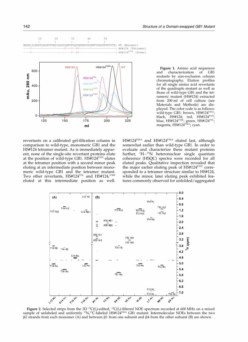
revertants on a calibrated gel-filtration column incomparison to wild-type, monomeric GB1 and theHS#124 tetramer mutant. As is immediately appar-ent, none of the single-site revertant proteins eluteat the position of wild-type GB1. HS#124F33Y elutesat the tetramer position with a second minor peakeluting at an intermediate position between mono-meric wild-type GB1 and the tetramer mutant.Two other revertants, HS#124V5L and HS#124,V30F
eluted at this intermediate position as well.
HS#124F26A and HS#124F34A eluted last, althoughsomewhat earlier than wild-type GB1. In order toevaluate and characterize these mutant proteinsfurther, 1H–15N heteronuclear single quantumcoherence (HSQC) spectra were recorded for alleluted peaks. Qualitative inspection revealed thatthe major earlier eluting peak of HS#124F33Y corre-sponded to a tetramer structure similar to HS#124,while the minor, later eluting peak exhibited fea-tures commonly observed for unfolded/aggregated
Figure 1. Amino acid sequencesand characterization of GB1mutants by size-exclusion columnchromatography. Elution profilesfor all single amino acid revertantsof the quadruple mutant as well asthose of wild-type GB1 and the tet-rameric mutant (HS#124) extractedfrom 200 ml of cell culture (seeMaterials and Methods) are dis-played. The color code is as follows:wild-type GB1; brown, HS#124F26A;black, HS#124; red, HS#124F33Y;blue, HS#124V30F; green, HS#124V5L;magenta, HS#124F34A; cyan.
Figure 2. Selected strips from the 3D 13C(f2)-edited, 12C(f3)-filtered NOE spectrum recorded at 600 MHz on a mixedsample of unlabeled and uniformly 15N,13C-labeled HS#124F26A GB1 mutant. Intermolecular NOEs between the twob2 strands from each monomer (A) and between b1 from one subunit and b4 from the other subunit (B) are shown.
142 Structure of a Domain-swapped GB1 Mutant
protein. Similarly, the spectra of HS#124V5L,HS#124V30F and HS#124F34A resembled those ofunfolded/aggregated protein and did not containsharp, resolved resonances that would indicate awell-structured protein. Only HS#124F26A yielded awell-dispersed spectrum, characteristic of a uniqueand stable architecture for this variant. We there-fore selected HS#124F26A (or L5V/F30V/Y33F/A34F-GB1) for structure determination.
Structure determination
Virtually complete 1H, 13C and 15N resonanceassignments for the quadruple mutant wereobtained using heteronuclear, multidimensionalNMR spectroscopy. Initial NMR and nuclear Over-hauser effect (NOE) analysis of a uniformly13C/15N-labeled mutant allowed us to readilyidentify the a-helix and b-sheet, indicating thatthe mutant exhibited essentially the same second-ary structure as wild-type GB1. Backbone torsionangle prediction using the chemical shifts of themutant as input for calculating torsion angles withthe program TALOS5 showed good agreementwith the angles of the wild-type structure for allbut two residues, Val39 and Asp40. This suggestedthat the regular secondary structure elements (a-helix and b-strands) are conserved in the mutantstructure, but that loop structures, especially theloop involving Val39 and Asp40, are differentfrom wild-type GB1. These two residues arelocated in a linker connecting the a-helix and thesecond b-hairpin. The linker exhibits an extendedconformation in wild-type GB1, whereas in themutant Val39 and Asp40 are predicted by TALOSto reside in a turn conformation. Furthermore, theb2 strand (Leu12–Thr18) that resides at an edge ofthe four-stranded b-sheet in the wild-type struc-ture was found to be H-bonded anti-parallel to anadditional b-strand. Slowly exchanging amide pro-tons were observed for Lys13, Glu15 and Thr17 inb2, in contrast to the fast exchange behavior forthese residues in wild-type GB1. An inter-molecu-lar interface between b2 and the same strand fromthe other monomer unit was identified and con-firmed by NOE analysis of 3D 13C-edited/12C-fil-tered and 13C-edited/12C,14N-filtered NOESYspectra on a mixed sample of unlabeled and15N,13C-labeled protein (Figure 2(A)). In additionto this interface, a second dimer interface wasfound: inter-molecular NOEs were observedbetween the b1 strand (Gln2-Asn8) of one mono-mer unit and the b4 strand (Thr51-Thr55) of theother monomer, forming a parallel b-sheet (Figure2(B)). In wild-type GB1, strands b1 and b4 are thecentral strands of the four-stranded b-sheet, andtheir inter-molecular arrangement in the mutantstrongly suggested domain swapping.
Structures were calculated with CNS6 basedon 1035 inter-proton distances, 72 hydrogen bonddistances and 148 dihedral angles constraints permonomeric subunit. All experimental constraintsused in the final structure calculations and perti-
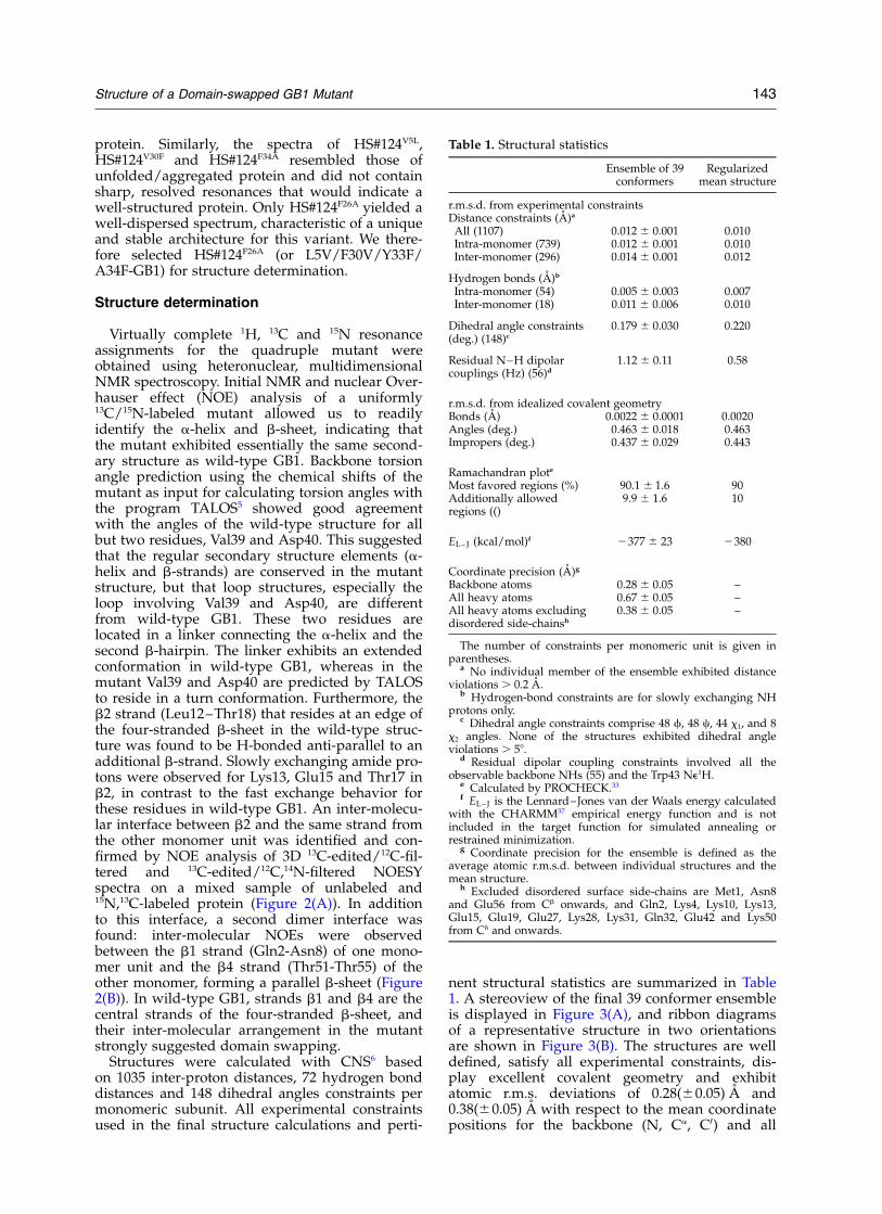
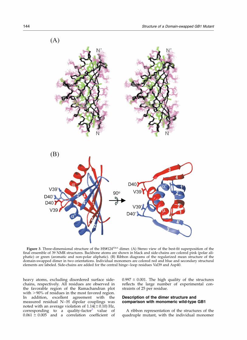
nent structural statistics are summarized in Table1. A stereoview of the final 39 conformer ensembleis displayed in Figure 3(A), and ribbon diagramsof a representative structure in two orientationsare shown in Figure 3(B). The structures are welldefined, satisfy all experimental constraints, dis-play excellent covalent geometry and exhibitatomic r.m.s. deviations of 0.28(^0.05) A and0.38(^0.05) A with respect to the mean coordinatepositions for the backbone (N, Ca, C0) and all
Table 1. Structural statistics
Ensemble of 39conformers
Regularizedmean structure
r.m.s.d. from experimental constraintsDistance constraints (A)a
All (1107) 0.012 ^ 0.001 0.010Intra-monomer (739) 0.012 ^ 0.001 0.010Inter-monomer (296) 0.014 ^ 0.001 0.012
Hydrogen bonds (A)b
Intra-monomer (54) 0.005 ^ 0.003 0.007Inter-monomer (18) 0.011 ^ 0.006 0.010
Dihedral angle constraints(deg.) (148)c
0.179 ^ 0.030 0.220
Residual N–H dipolarcouplings (Hz) (56)d
1.12 ^ 0.11 0.58
r.m.s.d. from idealized covalent geometryBonds (A) 0.0022 ^ 0.0001 0.0020Angles (deg.) 0.463 ^ 0.018 0.463Impropers (deg.) 0.437 ^ 0.029 0.443
Ramachandran plote
Most favored regions (%) 90.1 ^ 1.6 90Additionally allowedregions (()
9.9 ^ 1.6 10
EL– J (kcal/mol)f 2377 ^ 23 2380
Coordinate precision (A)g
Backbone atoms 0.28 ^ 0.05 –All heavy atoms 0.67 ^ 0.05 –All heavy atoms excludingdisordered side-chainsh
0.38 ^ 0.05 –
The number of constraints per monomeric unit is given inparentheses.
a No individual member of the ensemble exhibited distanceviolations . 0.2 A.
b Hydrogen-bond constraints are for slowly exchanging NHprotons only.
c Dihedral angle constraints comprise 48 f, 48 c, 44 x1, and 8x2 angles. None of the structures exhibited dihedral angleviolations . 58.
d Residual dipolar coupling constraints involved all theobservable backbone NHs (55) and the Trp43 Ne1H.
e Calculated by PROCHECK.33f EL– J is the Lennard–Jones van der Waals energy calculated
with the CHARMM37 empirical energy function and is notincluded in the target function for simulated annealing orrestrained minimization.
g Coordinate precision for the ensemble is defined as theaverage atomic r.m.s.d. between individual structures and themean structure.
h Excluded disordered surface side-chains are Met1, Asn8and Glu56 from Cb onwards, and Gln2, Lys4, Lys10, Lys13,Glu15, Glu19, Glu27, Lys28, Lys31, Gln32, Glu42 and Lys50from Cd and onwards.
Structure of a Domain-swapped GB1 Mutant 143
heavy atoms, excluding disordered surface side-chains, respectively. All residues are observed inthe favorable region of the Ramachandran plotwith .90% of residues in the most favored region.In addition, excellent agreement with themeasured residual N–H dipolar couplings wasnoted with an average violation of 1.14(^0.10) Hz,corresponding to a quality-factor7 value of0.061 ^ 0.005 and a correlation coefficient of
0.997 ^ 0.001. The high quality of the structuresreflects the large number of experimental con-straints of 25 per residue.
Description of the dimer structure andcomparison with monomeric wild-type GB1
A ribbon representation of the structures of thequadruple mutant, with the individual monomer
Figure 3. Three-dimensional structure of the HS#124F26A dimer. (A) Stereo view of the best-fit superposition of thefinal ensemble of 39 NMR structures. Backbone atoms are shown in black and side-chains are colored pink (polar ali-phatic) or green (aromatic and non-polar aliphatic). (B) Ribbon diagrams of the regularized mean structure of thedomain-swapped dimer in two orientations. Individual monomers are colored red and blue and secondary structuralelements are labeled. Side-chains are added for the central hinge–loop residues Val39 and Asp40.
144 Structure of a Domain-swapped GB1 Mutant
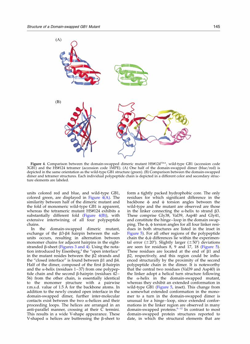
units colored red and blue, and wild-type GB1,colored green, are displayed in Figure 4(A). Thesimilarity between half of the dimeric mutant andthe fold of monomeric wild-type GB1 is apparent,whereas the tetrameric mutant HS#124 exhibits asubstantially different fold (Figure 4(B)), withextensive intertwining of all four polypeptidechains.
In the domain-swapped dimeric mutant,exchange of the b3-b4 hairpin between the sub-units occurs, resulting in alternation betweenmonomer chains for adjacent hairpins in the eight-stranded b-sheet (Figures 3 and 4). Using the nota-tion introduced by Eisenberg,8 the “open interface”in the mutant resides between the b2 strands andthe “closed interface” is found between b1 and b4.Half of the dimer, composed of the first b-hairpinand the a-helix (residues 1–37) from one polypep-tide chain and the second b-hairpin (residues 42–56) from the other chain, is essentially identicalto the monomer structure with a pairwiser.m.s.d. value of 1.5 A for the backbone atoms. Inaddition to the newly created open interface in thedomain-swapped dimer, further inter-molecularcontacts exist between the two a-helices and theirproceeding loops. The helices are arranged in ananti-parallel manner, crossing at their C termini.This results in a wide V-shape appearance. TheseV-shaped a helices are buttressing the b-sheet to
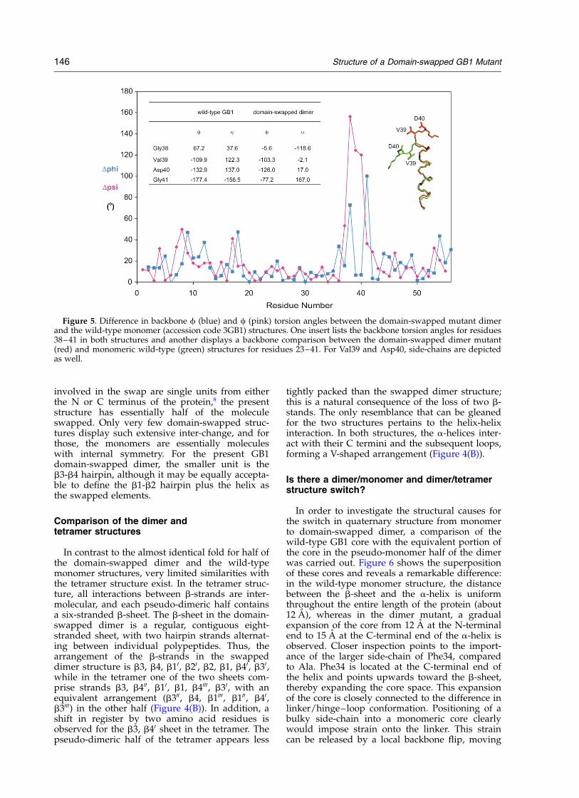
form a tightly packed hydrophobic core. The onlyresidues for which significant difference in thebackbone f and c torsion angles between thewild-type and the mutant are observed are thosein the linker connecting the a-helix to strand b3.These comprise Gly38, Val39, Asp40 and Gly41,and constitute the hinge–loop in the domain swap-ping. The f, c torsion angles for all four linker resi-dues in both structures are listed in the inset inFigure 5). For all other regions of the polypeptidechain the f,c differences lie within the experimen-tal error (^208). Slightly larger (^508) deviationsare seen for residues 8, 9 and 17, 18 (Figure 5).These residues are located at the end of b1 andb2, respectively, and this region could be influ-enced structurally by the proximity of the secondpolypeptide chain in the dimer. It is noteworthythat the central two residues (Val39 and Asp40) inthe linker adopt a helical turn structure followingthe a-helix in the domain-swapped mutant,whereas they exhibit an extended conformation inwild-type GB1 (Figure 5, inset). This change froma somewhat extended conformation in the mono-mer to a turn in the domain-swapped dimer isunusual for a hinge–loop, since extended confor-mations in the linker region are observed in manydomain-swapped proteins.8–12 In contrast to mostdomain-swapped protein structures reported todate, in which the structural elements that are
Figure 4. Comparison between the domain-swapped dimeric mutant HS#124F26A, wild-type GB1 (accession code3GB1) and the HS#124 tetramer (accession code 1MPE). (A) One half of the domain-swapped dimer (blue/red) isdepicted in the same orientation as the wild-type GB1 structure (green). (B) Comparison between the domain-swappeddimer and tetramer structures. Each individual polypeptide chain is depicted in a different color and secondary struc-ture elements are labeled.
Structure of a Domain-swapped GB1 Mutant 145
involved in the swap are single units from eitherthe N or C terminus of the protein,8 the presentstructure has essentially half of the moleculeswapped. Only very few domain-swapped struc-tures display such extensive inter-change, and forthose, the monomers are essentially moleculeswith internal symmetry. For the present GB1domain-swapped dimer, the smaller unit is theb3-b4 hairpin, although it may be equally accepta-ble to define the b1-b2 hairpin plus the helix asthe swapped elements.
Comparison of the dimer andtetramer structures
In contrast to the almost identical fold for half ofthe domain-swapped dimer and the wild-typemonomer structures, very limited similarities withthe tetramer structure exist. In the tetramer struc-ture, all interactions between b-strands are inter-molecular, and each pseudo-dimeric half containsa six-stranded b-sheet. The b-sheet in the domain-swapped dimer is a regular, contiguous eight-stranded sheet, with two hairpin strands alternat-ing between individual polypeptides. Thus, thearrangement of the b-strands in the swappeddimer structure is b3, b4, b10, b20, b2, b1, b40, b30,while in the tetramer one of the two sheets com-prise strands b3, b400, b10, b1, b4000, b30, with anequivalent arrangement (b300, b4, b1000, b100, b40,b3000) in the other half (Figure 4(B)). In addition, ashift in register by two amino acid residues isobserved for the b3, b40 sheet in the tetramer. Thepseudo-dimeric half of the tetramer appears less
tightly packed than the swapped dimer structure;this is a natural consequence of the loss of two b-stands. The only resemblance that can be gleanedfor the two structures pertains to the helix-helixinteraction. In both structures, the a-helices inter-act with their C termini and the subsequent loops,forming a V-shaped arrangement (Figure 4(B)).
Is there a dimer/monomer and dimer/tetramerstructure switch?
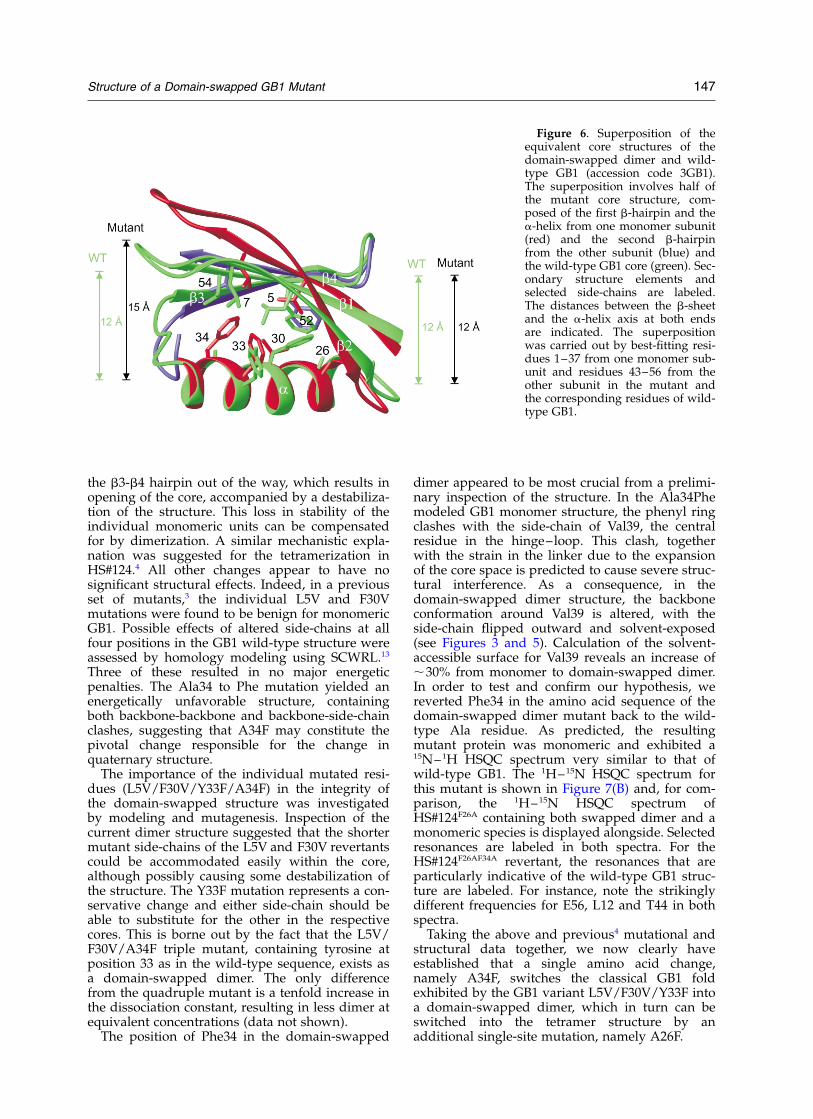
In order to investigate the structural causes forthe switch in quaternary structure from monomerto domain-swapped dimer, a comparison of thewild-type GB1 core with the equivalent portion ofthe core in the pseudo-monomer half of the dimerwas carried out. Figure 6 shows the superpositionof these cores and reveals a remarkable difference:in the wild-type monomer structure, the distancebetween the b-sheet and the a-helix is uniformthroughout the entire length of the protein (about12 A), whereas in the dimer mutant, a gradualexpansion of the core from 12 A at the N-terminalend to 15 A at the C-terminal end of the a-helix isobserved. Closer inspection points to the import-ance of the larger side-chain of Phe34, comparedto Ala. Phe34 is located at the C-terminal end ofthe helix and points upwards toward the b-sheet,thereby expanding the core space. This expansionof the core is closely connected to the difference inlinker/hinge–loop conformation. Positioning of abulky side-chain into a monomeric core clearlywould impose strain onto the linker. This straincan be released by a local backbone flip, moving
Figure 5. Difference in backbone f (blue) and c (pink) torsion angles between the domain-swapped mutant dimerand the wild-type monomer (accession code 3GB1) structures. One insert lists the backbone torsion angles for residues38–41 in both structures and another displays a backbone comparison between the domain-swapped dimer mutant(red) and monomeric wild-type (green) structures for residues 23–41. For Val39 and Asp40, side-chains are depictedas well.
146 Structure of a Domain-swapped GB1 Mutant
the b3-b4 hairpin out of the way, which results inopening of the core, accompanied by a destabiliza-tion of the structure. This loss in stability of theindividual monomeric units can be compensatedfor by dimerization. A similar mechanistic expla-nation was suggested for the tetramerization inHS#124.4 All other changes appear to have nosignificant structural effects. Indeed, in a previousset of mutants,3 the individual L5V and F30Vmutations were found to be benign for monomericGB1. Possible effects of altered side-chains at allfour positions in the GB1 wild-type structure wereassessed by homology modeling using SCWRL.13
Three of these resulted in no major energeticpenalties. The Ala34 to Phe mutation yielded anenergetically unfavorable structure, containingboth backbone-backbone and backbone-side-chainclashes, suggesting that A34F may constitute thepivotal change responsible for the change inquaternary structure.
The importance of the individual mutated resi-dues (L5V/F30V/Y33F/A34F) in the integrity ofthe domain-swapped structure was investigatedby modeling and mutagenesis. Inspection of thecurrent dimer structure suggested that the shortermutant side-chains of the L5V and F30V revertantscould be accommodated easily within the core,although possibly causing some destabilization ofthe structure. The Y33F mutation represents a con-servative change and either side-chain should beable to substitute for the other in the respectivecores. This is borne out by the fact that the L5V/F30V/A34F triple mutant, containing tyrosine atposition 33 as in the wild-type sequence, exists asa domain-swapped dimer. The only differencefrom the quadruple mutant is a tenfold increase inthe dissociation constant, resulting in less dimer atequivalent concentrations (data not shown).
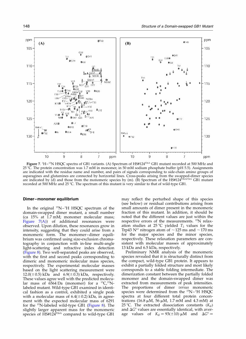
The position of Phe34 in the domain-swapped
dimer appeared to be most crucial from a prelimi-nary inspection of the structure. In the Ala34Phemodeled GB1 monomer structure, the phenyl ringclashes with the side-chain of Val39, the centralresidue in the hinge–loop. This clash, togetherwith the strain in the linker due to the expansionof the core space is predicted to cause severe struc-tural interference. As a consequence, in thedomain-swapped dimer structure, the backboneconformation around Val39 is altered, with theside-chain flipped outward and solvent-exposed(see Figures 3 and 5). Calculation of the solvent-accessible surface for Val39 reveals an increase of,30% from monomer to domain-swapped dimer.In order to test and confirm our hypothesis, wereverted Phe34 in the amino acid sequence of thedomain-swapped dimer mutant back to the wild-type Ala residue. As predicted, the resultingmutant protein was monomeric and exhibited a15N–1H HSQC spectrum very similar to that ofwild-type GB1. The 1H–15N HSQC spectrum forthis mutant is shown in Figure 7(B) and, for com-parison, the 1H–15N HSQC spectrum ofHS#124F26A containing both swapped dimer and amonomeric species is displayed alongside. Selectedresonances are labeled in both spectra. For theHS#124F26AF34A revertant, the resonances that areparticularly indicative of the wild-type GB1 struc-ture are labeled. For instance, note the strikinglydifferent frequencies for E56, L12 and T44 in bothspectra.Taking the above and previous4 mutational and
structural data together, we now clearly haveestablished that a single amino acid change,namely A34F, switches the classical GB1 foldexhibited by the GB1 variant L5V/F30V/Y33F intoa domain-swapped dimer, which in turn can beswitched into the tetramer structure by anadditional single-site mutation, namely A26F.
Figure 6. Superposition of theequivalent core structures of thedomain-swapped dimer and wild-type GB1 (accession code 3GB1).The superposition involves half ofthe mutant core structure, com-posed of the first b-hairpin and thea-helix from one monomer subunit(red) and the second b-hairpinfrom the other subunit (blue) andthe wild-type GB1 core (green). Sec-ondary structure elements andselected side-chains are labeled.The distances between the b-sheetand the a-helix axis at both endsare indicated. The superpositionwas carried out by best-fitting resi-dues 1–37 from one monomer sub-unit and residues 43–56 from theother subunit in the mutant andthe corresponding residues of wild-type GB1.
Structure of a Domain-swapped GB1 Mutant 147
Dimer–monomer equilibrium
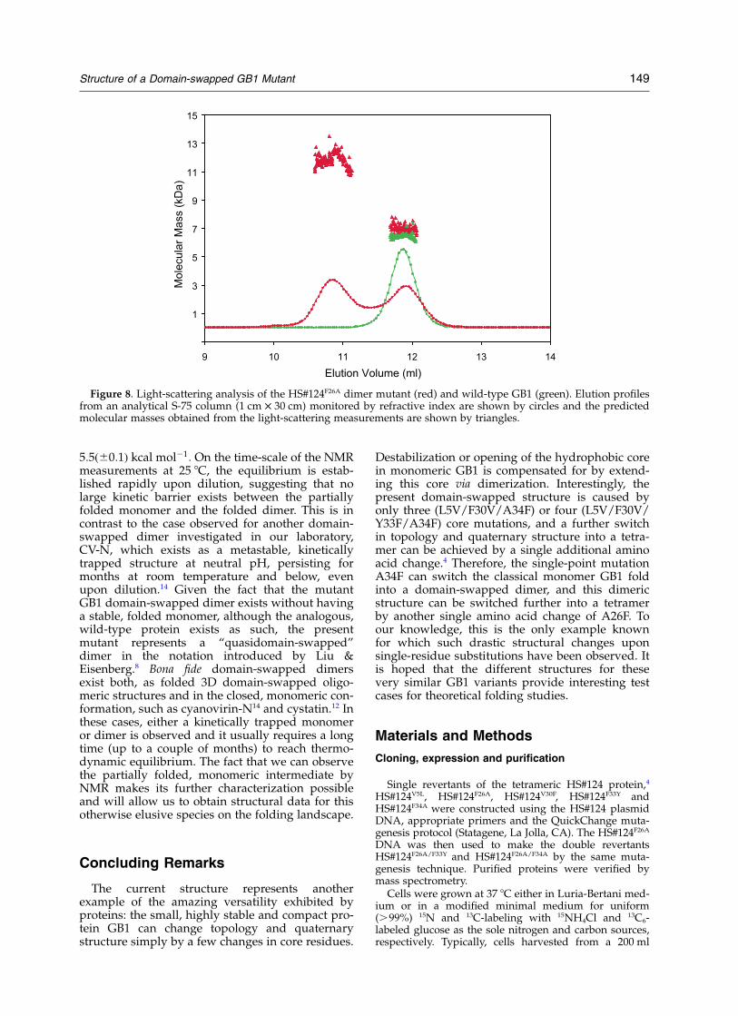
In the original 15N–1H HSQC spectrum of thedomain-swapped dimer mutant, a small number(ca 15% at 1.7 mM; monomer molecular mass;Figure 7(A)) of additional resonances wereobserved. Upon dilution, these resonances grow inintensity, suggesting that they could arise from amonomeric form. The monomer–dimer equili-brium was confirmed using size-exclusion chroma-tography in conjunction with in-line multi-anglelight-scattering and refractive index detection(Figure 8). Two well-separated peaks are observed,with the first and second peaks corresponding todimeric and monomeric molecular mass species,respectively. The experimental molecular massesbased on the light scattering measurement were12.0(^0.5) kDa and 6.9(^0.3) kDa, respectively.These values agree well with the predicted molecu-lar mass of 6564 Da (monomer) for a 13C,15N-labeled mutant. Wild-type GB1 examined in identi-cal fashion as a control, exhibited a single peakwith a molecular mass of 6.4(^0.2) kDa, in agree-ment with the expected molecular mass of 6291for the 15N-labeled wild-type GB1 (Figure 8). Theslightly larger apparent mass for the monomericspecies of HS#124F26A compared to wild-type GB1
may reflect the perturbed shape of this species(see below) or residual contributions arising fromsmall amounts of dimer present in the monomericfraction of this mutant. In addition, it should benoted that the different values are just within therespective errors of the measurements. 15N relax-ation studies at 25 8C yielded T2 values for theTrp43 Ne1 nitrogen atom of ,125 ms and ,170 msfor the major species and the minor species,respectively. These relaxation parameters are con-sistent with molecular masses of approximately13 kDa and 6.5 kDa, respectively.
Preliminary NMR analysis of the monomericspecies revealed that it is structurally distinct fromthe compact, wild-type GB1 protein. It appears toexhibit a partially folded structure and most likelycorresponds to a stable folding intermediate. Thedimerization constant between the partially foldedmonomer and the domain-swapped dimer wasextracted from measurements of peak intensities.The proportions of dimer versus monomericspecies were determined from the 15N–1H HSQCspectra at four different total protein concen-trations (16.8 mM, 56 mM, 1.7 mM and 4.3 mM) at25 8C. The extracted dissociation constants !Kd"and DG8 values are essentially identical, with aver-age values of Kd # 93!^10" mM and DG8 #
Figure 7. 1H–15N HSQC spectra of GB1 variants. (A) Spectrum of HS#124F26A GB1 mutant recorded at 500 MHz and25 8C. The protein concentration was 1.7 mM in monomer, in 50 mM sodium phosphate buffer (pH 5.5). Assignmentsare indicated with the residue name and number, and pairs of signals corresponding to side-chain amino groups ofasparagines and glutamines are connected by horizontal lines. Cross-peaks arising from the swapped-dimer speciesare indicated by (d) and those from the monomeric species by (m). (B) Spectrum of the HS#124F26AF34A GB1 mutantrecorded at 500 MHz and 25 8C. The spectrum of this mutant is very similar to that of wild-type GB1.
148 Structure of a Domain-swapped GB1 Mutant
5:5!^0:1" kcal mol21: On the time-scale of the NMRmeasurements at 25 8C, the equilibrium is estab-lished rapidly upon dilution, suggesting that nolarge kinetic barrier exists between the partiallyfolded monomer and the folded dimer. This is incontrast to the case observed for another domain-swapped dimer investigated in our laboratory,CV-N, which exists as a metastable, kineticallytrapped structure at neutral pH, persisting formonths at room temperature and below, evenupon dilution.14 Given the fact that the mutantGB1 domain-swapped dimer exists without havinga stable, folded monomer, although the analogous,wild-type protein exists as such, the presentmutant represents a “quasidomain-swapped”dimer in the notation introduced by Liu &Eisenberg.8 Bona fide domain-swapped dimersexist both, as folded 3D domain-swapped oligo-meric structures and in the closed, monomeric con-formation, such as cyanovirin-N14 and cystatin.12 Inthese cases, either a kinetically trapped monomeror dimer is observed and it usually requires a longtime (up to a couple of months) to reach thermo-dynamic equilibrium. The fact that we can observethe partially folded, monomeric intermediate byNMR makes its further characterization possibleand will allow us to obtain structural data for thisotherwise elusive species on the folding landscape.
Concluding Remarks
The current structure represents anotherexample of the amazing versatility exhibited byproteins: the small, highly stable and compact pro-tein GB1 can change topology and quaternarystructure simply by a few changes in core residues.
Destabilization or opening of the hydrophobic corein monomeric GB1 is compensated for by extend-ing this core via dimerization. Interestingly, thepresent domain-swapped structure is caused byonly three (L5V/F30V/A34F) or four (L5V/F30V/Y33F/A34F) core mutations, and a further switchin topology and quaternary structure into a tetra-mer can be achieved by a single additional aminoacid change.4 Therefore, the single-point mutationA34F can switch the classical monomer GB1 foldinto a domain-swapped dimer, and this dimericstructure can be switched further into a tetramerby another single amino acid change of A26F. Toour knowledge, this is the only example knownfor which such drastic structural changes uponsingle-residue substitutions have been observed. Itis hoped that the different structures for thesevery similar GB1 variants provide interesting testcases for theoretical folding studies.
Materials and Methods
Cloning, expression and purification
Single revertants of the tetrameric HS#124 protein,4
HS#124V5L, HS#124F26A, HS#124V30F, HS#124F33Y andHS#124F34A were constructed using the HS#124 plasmidDNA, appropriate primers and the QuickChange muta-genesis protocol (Statagene, La Jolla, CA). The HS#124F26A
DNA was then used to make the double revertantsHS#124F26A/F33Y and HS#124F26A/F34A by the same muta-genesis technique. Purified proteins were verified bymass spectrometry.
Cells were grown at 37 8C either in Luria-Bertani med-ium or in a modified minimal medium for uniform(.99%) 15N and 13C-labeling with 15NH4Cl and 13C6-labeled glucose as the sole nitrogen and carbon sources,respectively. Typically, cells harvested from a 200 ml
Figure 8. Light-scattering analysis of the HS#124F26A dimer mutant (red) and wild-type GB1 (green). Elution profilesfrom an analytical S-75 column (1 cm £ 30 cm) monitored by refractive index are shown by circles and the predictedmolecular masses obtained from the light-scattering measurements are shown by triangles.
Structure of a Domain-swapped GB1 Mutant 149
culture were suspended in 8-10 ml of PBS (17 mMKH2PO4, 50 mM Na2HPO4, 1.5 M NaCl, pH 7.4), heatedat 80 8C for five minutes, immediately chilled on ice forten minutes followed by centrifugation at 16,000 rpm(SS-34 rotor) for 30 minutes at 4 8C. The supernatantwas passed through a 0.45 mm pore size syringe filter,dialyzed against an excess of deionized water in thecold room, adjusted to 50 mM sodium phosphate (pH5.5), concentrated using Centriprep YM-3 devices (Milli-pore corp., Bedford, MA) and loaded onto a Superdex-75 column (2.6 cm £ 60 cm; Amersham Biosciences, Pis-cataway, NJ) equilibrated in 50 mM sodium phosphatebuffer (pH 5.5), at a flow-rate of 3 ml/minute at roomtemperature. Peak fractions were combined and concen-trated (Centriprep YM-3), estimated for protein concen-tration and stored at 4 8C until further use or subjectedto an additional reverse-phase HPLC purification stepfor unlabeled mutant proteins. Unlabeled mutant pro-teins were bound to POROS R2 resin and eluted using alinear gradient of aqueous 0–60% (v/v) acetonitrile,0.05% (v/v) trifluoroacetic acid over a period of 16 min-utes at a flow-rate of 4 ml/minute. Peak fractions werecombined and dialyzed excessively against 50 mMsodium phosphate buffer (pH 5.5), 0.02% (w/v) NaN3,concentrated and the protein concentration was deter-mined spectrophotometrically using an extinction coeffi-cient of 8250 M21 cm21 at 280 nm.
NMR spectroscopy and structure determination
All spectra for structure determination were recordedat 25 8C on NMR samples containing ca 1.7 mM protein(monomer molecular mass), 50 mM sodium phosphatebuffer (pH 5.5) using Bruker DRX800, DMX750,DRX600, DMX600 and DMX500 spectrometers, equippedwith 5 mm, triple resonance, three axes gradient probesor z-axis gradient cryoprobes. Spectra were processedwith NMRPipe15 and analyzed using NMRDraw andPIPP, CAPP and STAPP.16 1H, 13C and 15N sequentialassignments and NOE data analysis were carried outusing heteronuclear multi-dimensional experiments rou-tinely used in our laboratory.17–19 3D HNCACB,20
CBCA(CO)NH,21 H(CCO)HN-TOCSY and C(CCO)NH-TOCSY22,23 experiments were employed for sequentialassignments. NOE spectra to derive inter-proton dis-tance constraints included 2D NOESY and 12C,14N-fil-tered, and 3D 13C-edited, 15N-edited, 13C-edited/12C-filtered, and 13C-edited/12C,14N-filtered NOEexperiments,24,25 recorded using mixing times rangingfrom 80 to 100 ms. Approximate inter-proton distanceconstraints were grouped into four distance ranges: 1.8–2.7 A, 1.8–3.3 A, 1.8–5.0 A and 1.8–6.0 A, correspondingto strong, medium, weak and very weak NOEs, respect-ively. In addition, 0.5 A was added to the upper limit ofinter-proton distance constraints involving methylgroups. For hydrogen bonds, distance constraints of1.5–2.5 A (H–O) and 2.5–3.5 A (N–O) were employed.Torsion angle constraints were obtained by measuring3JN–Hb, 3JHa–Hb, 3JN–Cg, 3JC0 –Cg and 3JCa–Cd couplings usingquantitative J correlation spectroscopy.26–28 TALOS5 wasused for extracting backbone torsion angle (f,c) con-straints. Residual HN dipolar couplings were measuredusing in-phase/anti-phase 1H–15N HSQC experiments29
on protein samples in magnetically oriented (using17 mg/ml of pf1 phage solution) and isotropic media(without phage). The measured residual HN dipolarcouplings ranged from 216 Hz to 37 Hz. Slowly exchan-ging amide protons were detected in the 1H–15N HSQCspectrum of lyophilized protein freshly dissolved in
2H2O at 15 8C. All experimentally determined distance,torsion angle and residual dipolar coupling constraints(Table 1) were applied in a simulated annealing protocolusing the program CNS.6 The equivalence of the mono-meric subunits was ensured using non-crystallographicsymmetry constraints by implementing three axes ofsecond order for the dimer via a special symmetry NOEclass.30,31 An ensemble of 100 structures was generatedand the final family of 39 structures, the major structuralfamily with lowest CNS target function values, wasselected. The regularized mean structure was calculatedby restrained energy minimization of the geometricmean of the final 39 structures. Quality factor values forthe residual N–H dipolar couplings were calculatedusing PALES.32 The quality of the structures was ana-lyzed by CNS,6 PROCHECK,33 and MOLMOL.34 Allstructure Figures were generated with MOLMOL.34
Determination of the dissociation constant of thedimeric GB1 mutant
The relative amounts of dimeric and monomericspecies were determined from 15N–1H HSQC peak inten-sities. Volume or peak height measurements yieldedessentially the same values (^5% variation). The Trp43Ne1H and Ala 26 backbone NH resonances that exist aswell resolved, isolated peaks in the domain-swappeddimer and the partially folded monomer were used forquantification. Final fractions were calculated by aver-aging values from both resonances using the both deter-minations (volume and height) for four different proteinconcentrations (17 mM, 56 mM, 1.70 mM and 4.28 mM).The dissociation constant !Kd" and free energy of dis-sociation !DG8" were calculated using the followingequations:
D$ 2M
Kd # $M%2=$D% # 2Ptf2M=fD # 2Ptf
2M=!12 fM"
DG8 # 2RT ln Kd
where [M] and [D] are the molar concentrations of thepartially folded monomer and domain-swapped dimer,respectively, Pt is the total protein concentration (mono-mer molecular mass), fM and fD are the measured frac-tions of monomer and dimer, respectively, R is the gasconstant (1.987 cal deg21·mol21), and T is the absolutetemperature. DG8 was calculated by normalizing all reac-tants to the standard concentration of 1 M before apply-ing the logarithm.35,36
Size-exclusion chromatography and multi-angle light-scattering
Light-scattering data were obtained using an analyti-cal Superdex-75 column (1.0 cm £ 30 cm; AmershamBiosciences, Piscataway, NJ) with in-line multi-anglelight-scattering (DAWN EOS, Wyatt Technology, Inc.)and refractive index detectors (OPTILAB DSP, WyattTechnology, Inc., Santa Barbara, CA). Protein (90 mg at aconcentration of ,0.18 mM) was applied to the pre-equi-librated S75 column at a flow-rate of 0.5 ml/minuteat room temperature and eluted with 20 mM sodiumphosphate buffer (pH 5.5) containing 0.02% (w/v)sodium azide.
150 Structure of a Domain-swapped GB1 Mutant
Protein Data Bank accession numbers
The atomic coordinates of the 39 conformers of theNMR structure, the corresponding regularized meanstructure, together with the experimental distance andangle constrains, have been deposited in the RCSBProtein Data Bank,† accession numbers 1QIO. NMRassignments for the HS#124F26A mutant of GB1 havebeen deposited at the Biomolecular Magnetic ResonanceDatabank,‡ access code 5875.
Acknowledgements
We thank Drs D. Garrett and F. Delaglio for soft-ware, and J. Baber for technical support. This workwas supported, in part, by the Intramural AIDSTargeted Antiviral Program of the Office of theDirector of the National Institute of Health (toA.M.G.).
References
1. Gronenborn, A. M., Filpula, D. R., Essig, N. Z.,Achari, A., Whitlow, M., Wingfield, P. T. & Clore,G. M. (1991). A novel, highly stable fold of the immu-noglobulin binding domain of streptococcal proteinG. Science, 253, 657–661.
2. Gallagher, T., Alexander, P., Bryan, P. & Gilliland,G. L. (1994). Two crystal structures of the B1 immu-noglobulin-binding domain of streptococcal proteinG and comparison with NMR. Biochemistry, 33,4721–4729.
3. Gronenborn, A. M., Frank, M. K. & Clore, G. M.(1996). Core mutants of the immunoglobulin bindingdomain of streptococcal protein G: stability andstructural integrity. FEBS Letters, 398, 312–316.
4. Frank, M. K., Dyda, F., Dobrodumov, A. & Gronen-born, A. M. (2002). Core mutations switch mono-meric protein GB1 into an intertwined tetramer.Nature Struct. Biol. 9, 877–885.
5. Cornilescu, G., Delaglio, F. & Bax, A. (1999). Proteinbackbone angle restraints from searching a databasefor chemical shift and sequence homology. J. Biomol.NMR, 13, 289–302.
6. Brunger, A. T., Adams, P. D., Clore, G. M., DeLano,W. L., Gros, P., Grosse-Kunstleve, R. W. et al. (1998).Crystallography & NMR system: a new softwaresuite for macromolecular structure determination.Acta Crystallog. sect. D, 54, 905–921.
7. Cornilescu, G., Marquardt, J. L., Ottiger, M. & Bax, A.(1998). Validation of protein structure from anisotro-pic carbonyl chemical shifts in a dilute liquid crystal-line phase. J. Am. Chem. Soc. 120, 6836–6837.
8. Liu, Y. & Eisenberg, D. (2002). 3D domain swapping:as domains continue to swap. Protein Sci. 11,1285–1299.
9. Schlunegger, M. P., Bennett, M. J. & Eisenberg, D.(1997). Oligomer formation by 3D domain swapping:a model for protein assembly and misassembly.Advan. Protein Chem. 50, 61–122.
10. Yang, F., Bewley, C. A., Louis, J. M., Gustafson, K. R.,Boyd, M. R., Gronenborn, A. M. et al. (1999). Crystalstructure of cyanovirin-N, a potent HIV-inactivatingprotein, shows unexpected domain swapping. J. Mol.Biol. 288, 403–412.
11. Janowski, R., Kozak, M., Jankowska, E., Grzonka, Z.,Grubb, A., Abrahamson, M. & Jaskolski, M. (2001).Human cystatin C, an amyloidogenic protein,dimerizes through three-dimensional domain swap-ping. Nature Struct. Biol. 8, 316–320.
12. Staniforth, R. A., Giannini, S., Higgins, L. D., Conroy,M. J., Hounslow, A. M., Jerala, R. et al. (2001). Three-dimensional domain swapping in the folded andmolten-globule states of cystatins, an amyloid-form-ing structural superfamily. EMBO J. 20, 4774–4781.
13. Dunbrack, R. L. & Cohen, F. E. (1997). Bayesian stat-istical analysis of protein side-chain rotamer prefer-ences. Protein Sci. 6, 1661–1681.
14. Barrientos, L. G., Louis, J. M., Botos, I., Mori, T., Han,Z. Z., O’Keefe, B. R. et al. (2002). The domain-swapped dimer of cyanovirin-N is in a metastablefolded state: reconciliation of X-ray and NMR struc-tures. Structure, 10, 673–686.
15. Delaglio, F., Grzesiek, S., Vuister, G. W., Zhu, G., Pfei-fer, J. & Bax, A. (1995). Nmrpipe—a multidimen-sional spectral processing system based on UnixPipes. J. Biomol. NMR, 6, 277–293.
16. Garrett, D. S., Powers, R., Gronenborn, A. M. &Clore, G. M. (1991). A Common-sense approach topeak picking in 2-dimensional, 3-dimensional, and4-dimensional spectra using automatic computer-analysis of contour diagrams. J. Magn. Reson. 95,214–220.
17. Clore, G. M. & Gronenborn, A. M. (1991). 2-dimen-sional, 3-dimensional, and 4-dimensional NMRmethods for obtaining larger and more precise 3-dimensional structures of proteins in solution. Annu.Rev. Biophys. Biophys. Chem. 20, 29–63.
18. Clore, G. M. & Gronenborn, A. M. (1998). Determin-ing the structures of large proteins and protein com-plexes by NMR. Trends Biotechnol. 16, 22–34.
19. Bax, A. & Grzesiek, S. (1993). MethodologicalAdvances in Protein NMR. Accts Chem. Res. 26,131–138.
20. Wittekind, M. & Mueller, L. (1993). HNCACB, ahigh-sensitivity 3D NMR experiment to correlateamide-proton and nitrogen resonances with thealpha-carbon and beta-carbon resonances in pro-teins. J. Magn. Reson. ser. B, 101, 201–205.
21. Grzesiek, S. & Bax, A. (1992). Correlating backboneamide and side-chain resonances in larger proteinsby multiple relayed triple resonance NMR. J. Am.Chem. Soc. 114, 6291–6293.
22. Logan, T. M., Olejniczak, E. T., Xu, R. X. & Fesik, S. W.(1993). A general-method for assigning NMR-spectraof denatured proteins using 3D HC(CO)NH-TOCSYtriple resonance experiments. J. Biomol. NMR, 3,225–231.
23. Lin, Y. X. & Wagner, G. (1999). Efficient side-chainand backbone assignment in large proteins: appli-cation to tGCN5. J. Biomol. NMR, 15, 227–239.
24. Ikura, M. & Bax, A. (1992). Isotope-filtered 2D NMRof a protein peptide complex-study of a skeletal-muscle myosin light chain kinase fragment boundto calmodulin. J. Am. Chem. Soc. 114, 2433–2440.
25. Lee, W., Revington, M. J., Arrowsmith, C. & Kay, L. E.(1994). A pulsed-field gradient isotope-filtered 3DC-13 HMQC-NOESY experiment for extracting
†www.rcsb.org‡www.bmrb.wisc.edu
Structure of a Domain-swapped GB1 Mutant 151
intermolecular NOE contacts in molecular-com-plexes. FEBS Letters, 350, 87–90.
26. Bax, A., Vuister, G. W., Grzesiek, S., Delaglio, F.,Wang, A. C., Tschudin, R. & Zhu, G. (1994). Measure-ment of homonuclear and heteronuclear J-couplingsfrom quantitative J-correlation. Nucl. Magn. Reson.Pt C, 239, 79–105.
27. Grzesiek, S., Kuboniwa, H., Hinck, A. P. & Bax, A.(1995). Multiple-quantum line narrowing formeasurement of H-alpha-H-beta J-couplings in isoto-pically enriched proteins. J. Am. Chem. Soc. 117,5312–5315.
28. Hu, J. S., Grzesiek, S. & Bax, A. (1997). Two-dimen-sional NMR methods for determining (chi 1) anglesof aromatic residues in proteins from three-bondJ(C0C gamma) and J(NC gamma) couplings. J. Am.Chem. Soc. 119, 1803–1804.
29. Ottiger, M., Delaglio, F. & Bax, A. (1998). Measure-ment of J and dipolar couplings from simplifiedtwo-dimensional NMR spectra. J. Magn. Reson. 131,373–378.
30. ODonoghue, S. I., King, G. F. & Nilges, M. (1996).Calculation of symmetric multimer structures fromNMR data using a priori knowledge of the monomerstructure, co-monomer restraints, and interface map-ping: the case of leucine zippers. J. Biomol. NMR, 8,193–206.
31. Nilges, M. (1993). A calculation strategy for the struc-
ture determination of symmetrical dimers by H-1-NMR. Proteins: Struct. Funct. Genet. 17, 297–309.
32. Zweckstetter, M. & Bax, A. (2000). Prediction of steri-cally induced alignment in a dilute liquid crystallinephase: aid to protein structure determination byNMR. J. Am. Chem. Soc. 122, 3791–3792.
33. Laskowski, R. A., MacArthur, M. W., Moss, D. S. &Thornton, J. M. (1993). PROCHECK—a program tocheck the stereochemical quality of protein struc-tures. J. Appl. Crystallog. 26, 283–291.
34. Koradi, R., Billeter, M. & Wuthrich, K. (1996). MOL-MOL: a program for display and analysis of macro-molecular structures. J. Mol. Graph. 14, 51–55.
35. Backes, H., Berens, C., Helbl, V., Walter, S., Schmid,F. X. & Hillen, W. (1997). Combinations of the alpha-helix-turn-alpha-helix motif of TetR with respectiveresidues from LacI or 434Cro: DNA recognition,inducer binding, and urea-dependent denaturation.Biochemistry, 36, 5311–5322.
36. Dams, T. & Jaenicke, R. (1999). Stability and foldingof dihydrofolate reductase from the hyperthermo-philic bacterium Thermotoga maritima. Biochemistry,38, 9169–9178.
37. Brooks, B. R., Bruccoleri, R. E., Olafson, B. D., States,D. J., Swaminathan, S. & Karplus, M. (1983).CHARMM—a program for macromolecular energy,minimization, and dynamics calculations. J. Comput.Chem. 4, 187–217.
Edited by M. F. Summers
(Received 20 May 2003; received in revised form 11 July 2003; accepted 11 July 2003)
152 Structure of a Domain-swapped GB1 Mutant