Embed Size (px)
Citation preview
3030International Journal of Scientific Study | January 2021 | Vol 8 | Issue 10
A Prospective Study Comparing Prevalence of Different Antiphospholipid Antibodies in Pregnant Women with Recurrent Pregnancy LossMonika Garg1, Meena Samant2, Neha Quarrat Ain1, Omprakash Bisore3
1RSO, Department of Obstetrics and Gynaecology, Kurji Holi Family Hospital, Patna, Bihar, India, 2HOD, Department of Obstetrics and Gynaecology, Kurji Holi Family Hospital, Patna, Bihar, India, 3RSO, Department of Surgery, Shyam Shah Medical College, Rewa, Madhya Pradesh, India
INTRODUCTION
Recurrent pregnancy loss (RPL) is a common problem in obstetrics. It affects about 5–15% of all pregnancies worldwide.[1] It affects between 1 in 300 and 1 in 100 couples worldwide.[2] RPL is encountered in 5% of couples with two or more losses and in around 1–2% of couples with three or more losses.[3]
Original Article
AbstractIntroduction: Prospective study comparing prevalence of different antiphospholipid antibodies (APAs) in pregnant women with recurrent pregnancy loss.
Materials and Methods: The patients recruited in the study would include those attending the OBG outpatient department at Kurji Holy Family Hospital, Patna. After thorough history and clinical examination, routine blood investigations were sent. Patients were counseled about benefits of APLA test and its implications in pregnancy outcome. Consent was taken and patients were informed that if test is positive than she would have to undergo repeat test 12 weeks later for confirmation of diagnosis. Patients blood were collected into two separate bulbs one in plane bulb (2 ml blood) by which b2GP1 and ACL antibodies were detected by ELISA and other is citrate bulb (containing 0.2 ml of trisodium citrate 3.2%+1.8 ml of freshly collected blood) which detect LA.
Results: Among 120 patients, 23 patients (19.1%) were positive for APLA antibodies among which 11 cases were (9.16%) positive for lupus anticoagulant, 6 cases were anticardiolipin antibodies. Out of 6 cases, 2 (1.6%) cases positive for anticardiolipin antibodies IgG and 4 (3.33%) were positive for IgM. The positivity of anti-beta-2 glycoprotein 1 in our study was 5% in which 4 cases (3.33%) positive for IgG and 2 (1.6%) cases were positive for IgM antibodies. Among 23 patients of APLA positive, 16 were started on LMWH and 14 patients of APLA negative were also started on LMWH the outcome was not statistically significant. Among APLA positive out of 23, 12 cases delivered by LSCS, 7 by PTVD, 3 by FTVD, and 1 case missed and the indication of LSCS as was fetal distress in 4 women, severe oligohydramnios in 3 cases, 2 for severe preeclampsia, 2 for abruption, and 1 for failed induction. We also followed up the babies of these patients. Ninety-three babies were healthy and given to mother in which 14 babies were of APLA-positive mother, 20 neonates were admitted to NICU for prematurity or fetal distress out of which 6 patients of APLA-positive women, 3 had IUD 2 of which APLA positive, 3 neonatal death in which 1 neonate of APLA-positive women. We also note the complications among women in which preeclampsia was present in seven patients out which six were APLA positive. Among APLA positive, six patients had IUGR, five patients had abruption, one had vascular complications, and seven had severe oligohydramnios. Antithrombotic interventions are essential to have a favorable outcome in high-risk pregnancies in association with APLA antibodies and thus the need of these high-risk pregnancies for APLA.
Conclusion: The association of APLA antibodies in high-risk pregnancy with a history of recurrent miscarriages was found to be 19.1% in the present study. The incidence of APLA antibodies in general population is 5–20%, it is proved fact that the APAs interfere with normal development of the uteroplacental circulation to cause both early and late pregnancy loss. Based on the concept of APAs induced thrombophilia and placental thrombosis, antithrombotic interventions have been widely applied to reduce the incidence of miscarriages and fetal loss. The outcome of high-risk pregnancies in APLA syndrome is considerably improved by initiation of therapies using aspirin, unfractionated heparin, and/or low-molecular-weight heparin. The antiphospholipids have been the most important cause for recurrent fetal loss, thus, many pregnancies can be saved if diagnosis and treated adequately. This can be done by routine screening for the antiphospholipids antibodies in pregnant women with a bad obstetric history and unexplained fetal loss. Close antenatal surveillance and planned delivery of these pregnancies in a unit with specialist obstetrics and neonatal intensive care facilities are indicated.
Key words: ACA(anticardiolipin antibody), Antibeta2GLP1, APLA (antiphospholipid antibody), RPL (recurrent pregnancy loss)
Access this article online
www.ijss-sn.com
Month of Submission : 11-2020 Month of Peer Review : 11-2020 Month of Acceptance : 12-2020 Month of Publishing : 01-2021
Corresponding Author: Dr. Monika Garg, RSO, Department of Obstetrics and Gynaecology, Kurji Holi Family Hospital, Patna, Bihar, India
Print ISSN: 2321-6379Online ISSN: 2321-595X
Garg, et al.: Prevalence of APLA in RPL
3131 International Journal of Scientific Study | January 2021 | Vol 8 | Issue 10
The Royal College of Obstetricians and Gynecologists and the European Society of Human Reproduction and Embryology define recurrent miscarriage as three or more consecutive losses before 24 weeks gestation[4] Due to an increasing number of childless couples, the improved availability of diagnostic tests, and most importantly the minimal difference in the prognostic value between two and three losses, the American Society for Reproductive Medicine updated the definition of RPL to two or more clinical pregnancy losses, before 20 weeks period of gestation, documented by either ultrasonography or approved in a histopathologic examination.[4]
A large number of etiological factors are associated with RPL, in approximately two-third of cases, the cause is known to be genetic error, anatomic abnormalities of the reproductive tract, hormonal abnormalities, infection or immunologic factors, or systemic disease, whereas, idiopathic in one-third of all cases.[5]
There is a great deal of interest in the role inherited thrombophilia in RPL. Thrombophilias can be hereditary thrombophilias (HTs) or acquired thrombophilias (ATs). The most common HT is due to Factor V Leiden, prothrombin gene mutation, protein C deficiency, protein S deficiency, and antithrombin III deficiency accounting for other causes.[6] ATs are mostly attributed to antiphospholipid syndrome (APS) which is encountered in 5–20% of patients with RPL.
Antiphospholipid antibodies (APAs) are group of antibodies that bind to negatively charged phospholipids. APAs are heterogeneous group of autoantibodies directed against different antigens, predominantly anionic phospholipids, or phospholipids containing structures.[7] APAs that associated with APS are anticardiolipin antibodies (ACAs) or antibodies against other negatively charged phospholipids such as phosphatidylserine, phosphatidylinositol, phosphatidic acid, and phosphatidylglycerol.[8] Lupus anticoagulants (LAs) which are immunoglobulins directed against plasma proteins such as prothrombin or annexin V that are bound to phospholipids. Anti-b2 glycoprotein 1 is antibodies which recognize a plasma protein known as apolipoprotein H or beta-2 glycoprotein I and has higher specificity than ACA for thrombosis.[9]
The presence of the APAs was shown to associate with recurrent miscarriage due to thrombosis of the uteroplacental vasculature and subsequent placental infarct.
There is an increasing burden of RPL in the society and APS being one of the undisputed treatable causes for RPL. This study aims to evaluate the significance of the antibody profiles of APS in relation to RPL.
MATERIALS AND METHODS
Study DesignThe patients recruited in the study were included those attending the OBG outpatient department at Kurji Holy Family Hospital, Patna.
Study PopulationInclusion criteriaHistory of two or more previous spontaneous pregnancy losses with• Ultrasound confirmed pregnancy with intrauterine
gestation sac• Less than 20 weeks of gestation • With or without fetal cardiac activity.
Exclusion criteriaThe following criteria were excluded from the study:• Previous medical termination of pregnancy• Previous ectopic pregnancy• Previous pregnancy losses of more than 20 weeks
gestation• Trauma-induced previous pregnancy loss.
Data CollectionAfter thorough history and clinical examination, routine blood investigations were send. Patients were counseled about benefits of APLA test and its implications in pregnancy outcome. Consent was taken and patients were informed that if test is positive then she would have to undergo repeat test 12 weeks later for confirmation of diagnosis. Patients blood were collected into two separate bulbs one in plane bulb (2 ml blood) by which b2GP1 and ACL antibodies were detected by ELISA and other is citrate bulb (containing 0.2 ml of trisodium citrate 3.2% + 1.8 ml of freshly collected blood) which detect LA.
Sample SizeAbout 4000 cases come in OPD per year, out of which about 60–70 patients are of RPL. Now using Raosoft formula with 95% confidence level and 5% margin of error, sample comes out to be 60.
Y=z(c/100)2r (100-r) n=Ny/(N-1)E2+yE=sqrt[(N-n)y/n(N-1)]
For 2 years of study, my sample was about 120.
Study DurationThis study was from November 2017 to October 2019.
MethodologyWritten and informed consent was taken from all the patients after brief explanation of the procedure. Ethical
Garg, et al.: Prevalence of APLA in RPL
3232International Journal of Scientific Study | January 2021 | Vol 8 | Issue 10
clearance was obtained from Institute’s Ethical Clearance Committee.
A detailed history of patients will be take based on set questionnaires about pregnancy loss like weeks at which abortion occurs, was pregnancy confirmed or not any history of curettage, hypertension, DM, thyroid disorders, etc. Detailed general and gynecological examination findings were taken. Routine blood investigations were sent along with investigations for APLAs which included LA, anticardiolipin antibody (ACA), and anti-β2 glycoprotein 1 (Anti-β2GP1Ab). If any of the above-mentioned tests for APAs will come positive for a patient, a repeat of that particular test will be done after 12 weeks, since the diagnosis of APS requires a test to be positive on two or more occasions at least 12 weeks apart. LA will be measured using dilute Russell viper venom test using the principle of electromechanical clot detection. Normal values are between 32 and 42 s with higher values suggestive of antibody positivity. Serum ACA levels were tested by enzyme immune assay method. Values >15GPL for IgG antibody subtype and >12.5 MPL for IgM antibody subtype will be taken as positive. Serum anti-β2GP1Ab levels will be tested by enzyme immune assay method. Values >20 SGU for IgG type and >20 SMU for IgM type antibody will be considered to be positive. Statistical analyses will be done using SPSS version 18. Prevalence data will be noted as percentages or proportions. Categorical variables were compared using Fisher’s exact test while continuous variables were compared using Student’s t-test. P < 0.05 was taken to be statistically significant. Consent was taken from all the participants on consent forms written in the language comfortable to them.
RESULTS
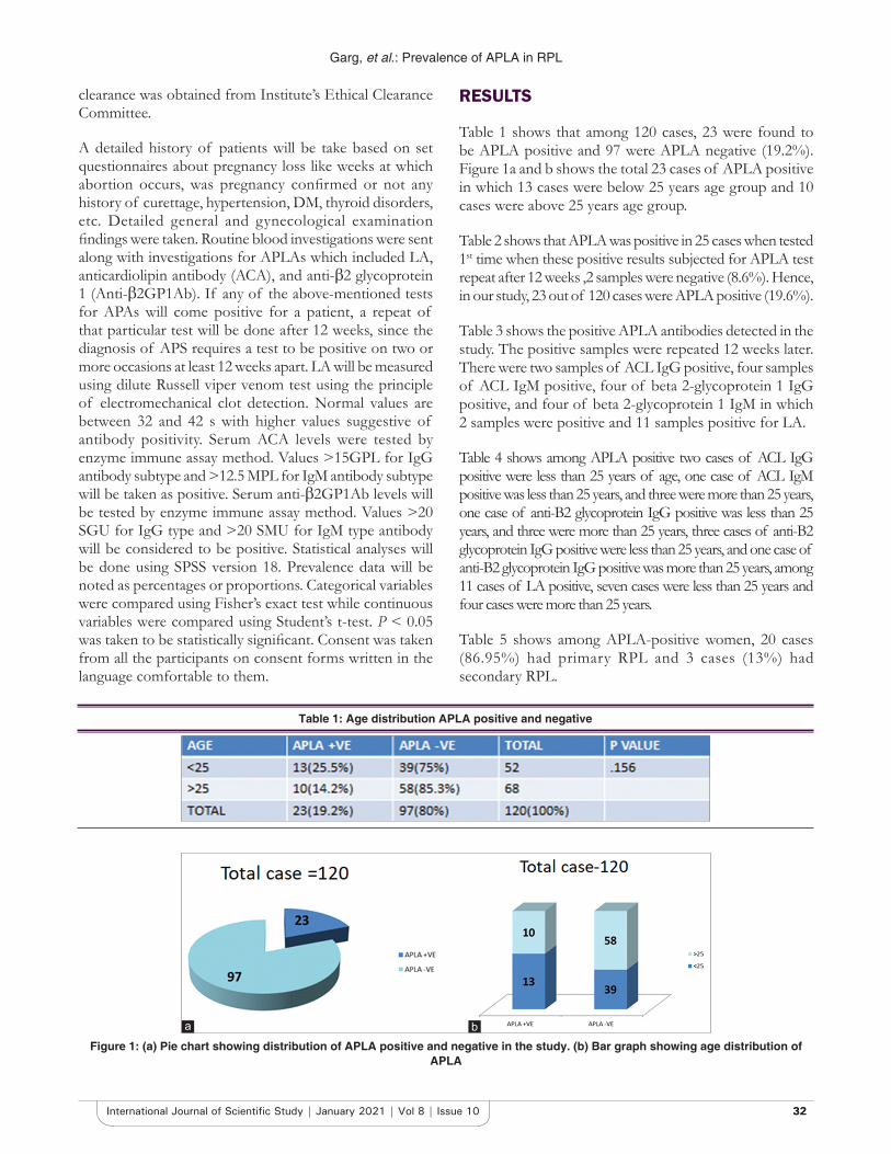
Table 1 shows that among 120 cases, 23 were found to be APLA positive and 97 were APLA negative (19.2%). Figure 1a and b shows the total 23 cases of APLA positive in which 13 cases were below 25 years age group and 10 cases were above 25 years age group.
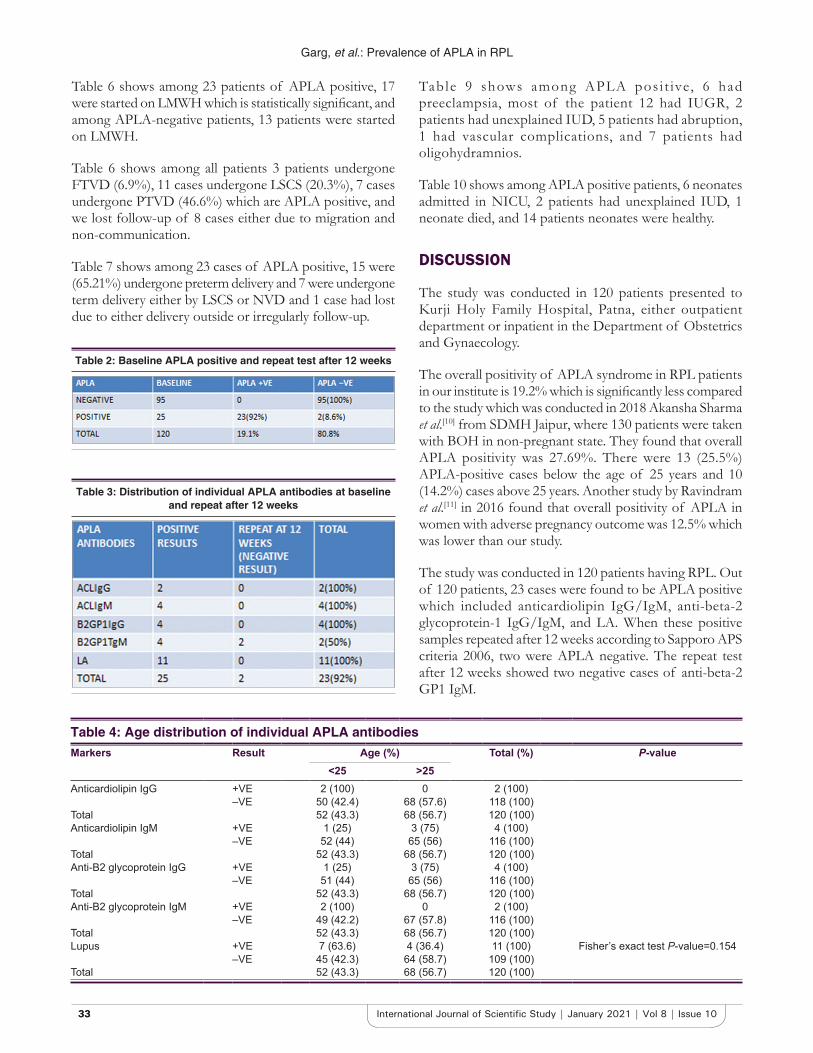
Table 2 shows that APLA was positive in 25 cases when tested 1st time when these positive results subjected for APLA test repeat after 12 weeks ,2 samples were negative (8.6%). Hence, in our study, 23 out of 120 cases were APLA positive (19.6%).
Table 3 shows the positive APLA antibodies detected in the study. The positive samples were repeated 12 weeks later. There were two samples of ACL IgG positive, four samples of ACL IgM positive, four of beta 2-glycoprotein 1 IgG positive, and four of beta 2-glycoprotein 1 IgM in which 2 samples were positive and 11 samples positive for LA.
Table 4 shows among APLA positive two cases of ACL IgG positive were less than 25 years of age, one case of ACL IgM positive was less than 25 years, and three were more than 25 years, one case of anti-B2 glycoprotein IgG positive was less than 25 years, and three were more than 25 years, three cases of anti-B2 glycoprotein IgG positive were less than 25 years, and one case of anti-B2 glycoprotein IgG positive was more than 25 years, among 11 cases of LA positive, seven cases were less than 25 years and four cases were more than 25 years.
Table 5 shows among APLA-positive women, 20 cases (86.95%) had primary RPL and 3 cases (13%) had secondary RPL.
Table 1: Age distribution APLA positive and negative
Figure 1: (a) Pie chart showing distribution of APLA positive and negative in the study. (b) Bar graph showing age distribution of APLA
ba
Garg, et al.: Prevalence of APLA in RPL
3333 International Journal of Scientific Study | January 2021 | Vol 8 | Issue 10
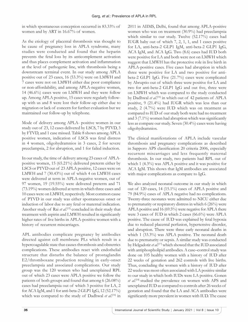
Table 4: Age distribution of individual APLA antibodiesMarkers Result Age (%) Total (%) P-value
<25 >25Anticardiolipin IgG +VE 2 (100) 0 2 (100)
–VE 50 (42.4) 68 (57.6) 118 (100)Total 52 (43.3) 68 (56.7) 120 (100)Anticardiolipin IgM +VE 1 (25) 3 (75) 4 (100)
–VE 52 (44) 65 (56) 116 (100)Total 52 (43.3) 68 (56.7) 120 (100)Anti-B2 glycoprotein IgG +VE 1 (25) 3 (75) 4 (100)
–VE 51 (44) 65 (56) 116 (100)Total 52 (43.3) 68 (56.7) 120 (100)Anti-B2 glycoprotein IgM +VE 2 (100) 0 2 (100)
–VE 49 (42.2) 67 (57.8) 116 (100)Total 52 (43.3) 68 (56.7) 120 (100)Lupus +VE 7 (63.6) 4 (36.4) 11 (100) Fisher’s exact test P-value=0.154
–VE 45 (42.3) 64 (58.7) 109 (100)Total 52 (43.3) 68 (56.7) 120 (100)
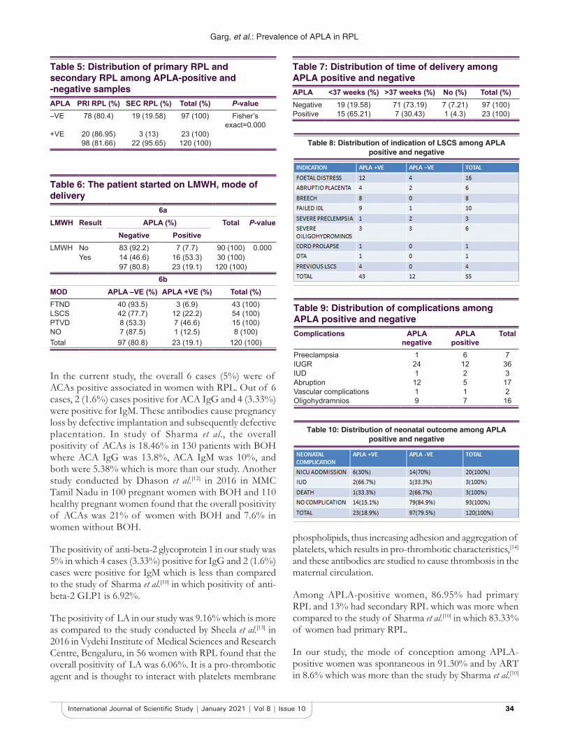
Table 6 shows among 23 patients of APLA positive, 17 were started on LMWH which is statistically significant, and among APLA-negative patients, 13 patients were started on LMWH.
Table 6 shows among all patients 3 patients undergone FTVD (6.9%), 11 cases undergone LSCS (20.3%), 7 cases undergone PTVD (46.6%) which are APLA positive, and we lost follow-up of 8 cases either due to migration and non-communication.
Table 7 shows among 23 cases of APLA positive, 15 were (65.21%) undergone preterm delivery and 7 were undergone term delivery either by LSCS or NVD and 1 case had lost due to either delivery outside or irregularly follow-up.
Table 9 shows among APLA pos i t ive, 6 had preeclampsia, most of the patient 12 had IUGR, 2 patients had unexplained IUD, 5 patients had abruption, 1 had vascular complications, and 7 patients had oligohydramnios.
Table 10 shows among APLA positive patients, 6 neonates admitted in NICU, 2 patients had unexplained IUD, 1 neonate died, and 14 patients neonates were healthy.
DISCUSSION
The study was conducted in 120 patients presented to Kurji Holy Family Hospital, Patna, either outpatient department or inpatient in the Department of Obstetrics and Gynaecology.
The overall positivity of APLA syndrome in RPL patients in our institute is 19.2% which is significantly less compared to the study which was conducted in 2018 Akansha Sharma et al.[10] from SDMH Jaipur, where 130 patients were taken with BOH in non-pregnant state. They found that overall APLA positivity was 27.69%. There were 13 (25.5%) APLA-positive cases below the age of 25 years and 10 (14.2%) cases above 25 years. Another study by Ravindram et al.[11] in 2016 found that overall positivity of APLA in women with adverse pregnancy outcome was 12.5% which was lower than our study.
The study was conducted in 120 patients having RPL. Out of 120 patients, 23 cases were found to be APLA positive which included anticardiolipin IgG/IgM, anti-beta-2 glycoprotein-1 IgG/IgM, and LA. When these positive samples repeated after 12 weeks according to Sapporo APS criteria 2006, two were APLA negative. The repeat test after 12 weeks showed two negative cases of anti-beta-2 GP1 IgM.
Table 2: Baseline APLA positive and repeat test after 12 weeks
Table 3: Distribution of individual APLA antibodies at baseline and repeat after 12 weeks
Garg, et al.: Prevalence of APLA in RPL
3434International Journal of Scientific Study | January 2021 | Vol 8 | Issue 10
phospholipids, thus increasing adhesion and aggregation of platelets, which results in pro-thrombotic characteristics,[14] and these antibodies are studied to cause thrombosis in the maternal circulation.
Among APLA-positive women, 86.95% had primary RPL and 13% had secondary RPL which was more when compared to the study of Sharma et al.[10] in which 83.33% of women had primary RPL.
In our study, the mode of conception among APLA-positive women was spontaneous in 91.30% and by ART in 8.6% which was more than the study by Sharma et al.[10]
Table 7: Distribution of time of delivery among APLA positive and negativeAPLA <37 weeks (%) >37 weeks (%) No (%) Total (%)Negative 19 (19.58) 71 (73.19) 7 (7.21) 97 (100)Positive 15 (65.21) 7 (30.43) 1 (4.3) 23 (100)
Table 9: Distribution of complications among APLA positive and negative Complications APLA
negativeAPLA
positiveTotal
Preeclampsia 1 6 7IUGR 24 12 36IUD 1 2 3Abruption 12 5 17Vascular complications 1 1 2Oligohydramnios 9 7 16
Table 5: Distribution of primary RPL and secondary RPL among APLA-positive and -negative samplesAPLA PRI RPL (%) SEC RPL (%) Total (%) P-value–VE 78 (80.4) 19 (19.58) 97 (100) Fisher’s
exact=0.000+VE 20 (86.95) 3 (13) 23 (100)
98 (81.66) 22 (95.65) 120 (100)
In the current study, the overall 6 cases (5%) were of ACAs positive associated in women with RPL. Out of 6 cases, 2 (1.6%) cases positive for ACA IgG and 4 (3.33%) were positive for IgM. These antibodies cause pregnancy loss by defective implantation and subsequently defective placentation. In study of Sharma et al., the overall positivity of ACAs is 18.46% in 130 patients with BOH where ACA IgG was 13.8%, ACA IgM was 10%, and both were 5.38% which is more than our study. Another study conducted by Dhason et al.[12] in 2016 in MMC Tamil Nadu in 100 pregnant women with BOH and 110 healthy pregnant women found that the overall positivity of ACAs was 21% of women with BOH and 7.6% in women without BOH.
The positivity of anti-beta-2 glycoprotein 1 in our study was 5% in which 4 cases (3.33%) positive for IgG and 2 (1.6%) cases were positive for IgM which is less than compared to the study of Sharma et al.[10] in which positivity of anti-beta-2 GLP1 is 6.92%.
The positivity of LA in our study was 9.16% which is more as compared to the study conducted by Sheela et al.[13] in 2016 in Vydehi Institute of Medical Sciences and Research Centre, Bengaluru, in 56 women with RPL found that the overall positivity of LA was 6.06%. It is a pro-thrombotic agent and is thought to interact with platelets membrane
Table 10: Distribution of neonatal outcome among APLA positive and negative
Table 8: Distribution of indication of LSCS among APLA positive and negative
Table 6: The patient started on LMWH, mode of delivery
6aLMWH Result APLA (%) Total P-value
Negative PositiveLMWH No 83 (92.2) 7 (7.7) 90 (100) 0.000
Yes 14 (46.6) 16 (53.3) 30 (100)97 (80.8) 23 (19.1) 120 (100)
6bMOD APLA –VE (%) APLA +VE (%) Total (%)FTND 40 (93.5) 3 (6.9) 43 (100)LSCS 42 (77.7) 12 (22.2) 54 (100)PTVD 8 (53.3) 7 (46.6) 15 (100)NO 7 (87.5) 1 (12.5) 8 (100)Total 97 (80.8) 23 (19.1) 120 (100)
Garg, et al.: Prevalence of APLA in RPL
3535 International Journal of Scientific Study | January 2021 | Vol 8 | Issue 10
in which spontaneous conception occurred in 83.33% of women and by ART in 16.67% of women.
As the etiology of placental thrombosis was thought to be cause of pregnancy loss in APLA syndrome, many studies were conducted and found that the heparin prevents the fetal loss by its anticomplement activation and thus places complement activation and inflammation at the level of pathogenic line, with thrombosis being a downstream terminal event. In our study among APLA positive out of 23 cases, 16 (53.3%) were on LMWH and 7 cases were not on LMWH either due poor compliance or non-affordability, and among APLA-negative women, 14 (46.6%) cases were on LMWH and they were follow up. Among APLA positive, 15 cases were regularly follow-up with us and 8 were lost their follow-up either due to migration or lack of concern for further evaluation but we maintained our follow-up by telephone.
Mode of delivery among APLA- positive women in our study out of 23, 12 cases delivered by LSCS, 7 by PTVD, 3 by FTVD, and 1 case missed. Table 8 shows among APLA positive women, indication of LSCS was fetal distress in 4 women, oligohydramnios in 3 cases, 2 for severe preeclampsia, 2 for abruption, and 1 for failed induction.
In our study, the time of delivery among 23 cases of APLA-positive women, 15 (65.21%) delivered preterm either by LSCS or PTVD out of 23 APLA positive, 12 cases were on LMWH and 7 (30.43%) out of which 4 on LMWH cases were delivered at term in APLA-negative women, out of 97 women, 19 (19.55%) were delivered preterm and 71 (73.19%) women delivered at term in which three cases and 10 cases were on LMWH, respectively. Most common cause of PTVD in our study was either spontaneous onset or induction of labor due to any fetal or maternal indication. Another study of Rai et al.[15] concluded in their study that treatment with aspirin and LMWH resulted in significantly higher rates of live births in APLA-positive women with a history of recurrent miscarriages.
APL antibodies complicate pregnancy by antibodies directed against cell membrane PLs which result in a hypercoagulable state that causes thrombosis and obstetrics complications. These antibodies react with endothelial structure that disturbs the balance of prostaglandin E2/thromboxane production resulting in early-onset preeclampsia and associated complications. Our study group was the 120 women who had unexplained RPL out of which 23 cases were APLA positive we follow the patients of both groups and found that among 6 (26.08%) cases had preeclampsia out of which 3 positive for LA, 2 for ACA IgM, and 1 for anti-beta-2 GLP1 IgG, 12 (52.17%) which was compared to the study of Dadhwal et al.[16] in
2011 in AIIMS, Delhi, found that among APLA-positive women who was on treatment (30.9%) had preeclampsia which similar to our study. Twelve (52.17%) cases had IUGR baby out of which 7, 2, 1, 1, and 1 cases positive for LA, anti-beta-2 GLP1 IgM, anti-beta-2 GLP1 IgG, ACA IgM, and ACA IgG. Two (8.6) cases had IUD both were positive for LA and both were not on LMWH which suggest that LMWH has the protective role in live birth in APLA-positive cases. Five cases had abruption in which three were positive for LA and two positive for anti-beta-2 GLP1 IgG. Five (21.7%) cases were complicated by Abruptio out of which three were positive for LA and two for anti-beta-2 GLP1 IgG and out five, three were on LMWH which was compared to the study conducted by Dadhwal et al.[16] in which out of 42 patients of APLA positive, 9 (21.4%) had IUGR which was less than our study, 2 (4.7%) were IUD which was on treatment as compared to IUD of our study both were had no treatment and 3 (7.1%) women had abruption which was significantly less as compare our study. Seven (30.4%) cases were having oligohydramnios.
The clinical manifestations of APLA include vascular thrombosis and pregnancy complications as described in Sapporo APS classification 20 criteria 2006, especially recurrent miscarriages and less frequently maternal thrombosis. In our study, two patients had RPL out of which 1 (4.3%) was APLA positive and it was positive for ACA IgM. This shows that IgM antibodies are associated with major complications as compare to IgG.
We also analyzed neonatal outcome in our study in which out of 120 cases, 14 (15.1%) cases of APLA positive and 79 (84.9%) cases of APLA negative had no complications. Twenty-three neonates were admitted to NICU either due to prematurity or respiratory distress in which 6 (26%) were APLA positive and 16 (69.5%) were negative for APLA there were 3 cases of IUD in which 2 cases (66.6%) were APLA positive. The cause of IUD was explained by fetal hypoxia due to reduced placental perfusion, hypertensive disorder, and abruption. There were three early neonatal deaths in which 1 (33.3%) was APLA positive. The neonatal death due to prematurity or sepsis. A similar study was conducted by Helgadotir et al.[17] which showed that the IUD associated with antiphospholipid antibodies. A case–control study was done on 105 healthy women with a history of IUD after 22 weeks of gestation and 262 controls with live births. Thus, concluding the women with a history of IUD after 22 weeks was most often associated with LA positive similar to our study in which both IUDs were LA positive. Gonen et al.[18] studied the prevalence on women with APS and unexplained IUD as compared to controls after 26 weeks of gestation and found that the LA and ACA antibodies were significantly more prevalent in women with IUD. The cause
Garg, et al.: Prevalence of APLA in RPL
3636International Journal of Scientific Study | January 2021 | Vol 8 | Issue 10
of fetal IUD in APLA syndrome is initiation of activation of endothelial cells, monocytes, and platelets, causing an overproduction of tissue factor and thromboxane A2. This complement activation might have a central role. These factors result in typical changes in the hemostatic system during normal pregnancy, results in the hypercoagulable state leading to thrombosis that is presumed to provoke may of the pregnancy complications associated with APS including intrauterine fetal death of the fetus.
CONCLUSION
The association of APLA antibodies in high-risk pregnancy with a history of recurrent miscarriages was found to be 19.1% in the present study. The incidence of APLA antibodies in general population is 5–20%, it is proved fact that the APAs interfere with normal development of the uteroplacental circulation to cause both early and late pregnancy losses. Based on the concept of APAs induced thrombophilia and placental thrombosis, antithrombotic interventions have been widely applied to reduce the incidence of miscarriages and fetal loss. The outcome of high-risk pregnancies in APLA syndrome is considerably improved by initiation of therapies using aspirin, unfractionated heparin, and/or low-molecular-weight heparin. The antiphospholipids have been the most important cause for recurrent fetal loss, thus, many pregnancies can be saved if diagnosis and treated adequately. This can be done by routine screening for the antiphospholipid antibodies in pregnant women with a bad obstetric history and unexplained fetal loss. Close antenatal surveillance and planned delivery of these pregnancies in a unit with specialist obstetrics and neonatal intensive care facilities are indicated.
REFERENCES
1. Asherson RA, Khamashta MA, Amengual O, Koike T. The primary antiphospholipid syndrome. Medicine 2006;68:584-9.
2. Lathi RB, Schust DJ. Recurrent pregnancy loss. In: Berek JS, editor. Berek and Novak Gynecology. 15th ed. Philadelphia, PA: Lippincott Williams and Wilkins; 2015. p. 1190.
3. Jauniaux E, Farquharson RG, Christiansen OB, Exalto N. Evidence-based guidelines for the investigation and medical treatment of recurrent miscarriage. Hum Reprod 2006;21:2216-22.
4. Practice Committee of the American Society for Reproductive Medicine. Definitions of infertility and recurrent pregnancy loss. Fertil Steril 2013;99:63-67.
5. Cosgriff TM, Martin BA, Fishel LA. Low functional and high antigenic antithrombin III level in a patient with the lupus anticoagulant and recurrent thrombosis. Rheumatol J 2009;24:94-6.
6. De Stefano V, Chiusolo P, Paciaroni K, Leone G. Epidemiology of factor V Leiden: Clinical implications. Semin Thromb Hemost 1998;24:367-79.
7. D’Cruz DP, Khamashta MA, Hughes GR. Systemic lupus erythematosus. Lancet 2007;369:587-96.
8. Cordts PR, Gawley TS. Anatomic and physiologic changes in lower extremity venous hemodynamic associated with pregnancy. Rheumatol J 2004;24:763-7.
9. Branch WD, Silver RM, Blackwell JL. Outcome of treated pregnancies in women with antiphospholipid syndrome. Obstet Gynecol 2009;80:614.
10. Akansha S, Santosh Y, Poonam Y. Study of antiphospholipid syndrome in patient with bad obstetrics history. Int J Clin Obstet Gynaecol 2018;2:24-8.
11. Ravindran RK, Vaman JV, Nirmala C, Sujatha TL, Geetha MI. Study to assess the prevalence of antiphospholipid syndrome (APS) among women with adverse pregnancy outcome in a tertiary care centre. OBG Rev J Obstet Gynecol 2016;2:11-5.
12. Dhason T. Role of anticardiolipin antibodies in bad obstetric history detected by ELISA test in a tertiary care centre. J Immunol Clin Microbiol 2017;2:47-3.
13. Sheela HS, Lakshmidevi M, Koganti B, Venkatesh S. The study of antiphospholipid antibodies in recurrent pregnancy loss. Int J Reprod Contracept Obster Gynecol 2017;6:5135-40.
14. Zolghadri J, Gharesi-Fard B, Parsanezhad ME, Alborzi S. The prevalence of antiphospholipid syndrome in recurrent pregnancy loss: A report from South of Iran. Med J Islam Republic Iran 2004;18:1383.
15. Rai R, Cohen H, Dave M, Regan L. Randomised controlled trial of apirin and aspirin plus heparin in pregnant women with recurrent miscarriages associated with antiphospholipid antibodies BMJ 1997;314:253-7.
16. Dadhwal V, Sharma AK, Deka D, Gupta B, Mittal S. The obstetric outcome following treatment in a cohort of patients with antiphospholipid antibody syndrome in a tertiary care center. J Postgrad Med 2011;57:16-9.
17. Helgadotir L, Skjeldestad F, Jacobsen A, Sandset P, Jacobsen E. The association of antiphospholipid antibodies with intrauterine foetal death: A case control study. Thromb Res 2012;130:32-7.
18. Gonen R, Lavi N, Attias D, Schliamser L, Borochowitz Z, Toubi E, et al. Absence of association of inherited thrombophilia with unexplained third trimester intrauterine foetal death. Am J Obstet Gynecol 2005;192:742-6.
How to cite this article: Garg M, Samant M, Ain NQ, Bisore O. A Prospective Study Comparing Prevalence of Different Antiphospholipid Antibodies in Pregnant Women with Recurrent Pregnancy Loss. Int J Sci Stud 2021;8(10):30-36.
Source of Support: Nil, Conflicts of Interest: None declared.