Embed Size (px)
Citation preview

Physiotherapy 90 (2004) 120–124
A pilot study measuring mixed venous carbon dioxide levelsin students with and without a diagnosis of asthma
Anne Bruton∗, Richard Clark
School of Health Professions and Rehabilitation Sciences, University of Southampton, Highfield, Southampton SO17 1BJ, UK
Abstract
Objectives To pilot a protocol for measuring mixed venous carbon dioxide levels in individuals with asthma and establish likely values andvariance for future power calculations.Design Preliminary pilot study comparing two groups of individuals defined as asthmatic and non-asthmatic.Setting Lung function laboratory in a regional teaching hospital.Participants Eleven university students, five with a diagnosis of asthma and six with no known disorders.Measurements Mixed venous carbon dioxide levels; spirometry.Results Mixed venous carbon dioxide levels in the students with asthma ranged from 5.08 to 5.71 kPa (mean 5.35 kPa), and in the healthystudents they ranged from 5.62 to 6.45 kPa (mean 6.01 kPa). The mean difference in mixed venous carbon dioxide levels between the twogroups was 0.66 kPa (95% confidence interval 0.28–1.03).Conclusion This pilot study has demonstrated that a protocol using non-invasive mixed venous carbon dioxide measures is acceptable topeople with asthma. It has also added to the evidence suggesting that asthmatic individuals have lower levels of carbon dioxide than thehealthy population, even when they are stable and asymptomatic.© 2004 Chartered Society of Physiotherapy. Published by Elsevier Ltd. All rights reserved.
Keywords: Asthma; Carbon Dioxide; Pilot study
Introduction
Physiotherapists frequently use breathing control tech-niques to treat patients with symptoms of overbreathing, in-cluding asthma, chronic obstructive pulmonary disease andhyperventilation syndrome. Despite the frequency of theiruse, there is little published data on the physiological effectsof breathing control techniques on the respiratory system.Physiotherapists use these techniques based on the hypoth-esis that they will result in a rise in patients’ carbon dioxide(CO2) levels[1], and that this will ‘desensitise’ patients toCO2 and hence reduce the sensation of breathlessness. Thishypothesis suggests a belief that individuals with symptomsof hyperventilation are hypocapnic (have low CO2 levels)compared to the healthy population. During acute episodesof asthma, hyperventilation leading to hypocapnia is welldocumented[2–4], but only two studies have suggested that
∗ Corresponding author. Tel.:+44 23 8059 5283;fax: +44 23 8059 5303.
E-mail address: [email protected] (A. Bruton).
patients with asthma are hypocapnic when their asthma isstable[5,6].
Animal studies have found that reduction in alveolarCO2 produces an increase in airway resistance by in-ducing bronchospasm and increasing the permeability ofmicrovessels in the airway[7]. Although hypocapnia is aconsistent finding in acute asthma, it is not certain whetherit has any clinically relevant pathogenic role. Proponentsof the Buteyko breathing technique[8] would suggestthat this is the case. Also, in 1968 it was hypothesised inthe New England Journal of Medicine [9] that hypocap-nia during an asthma attack could perpetuate the bron-chospasm and lead to a cycle of progressive hypocapniaand increasing bronchospasm. There is a body of exper-imental evidence that supports this hypothesis. There isin vitro animal evidence suggesting that low CO2 causesbronchoconstriction[7] while a high CO2 acts directly onthe airway smooth muscle to cause bronchodilatation[10].However, the mechanism for the bronchoconstriction is stilluncertain and may relate to the degree of hypocapnia. Ster-ling [11] found that when end tidal CO2 was less than about
0031-9406/$ – see front matter © 2004 Chartered Society of Physiotherapy. Published by Elsevier Ltd. All rights reserved.doi:10.1016/j.physio.2004.05.003
A. Bruton, R. Clark / Physiotherapy 90 (2004) 120–124 121
30 mmHg (4 kPa) the bronchoconstriction was mediated viathe autonomic nervous system, through the vagus nerve, butthat when end tidal CO2 was less than 15 mmHg (2 kPa) itwas mediated by direct effect on the airway muscle.
There is also support for the association between hypocap-nia and bronchoconstriction from experimental evidencefrom humans[5,11–14]. Elshout et al.[5] studied the effectsof hypercapnia and hypocapnia on respiratory resistancein both normal and asthmatic subjects. They found thata reduction in end tidal CO2 of only 1 kPa caused an in-crease in resistance by 13% and a fall in reactance by 45%,while the same reduction in CO2 had no effect on healthysubjects. Conversely an increase in end tidal CO2 of only1 kPa resulted in a significant fall in airway resistance inboth asthmatic and normal subjects. Bayindir et al.[15]have reported on the adverse effects of hypocapnia duringcardiopulmonary bypass, which led to an increase in airwayresistance and a reduction in lung compliance.
Aim
The purpose of this pilot study was to trial an alternativemethodology for the measurement of CO2 levels and to de-termine the likely values and variance using this technique.This data would then be used to assist in producing powercalculations for a more definitive study to determine whetherindividuals with asthma have lower CO2 levels than healthyindividuals. This study was presented at the World Confed-eration for Physical Therapy conference in 2003[16] and isnow reported more fully here.
Method
Sample size
As this was a preliminary pilot study, power calculationswere not appropriate to determine sample size and the aimwas to recruit a convenience sample of 12 participants.
Recruitment
Participants were recruited via posters placed in studentareas around the University of Southampton. Inclusion cri-teria were that all participants should be aged 18 or over,non-smokers, and asymptomatic from respiratory tract infec-tions. Inclusion criteria for the asthma participants were thatthey considered themselves to be mildly or moderately asth-matic, had been previously diagnosed with asthma by a med-ical practitioner, and regularly used inhaled bronchodilatorsto relieve their symptoms. Inclusion criteria for the healthyparticipants were that they considered themselves to be ingood health, to have no symptoms of asthma or hay fever,and to have no medical diagnosis of any other respiratorydisease. Interested volunteers contacted the researcher via
e-mail. All interested parties were sent a detailed informa-tion sheet about the study. Six students with asthma and fivehealthy students were recruited and a convenient time fortesting was arranged.
Procedure
All participants attended the lung function department ofa regional general hospital for data collection. After an op-portunity to re-read the information sheet and ask any ques-tions, each participant signed a consent form. Data collec-tion consisted of
(1) Basic demographic data (gender, age, and height).(2) Spirometry measures (FEV1 and FVC) carried out in ac-
cordance with standardised guidelines[17] using a Sen-sormedics rolling seal spirometer. This was connectedonline to an Archimedes’ computer via a 10-bit, A–Dconverter.
(3) Resting mixed venous carbon dioxide (MVCO2).
The possible techniques for measuring CO2 levels includearterial blood CO2, capillary blood CO2, end-tidal CO2, andMVCO2. The first two were deemed to be too invasive fora pilot study and may actually induce hyperventilation[18].End-tidal CO2 is a non-invasive measure of alveolar CO2which may not reflect arterial CO2 if there are any alteredventilation/perfusion ratios within the lungs. MVCO2 waschosen as the method of measurement following advice fromthe head of the lung function department. This method wasbelieved to be the most reproducible non-invasive procedureavailable and provides an indirect measure of arterial CO2[19]. Based on the method first described by Campbell andHowell [20], this measures the equilibration of CO2 levelsbetween air in the lungs and a rebreathing bag, during ashort period of rebreathing.
Mixed venous CO2 measurement
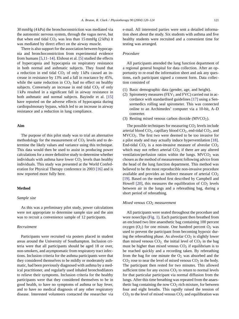
All participants were seated throughout the procedure andwore noseclips (Fig. 1). Each participant then breathed froman enclosed two litre anaesthetic bag containing 100 percentoxygen (O2) for one minute. One hundred percent O2 wasused to prevent the participant from becoming hypoxic dur-ing the rebreathing phase. As alveolar CO2 is slightly lowerthan mixed venous CO2, the initial level of CO2 in the bagmust be higher than mixed venous CO2 if equilibrium is tobe reached quickly and a recording taken. By rebreathingfrom the bag for one minute the O2 was absorbed and theCO2 rose to near the level of mixed venous CO2 in the body.The participant then rested for two minutes. This allowedsufficient time for any excess CO2 to return to normal levelsfor that particular participant via normal diffusion from thelungs. After this time breathing was repeated from the anaes-thetic bag containing the now CO2 rich mixture, for betweenfour and eight breaths. This rapidly raised the tension ofCO2 to the level of mixed venous CO2 and equilibration was

122 A. Bruton, R. Clark / Physiotherapy 90 (2004) 120–124
Fig. 1. Photograph of mock subject undergoing mixed venous carbon dioxide measurement.
briefly achieved between the CO2 in the bag and MVCO2in the body. In order to acquire the data on CO2 levels, themouthpiece from which the subject breathed from the baghad a Jaeger linear rapid CO2 analyser attached. This datawas then recorded onto a 10-inch flat bed chart recorder.
Data analysis
All data were transferred into SPSS for Windows (version11.0) for analysis. Descriptive statistics were used to de-scribe the participants’ baseline characteristics and outcomemeasures.
Results
Table 1provides the descriptive data for the participants.On average, the healthy students were taller and heavier
Table 1Descriptive statistics (mean and S.D.) with the mean difference between the two groups’ characteristics and outcome measures
Variable Asthmatic group (n = 5) Non-asthmatic group (n = 6) Mean difference
Age (years) 21 (0. 5) 26 (6.6) 5.2Gender (M/F) 1/4 5/1 N/AHeight (cm) 169 (9.9) 177 (6.1) 8.3Weight (kg) 68 (14.9) 78 (7.6) 9.6FEV1 (l) 3.54 (0.99) 4.52 (0.59) 0.97Predicted FEV1 (%) 98 (10.40) 107 (4.48) 8.43FVC (l) 4.67 (1.74) 5.74 (0.87) 1.07FEV1/FVC (%) 77.5 (6.12) 79.2 (7.13) 1.61MVCO2 (kPa) 5.35 (0.23) 6.01 (0.30) 0.66
FEV1 forced expiratory volume in 1 s; FVC forced vital capacity; MVCO2 mixed venous carbon dioxide.
than those with asthma. In the students with asthma, per-centage predicted FEV1 ranged from 84% to 112% (mean98%) and mixed venous CO2 levels ranged from 5.08 to5.71 kPa (mean 5.35 kPa). in the healthy students percentagepredicted FEV1 ranged from 102% to 115% (mean 107%)and mixed venous CO2 levels ranged from 5.62 to 6.45 kPa(mean 6.01 kPa). The mean difference in mixed venous CO2levels was 0.66 kPa (95% confidence interval 0.28–1.03).Fig. 2 provides box and whisker plots of the mixed venousCO2 measures from the two groups, giving a visual repre-sentation of the data.
Discussion
This preliminary pilot study has tested an indirect methodfor assessing arterial CO2 in individuals with a diagnosis ofasthma. However, there was no formal determination of a

A. Bruton, R. Clark / Physiotherapy 90 (2004) 120–124 123
Fig. 2. Box and whisker plot of mixed venous carbon dioxide measuresin each group.
diagnosis of asthma in the asthma group, or of good health inthe control group. Participants’ own statements about theirhealth status were accepted, which may have led to someparticipants being assigned to the ‘wrong’ group. However,there is no universally accepted objective test for the diag-nosis of asthma[21,22] and these results do provide somesupport for the findings from other workers that stable asth-matic patients have lower CO2 levels than the healthy popu-lation. In the study by Osborne et al.[6], they found that 23asymptomatic asthma patients had arterial blood CO2 levelsof 4.96(0.43) kPa versus 5.27(0.38) kPa in matched controlswith a mean difference of 0.31 kPa (95% confidence interval0.06–0.56). The figures in this pilot study are higher thanthose reported by Osborne et al.[6], but it is known thatmixed venous CO2 measured by the rebreathing method islikely to exceed arterial CO2 by 1–2 kPa[19].
If one assumes that this hypocapnia is due to hyperventila-tion, these findings also support the recent work by Thomaset al. [23], suggesting that a significant proportion of indi-viduals diagnosed with asthma have dysfunctional breath-ing. It is therefore an attractive proposition that, by retrain-ing breathing patterns to reduce hyperventilation in suchindividuals, it should be possible to raise CO2 levels andreverse the bronchoconstrictive effects of hypocapnia. Atpresent, however, although there is some evidence that var-ious forms of breathing retraining (e.g. yoga[24], Buteyko[25] and physiotherapy[26]) have had some beneficial ef-fects on asthma patients, it is uncertain what mechanism pro-duces these effects. Apart from the CO2 hypothesis, otherpotential mechanisms include increased feelings of masteryor control, relaxation, or non-specific attention effects. Arecent randomised controlled trial of the Buteyko breathingtechnique by Cooper et al.[25] found that Buteyko breath-ing improved asthma symptoms and reduced bronchodilatoruse, but without affecting airway calibre or responsiveness.Thomas et al.[26] have reported on a study of physiother-
apy breathing retraining for asthma patients and found im-proved quality of life scores. However, neither of these stud-ies shed any light on the mechanism producing the improve-ments generated. Very few studies have included measuresof CO2 in their outcomes. One exception is the randomisedcontrolled trial of Buteyko by Bowler et al.[27]. They foundno significant differences in means between the interventionand control groups post intervention, but unfortunately donot report any within subject changes. In a controlled trial ofButeyko currently only published in abstract form, Abram-son et al.[28] reported no changes in lung function, but didfind some increase in end tidal CO2 in one group and somereduction in response to CO2 in another. They employed afactorial design involving four groups, one receiving ‘full’Buteyko (Buteyko practitioner plus Buteyko video), two re-ceiving ‘partial’ Buteyko (Buteyko practitioner plus placebovideo, or Buteyko video plus placebo educator) and one re-ceiving no Buteyko (placebo video plus placebo educator).Their findings are interesting but the complexity of their de-sign makes interpretation difficult.
Future research
Further work is needed to establish the role of CO2 inasthma and other conditions with symptoms of overbreath-ing. We need to establish if there are genuine, consistentdifferences in resting CO2 levels between people with suchconditions and the healthy population. Another area of in-terest is individual response to CO2, as it is possible that notonly may resting levels of CO2 differ significantly, but sen-sitivity to changes in CO2 may also do so. There is also aneed for studies investigating claims that specific breathingtechniques are able to raise CO2 levels within individuals.
Conclusion
The gold standard measure for CO2 levels is arterial bloodgas analysis, but mixed venous CO2 is an accepted alterna-tive [19] that provides a reproducible and reliable indirectmeasurement of arterial CO2. This pilot study has demon-strated that a protocol using non invasive mixed venous CO2measures is acceptable to people with asthma. It has alsoadded to the evidence suggesting that asthmatic individualshave lower levels of CO2 than the healthy population, evenwhen they are stable and asymptomatic. However, the num-ber of participants was very small, so it is possible that theywere not representative of the wider population. Previousworkers have published indirect evidence that a significantproportion of the stable asthmatic population have symptomsof hyperventilation. Physiotherapists and others frequentlyuse breathing retraining techniques to alleviate these symp-toms with some reported benefits. While these benefits maybe related to raising CO2 levels, as has been hypothesised,as yet there is no convincing evidence that this is the case.

124 A. Bruton, R. Clark / Physiotherapy 90 (2004) 120–124
Ethical approval
Southampton and South West Hampshire LREC (LocalResearch Ethics Committee).
Acknowledgements
The authors are indebted to John Heath of the PulmonaryFunction Laboratory, Southampton General Hospital for hisassistance in the recording and analysing of the mixed ve-nous carbon dioxide data.
References
[1] Pryor JA, Webber BA. Physiotherapy for respiratory and cardiacproblems. Edinburgh: Churchill Livingstone; 1998.
[2] Laffey JG, Kavanagh BP. Hypocapnia. New Engl J Med 2002;347:43–53.
[3] Rodriguez-Roisin R. Gas exchange abnormalities in asthma. Lung1990;168(Suppl):599–605.
[4] Odhiambo JA, Chwala RD. Arterial blood gases and acid-base sta-tus of adult patients presenting with acute severe asthma at Keny-atta National Hospital, Nairobi. East African Med J 1992;69:319–22.
[5] van den Elshout FJ, van Herwaarden CL, Folgering HT. Effects ofhypercapnia and hypocapnia on respiratory resistance in normal andasthmatic subjects. Thorax 1991;46:28–32.
[6] Osborne CA, O’Connor BJ, Lewis A, Kanabar V, GardnerWN. Hyperventilation and asymptomatic chronic asthma. Thorax2000;55:1016–22.
[7] Reynolds AM, McEvoy RD. Tachykinins mediate hypocapnia-induced bronchoconstriction in guinea pigs. J Appl Physiol 1989;67:2454–60.
[8] Stalmatski A. Freedom from asthma: Buteyko’s revolutionary treat-ment. London: Kyle Cathie Ltd.; 1997.
[9] Hypoxemia and hypocapnia in asthma. New Engl J Med 1968;278:1068.
[10] D’Angelo E, Calderini IS, Tavola M. The effects of CO2 on respi-ratory mechanics in anesthetized paralyzed humans. Anesthesiology2001;94:604–10.
[11] Sterling GM. The mechanism of bronchoconstriction due to hypocap-nia in man. Clin Sci 1968;34:277–85.
[12] Newhouse MT, Becklake MR, Macklem PT, McGregor M. Effectof alterations in end-tidal CO2 tension on flow resistance. J ApplPhysiol 1964;19:745–9.
[13] O’Cain CF, Hensley MJ, McFadden Jr ER, Ingram Jr RH, Patternand mechanism of airway response to hypocapnia in normal subjects.J Appl Physiol 1979;47:8–12.
[14] Jamison JP, Glover PJ, Wallace WF. Comparison of the effects of in-haled ipratropium bromide and salbutamol on the bronchoconstrictorresponse to hypocapnic hyperventilation in normal subjects. Thorax1987;42:809–14.
[15] Bayindir O, Akpinar B, Ozbek U, Cakali E, Pekcan U, BulutcuF, et al. The hazardous effects of alveolar hypocapnia on lungmechanics during weaning from cardiopulmonary bypass. Perfusion2000;15:27–31.
[16] Clark R, Bruton A. Resting mixed venous carbon dioxide levels inasthmatics and non-asthmatics: a pilot study investigating Buteyko’stheory. In: The Fourteenth International World Confederation forPhysical Therapy Congress, 2003.
[17] American Thoracic Society. Standardization of spirometry, 1994 up-date. Am J Respir Crit Care Med 1995;152:1107–36.
[18] Gardner WN. The pathophysiology of hyperventilation disorders.Chest 1996;109:516–34.
[19] Lumb AB. Nunn’s applied respiratory physiology. Oxford: Butter-worth Heineman; 2000.
[20] Campbell EJ, Howell JBL. Simple rapid methods of estimating ar-terial and mixed venous PCO2. BMJ 1960;1:458–62.
[21] British guidelines on the management of asthma. Thorax 2003;58(Suppl 1):i1–94.
[22] Britton J, Lewis S. Objective measures and the diagnosis of asthma.We need a simple diagnostic test—but don’t have one yet. BMJ1998;317:227–8.
[23] Thomas M, McKinley RK, Freeman E, Foy C. Prevalence of dys-functional breathing in patients treated for asthma in primary care:cross sectional survey. BMJ 2001;322:1098–100.
[24] Manocha R, Marks GB, Kenchington P, Peters D, Salome CM.Sahaja yoga in the management of moderate to severe asthma: arandomised controlled trial. Thorax 2002;57:110–5.
[25] Cooper S, Oborne J, Newton S, Harrison V, Thompson CJ, LewisS, et al. Effect of two breathing exercises (Buteyko and pranayama)in asthma: a randomised controlled trial. Thorax 2003;58:674–9.
[26] Thomas M, McKinley RK, Freeman E, Foy C, Prodger P, PriceD. Breathing retraining for dysfunctional breathing in asthma: arandomised controlled trial. Thorax 2003;58:110–5.
[27] Bowler SD, Green A, Mitchell CA. Buteyko breathing techniquesin asthma: a blinded randomised controlled trial. Med J Aust1998;169:575–8.
[28] Abramson MJ, Borg B, Doran C, Giorlando F, Hartley F, Jack S, etal. A randomised controlled trial of the Buteyko method for asthma.In: Australian Asthma Conference, Melbourne, 2004 (abstract).