Embed Size (px)
Citation preview
RESEARCH ARTICLE Open Access
A pancreatic exocrine-like cell regulatorycircuit operating in the upper stomach ofthe sea urchin Strongylocentrotuspurpuratus larvaMargherita Perillo1,3, Yue Julia Wang2, Steven D. Leach2 and Maria Ina Arnone1*
Abstract
Background: Digestive cells are present in all metazoans and provide the energy necessary for the wholeorganism. Pancreatic exocrine cells are a unique vertebrate cell type involved in extracellular digestion of a widerange of nutrients. Although the organization and regulation of this cell type is intensively studied in vertebrates, itsevolutionary history is still unknown. In order to understand which are the elements that define the pancreaticexocrine phenotype, we have analyzed the expression of genes that contribute to specification and function of thiscell-type in an early branching deuterostome, the sea urchin Strongylocentrotus purpuratus.
Results: We defined the spatial and temporal expression of sea urchin orthologs of pancreatic exocrine genes anddescribed a unique population of cells clustered in the upper stomach of the sea urchin embryo where exocrinemarkers are co-expressed. We used a combination of perturbation analysis, drug and feeding experiments andfound that in these cells of the sea urchin embryo gene expression and gene regulatory interactions resemble thatof bona fide pancreatic exocrine cells. We show that the sea urchin Ptf1a, a key transcriptional activator of digestiveenzymes in pancreatic exocrine cells, can substitute for its vertebrate ortholog in activating downstream genes.
Conclusions: Collectively, our study is the first to show with molecular tools that defining features of a vertebratecell-type, the pancreatic exocrine cell, are shared by a non-vertebrate deuterostome. Our results indicate that thefunctional cell-type unit of the vertebrate pancreas may evolutionarily predate the emergence of the pancreas as adiscrete organ. From an evolutionary perspective, these results encourage to further explore the homologs of othervertebrate cell-types in traditional or newly emerging deuterostome systems.
Keywords: Strogylocentrotus purpuratus, Pancreas, Ptf1a, Carboxypeptidase, Pancreatic lipase, Amylase
BackgroundThe ability of a species to survive in a new environment isstrictly related to its capacity to successfully digest and as-similate different food sources. A characteristic that isshared by all the living forms is the ability to digest largemolecules available in their habitat to produce energy. Di-gestion relies on the function of enzymes that are able tobreak down proteins, lipids and sugars in the diet. Thenumber of specialized enzyme-producing cell types
involved in food digestion increases with the complexityof the animal body and the variety of the meal. Metazoanstypically have specialized exocrine cell types for the secre-tion of digestive enzymes. For example, gland cells thatproduce and release zymogens in the gastric cavity arescattered along the hydra gastroepidermis [1, 2] and alongthe digestive filaments of corals [3]. Cells rich in granularinclusions involved in extracellular digestion are localizedalong the foregut and in the gut caeca of different speciesof flat worms [4–6]. In different regions of the Drosophilamidgut there are enterocytes that secrete a vast array ofdigestive enzymes, with expression levels matching organ-ismal requirements [7]. A specialized organ geared toward
* Correspondence: [email protected] and Evolution of Marine Organisms, Stazione Zoologica AntonDohrn, Napoli 80121, ItalyFull list of author information is available at the end of the article
© 2016 Perillo et al. Open Access This article is distributed under the terms of the Creative Commons Attribution 4.0International License (http://creativecommons.org/licenses/by/4.0/), which permits unrestricted use, distribution, andreproduction in any medium, provided you give appropriate credit to the original author(s) and the source, provide a link tothe Creative Commons license, and indicate if changes were made. The Creative Commons Public Domain Dedication waiver(http://creativecommons.org/publicdomain/zero/1.0/) applies to the data made available in this article, unless otherwise stated.
Perillo et al. BMC Evolutionary Biology (2016) 16:117 DOI 10.1186/s12862-016-0686-0

the production of digestive enzymes and hormones, thepancreas, appeared uniquely in the vertebrate lineage. Thepancreas is a complex organ evolved mainly for food di-gestion (exocrine cells) and maintaining blood sugar levels(endocrine cells). The exocrine pancreas constitutes themajority of the mass of the pancreas and it is composedby exocrine cells grouped into acini that secrete enzymesinto the intestine [8]. The pancreas originates early in de-velopment from progenitor cells located within the endo-dermal epithelium. Two members of the basic helix-loop-helix (bHLH) family of protein are known to be criticalfor the exocrine pancreas differentiation and identity,Ptf1a (pancreatic transcriptional factor 1) and Mist1 [9, 10].Ptf1a has a double role, first in early pancreas specification,and later in acinar cell differentiation and maintenance[11–13], while Mist1 is necessary for full maturation of theacinar cell phenotype [14]. In the mouse, it has been foundthat in the absence of Hnf1β, Ptf1a expression is not in-duced, leading to defective specification and reduction ofthe ventral and dorsal pancreas [15]. Furthermore, severalsignaling factors are involved in balancing exocrine cell pro-liferation and differentiation. Among all, Notch appears toprevent pancreatic exocrine development, while FGFsignaling mediates growth, morphogenesis and differ-entiation of exocrine cells [16–18]. In both mouseand zebrafish, Notch and its target genes inhibit theactivity of the Ptf1 complex in the exocrine cells, andloss of Notch function results in accelerated develop-ment of exocrine pancreas [19].One of the most abundant and evolutionary conserved
miRNA involved in pancreas development is miR-375.MiR-375 negatively regulates glucose-stimulated insulinexocytosis by targeting myothrophin mRNA, a protein in-volved in insulin secrection in pancreatic β-cells [20]. Aloss of function approach in zebrafish revealed that miR-375 is essential for the formation of insulin producing β-cells because its knockdown mainly results in malformationof the endocrine pancreas [21]. In addition, studies on micelacking miR-375 demonstrated that it controls β- and α-cells mass by regulating genes involved in cellular growthand differentiation [22].Echinoderms—a group of animals that comprise sea
urchins, sea stars, sea lilies, brittle stars and sea cucum-bers- belong to the deuterostome clade. The relativelysimple development of sea urchin embryos, coupled withthe availability of molecular tools for gene perturbation,enable comparative studies on cell specification anddevelopmental mechanisms with other deuterostomes[23]. In particular, echinoderm systems present, in thecomparison with vertebrate deuterostomes, the great ad-vantage of a simplified gene toolkit, since this group splitbefore the duplication events occurred at the emergenceof Craniata. Importantly for this study, Strongylocentrotuspurpuratus has an indirect development that generates a
bilateral free-swimming larva whose behavior is centeredon feeding.The sea urchin larval gut is a simple tripartite tract
composed of a muscular esophagus that exhibits strongcontractions, a large spherical stomach with sphinctersat both openings, and a small tubular intestine, whichexits through the anus. In the stomach of sea urchin lar-vae, there are cells with the morphological features ofzymogen secreting cells that are able to synthesize di-gestive enzymes, such as β-glucanase and α-amylase[24–27]. Gastric exocrine cells have been described alsoin the stomach of the adult form of S. purpuratus. Thesecells are highly concentrated in the upper stomach andelectron microscopy analyses reveal that they are strik-ingly similar to mammalian zymogen cells, such as cellsof the gastric mucosa and pancreatic acinar cells [28].Although pancreatic cell types have been extensively
characterized in the main vertebrate model systems,most of the studies in non-vertebrate animals have ex-amined gut cells at the morphological level only andmolecular information is still very scarce. Therefore, afew data are currently available on the genes/geneticcharacterization of the pancreatic exocrine-like cell typesin non-vertebrate metazoans. In this study, we use a can-didate gene approach to identify and characterize thepancreatic exocrine cell type in the sea urchin embryoand larva. We defined a unique population of cells clus-tered in the sea urchin larva upper stomach that re-sponds to food and expresses the homologs of thepancreatic genes Ptf1a and Mist1 together with at leastthree digestive enzymes. Our findings are a first step tounderstand the evolution of pancreatic exocrine cells.We report that the components of the specificationpathway and expression for zymogen genes are active inan early branching deuterostome, which suggests thatthe exocrine cell type may predate the evolution of thevertebrate pancreas.
MethodsAnimal husbandry, embryo and larva cultures, feedingexperimentsAdult S. purpuratus were obtained from Patrick Leahy(Kerchoff Marine Laboratory, California Institute ofTechnology, Pasadena, CA, USA) and housed in circu-lating seawater aquaria at the Stazione Zoologica AntonDohrn of Naples. Adult sea urchin maintenance, embryoand larva cultures and feeding experiments were per-formed as previously described [29].
RNA whole mount in situ hybridizationFor fluorescent whole mount in situ hybridization(FISH), we followed the protocol outlined in Cole et al.2009 with the modification described in [30]. Signal wasdeveloped with fluorophore-conjugated tyramide (1:400
Perillo et al. BMC Evolutionary Biology (2016) 16:117 Page 2 of 15

reagent diluents, Perkin Elmer) following instructions.For all the genes, labeled probes were transcribed fromlinearized DNA as described in [31]. SpmiR-375 probehas been synthesized and DIG labelled from Exiqon, andthe sequence is: 5’/DigN/TGACGCGAGCCGAACGAA-CAAA/3’DigN/. The double FISH procedure forSpCpa2L and SpmiR-375 was performed as cited withthe only exception that the miRNA probe concentrationwas 0025 pmol/μl and the samples were hybridized5 days at 42 °C. mir375 probe has already been used byChristodoulou et al. 2010. Primers used to amplify theriboprobes are summarized in Additional file 1. Tem-plates of all the probes were sequenced prior to probegeneration and cloned in the pGEM®-T Easy Vector(Promega, Madison, WI, USA). Sense probes were syn-thesized to test the specificity of the antisense probe sig-nal. FISH was imaged with a Zeiss 510 Meta confocalmicroscope.
DAPT treatmentThe γ-secretase inhibitor DAPT (N-[N-(3,5-difluorophe-nacetyl)-L-alanyl]-S-phenylglycine t-butyl ester, Sigma–Aldrich, St. Louis, MO) [32] was dissolved in DMSOand added at 17 h to a final concentration of 8 μM, toavoid the early and toxic effects of the drug, as describedin Materna et al. 2012. A corresponding volume ofDMSO was added as control.
Perturbation experiments with MO injectionFor each experiment and for each morpholino oligo-nucleotide (MO), 200 to 400 eggs were injected with ap-proximately 2-4pl of oligonucleotide injection solutionand each experiment was repeated three times. As anegative control, fertilized eggs were injected with thestandard control morpholino (GeneTools, Philomath,OR) and compared side-by-side with knockdown em-bryos. The injection of the standard control morpholinodid not have any effect on the development of embryos.In a pilot experiment, different concentrations of theMASOs were injected and the morphant’s phenotypewas observed in order to test for the right concentrationand avoid secondary effects or MASO toxicity. The high-est concentration that was not toxic for the embryos wasused in each experiment. The injected embryos were cul-tured at 15 °C until 50 h and 67 h of development, whenthey were fixed and used to analyze the expression of targetgenes in FISH or qPCR experiments. A translation-blocking antisense MO against SpHnf1a was used at a finalconcentration in the injection solution of 200 μM (5’-CTAGTTCGTCACCCGAATGCAGCAT-3’, first tested byPeterson and Davidson 2011). A translation-blocking anti-sense MO against SpPtf1a was newly designed and used ata final concentration of 300 μM (SpPtf1a_MO1: 5’-ATATTTTCCATAGTGATCTCTGAGT-3’). A second
translation-blocking antisense MO against SpPtf1a wasinjected at a final concentration of 150 μM and used toconfirm the specific effect of SpPtf1a MO_1(SpPtf1a_MO2: 5’-GCGCAGGTGGATTATCAAATTGTTC-3’).
RNA extraction and quantitative real-time PCR (qPCR)For qPCR analysis of temporal expression profiles, totalRNA was isolated from cultures derived from threemixed batches and extracted with Eurozol (EuroClone,Celbio, Milan, Italy). For SpPtf1a morphants and relativecontrols and for feeding experiments, RNA was ex-tracted using the RNAquous kit (Ambion). For all theexperiments, samples were treated with DNase I(Ambion) to remove DNA contamination as describedby the manufacturer. First-strand cDNA was synthesizedfrom total RNA using the SuperScript VILO™ cDNASynthesis Kit (Invitrogen) following the manufacturer’sinstructions. Reactions were performed as described in[33] using the ViiA7 REAL TIME PCR with SYBR Greenchemistry (Applied Biosystems, Foster City, CA). For allqPCR experiments, ubiquitin mRNA was used as a refer-ence because remains relatively constant during develop-ment [34]. For knockdown and feeding experiments,three biological replicates were analyzed, ddCt valueswere calculated between experiment and control em-bryos and converted to fold differences using ubiquitinas a reference (FD = 2 ΔΔCt). A threshold of 2-fold differ-ence was chosen as a significant change. Primer setswere chosen to amplify products 100 to 200 bp in lengthand sequences are reported in Additional file 1. qPCRexperiments in different feeding conditions and the rela-tive statistics were performed as previously described inPerillo and Arnone 2014.
Luciferase assayThe coding sequences of E47, Rbpl, rnPtf1a and spPtf1awere cloned into the pCDNA3.1 backbone. Ptf1a Lucif-erase assay was conducted as previous described [35].Briefly, 293 t or Hela cells were plated at a density of1x105 cells/well in a 24-well plate and transfected with512.5 ng total DNA/well using Fugene 6 transfection re-agent (Promega). For individual constructs, the followingconcentrations were used (per well): 12.5 ng renilla(pRL-TK, Promega), 125 ng 4x ptf1a:luciferase reporter(contains 4x Ptf1a binding motif cloned from rat Chymo-trypsinogen B promoter, gift of Dr. Masashi Kawaichi),125 ng human E47, 125 ng Rbpl and 125 ng of either ratrnPtf1a or sea urchin spPtf1a. Different combinations ofplasmids were cotransfected and pcDNA3.1 (Invitrogen)was used as filler DNA to make the total amount ofDNA per well uniform across different conditions. Twodays post-transfection, cells were harvested and analyzedfor firefly and renilla luciferase activities using the Dual-
Perillo et al. BMC Evolutionary Biology (2016) 16:117 Page 3 of 15

luciferase reporter assay system (Promega) per manufactor’sprotocol.
Bioinformatics analysisTo identify putative Ptf1a binding sites on sea urchingenes of interest, we used the list of Ptf1a binding sitesof the mouse acinar digestive enzyme gene promoterspublished by Masui et. al. (2007). From this list we builtbase frequency tables for the E-box and TC-box separ-ately, i.e. counts the number of times each base occursat each position. The base frequency table has four rows(one row for each letter: A, C, G and T) and the numberof columns is equal to the motif length. Then, we calcu-lated the weight matrices for the E-box and TC-box, thematrices have the same number of rows and columns asthe corresponding frequency tables, with the value ateach position being the natural logarithm of the valuefrom the frequency table divided by the number ofsequences, i.e. the weight matrices contain the estimatesof the log-probabilities of each base occurring at eachposition in true binding sites, based on the sample ofknown sites. Since the gap between the two boxes isabout one or two helical DNA-turns, we introduced apenalty function based on a cosine function, whoseperiod and phase shift are 10.5 and 21 base pairs separ-ately. Thus for a given sequence, we can calculate amatching score based on the sum of the weights thatcorrespond to the sequence, which should be equal tothe log-probability of seeing that sequence given that itis a binding site, and also the gap penalty function forthe distance between the boxes.
ResultsCharacterization of regulatory and differentiationsignatures for sea urchin pancreatic exocrine-like cellsWe identified sea urchin orthologs of regulatory geneswith a known conserved role in pancreatic exocrine celldevelopment. The embryonic temporal expression of thetranscriptional factors (TFs) analyzed in this study agreeswith data already published from other authors [36, 37],but our analysis includes also late larval stages.The hepatocyte nuclear factor (HNF) gene family is
part of the HNF homeobox class and includes HNF1αand HNF1β, two paralogs with known interchangeablefunctions in several contexts [38, 39]. The sea urchingenome has only one homolog named SpHnf1. Its ex-pression begins in the presumptive endoderm at blastulastage and by mid-gastrula appears confined to the gut[37]. To clarify its later functions in development, wedefined the spatial expression of SpHnf1 in post-gastrulastage embryos. During gastrulation, SpHnf1 is expressedthroughout the hindgut and the midgut, but particularlyenriched in a group of cells just below the cardiacsphincters (Fig. 1a). This expression fades away in the
early pluteus (Fig. 1b) and it is not detectable in olderstages, consistently with the low quantitative level of ex-pression exhibited by this gene at larval stages (Fig. 1g).The sea urchin ortholog of Ptf1a [36] has two distinct
domains of expression. At gastrula stage (48hpf, Fig. 1c),transcripts start to accumulate in cells situated right-lateral toward the dorsal side of the midgut, next to thecardiac sphincter, and some hours later it equally expandson both sides. The second domain of expression is in theectoderm, particularly at the apical organ and in scatteredcells of the ciliary band. Throughout the larval stages, thestomach cells increase in number, and we also observe anincrease in SpPtf1a positive cells (Fig. 1d). The expressionof another bHLH gene, SpMist1 [36], starts at late gastrulastage in the upper stomach and in scattered cells of theapical organ and ciliary band (Fig. 1e, f ). SpMist1 gene ex-pression increases during gastrulation (2 days), althoughat early pluteus stage (3 days) SpPtf1a decreases whileSpMist1 reaches its maximum expression (Fig. 1g). Simi-larly, the expression of both genes decreases at 7 days afterfertilization and increases again after a few days.We next identified the genes that most likely encode for
enzymes used in food digestion. We compared the pre-dicted ORFs of digestive enzyme gene families with theESTs of an annotated sea urchin late larva cDNA library(MPMGp691, http://owww.molgen.mpg.de/ag_seaurchin/,[40]). The carboxypeptidase family was the first to be ana-lyzed. Among 7 sea urchin orthologs, only SpCpa2Lshowed a significant representation in the above men-tioned larval cDNA library. As shown in Fig. 1h, SpCpa2Ltranscripts are expressed at gastrula stage in two groupsof cells in the upper midgut, next to the cardiac sphincter.In a temporal progression, SpCpa2L expression begins atthe upper lateral stomach cells and later it extends ven-trally from both sides until coalescing at the ventral mostcells, while leaving a gap at the dorsal most part of thestomach (Fig. 1i and k). Based on the EST abundance atlarval stages, SpPnlp2 and SpPnlp5 were selected amongthe lipases and blast analysis revealed that they encode thesame protein, hereby named SpPnlp2/5. Likewise, SpAmy3was selected among the amylases. For both genes, tran-scripts are detectable one day after SpCpa2L starts to beexpressed, and they are produced in the same above de-scribed cells of the upper stomach (Fig. 1j-m). Consist-ently, the levels of expression of these three genes steadilyincrease during development and it reaches a peak at10 days (Fig. 1n).
Pancreatic exocrine-like cells are clustered in the sea urchinlarva upper stomachOur examination of the expression of individual pancre-atic regulatory and terminal differentiation genes suggeststhat they are expressed within a common exocrine-likecell population. To further test this premise, we tested co-
Perillo et al. BMC Evolutionary Biology (2016) 16:117 Page 4 of 15

expression of these factors by double fluorescent in situ.The endodermal expression of SpPtf1a is initially confinedto a group of ~3 cells in the midgut within the broadSpHnf1 domain (Fig. 2a). We next asked if other homologsof pancreatic exocrine genes were co-expressed in thosecells. The first digestive enzyme that appears in theSpPtf1a and SpHnf1 positive domain is SpCpa2L (Fig. 2b).At gastrula stage, a group of cells in the dorsal-right mid-gut co-express SpCpa2L and SpPtf1a. Some hours later, atthe pluteus stage, cells expressing these two markers ap-pear ventrally, forming a belt-like structure (Fig. 2c; seealso Fig. 2I in [41] for co-expression of these two genes at
the pluteus stage). Remarkably, only a few cells of the dor-sal stomach do not express these markers. Consideringthe high expression of SpCpa2L transcripts, all confinedto the Ptf1a positive cells (SpPtf1a+), we used this geneexpression as a marker for the pancreatic exocrine-likecells. Thus, we analyzed SpMist, SpPnlp2/5 and SpAmy3spatial expressions relative to SpCpa2L at larval stages. Asshown in Fig. 2c-e, all the orthologs of exocrine genes thatwe considered in this study are co-expressed in the pan-creatic exocrine-like cells. Remarkably, the same cells ex-press the evolutionary conserved pancreatic microRNAmiRNA-375. Figure 2f shows that miRNA-375 is strongly
Fig. 1 Expression analysis of regulatory and terminal differentiation orthologs of known pancreatic genes. mRNA localization of sea urchinregulatory (a-f, green) and terminal differentiation (h-m, magenta) genes. For all the figures in this paper, every picture is a full projection ofmerged confocal Z stacks and nuclei are stained with DAPI and depicted in blue. Red circles in f show Mist1 RNA localization in alternating cellsof the apical organ. Inset in panel k is a representative single confocal section of the upper stomach of a late larva showing that SpCpa2Ltranscripts are abundantly expressed in the entire cell. g, n Temporal expression profiles of pancreatic regulatory and terminal differentiationgenes during sea urchin development. The graphs show the relative transcript abundance normalized against ubiquitin mRNA. The results areexpressed as percentage of the maximum value, corresponding to the stage with the highest level of expression. Standard deviations of threetechnical replicas are all <0.5. For the sake of simplicity, for each panel, the species in the gene name has been omitted. Abbreviations: d, days;dv, dorsal view; h, hours post fertilization; vv, ventral view
Perillo et al. BMC Evolutionary Biology (2016) 16:117 Page 5 of 15

expressed throughout the ciliary band and in the stomach,where it is localized exclusively in the pancreatic exocrine-like cells. In addition, this gene is expressed in the coelomicpouches and the probe signal is stronger in the right one.
Notch signaling inhibits differentiation towardspancreatic exocrine cell fateGiven its known role in pancreatic differentiation, wenext tested the role of the Notch signaling pathway insea urchin pancreatic exocrine-like cell differentiation,using the Notch inhibitor DAPT. The first striking effectof this treatment is the expansion of the SpPtf1a + cellsin the gastrula and larva upper midgut/stomach (Fig. 3).Notably, the SpPtf1a expansion in the treated embryosnever overcomes the cardiac sphincter border. Despitethe strong effect on the endodermal cells, ectodermalSpPtf1a expression is not affected. Moreover, 35 % of theanalyzed plutei showed an ectopic expression of SpPtf1ain a small group of cells in the anus (Fig. 3d, red arrow).Conversely, the expression of the early endodermal geneSpHnf1 is not affected by the DAPT treatment at gas-trula stage (Fig. 3a, b), in agreement with the previouslyreported data at blastula stage [42]. When Notch signal-ing is perturbed, the expression of SpMist1 exhibits op-posite effects than that of SpPtf1a. At the pluteus stage,the gene expression in the stomach is unaltered, while inthe esophagus and in the apical organ SpMist1 is turnedon also in cells that are adjacent to the wild typeSpMist1+ cells(Fig. 3e and f and arrows in Fig. 3f ).Many modulators of Notch signaling are known, among
which there is the O-fucose β-1,3 N-acetylglucosaminyl-transferase Fringe, whose role is to potentiate Notch acti-vation by Delta [43]. Since a Notch signaling requiringFringe is involved in specification of endoderm and
boundary formation during sea urchin embryonic devel-opment [44], we asked if Notch inhibition affects Fringeexpression also in the pluteus larva. The sea urchin ortho-log of Fringe, SpFng, is widely expressed in the pluteusmesoderm, ectoderm and endoderm (Fig. 3g). Our ana-lysis demonstrated that perturbation of Notch signalingcauses a consistent reduction of SpFng expression in allthe tissues where it is normally expressed (Fig. 3h), evenin the pluteus.
Transcriptional control of sea urchin pancreaticexocrine-like cell developmentWe next asked if the TFs expressed early in developmentare involved in a gene regulatory module that specifies pan-creatic exocrine cells. To this aim, specific morpholino oli-gonucleotides were injected in the sea urchin fertilized eggsto block either SpHnf1 or SpPtf1a translation. SpHnf1 MOsequence specificity has already been demonstrated by Pe-terson and Davidson (2001), and we used that same mor-pholino in our experiments. In SpHnf1 morphants, SpPtf1aexpression in the midgut is downregulated, while its expres-sion in the ectoderm remains unaltered (Fig. 4a, b). Fluores-cent in situ hybridization (FISH) or quantitative PCR(qPCR) analyses were performed on larvae to test the ef-fects of SpPtf1a MO perturbation on putative target genes.We observed that the spatial expression of SpCpa2L,SpPnlp2/5 and SpAmy3 is strongly reduced (Fig. 4e-h). ForSpCpa2L, which among the enzymes shows the highesttranscript expression at 3 days, changes at the transcriptlevels were also analyzed by qPCR analysis, confirmingthat its expression is strongly downregulated (Fig. 4k).Conversely, in SpPtf1a morphants additional groups ofSpPtf1a + cells appear in the upper stomach, both ven-trally and dorsally (Fig. 4j) and qPCR data confirm SpPtf1a
Fig. 2 Co-expression analysis of markers of pancreatic exocrine cell-types. Double FISH of selected pancreatic genes in the sea urchin embryosand larva (dv). On the right of each panel, split and combined channels of single confocal sections of the gut domain expressing the two genes(the region of the embryo shown in the right insets is underlined by a yellow square ) are provided to confirm that the two genes are indeedexpressed in the same cells
Perillo et al. BMC Evolutionary Biology (2016) 16:117 Page 6 of 15

upregulation (Fig. 4k). Additionally, in SpPtf1 morphantsthere is a modest increase of SpMist1 expression (Fig. 4k),and a slight increase of SpFng transcripts (Fig. 4i, j and k).Particularly, SpFng and SpPtf1 seem to be expressed byadjacent and not overlapping cells (Fig. 4j, insert). To test
if the effects of SpPtf1a MO were specific, we injected asecond MO to block SpPtf1a translation (SpPtf1a MO_2)and we checked the expression of SpCpa2L, since thisgene is the first TD marker of this cell type to appear andalso the most abundant at pluteus stage. Also in this case,
Fig. 3 Spatial analysis of gene expression after Notch signaling perturbation. Hnf1 (a, b), Ptf1a (c, d), Mist1 (e, f) and Fng (g, h) transcriptlocalization tested by FISH in control animals (a, c, e, g) and in animals treated with DAPT (b, d, f, h). In all the experiments, Ptf1a (in green) wasused as second probe in double FISH (in a, b, e, f, g, h) since the increase of Ptf1a + cells in treated larvae confirms the phenotype. Red arrow in Dindicates Ptf1 ectopic expression in a few cells of the anus. Yellow circles in E show that Mist1 is expressed in alternated cells of the apical organ incontrol larvae, while arrows in F indicate that in treated larvae Mist1 is expressed in adjacent cells of the apical organ (yellow arrow) and in a few cellsof the esophagus (white arrow). For each analyzed gene, quantification of the phenotypes is shown on the right. Abbreviations: AO, apical organ; es,esophagus; lv, lateral view; MG, midgut; st, stomach, v, ventral
Perillo et al. BMC Evolutionary Biology (2016) 16:117 Page 7 of 15

we found that the SpCpa2L mRNA is downregulated inthe morphants (Fig. 4c and d), confirming that the twomorpholinos produce comparable effects.
SpPtf1a function in mammalian cellsPtf1a is part of the PTF1 heterotrimeric complex thatalso includes one of the common E proteins (E47) andRBPL [45]. To test the ability of the sea urchin Ptf1aprotein to substitute for mammalian Ptf1a in this com-plex, we performed luciferase essays in which eitherSpPtf1a or rat Ptf1a were co-transfected with mamma-lian E47 and RBPL, and evaluated for an ability to
transactivate a PTF1-responsive luciferase reporter con-taining four tandem repeats of the PTF1 binding sitefrom the rat chymotrypsinogen promoter (Obata et al.,2001). Figure 5a and b show that, in both 293 t andHeLa cells, SpPtf1a can effectively partner with mamma-lian PTF1 components to activate the rat chymotrypsin-ogen element, albeit with lower activity than rat Ptf1a(Fig. 5b). As a correlate of this conserved function, wehypothesized that the sea urchin orthologs of mamma-lian PTF1a target genes would also display similar PTF1aregulatory motifs. In the promoter of genes encodingmammalian pancreatic digestive enzymes, PTF1 has
Fig. 4 Analysis of gene expression after MO perturbation. Ptf1a mRNA localization in control embryos (a) and in embryos injected with MOdirected against the translation of SpHnf1 RNA (b). Cpa2L, Pnlp2/5, Amy3, Ptf1a and Fng transcripts were detected by single or double FISH incontrol larvae (c, e, g, i) and in larvae injected with two different MOs directed against the translation of SpPtf1a RNA (d, f, h, j). Note that f, h and jshow larvae injected with SpPtf1a MO1. To confirm the effects of MO1, figure d shows that SpCpa2L transcripts are absent also in larvae injected withSpPtf1a MO2. Inset in j is a representative single confocal section of the dorsal upper stomach of a late larva and the white arrow shows that Ptf1a +and Fng + cells are adjacent and not overlapping. k qPCR analysis showing the effects of SpPtf1a MO1 perturbation on transcript levels of selectedpancreatic genes at 70 h. Fold changes≥ -2 and≤ 2 are shaded in light grey and indicate non-significant changes in gene expression
Perillo et al. BMC Evolutionary Biology (2016) 16:117 Page 8 of 15

been found to bind to an extended bipartite DNA se-quence that consists of an E-box and a TC-box,spaced one or two helical DNA-turns apart. Masuiand coauthors have reported a list of PTF1-bindingsites of the acinar digestive enzyme gene promotersfor mouse [45]. We used the list of binding sites fromthat paper and built a position weight matrix for theE-box and TC-box, taking into consideration of the
distance between these two elements. This matrix wasused to look for Ptf1a binding sites in the promoters ofthe genes that were affected in SpPtf1a morpholino per-turbations. Interestingly, we identified canonical Ptf1abinding sites in sea urchin genes displaying in vivo spPtf1adependence, including SpFng, SpCpa2L, SpPnlp2/5 andSpAmy3. An alignment of the documented Ptf1a bindingsite in the rat Ctrb1 gene with the putative Ptf1a binding
Fig. 5 Sea urchin Ptf1a is active and can function together with mammalian Ptf1 partners. a 293 t cells were transiently transfected withluciferase reporter containing 4x Ptf1a-responsive element (vector indicated as 4x in the figure) and/or spPtf1a, E47, Rbpl construct; renilla vectorwas cotransfected for normalization. Results are displayed in the box plot as fold activation over the activity of 4x reporter alone. Transfectionswere done in triplicate and were repeated 5 times. Luciferase assays from all transfections show similar trend, one representative result is shown.t-test was carried out for group comparison. The bottom and top of the box are the first and third quartiles, and the band inside the box is themedian. The whisker represents 1.5 IQR (interquantile range, the differences between the third and first quantile). b SpPtf1a has lower activitycompared with mammalian Ptf1a in the in vitro luciferase assay. Experiment setting is the same as (a) except that rat rnPtf1a was transfected in a secondplate side-by-side for comparison. We tested SpPtf1a activity in multiple cell lines, one representative result from HeLa cells is shown. c Alignment of theclosest to the transcription start site (TSS) Ptf1a binding sites found by bioinformatic analysis on S. purpuratus (Sp) promoters with the canonical Ptf1abinding site of the RnCtbr1 gene (ref). nt, nucleotides
Perillo et al. BMC Evolutionary Biology (2016) 16:117 Page 9 of 15

sites in each of the S. purpuratus genes is shown inFig. 5c.
Pancreatic exocrine-like gene expression response todifferent feeding regimesWe next asked if pancreatic exocrine-like cells respond todifferent feeding regimes. To this aim, S. purpuratus em-bryos were grown and fed on algae until day 10 to reachthe normal larval morphology, then food was removedfrom the culture media. The culture was split into twobatches, of which one was fed for 24 h and the other wasfasted for the same time interval. First, we observed larvalmorphology and found that the different feeding regimesshow a clear effect on gut sizes. The stomach and intestineof larvae that were fed is bigger than the ones of the fastedlarvae, suggesting that the whole gut epithelium responds
to the presence of food (Fig. 6a, b). Second, we comparedby qPCR the expression levels of SpPtf1a and the previ-ously described digestive enzymes in feeding vs. starvingconditions (Fig. 6c). The analysis shows that the expressionof the digestive enzymes increases upon feeding. Differentlyfrom its putative target genes, SpPtf1a expression is higherin fasted larvae than in fed ones. To understand if the in-creased expression of those genes was due to the same pan-creatic exocrine-like cells or to other cells, we tested geneexpression through FISH analysis, confirming that thespatial expression of the above genes does not change infeeding or starving conditions (data not shown).
DiscussionIn the present study, we show that, in the sea urchinlarva, cells specialized in food digestion are clustered in
Fig. 6 Effect of different feeding regimes on pancreatic exocrine cell gene expression. a and (b) are bright field images of 15-day larvae that arerepresentative for the fed or starved conditions. Larvae are viewed from lateral right side, mouth up. Stomach diameter was measured from ventral todorsal, along the midline of the larval body. c Quantitative PCR analysis of SpPtf1a and digestive enzymes transcripts in feeding and fasting conditions.Data were normalized using ubiquitin as reference gene. Three biological replicas and three technical replicas were measured. The differences betweengroups that resulted significant by Student’s two tailed t-test are indicated as ** if p < 0.01 (highly significant)
Perillo et al. BMC Evolutionary Biology (2016) 16:117 Page 10 of 15
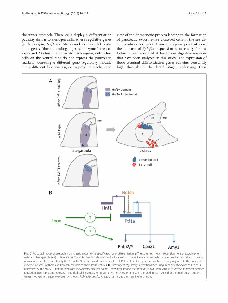
the upper stomach. These cells display a differentiationpathway similar to zymogen cells, where regulative genes(such as Ptf1a, Hnf1 and Mist1) and terminal differenti-ation genes (those encoding digestive enzymes) are co-expressed. Within this upper stomach region, only a fewcells on the ventral side do not express the pancreaticmarkers, denoting a different gene regulatory moduleand a different function. Figure 7a presents a schematic
view of the ontogenetic process leading to the formationof pancreatic exocrine-like clustered cells in the sea ur-chin embryo and larva. From a temporal point of view,the increase of SpPtf1a expression is necessary for thefollowing expression of at least three digestive enzymesthat have been analyzed in this study. The expression ofthese terminal differentiation genes remains constantlyhigh throughout the larval stage, underlying their
Fig. 7 Proposed model of sea urchin pancreatic exocrine-like specification and differentiation. a The schemes show the development of exocrine-likecells from late gastrula (left) to larva (right). The right drawing also shows the localization of putative endocrine cells that are positive for antibody stainingof a member of the insulin family (ILP-1+ cells). Note that we do not know if the ILP-1+ cells in the upper stomach are simply adjacent to the pancreaticexocrine-like cells or there are stomach cells which share both features. b Summary of regulatory interactions occurring in pancreatic exocrine-like cellsunraveled by this study. Different genes are shown with different colors. The wiring among the genes is shown with solid lines. Arrows represent positiveregulation, bars represent repression, and dashed lines indicate signaling events. Question marks in the food input means that the mechanism and thegenes involved in the pathway are not known. Abbreviations: fg, foregut; hg, hindgut; in, intestine; mo, mouth
Perillo et al. BMC Evolutionary Biology (2016) 16:117 Page 11 of 15

importance for larval development. SpPtf1a and SpMist1expression oscillate, and after initial activation, decrease,suggesting that these early TFs may be necessary to acti-vate digestive enzyme transcription but also other factorsare involved in maintaining the right level of enzymesduring feeding. Interestingly, the pancreatic exocrine-like cells also express the islet-specific microRNA-375. Aformer study showed that also miR-100, miR-125 andlet-7, markers of neurosecretory endocrine cells, are lo-calized in the same area of the sea urchin stomach [46].These cells seem to be of the pancreatic exocrine-liketype, although further analysis will be necessary to testco-expression.In the developing pancreas, Notch signaling represses
endocrine development and exocrine cell differentiation[16, 19, 47]. The early role of Notch signaling in the seaurchin embryos has been extensively studied [48–51]. Inthis study, we identified a putative recruitment of Notchsignaling at the late gastrula stage in blocking the differ-entiation of stomach cells towards an exocrine-like fatein the pluteus larva. We found that Notch signaling re-presses SpPtf1a expression in those cells that are adja-cent to the SpPtf1a + cells. Moreover, we observed anectopic expression of SpPtf1a in a group of cells of theanus, suggesting that the Notch signaling could be re-sponsible for the direct or indirect repression of SpPtf1aexpression in these intestinal cells. Nonetheless, we ob-served that when Notch signaling is blocked, SpMist1ectopically accumulates in the cells of the apical organthat are adjacent to SpMist1+ cells. One possibility isthat a secretory fate is repressed by a Notch lateral-inhibition mechanism in ectodermal cells that are com-mitted to a not-secretory fate. Alternatively, this ectoder-mal effect may be due to an extra round of cell division.In a similar way, our observations suggest that Notchsignaling prevents the exocrine fate of a group of cells ofthe esophagus. In some cases, we observed that whenNotch signaling is blocked, pancreatic genes that areexpressed in both the ectoderm and the endoderm loseexpression in a specific territory, and not in the other.For instance, the transcription of SpPtf1a in the ecto-derm and that of SpMist1 in the stomach do not changein DAPT treated plutei, similarly to what Materna andDavidson observed for SpShr2 and SpDelta in the earlyembryo [42]. Our data suggest that Notch signalingcould have different effects not only during early devel-opment, but it can also finely regulate cell differentiationduring development in the sea urchin larva. Nonetheless,to deeply analyze the fine mechanisms that underlieregulation of midgut fates, further knockdown experi-ments of the Notch signaling machinery will be import-ant to enhance our understanding in the future.The Ptf1a transcriptional complex controls maturation
of pancreatic exocrine cells and many efforts has been
made to identify its cofactors and targets [52, 53]. In thepancreas, Hnf1a activates the transcription of Ptf1a,which is part of a transcriptional complex that controlspancreatic exocrine cell maturation and regulates theproduction of digestive enzymes in acinar cells [13, 54].Here we found that this gene regulatory module is con-served in an early-branching deuterostome. Based onthe homology with mammalian binding sequences, webioinformatically identified several putative Ptf1a bind-ing sites on the promoters of the sea urchin digestive en-zymes which transcription is strongly downregulated inPtf1a perturbed embryos. We also showed that SpPtf1acan bind mammalian target genes to activate transcrip-tion. However, further in vivo experiments are necessaryto test the functionality of these binding sites predictedin silico. In this study, we also demonstrated thatSpPtf1a represses its own gene expression in some stom-ach cells. We reported that in SpPtf1a morphants, moreSpPtf1a + cells appear as single pouches at the ventraland dorsal stomach in cells where normally the gene isnever expressed. These cells appear as a distinct groupof cells that alternate with cells where SpPtf1a is absent,like a chessboard (Fig. 4i). Conversely, blocking theNotch pathway results in a continuous ring of Ptf1a +cells (Fig. 4l). Hence, the presence of different mecha-nisms supports the role of a Notch lateral-inhibitionmodel in the stomach domain. Another finding thatemerged from our data is that SpPtf1a may repress theexpression of SpFng. Due to its role, the Fringe orthologis widely expressed in the embryos, and this could ex-plain the borderline qPCR values in SpPtf1a morphants.However, our bioinformatic analysis found binding sitesfor Ptf1a in the SpFng promoter region. To understandif the effect of DAPT treatment on SpFng is specific foran exocrine cell fate, further experiments should definewhether Ptf1a interacts with corepressor proteins, as inthe case of Nkx6 in mammals [55], to block SpFng tran-scription and consequentially Notch activation, in anegative feedback loop manner.A summary of the above described putative gene inter-
actions and the proposed gene model leading to specifi-cation and differentiation of sea urchin pancreaticexocrine-like cells are schematized in Fig. 7b.The sea urchin larva is a very plastic organism, able to
adapt its metabolic and growth programs to changes inthe environmental energy context, e.g. arms and ciliaryband growth is influenced by different food availability[56–61]. Feeding has a central role in the life of sea ur-chin larvae, since they may live up to several months inthe plankton, therefore they have to be considered asfully developed animals, even if their body axis will berearranged after metamorphosis. In a previous study, wefound that in the sea urchin larva, a member of the insu-lin family is expressed in a feeding-dependent fashion,
Perillo et al. BMC Evolutionary Biology (2016) 16:117 Page 12 of 15

opening new questions on whether other genes are influ-enced by food assumption or deprivation [29]. As ex-pected during feeding, when more digestive enzymes arenecessary, we found that the transcription of a carboxi-peptidase, a pancreatic lipase and an amylase are signifi-cantly increased, whereas SpPtf1a expression showed anopposite behavior. It will be important to further investi-gate which other factors are involved in the binding of thePtf1a complex to its targets. Previous transcriptomic ana-lysis reported that SpCpa2L and SpPnlp2/5 transcripts arestrongly reduced when SpLox is knocked down [62] and itis known that its vertebrate ortholog, Pdx1, regulates thedevelopment of digestive enzyme-producing acinar cellsand insulin expression [63, 64]. In the sea urchin, SpLox isexpressed in a group of cells between the lower stomachand the intestine (the pyloric sphincter) [65], in a gut do-main and in cells that are different from the herein char-acterized pancreatic exocrine-like cells. Thus, a possiblescenario that could be worth testing in further experi-ments is that SpLox could act through a hypothetic signal-ing pathway to eventually regulate the expression ofdigestive enzymes, accordingly with food availability.The pancreatic exocrine-like cells that we describe are
localized in the same area where there are also endocrine-like cells producing a member of the insulin family, ILP1[29]. We cannot rule out the possibility that these two celltypes are just in close proximity and do not represent aunique cell-type sharing both functions. Generation ofantibodies for the digestive enzymes will be necessary totest if these exocrine markers are coexpressed in the ILP+cells, but we can at least conclude that both cell types arelocalized in the same area of the stomach. In the cephalo-cordate amphioxus, endocrine and exocrine cells do notexhibit any gut specialization [8, 66–68]. In the hagfish, anislet-like organ is independent of exocrine acini, and, inthe teleost, endocrine islets are scattered among the morewidespread exocrine parenchyma [69]. Thus, two possibleevolutionary scenarios could be put forward. First, thepancreas and the exocrine and endocrine-like cells (closelylocalized) in the sea urchin gut are the result of conver-gent evolution, thus implying that this feature was lost inearly branching chordates. Second, the clustering of thesea urchin pancreatic exocrine-like cells could be the re-sult of an independent evolution from an ancestral pan-creatic cell-type. To test these hypotheses and to infer theevolutionary origin of the pancreas, homologs of pancre-atic genes should be studied in other echinoderms as wellas in other non-chordate deuterostomes, such as thehemichordates.
ConclusionsHere, we report that a gene regulatory module requiredfor vertebrate pancreatic exocrine cell development isshared by a non-chordate deuterostome. Our study,
together with previous studies [65, 70], supports the ideathat the sea urchin gut is a multifunctional organ and itis composed of highly differentiated cells, which in manyaspects are similar to vertebrates, therefore providing anamenable system in which to study development andfunction of gut cells.
Additional file
Additional file 1: Supplementary Table 1. (PDF 55 kb)
AcknowledgmentsThe authors wish to thank Giovanna Benvenuto for confocal microscopy,Marco Borra for quantitative Real Time PCR and Xiaogang Zhong forbioinformatics assistance. We would also like to thank Pat Leahy (KML,Caltech, Pasadena, CA) for providing adult S. purpuratus and DavideCaramiello (SZN) for animal maintenance. MP would like to thank Steven Z.Swartz for stimulating discussions and paper revisions. This work wassupported by MIUR (premiale PANTRAC to MIA) and NIH grant DK61215 (toSDL). M.P. has been supported by a SZN PhD fellowship and by a fellowshipfrom POR Campania FSE 2007-2013 Project-MODO, Model Organism.
Authors’ contributionsMIA and MP conceived the project, SDL and YJW designed the bioinformaticanalysis and the experiments in mammalian cells. MP performed theexperimental work, YJW performed the luciferase essays. The authorscontributed equally to the interpretation of the results and the writing of themanuscript. All authors read and approved the final manuscript.
Competing interestsThe authors declare that they have no competing interests.
Author details1Biology and Evolution of Marine Organisms, Stazione Zoologica AntonDohrn, Napoli 80121, Italy. 2Department of Surgery and the McKusickNathans Institute for Genetic Medicine, Johns Hopkins University, Baltimore,MD 21205, USA. 3Present address: Department of Biology, Boston College,Chestnut Hill, MA, USA.
Received: 8 April 2016 Accepted: 12 May 2016
References1. Chera S, de Rosa R, Miljkovic-Licina M, Dobretz K, Ghila L, Kaloulis K, Galliot B.
Silencing of the hydra serine protease inhibitor Kazal1 gene mimics thehuman SPINK1 pancreatic phenotype. J Cell Sci. 2006;119:846–57.
2. Lentz TL. Intramitochondrial glycogen granules in digestive cells of Hydra. JCell Biol. 1966;29:162–7.
3. Goldberg WM. Gastrodermal structure and feeding responses in thescleractinian Mycetophyllia reesi, a coral with novel digestive filaments.Tissue Cell. 2002;34:246–61.
4. Brennan GP, Ramasamy P. Ultrastructure of the gut caecal epithelium ofPricea multae (Monogenea: Polyopisthocotylea). Parasitol Res. 1996;82:312–8.
5. Jones MK, Hughes-Stamm SR, East RM, Cribb TH. Ultrastructure of the digestivetract of Gyliauchen nahaensis (Platyhelminthes, Digenea), an inhabitant of thehindgut of herbivorous fishes. J Morphol. 2000;246:198–211.
6. Brennan GP, Ramasamy P. Ultrastructure of the surface structures andelectron immunogold labeling of peptide immunoreactivity in the nervoussystem of Pseudothoracocotyla indica (Polyopisthocotylea: Monogenea).Parasitol Res. 1996;82:638–46.
7. Lemaitre B, Miguel-Aliaga I. The digestive tract of Drosophila melanogaster.Annu Rev Genet. 2013;47:377–404.
8. Slack JM. Developmental biology of the pancreas. Development.1995;121:1569–80.
9. Krapp A, Knöfler M, Ledermann B, Bürki K, Berney C, Zoerkler N, HagenbüchleO, Wellauer PK. The bHLH protein PTF1-p48 is essential for the formation ofthe exocrine and the correct spatial organization of the endocrine pancreas.Genes Dev. 1998;12:3752–63.
Perillo et al. BMC Evolutionary Biology (2016) 16:117 Page 13 of 15
10. Lemercier C, To RQ, Swanson BJ, Lyons GE, Konieczny SF. Mist1: a novelbasic helix-loop-helix transcription factor exhibits a developmentallyregulated expression pattern. Dev Biol. 1997;182:101–13.
11. Cockell M, Stevenson BJ, Strubin M, Hagenbüchle O, Wellauer PK.Identification of a cell-specific DNA-binding activity that interacts with atranscriptional activator of genes expressed in the acinar pancreas. Mol CellBiol. 1989;9:2464–76.
12. Kawaguchi Y, Cooper B, Gannon M, Ray M, MacDonald RJ, Wright CVE. Therole of the transcriptional regulator Ptf1a in converting intestinal topancreatic progenitors. Nat Genet. 2002;32:128–34.
13. Krapp A, Knöfler M, Frutiger S, Hughes GJ, Hagenbüchle O, Wellauer PK.The p48 DNA-binding subunit of transcription factor PTF1 is a newexocrine pancreas-specific basic helix-loop-helix protein. EMBO J.1996;15:4317–29.
14. Pin CL, Rukstalis JM, Johnson C, Konieczny SF. The bHLH transcription factorMist1 is required to maintain exocrine pancreas cell organization and acinarcell identity. J Cell Biol. 2001;155:519–30.
15. Haumaitre C, Barbacci E, Jenny M, Ott MO, Gradwohl G, Cereghini S. Lack ofTCF2/vHNF1 in mice leads to pancreas agenesis. Proc Natl Acad Sci U S A.2005;102:1490–5.
16. Hald J, Hjorth JP, German MS, Madsen OD, Serup P, Jensen J. ActivatedNotch1 prevents differentiation of pancreatic acinar cells and attenuateendocrine development. Dev Biol. 2003;260:426–37.
17. Kim SK, Hebrok M. Intercellular signals regulating pancreas developmentand function. Genes Dev. 2001;15:111–27.
18. Norgaard GA, Jensen JN, Jensen J. FGF10 signaling maintains the pancreaticprogenitor cell state revealing a novel role of Notch in organ development.Dev Biol. 2003;264:323–38.
19. Esni F, Ghosh B, Biankin AV, Lin JW, Albert MA, Yu X, MacDonald RJ, Civin CI,Real FX, Pack MA, Ball DW, Leach SD. Notch inhibits Ptf1 function and acinarcell differentiation in developing mouse and zebrafish pancreas.Development. 2004;131:4213–24.
20. Poy MN, Eliasson L, Krutzfeldt J, Kuwajima S, Ma X, Macdonald PE, Pfeffer S,Tuschl T, Rajewsky N, Rorsman P, Stoffel M. A pancreatic islet-specificmicroRNA regulates insulin secretion. Nature. 2004;432:226–30.
21. Kloosterman WP, Lagendijk AK, Ketting RF, Moulton JD, Plasterk RHA.Targeted inhibition of miRNA maturation with morpholinos reveals a rolefor miR-375 in pancreatic islet development. PLoS Biol. 2007;5:e203.
22. Poy MN, Hausser J, Trajkovski M, Braun M, Collins S, Rorsman P, Zavolan M,Stoffel M. miR-375 maintains normal pancreatic alpha- and beta-cell mass.Proc Natl Acad Sci U S A. 2009;106:5813–8.
23. McClay DR. Evolutionary crossroads in developmental biology: sea urchins.Development. 2011;138:2639–48.
24. Burke RD. Structure of the digestive tract of the pluteus larva ofDendraster excentricus (Echinodermata: Echinoida). Zoomorphology. 1981;98:209–25.
25. Vacquier VD. The appearance of -1,3-glucanohydrolase activity during thedifferentiation of the gut of sand dollar plutei. Dev Biol. 1971;26:1–10.
26. Vacquier VD, Korn LJ, Epel D. The appearance of -amylase activity duringgut differentiation in sand dollar plutei. Dev Biol. 1971;26:393–9.
27. Vacquier VD. The effects of glucose and lithium chloride on theappearance of -1,3-glucanohydrolase activity in sand dollar plutei. DevBiol. 1971;26:11–6.
28. Holland ND, Lauritis JA: The fine structure of the gastric exocrine cells of thepurple sea urchin, Strongylocentrotus purpuratus. Trans Amer Microscop Soc.1968;87:201-209.
29. Perillo M, Arnone MI. Characterization of insulin-like peptides (ILPs) in thesea urchin Strongylocentrotus purpuratus: insights on the evolution of theinsulin family. Gen Comp Endocrinol. 2014;205:68–79.
30. Andrikou C, Iovene E, Rizzo F, Oliveri P, Arnone MI. Myogenesis in the seaurchin embryo: the molecular fingerprint of the myoblast precursors.Evodevo. 2013;4:33.
31. Andrikou C, Pai C-Y, Su Y-H, Arnone MI: Logics and properties of a geneticregulatory program that drives embryonic muscle development in anechinoderm. Elife. 2015;4.
32. Hughes JN, Dodge N, Rathjen PD, Rathjen J. A novel role for gamma-secretase in the formation of primitive streak-like intermediates from EScells in culture. Stem Cells. 2009;27:2941–51.
33. Rast JP, Amore G, Calestani C, Livi CB, Ransick A, Davidson EH. Recovery ofdevelopmentally defined gene sets from high-density cDNA macroarrays.Dev Biol. 2000;228:270–86.
34. Nemer M, Rondinelli E, Infante D, Infante AA. Polyubiquitin RNAcharacteristics and conditional induction in sea urchin embryos. Dev Biol.1991;145:255–65.
35. Dong PDS, Provost E, Leach SD, Stainier DYR. Graded levels of Ptf1adifferentially regulate endocrine and exocrine fates in the developingpancreas. Genes Dev. 2008;22:1445–50.
36. Howard-Ashby M, Materna SC, Brown CT, Chen L, Cameron RA, Davidson EH.Gene families encoding transcription factors expressed in early developmentof Strongylocentrotus purpuratus. Dev Biol. 2006;300:90–107.
37. Howard-Ashby M, Materna SC, Brown CT, Chen L, Cameron RA, DavidsonEH. Identification and characterization of homeobox transcription factorgenes in Strongylocentrotus purpuratus, and their expression in embryonicdevelopment. Dev Biol. 2006;300:74–89.
38. Bach I, Mattei MG, Cereghini S, Yaniv M. Two members of an HNF1 homeoproteinfamily are expressed in human liver. Nucleic Acids Res. 1991;19:3553–9.
39. Booth H, Holland P: Annotation, nomenclature and evolution of four novelhomeobox genes expressed in the human germ line. Gene. 2007;387:7–14.
40. Poustka AJ, Groth D, Hennig S, Thamm S, Cameron A, Beck A, Reinhardt R,Herwig R, Panopoulou G, Lehrach H. Generation, annotation, evolutionaryanalysis, and database integration of 20,000 unique sea urchin EST clusters.Genome Res. 2003;13:2736–46.
41. Annunziata R, Perillo M, Andrikou C, Cole AG, Martinez P, Arnone MI. Patternand process during sea urchin gut morphogenesis: the regulatorylandscape. Genesis. 2014;52:251–68.
42. Materna SC, Davidson EH. A comprehensive analysis of Delta signaling inpre-gastrular sea urchin embryos. Dev Biol. 2012;364:77–87.
43. Moloney DJ, Panin VM, Johnston SH, Chen J, Shao L: Fringe is aglycosyltransferase that modifies Notch. Nature. 2000;406:369–375.
44. Peterson RE, McClay DR. A Fringe-modified Notch signal affectsspecification of mesoderm and endoderm in the sea urchin embryo.Dev Biol. 2005;282:126–37.
45. Masui T, Long Q, Beres TM, Magnuson MA, MacDonald RJ. Early pancreaticdevelopment requires the vertebrate Suppressor of Hairless (RBPJ) in thePTF1 bHLH complex. Genes Dev. 2007;21:2629–43.
46. Christodoulou F, Raible F, Tomer R, Simakov O, Trachana K, Klaus S, Snyman H,Hannon GJ, Bork P, Arendt D. Ancient animal microRNAs and the evolution oftissue identity. Nature. 2010;463:1084–8.
47. Murtaugh LC, Stanger BZ, Kwan KM, Melton DA. Notch signaling controlsmultiple steps of pancreatic differentiation. Proc Natl Acad Sci U S A.2003;100:14920–5.
48. Croce JC, McClay DR. Dynamics of Delta/Notch signaling on endomesodermsegregation in the sea urchin embryo. Development. 2010;137:83–91.
49. Sherwood DR, McClay DR: Identification and localization of a sea urchinNotch homologue: insights into vegetal plate regionalization and Notchreceptor regulation. Development. 1997;124:3363–3374.
50. Sherwood DR, McClay DR: LvNotch signaling plays a dual role in regulatingthe position of the ectoderm-endoderm boundary in the sea urchinembryo. Development. 2001;128:2221–32.
51. Materna SC, Swartz SZ, Smith J. Notch and Nodal control forkhead factorexpression in the specification of multipotent progenitors in sea urchin.Development. 2013;140:1796–806.
52. Thompson N, Gésina E, Scheinert P, Bucher P, Grapin-Botton A. RNAprofiling and chromatin immunoprecipitation-sequencing reveal that PTF1astabilizes pancreas progenitor identity via the control of MNX1/HLXB9 and anetwork of other transcription factors. Mol Cell Biol. 2012;32:1189–99.
53. Arda HE, Benitez CM, Kim SK. Gene regulatory networks governing pancreasdevelopment. Dev Cell. 2013;25:5–13.
54. Petrucco S, Wellauer PK, Hagenbüchle O. The DNA-binding activity oftranscription factor PTF1 parallels the synthesis of pancreas-specific mRNAsduring mouse development. Mol Cell Biol. 1990;10:254–64.
55. Schaffer AE, Freude KK, Nelson SB, Sander M. Nkx6 transcription factors andPtf1a function as antagonistic lineage determinants in multipotentpancreatic progenitors. Dev Cell. 2010;18:1022–9.
56. Adams DK, Sewell MA, Angerer RC, Angerer LM. Rapid adaptation to foodavailability by a dopamine-mediated morphogenetic response. NatCommun. 2011;2:592.
57. Heyland A, Reitzel AM, Hodin J. Thyroid hormones determine developmentalmode in sand dollars (Echinodermata: Echinoidea). Evol Dev. 2004;6:382–92.
58. Reitzel AM, Heyland A: Reduction in morphological plasticity in echinoidlarvae: relationship of plasticity with maternal investment and foodavailability. Evol Ecol Res. 2007;9:109–121.
Perillo et al. BMC Evolutionary Biology (2016) 16:117 Page 14 of 15

59. Strathmann RR, Fenaux L, Strathmann MF: Heterochronic developmentalplasticity in larval sea urchins and its implications for evolution ofnonfeeding larvae. Evolution. 1992;46:972–986.
60. Strathmann RR, Foley GP, Hysert AN. Loss and gain of the juvenile rudimentand metamorphic competence during starvation and feeding of bryozoanlarvae. Evol Dev. 2008;10:731–6.
61. Carrier TJ, King BL, Coffman JA. Gene Expression Changes Associated Withthe Developmental Plasticity of Sea Urchin Larvae in Response to FoodAvailability. Biol Bull. 2015;228:171–80.
62. Annunziata R, Martinez P, Arnone MI. Intact cluster and chordate-likeexpression of ParaHox genes in a sea star. BMC Biol. 2013;11:68.
63. Ohlsson H, Karlsson K, Edlund T. IPF1, a homeodomain-containingtransactivator of the insulin gene. EMBO J. 1993;12:4251–9.
64. Zhou Q, Law AC, Rajagopal J, Anderson WJ, Gray PA, Melton DA. Amultipotent progenitor domain guides pancreatic organogenesis. Dev Cell.2007;13:103–14.
65. Cole AG, Rizzo F, Martinez P, Fernandez-Serra M, Arnone MI. Two ParaHoxgenes, SpLox and SpCdx, interact to partition the posterior endoderm inthe formation of a functional gut. Development. 2009;136:541–9.
66. Falkmer S, Dafgård E, el-Salhy M, Engström W, Grimelius L, Zetterberg A.Phylogenetical aspects on islet hormone families: a minireview withparticular reference to insulin as a growth factor and to the phylogeny ofPYY and NPY immunoreactive cells and nerves in the endocrine andexocrine pancreas. Peptides. 1985;6 Suppl 3:315–20.
67. Lecroisey C, Le Pétillon Y, Escriva H, Lammert E, Laudet V. Identification,evolution and expression of an insulin-like peptide in the cephalochordateBranchiostoma lanceolatum. PLoS One. 2015;10:e0119461.
68. Pieler T, Chen Y. Forgotten and novel aspects in pancreas development.Biol Cell. 2006;98:79–88.
69. Youson JH, Al-Mahrouki AA. Ontogenetic and phylogenetic development ofthe endocrine pancreas (islet organ) in fish. Gen Comp Endocrinol.1999;116:303–35.
70. Annunziata R, Arnone MI. A dynamic regulatory network explains ParaHoxgene control of gut patterning in the sea urchin. Development.2014;141:2462–72.
• We accept pre-submission inquiries
• Our selector tool helps you to find the most relevant journal
• We provide round the clock customer support
• Convenient online submission
• Thorough peer review
• Inclusion in PubMed and all major indexing services
• Maximum visibility for your research
Submit your manuscript atwww.biomedcentral.com/submit
Submit your next manuscript to BioMed Central and we will help you at every step:
Perillo et al. BMC Evolutionary Biology (2016) 16:117 Page 15 of 15