Embed Size (px)
Citation preview

Pancreatic cancer
Dr. Geoff S Williams
Division of GastroenterologyDalhousie University

Objectives
At the completion of this talk the attendees will:
1. Be able to describe the presenting symptoms of pancreatic cancer
2. Have an understanding of the steps involved in diagnosis
3. Appreciate the challenges in managing pancreatic cancer

Pancreatic Anatomy
The pancreas is a soft, elongated, flattened organ
12 - 20 cm in length Weighs 70 - 110 g in
adults The pancreas lies
posterior the stomach
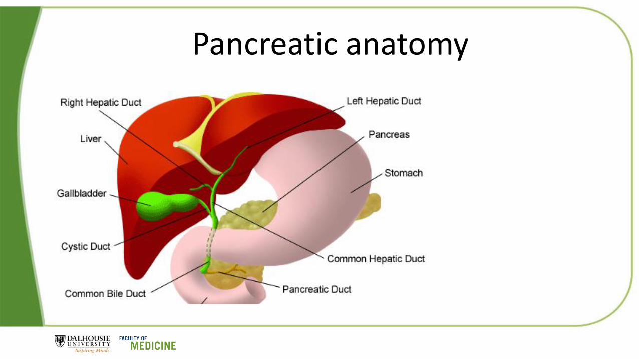
Pancreatic anatomy
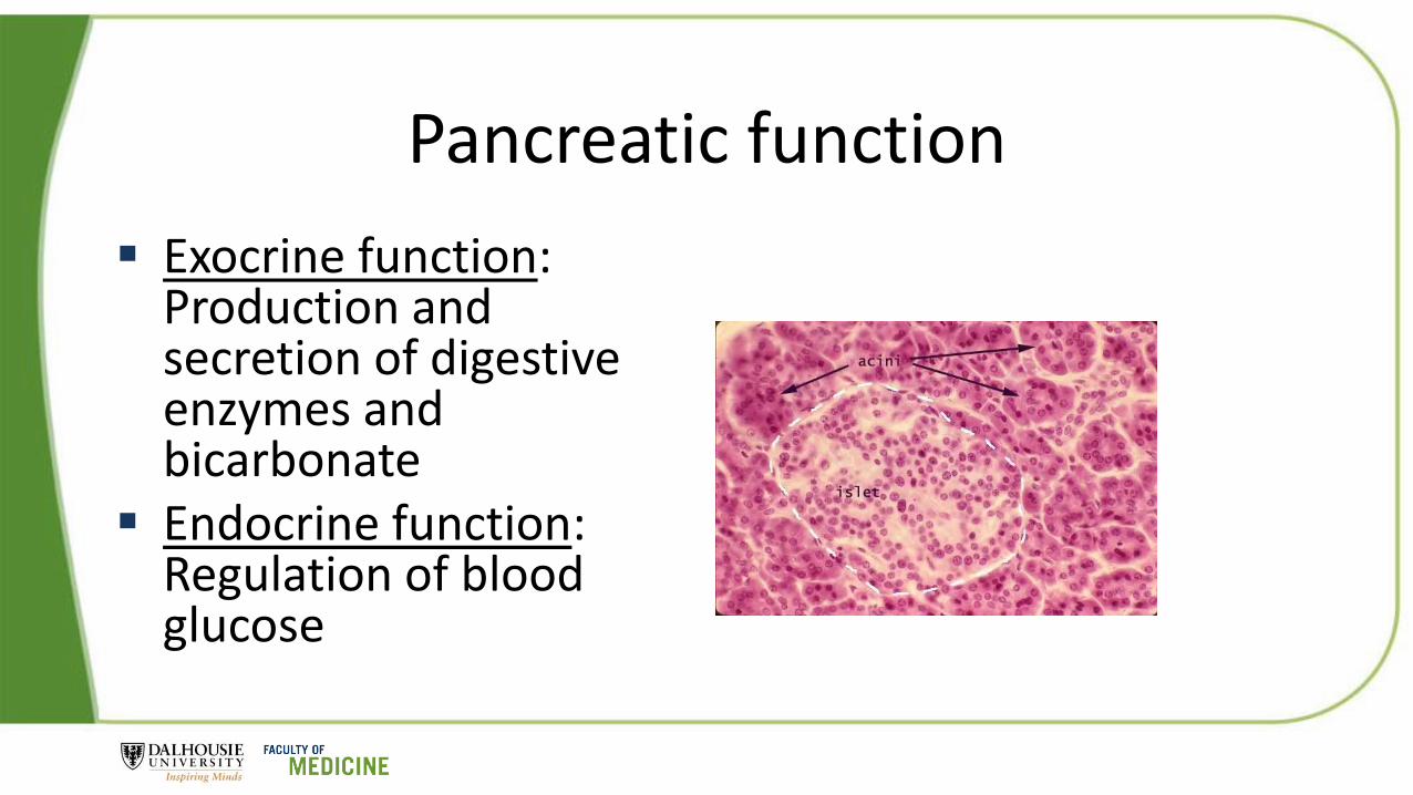
Pancreatic function
Exocrine function: Production and secretion of digestive enzymes and bicarbonate
Endocrine function: Regulation of blood glucose

The acinus

Pancreatic Enzymes
Lipase - Fat digestion
Amylase - Enzyme which digests starch and glycogen.
Proteases - Protein digestion
* When measuring ‘injury’ to the pancreas, we typically measure blood levels of lipase and amylase, but not proteases.

A case: Arnold Colhurst A.C. is a pleasant 64 year old Civil Engineer, planning to retire in 6
months One morning his wife commented that he “looked a little yellow” Upon looking at himself closely in the mirror, he indeed did note
yellowing of the sclera (whites of the eyes). He recalled his mother having jaundice when he was a child, but it
resolved and she lived to age of 84, dying from complications of a broken hip
A.C. booked an appointment with his Family Physician two days later

Family Doctor visit History
• He has had no pain. His stools are pale, and urine is dark (looks like Coca cola). He has lost perhaps 5 – 7 lbs over the last month, and attributed to eating healthier
Past Medical History• Borderline diabetic, Hypertension
Medications• Hydrochlorothiazide, Baby aspirin
Social history • Ex smoker, having quit a decade ago. Drinks Bottle of red wine on weekends
Family history• Mom with jaundice, cause unknown, but likely due to viral hepatitis

On examination
Vital signs normal
Scleral icterus
No Virchow’s nodes
Liver palpable in the mid clavicular line, 4 cm below costal margin (not tender)
No abdominal ascites
Otherwise normal

Lab tests arranged
Liver Enzymes
ALT 462 (n < 50)
AST 368 (n < 50)
ALP 720 (n < 90)
GGT 480 (n < 50)
Lipase 60 (n < 90)
Liver function tests
INR 1.8 (n < 1.2)
Albumin 28 (n : 35 – 42)
Total bilirubin 124 (n < 22)
Direct bilirubin 68 (n < 5)
Indirect bilirubin 56 ( n < 17)

The physiology of bile
Physiology Text Book 2003
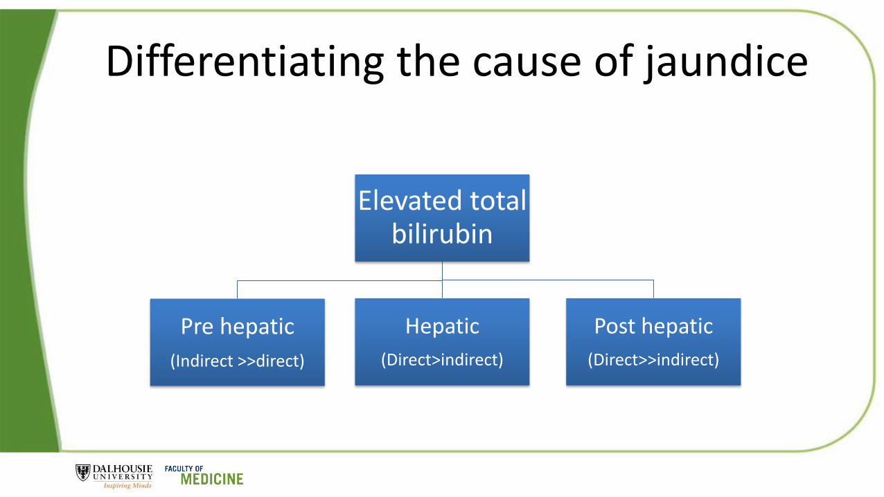
Differentiating the cause of jaundice
Elevated total bilirubin
Pre hepatic
(Indirect >>direct)
Hepatic
(Direct>indirect)
Post hepatic
(Direct>>indirect)

Approach to elevated indirect bilirubin
Elevated indirect bilirubin
Increased biliruibinproduction
Hemolysis
Impaired RBC synthesis
Impaired hepatic uptake
CHF
Portosystemic shunts
Drugs (eg. Rifampin)
Impaired bilirubin conjugation
Gilberts’s syndrome
Neonates
Hyper thyroidism
Chronic liver disease

Approach to elevated direct bilirubin
Ultrasound/CT
No obstruction
Hepatocellular
(ALT/AST >> ALP/GGT)
Cholestatic
(ALP/GGT >> ALT/AST)
Obstruction
Benign vsmalignant
causes

Where to go from here with Arnold?
Do an ultrasound?
Do a CT?
Do an MRI?
Do an ERCP?

Abdominal ultrasound
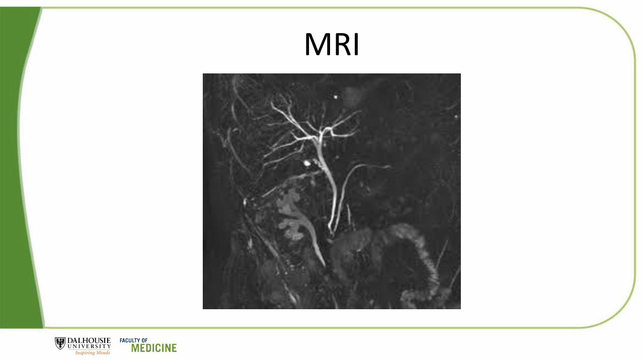
MRI

CT scan

Approaches to tissue acquisition
ERCP
Percutaneous biopsy
EUS
Open surgical

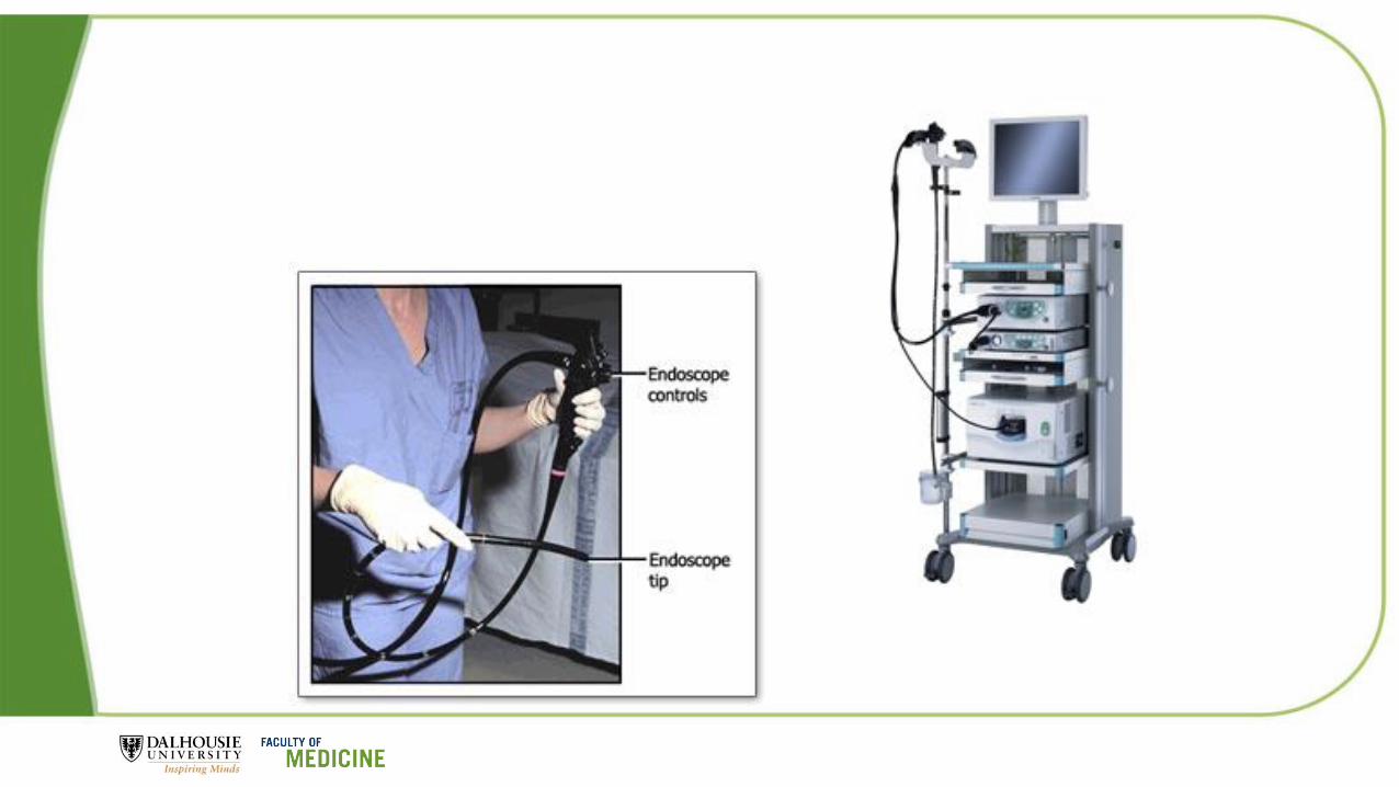
What is ERCP?
ERCP is an acronym which stands for Endoscopic Retrograde Cholangio-Pancreatography
It is a method for accessing the common bile duct and pancreatic ducts

The patient is sedated. A endoscope with a side-viewing camera is advanced through the mouth, down to the second part of the duodenum where the ampulla is identified. The ampulla is entered with a catheter and dye is injected and viewed with fluoroscopy (xray technology). To access the bile duct, the ampulla is cut open (sphincterotomy).

A: Normal ampulla B: A sphincterotomy has been performed to access the CBD. C: The ampulla is dilated with a balloon D: A stone is extracted with a basket.

Footnote
CBD
stent

ERCP

Brush cytology
Small brush used to obtain cells from a tumourinvading the bile duct
Yields a diagnosis 50 – 60 % of the time when cancer is present

Complications of ERCP
Pancreatitis (5 – 10%)
Bleeding (1%)
Perforation (0.5 – 1%)
Cholangitis (<1%)
Issues relating to sedation

Endoscopic Ultrasound

Radial echoendoscope

Linear echoendoscope

Technique
Air is the enemy in EUS
Balloon on tip (water filled) is used to create an echo window
Fluid can be instilled into lumen as well and air removed

EUS


FNA for solid tumors
Usually 3 - 5 passes made with needle
Sample smeared on a slide
Cytotechnologist evaluates on site
Diagnostic yield is > 90% when cancer is present

Cytology

Complications of EUS +/- FNA EUS alone
• Higher rates of sedation, longer procedures than standard endoscopy• Perforation (0.001%)• Bleeding (0.001)• Infection (1/ million)
With FNA• Perforation (0.1%)• Bleeding (0.5%)• Pancreatitis (0.6 – 2%)• Infection (low except if cystic lesion)

Percutaneous biopsy

Would you choose surgery?

Back to our patient…..
Underwent ERCP with brush cytology and a plastic stent was placed in the bile duct to treat the jaundice.
Cytology results: “Atypical, not diagnostic”
Referred for an EUS: Biopsy confirms adenocarcinoma of pancreas

Pancreatic Cancer
Most pancreatic cancers are adenocarcinomas (85%), arising from the ductal epithelium (exocrine pancreas)
Third most common GI cancer (behind colon and rectal cancers)
Most patients > 45 y/o
Poor prognosis:• 20% 5-year survival if surgically resectable
• <10 % 5-year survival if lymph nodes involved or other metastasis

Epidemiology of Pancreatic Cancer in Canada (2016 Estimates)
Annual pancreatic cancer diagnosed
5,200
2,600 men 2,600 women
Cases of Pancreatic Cancer
in Nova Scotia
150
70 men 80 women
Pancreatic Cancer is the 10th most common cancerin men and 9th in women in Canada.

Distribution of New Cancer Cases
Canadian Cancer Statistics 2016

Estimate Cancer Deaths by Sex in Canada
Canadian Cancer Statistics 2016

Risk factors
Non-modifiable risk factors
- Peutz Jeghers syndrome
- Familial pancreatitis
- Increases with age
- Men > women
- African American > white
- BRCA 2 positivity
Modifiable risk factors:- Chronic pancreatitis 15 X- Cigarette smoking 2 – 3 X- Obesity 2 X- Type 2 Diabetes 2 X- Alcohol >30 g per day 1.2 X- ?Occupational exposure

Symptoms
Symptoms depend on where in the pancreas the cancer develops
Pancreatic body or tail:• Weight loss• Abdominal pain
Head of pancreas• Jaundice with dark urine
and pale stools

Other symptoms that can occur Pain in the upper abdomen that radiates to the back which is new,
significant and persistent that is relieved by leaning forward Back pain Diabetes which is new-onset and not associated with weight gain Vague indigestion (dyspepsia) or abdominal discomfort (not
responding to prescribed medication) Loss of appetite Nausea and vomiting Pain when eating Steatorrhea (fatty stools that are often pale and smell foul)
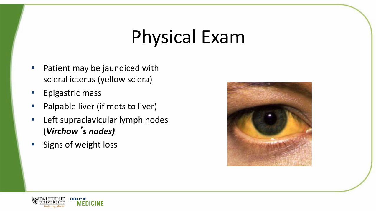
Physical Exam
Patient may be jaundiced with scleral icterus (yellow sclera)
Epigastric mass
Palpable liver (if mets to liver)
Left supraclavicular lymph nodes (Virchow’s nodes)
Signs of weight loss

Tumour markers (CA 19-9)
Carbohydrate Antigen 19 – 9 (CA 19-9)• Sensitivity 79 – 81%• Specificity 82 – 90%
CA 19-9 < 100 implies resectability CA 19-9 > 100 implies metastatic or locally invasive
disease Important limitations:
• Lack of Lewis blood group antigen in 5 – 10%• False positives in obstructive jaundice (10 – 60%)
Ballehaninna; J. Gastro Oncology 2012

How do we treat pancreatic cancer?
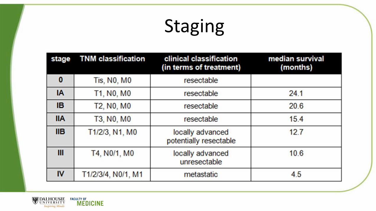
Staging

Surgery best option for cure
< 20% are considered curative by surgery
< 20% 5 year survival with surgery

Chemotherapy if non-resectable


Should I screen her for pancreatic cancer?
No screening protocol has ever been shown to prevent or alter natural history of pancreatic cancer
EXPERT opinion:• Hereditary chronic pancreatitis
• Peutz-Jeghers syndrome
• Family history of pancreatic cancer in three relatives, atleast one of which is first degree relative
• Known BRCA2 positive patient

Back to Arnold
Arnold had tumour in his liver and therefore was not a surgical ‘candidate’
He underwent Folfirinox chemotherapy and in follow up 14 months later he was doing well with no evidence of progression on CT