Embed Size (px)
Citation preview

January 2015
A one year old male with a right testicular tumor
Contributed by Muhammad Idrees, MD Associate Professor Indiana University School of Medicine/ IU Health Partners Department of Pathology and Laboratory Medicine Diplomate of the American Board of Pathology in Anatomic and Clinical Pathology
Subspecialty Certification: Cytopathology
Special Interest: Surgical pathology, Cytopathology, Urologic pathology
Clinical history and findings: One year old male with a right testicular tumor. At exploration it was identified that tumor was arising from epididymis and was fairly circumscribed.
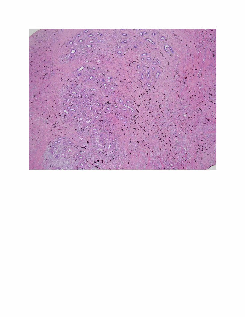
Gross examination: An orchiectomy was performed. At gross examination the tumor was well circumscribed and appeared to be arising from the epididymis. However, it was firmly tethered to the testis. The cut section showed a brown to black homogenous appearance. Microscopic photographs are below.
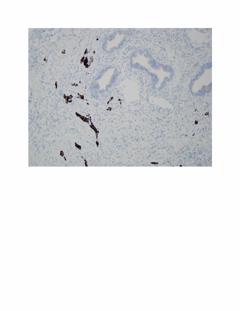
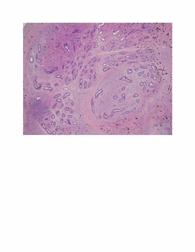
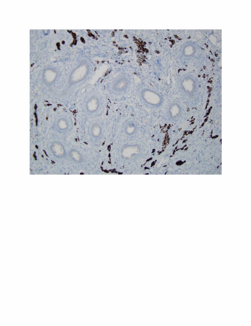
Microscopy and Immunohistochemistry:
Immunohistochemical stains showed positive expression of synaptophysin and NSE, while pancytokeratin, CD68, CK5/6, EMA, Melan A and HMB-45 were negative. See micrographs below.


Final diagnosis: Melanotic Neuroectodermal Tumor of Infancy
Discussion:
Synonyms: Retinal anlage tumor, melanotic hamartoma and melanotic progonoma.
Melanotic neuroectodermal tumor is an extremely rare neoplasm of testicular appendages with usual occurrence in the epididymis. The more common location in both sexes is maxilla.1 It occurs mostly in children less than one year of age; however it can be identified in older children.1-5 In a relatively large review of testicular appendages cases, 82% of the cases occurred in children less than 1 year of age.
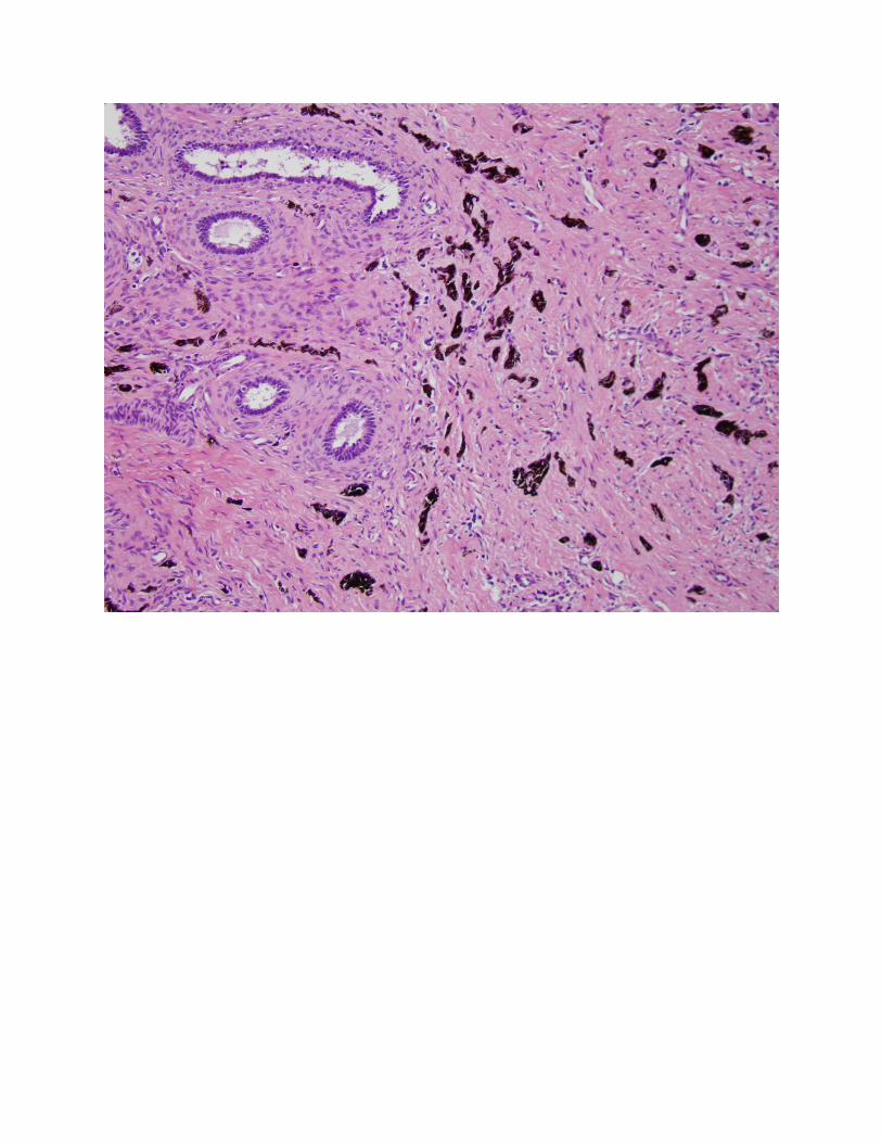
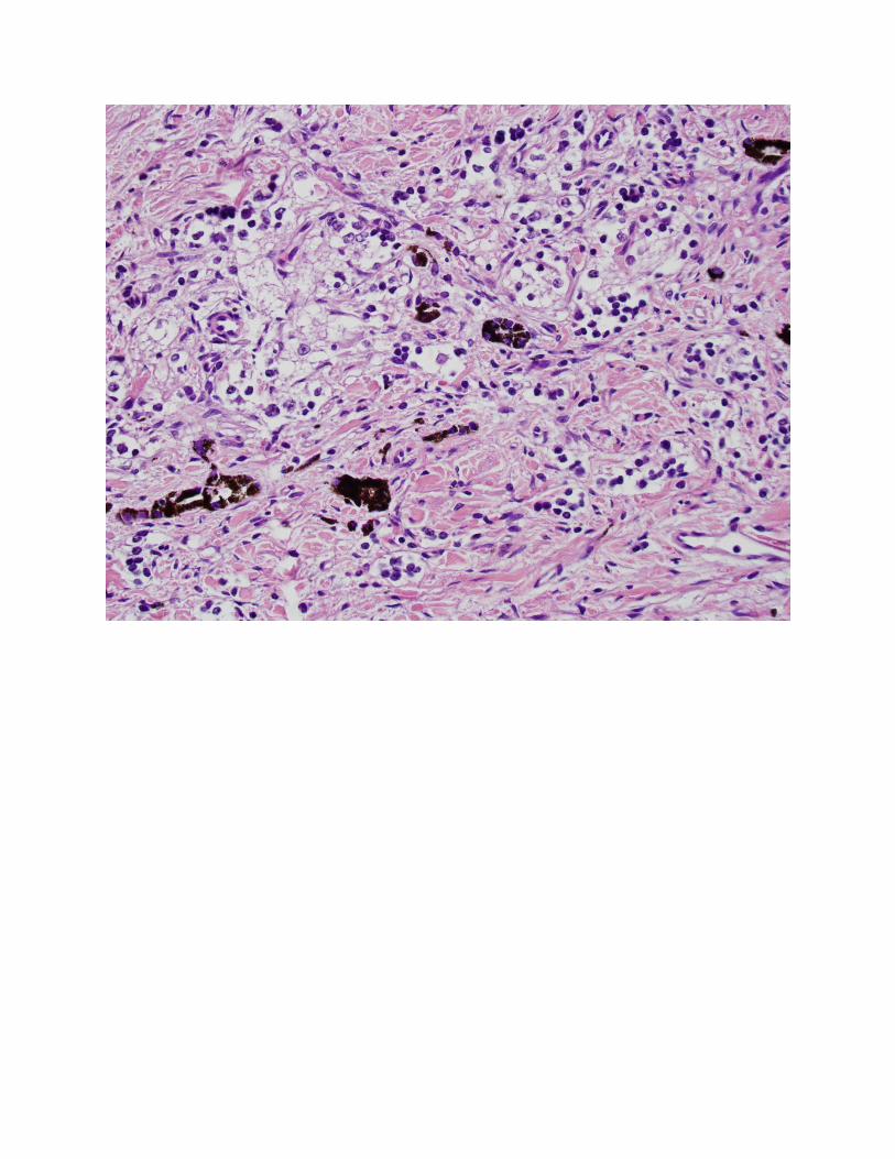
On gross examination, the tumor is usually circumscribed and less than 4 cm in size. The cut surface shows a dark brown to black homogenous appearance. Some tumors display focal or no pigmentation. Microscopically, the tumor is composed of large epithelioid cells admixed with numerous small hyperchromatic neuroblastic-appearing cells in variably sized nests, sheets and cords. These nests are typically dispersed in a dense fibrous stroma. Granules, containing melanin pigment are visible in the larger epithelioid cells. The cytoplasm of larger epithelioid appearing cells is clear while small cells contain scant cytoplasm. In many cases the cells can be seen infiltrating around the tubules which does

not indicate a malignant potential. The tumor cells especially small darker cells may exhibit prominent mitotic activity.
Immunohistochemical expression for synaptophysin and HMB-45 occurs in both cell components. The larger epithelioid cells are usually positive for cytokeratin.1, 6 Molecular markers for neuroblastoma, including N-myc amplification and 1p deletion, are absent in the small neuroblastic appearing cells. Additionally no translocations seen in Ewing׳s sarcoma/PNET and desmoplastic small round cell tumor are identified.7 To date only three cases arising in epididymis presented as metastasis in retroperitoneal lymph nodes via lymphatic route.8, 9 The rest of the reported cases showed a benign course.3, 10 There is no morphological criteria to establish a worse prognosis in epididymis cases; however in maxillary melanotic neuroectodermal tumor cases it is suggested that increased mitoses and Ki-67 staining may be helpful in identifying the aggressively behaving tumors.6
The major differential diagnosis of Melanotic neuroectodermal tumor is embryonal rhabdomyosarcoma. Melanotic neuroectodermal tumors occur in patients mostly less than 1 year of age while rhabdomyosarcoma occurs in slightly older children with the median age of 5 years. The two cell population of melanotic neuroectodermal tumor, with small neuroblastic cells and larger epithelioid cells resemble the primitive cellular component and differentiated rhabdomyoblasts, respectively, of embryonal rhabdomyosarcoma. Rhabdomyoblasts contain eosinophilic cytoplasm while epithelioid cells of melanotic neuroectodermal tumor contain clear cytoplasm. Rhabdomyosarcomas usually have more spindled cell morphology compared to melanotic neuroectodermal tumor. The presence of melanin pigment granules in cells is a prerequisite for the diagnosis of retinal anlage tumor. Muscle markers in conjunction with synaptophysin, HMB-45, and cytokeratin can also resolve this differential diagnosis. Other small cell tumors including neuroblastoma, undifferentiated sarcoma and desmoplastic round blue cell tumor may pose a diagnostic challenge.
References:
[1] Pettinato G, Manivel JC, d'Amore ES, Jaszcz W, Gorlin RJ: Melanotic neuroectodermal tumor of infancy. A reexamination of a histogenetic problem based on immunohistochemical, flow cytometric, and ultrastructural study of 10 cases. The American Journal of Surgical Pathology 1991, 15:233-45.
[2] Calabrese F, Danieli D, Valente M: Melanotic neuroectodermal tumor of the epididymis in infancy: case report and review of the literature. Urology 1995, 46:415-8.
[3] Jurincic-Winkler C, Metz KA, Klippel KF: Melanotic neuroectodermal tumor of infancy (MNTI) in the epididymis. A case report with immunohistological studies and special consideration of malignant features. Zentralblatt fur Pathologie 1994, 140:181-5.
[4] Kobayashi T, Kunimi K, Imao T, Ohkawa M, Komatsu K, Mizukami Y, Namiki M: Melanotic neuroectodermal tumor of infancy in the epididymis. Case report and literature review. Urologia Internationalis 1996, 57:262-5.
[5] Toda T, Sadi AM, Kiyuna M, Egawa H, Tamamoto T, Toyoda Z: Pigmented neuroectodermal tumor of infancy in the epididymis. A case report. Acta Cytologica 1998, 42:775-80.
[6] Barrett AW, Morgan M, Ramsay AD, Farthing PM, Newman L, Speight PM: A clinicopathologic and immunohistochemical analysis of melanotic neuroectodermal tumor of infancy. Oral Surgery, Oral Medicine, Oral Pathology, Oral Radiology, and Endodontics 2002, 93:688-98.
[7] Khoddami M, Squire J, Zielenska M, Thorner P: Melanotic neuroectodermal tumor of infancy: a molecular genetic study. Pediatric and developmental pathology : The Official Journal of the Society for Pediatric Pathology and the Paediatric Pathology Society 1998, 1:295-9.
[8] Johnson RE, Scheithauer BW, Dahlin DC: Melanotic neuroectodermal tumor of infancy. A review of seven cases. Cancer 1983, 52:661-6.
[9] Kruse-Losler B, Gaertner C, Burger H, Seper L, Joos U, Kleinheinz J: Melanotic neuroectodermal tumor of infancy: systematic review of the literature and presentation of a case. Oral surgery, Oral Medicine, Oral Pathology, Oral Radiology, and Endodontics 2006, 102:204-16.
[10] De Chiara A, Van Tornout JM, Hachitanda Y, Ortega JA, Shimada H: Melanotic neuroectodermal tumor of infancy. A case report of paratesticular primary with lymph node involvement. The American Journal of Pediatric Hematology/Oncology 1992, 14:356-60.