Embed Size (px)
Citation preview

Testicular tumors and Testicular tumors and STDsSTDs
Dr. Basu MDDr. Basu MD
Our topic Our topic A.A. Classification of Classification of
testicular tumor.testicular tumor.B.B. SeminomaSeminomaC.C. Embryonal carcinomaEmbryonal carcinomaD.D. Yolk sac tumor Yolk sac tumor E.E. ChoriocarcinomaChoriocarcinomaF.F. TeratomaTeratomaG.G. Diagnosis of these Diagnosis of these
tumorstumors
What you should know about a What you should know about a Testicular tumorTesticular tumor
AgeAgeGross and microscopyGross and microscopyMarkersMarkersClinical PresentationClinical Presentation
Classification of testicular tumor.Classification of testicular tumor.
1.1. Tumor arising from the Germ Tumor arising from the Germ cellscells
2.2. Tumor arising from Leydig Tumor arising from Leydig cells( produce endocrine cells( produce endocrine abnormality).abnormality).
3.3. Tumor arising from Sertoli cells.Tumor arising from Sertoli cells.
Tumor arising from the Germ Tumor arising from the Germ cellscells
1.1. Tumors with Tumors with one one histologicalhistological pattern patternA.A. SeminomaSeminomaB.B. Embryonal carcinomaEmbryonal carcinomaC.C. Yolk sac tumor Yolk sac tumor D.D. ChoriocarcinomaChoriocarcinomaE.E. TeratomaTeratoma
2.2. Tumor with Tumor with more than onemore than one histological patternhistological pattern
3.3. MiscellaneousMiscellaneous
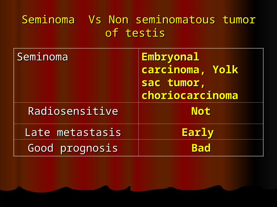
Seminoma Vs Non seminomatous tumor of Seminoma Vs Non seminomatous tumor of testis testis
Seminoma Seminoma Embryonal Embryonal carcinoma, Yolk carcinoma, Yolk sac tumor, sac tumor, choriocarcinomachoriocarcinoma
Radiosensitive Radiosensitive NotNot
Late metastasis Late metastasis Early Early
Good prognosis Good prognosis BadBad
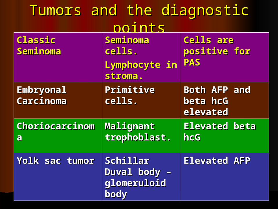
Tumors and the diagnostic Tumors and the diagnostic pointspoints
Classic Classic SeminomaSeminoma
Seminoma Seminoma cells.cells.
Lymphocyte Lymphocyte in stroma.in stroma.
Cells are Cells are positive for positive for PASPAS
Embryonal Embryonal CarcinomaCarcinoma
Primitive Primitive cells.cells.
Both AFP and Both AFP and beta hcG beta hcG elevatedelevated
ChoriocarcinomChoriocarcinomaa
Malignant Malignant trophoblast.trophoblast.
Elevated beta Elevated beta hcGhcG
Yolk sac tumorYolk sac tumor Schillar Duval Schillar Duval body – body – glomeruloid glomeruloid bodybody
Elevated AFP Elevated AFP
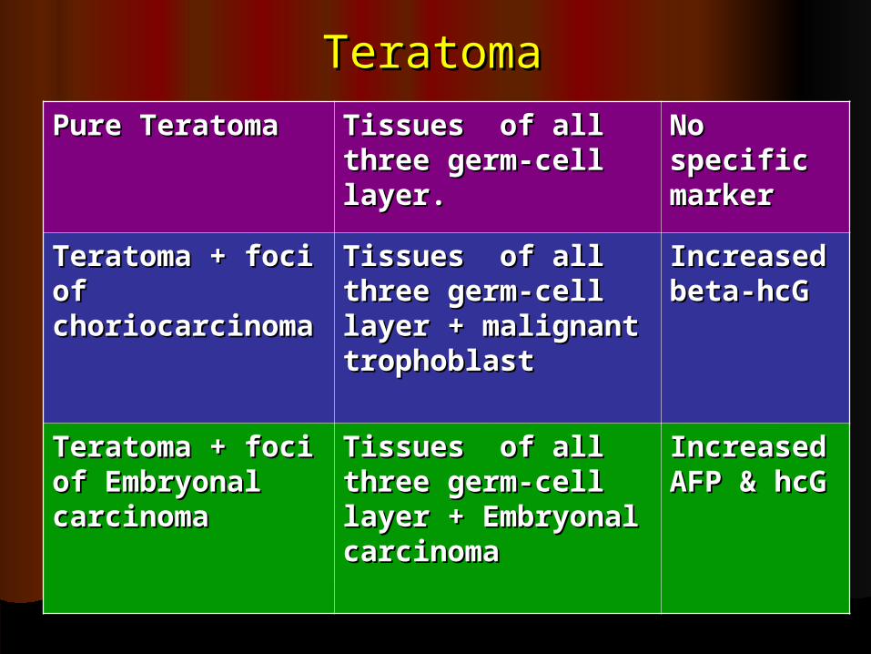
TeratomaTeratoma
Pure TeratomaPure Teratoma Tissues of all Tissues of all three germ-cell three germ-cell layer.layer.
No No specific specific markermarker
Teratoma + foci Teratoma + foci of of choriocarcinomachoriocarcinoma
Tissues of all Tissues of all three germ-cell three germ-cell layer + malignant layer + malignant trophoblasttrophoblast
Increased Increased beta-hcGbeta-hcG
Teratoma + foci Teratoma + foci of Embryonal of Embryonal carcinomacarcinoma
Tissues of all Tissues of all three germ-cell three germ-cell layer + Embryonal layer + Embryonal carcinomacarcinoma
Increased Increased AFP & hcG AFP & hcG
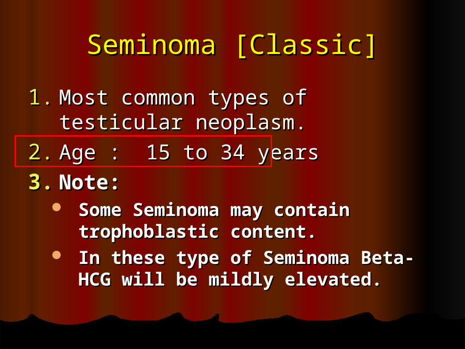
Seminoma [Classic]Seminoma [Classic]
1.1. Most common types of testicular Most common types of testicular neoplasm. neoplasm.
2.2. Age : 15 to 34 yearsAge : 15 to 34 years
3.3. Note:Note: Some Seminoma may contain Some Seminoma may contain
trophoblastic content.trophoblastic content. In these type of Seminoma Beta-In these type of Seminoma Beta-
HCG will be mildly elevated.HCG will be mildly elevated.
Variant of Variant of SeminomaSeminoma
Variant : Variant : Spermatocytic SeminomaSpermatocytic Seminoma In this case metastasis is rare, In this case metastasis is rare,
common in old people.common in old people. Three types of cell are seenThree types of cell are seen
1.1. large multinucleated cells, large multinucleated cells,
2.2. medium size cells and medium size cells and
3.3. small cells that reminiscent of small cells that reminiscent of spermatocytesspermatocytes
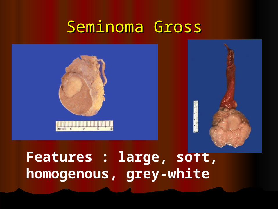
Seminoma Gross Seminoma Gross
Features : large, soft, homogenous, grey-white
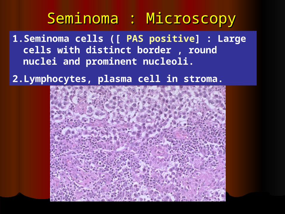
Seminoma : MicroscopySeminoma : Microscopy1.Seminoma cells ([ PAS positive] : Large
cells with distinct border , round nuclei and prominent nucleoli.
2.Lymphocytes, plasma cell in stroma.
Seminoma counterpart in Seminoma counterpart in OvaryOvary
DYSGERMINOMADYSGERMINOMA
Embryonal carcinomaEmbryonal carcinoma
Age : 20-30 years.Age : 20-30 years.FeaturesFeatures : : 1.1.Often multiple metastasis is Often multiple metastasis is
present at the time of diagnosis.present at the time of diagnosis.2.2.Often it contain other foci of Often it contain other foci of
Yolk sac tumor, teratoma Yolk sac tumor, teratoma and and Chorio-carcinomaChorio-carcinoma..
3.3.So both AFP and beta hcG will So both AFP and beta hcG will be elevated be elevated ( non specific) ( non specific)
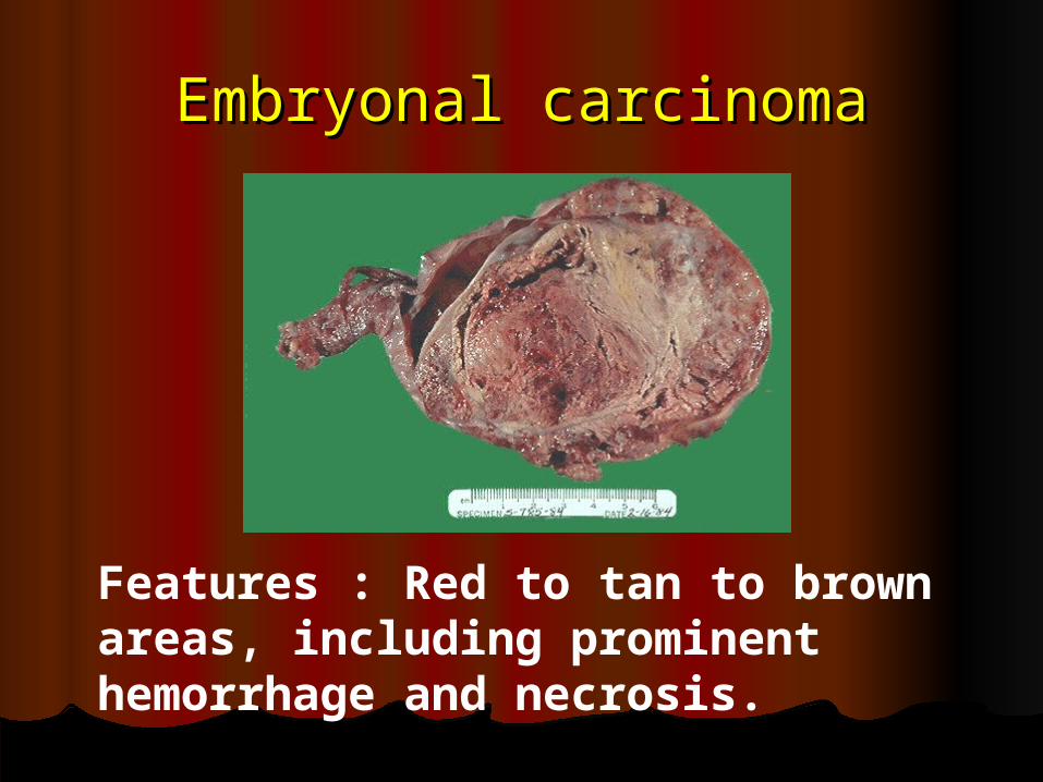
Embryonal carcinomaEmbryonal carcinoma
Features : Red to tan to brown areas, including prominent hemorrhage and necrosis.
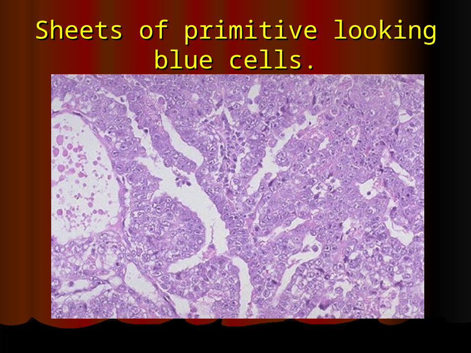
Sheets of primitive looking blue Sheets of primitive looking blue cells.cells.
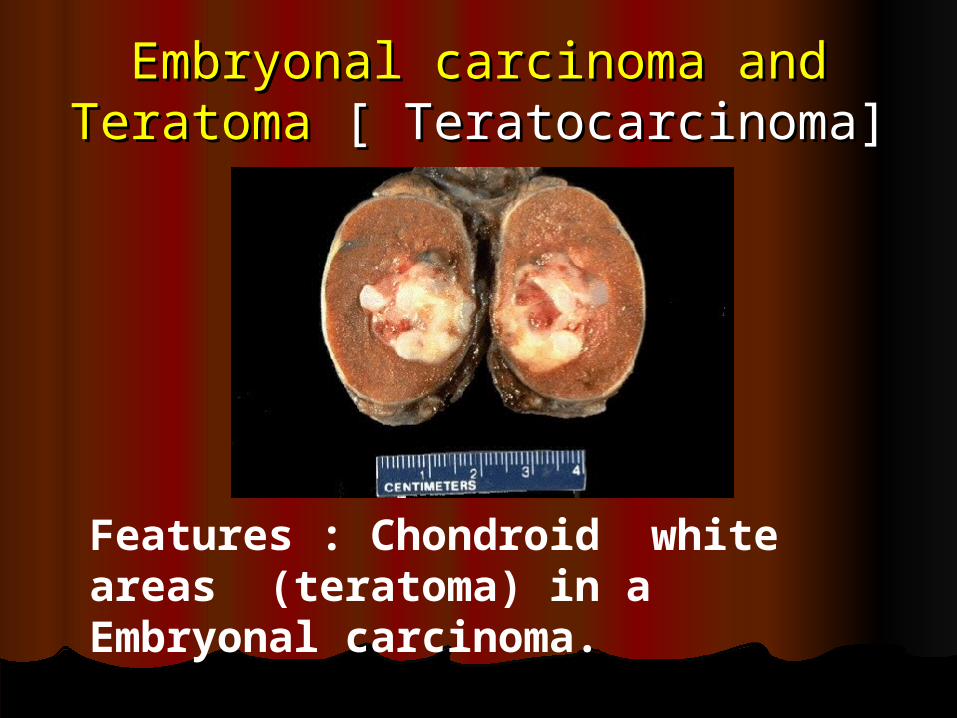
Embryonal carcinoma and Embryonal carcinoma and Teratoma Teratoma [ Teratocarcinoma][ Teratocarcinoma]
Features : Chondroid white areas (teratoma) in a Embryonal carcinoma.

Solid cystic mass in Ultra sound- Solid cystic mass in Ultra sound- TeratocarcinomaTeratocarcinoma
Teratoma in testisTeratoma in testis
Age = all agesAge = all agesAlmost always malignantAlmost always malignant ( unlike ( unlike
ovary – where it is usually ovary – where it is usually benign)benign)
Yolk sac tumor [ endodermal Yolk sac tumor [ endodermal sinus tumor]sinus tumor]
Age : 3 yearsAge : 3 yearsHistology : Presence of Histology : Presence of
Schiller –Duvall bodySchiller –Duvall body ( glomeruloid body)( glomeruloid body)
Specific Marker = Specific Marker = AFP AFP
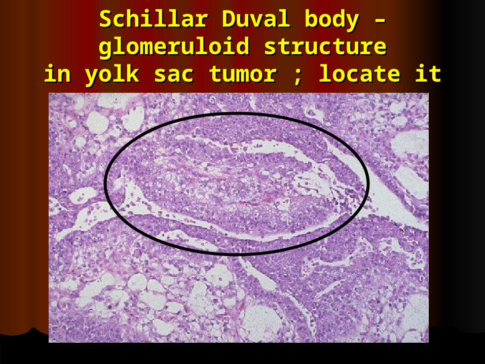
Schillar Duval body – Schillar Duval body – glomeruloid structureglomeruloid structure
in yolk sac tumor ; locate itin yolk sac tumor ; locate it
ChoriocarcinomaChoriocarcinoma
Age = 20 -30 Age = 20 -30 Pure Chorio carcinoma is rare in Pure Chorio carcinoma is rare in
testistestis.. It is alwaysIt is always mixed mixed with Teratoma, or with Teratoma, or
other tumor even with Seminoma.other tumor even with Seminoma. Histology : Malignant cyto and Histology : Malignant cyto and
syncytiotrophoblast without villous syncytiotrophoblast without villous formation.formation.
Specific Marker = beta hcGSpecific Marker = beta hcG
Mixed tumorMixed tumor
Add………….Add………….
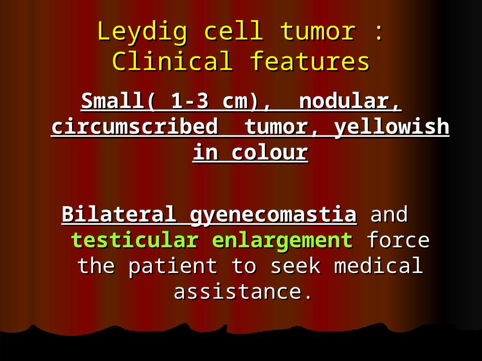
Leydig cell tumor : Clinical Leydig cell tumor : Clinical featuresfeatures
Small( 1-3 cm), nodular, Small( 1-3 cm), nodular, circumscribed tumor, yellowish circumscribed tumor, yellowish
in colourin colour
Bilateral gyenecomastiaBilateral gyenecomastia and and testicular enlargementtesticular enlargement force the force the patient to seek medical assistance. patient to seek medical assistance.
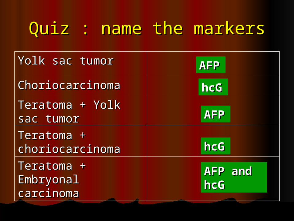
Quiz : name the markersQuiz : name the markers
Yolk sac tumorYolk sac tumor
ChoriocarcinomaChoriocarcinoma
Teratoma + Yolk sac Teratoma + Yolk sac tumortumor
Teratoma + Teratoma + choriocarcinomachoriocarcinoma
Teratoma + Teratoma + Embryonal carcinomaEmbryonal carcinoma
AFAFPPhchcGGAFAFPP
hchcGGAFP and AFP and hcGhcG
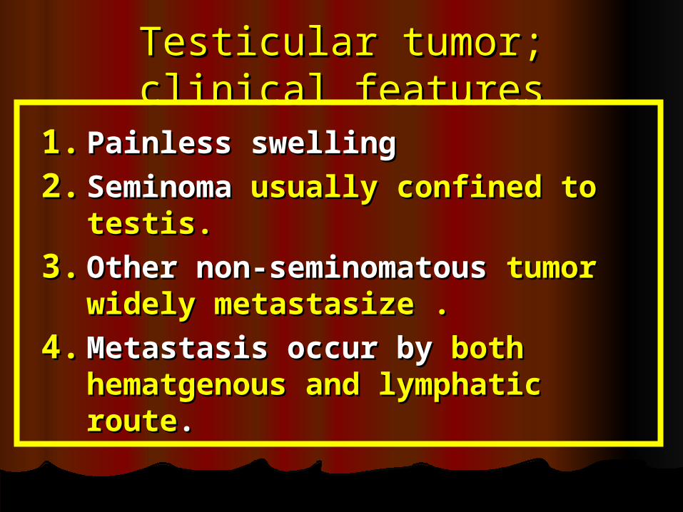
Testicular tumor; clinical Testicular tumor; clinical featuresfeatures
1.1. Painless swellingPainless swelling
2.2. Seminoma Seminoma usually confined to usually confined to testis.testis.
3.3. Other non-seminomatous Other non-seminomatous tumor tumor widely metastasize .widely metastasize .
4.4. Metastasis occur by Metastasis occur by both both hematgenous and lymphatic hematgenous and lymphatic routeroute..
Rest your eyes : Time for Rest your eyes : Time for Sexually transmitted diseaseSexually transmitted disease
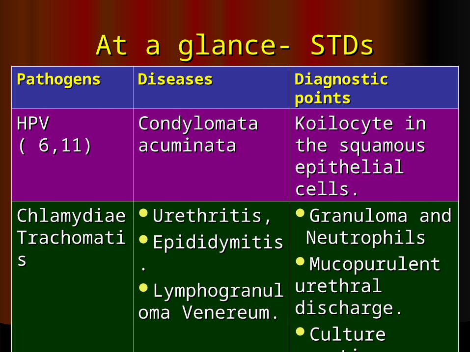
At a glance- STDsAt a glance- STDsPathogens Pathogens DiseasesDiseases Diagnostic pointsDiagnostic points
HPV ( 6,11)HPV ( 6,11) Condylomata Condylomata acuminataacuminata
Koilocyte in the Koilocyte in the squamous squamous epithelial cells.epithelial cells.
Chlamydiae Chlamydiae TrachomatiTrachomatiss
Urethritis, Urethritis, Epididymitis.Epididymitis.LymphogranulLymphogranuloma Venereum.oma Venereum.
Granuloma and Granuloma and Neutrophils NeutrophilsMucopurulent Mucopurulent urethral urethral discharge.discharge.Culture Culture negativenegative
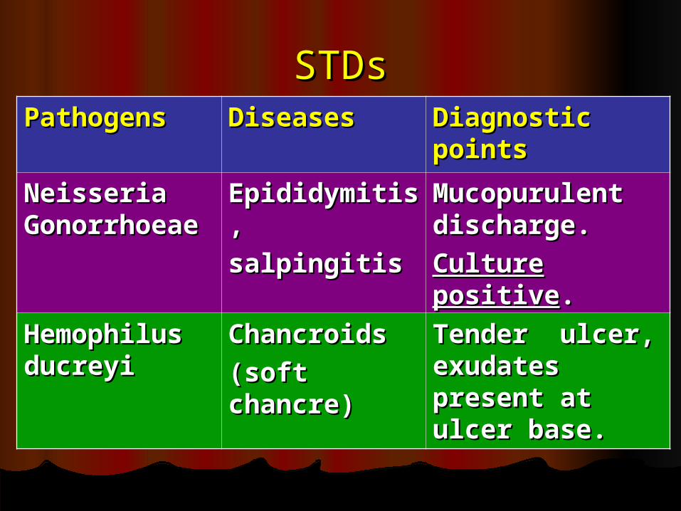
STDsSTDsPathogens Pathogens DiseasesDiseases Diagnostic Diagnostic
pointspoints
Neisseria Neisseria GonorrhoeaGonorrhoeaee
EpididymitisEpididymitis,,
salpingitis salpingitis
Mucopurulent Mucopurulent discharge.discharge.
Culture Culture positivepositive..
Hemophilus Hemophilus ducreyiducreyi
Chancroids Chancroids
(soft (soft chancre)chancre)
Tender ulcer, Tender ulcer, exudates exudates present at present at ulcer base.ulcer base.
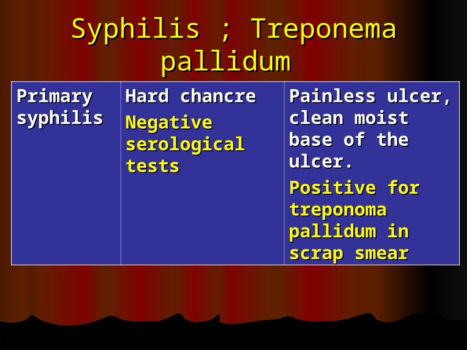
Syphilis ; Treponema Syphilis ; Treponema pallidum pallidum
Primary Primary syphilissyphilis
Hard chancreHard chancre
Negative Negative serological serological tests tests
Painless ulcer, Painless ulcer, clean moist clean moist base of the base of the ulcer. ulcer.
Positive for Positive for treponoma treponoma pallidum in pallidum in scrap smearscrap smear
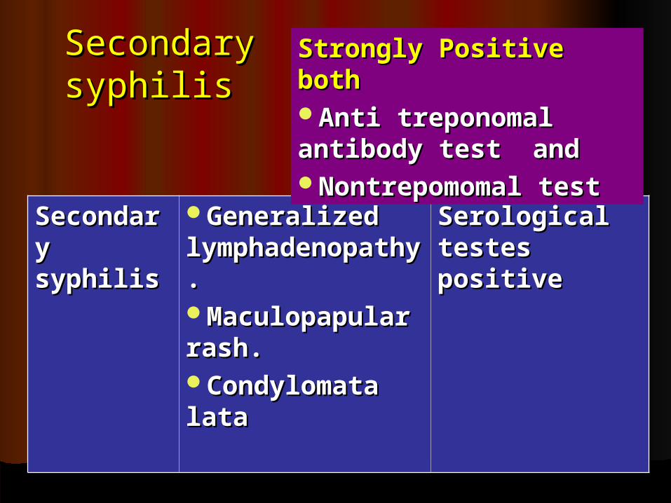
Secondary Secondary syphilissyphilis
SecondarSecondary y syphilissyphilis
Generalized Generalized lymphadenopatlymphadenopathy.hy.Maculopapular Maculopapular rash.rash.Condylomata Condylomata latalata
Serological Serological testes testes positivepositive
Strongly Positive both Strongly Positive both Anti treponomal Anti treponomal antibody test and antibody test and Nontrepomomal testNontrepomomal test
SyphilisSyphilis
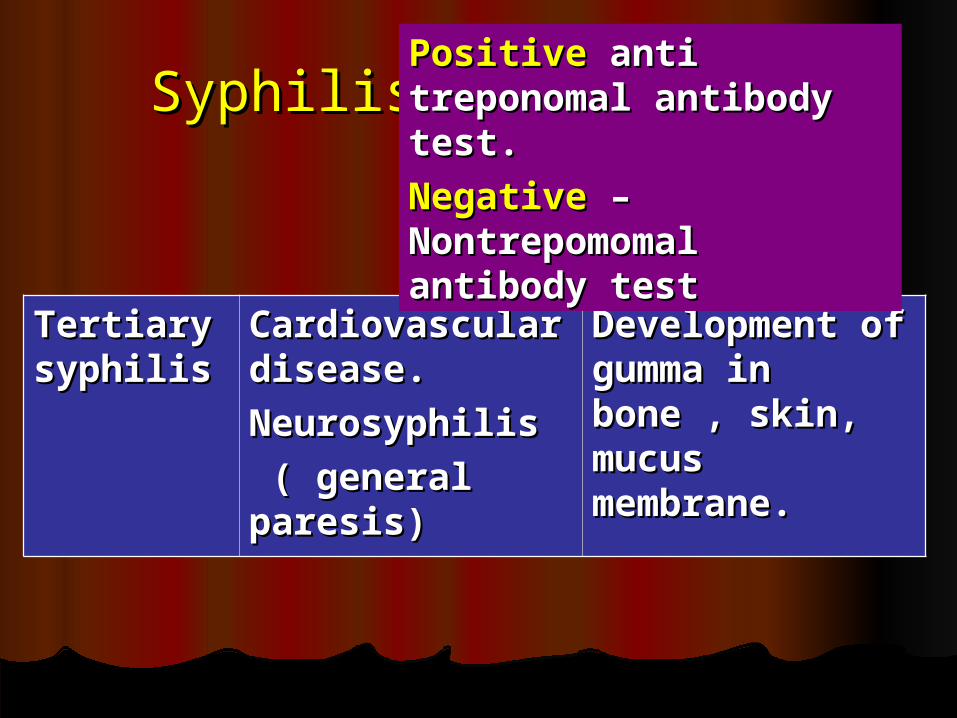
Tertiary Tertiary syphilissyphilis
Cardiovascular Cardiovascular disease.disease.
NeurosyphilisNeurosyphilis
( general ( general paresis)paresis)
Development Development of gumma in of gumma in bone , skin, bone , skin, mucus mucus membrane.membrane.
Positive Positive anti anti treponomal antibody treponomal antibody test.test.
Negative Negative – – Nontrepomomal Nontrepomomal antibody testantibody test
STDsSTDs
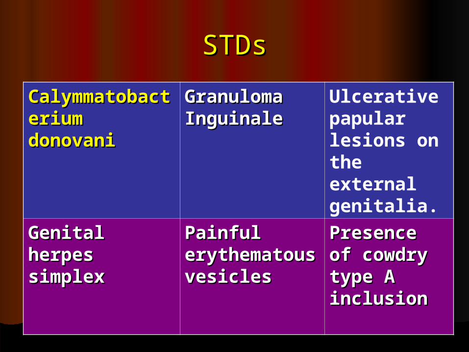
CalymmatobaCalymmatobacterium cterium donovanidonovani
Granuloma Granuloma InguinaleInguinale
Ulcerative papular lesions on the external genitalia.
Genital Genital herpes herpes simplexsimplex
Painful Painful erythematouerythematous vesicless vesicles
Presence Presence of cowdry of cowdry type A type A inclusioninclusion
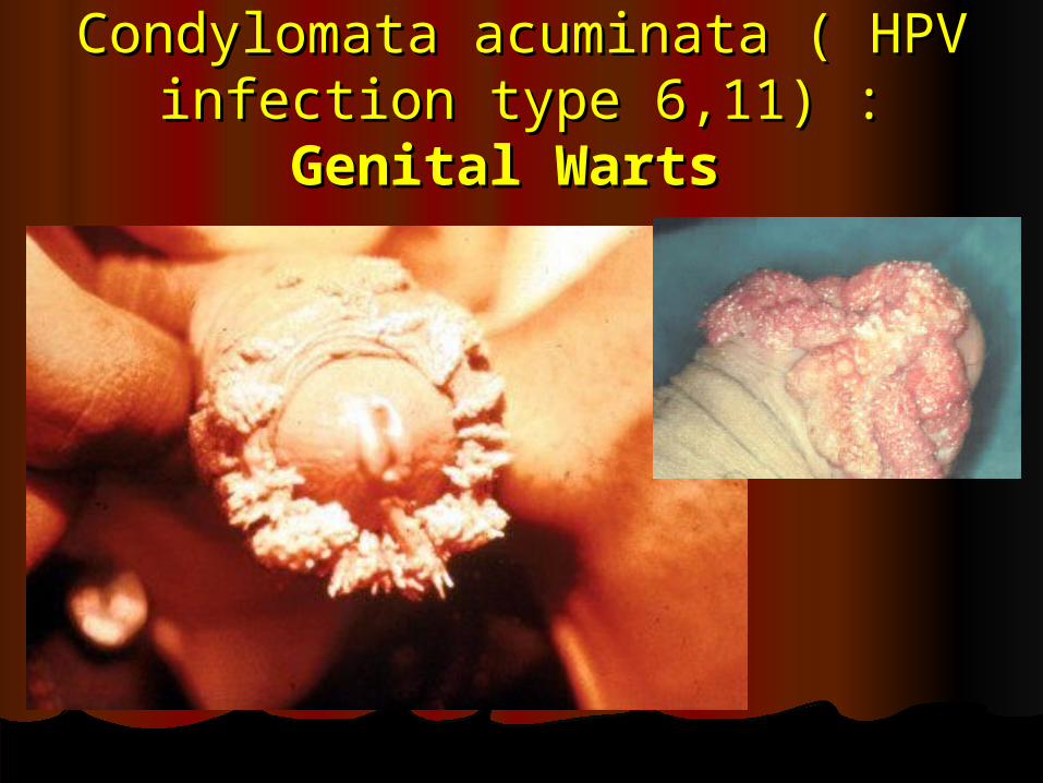
Condylomata acuminata ( HPV Condylomata acuminata ( HPV infection type 6,11) : infection type 6,11) : Genital Genital
Warts Warts
Gonorrhea ; clinical featuresGonorrhea ; clinical features
Male :Male : Epididymitis, may involve Epididymitis, may involve prostate.prostate.
Female : Female : salpingitis, infertilitysalpingitis, infertilityInfants ( during delivery) : Infants ( during delivery) :
Purulent infection of the eye : Purulent infection of the eye : Ophthalmia neonatorum).Ophthalmia neonatorum).
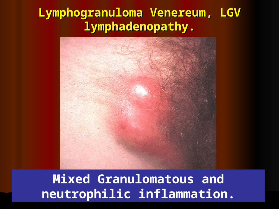
Lymphogranuloma Venereum, LGV Lymphogranuloma Venereum, LGV lymphadenopathy.lymphadenopathy.
Mixed Granulomatous and neutrophilic inflammation.
Diagnosis - Diagnosis - LGVLGV
Demonstration of organism in Biopsy Demonstration of organism in Biopsy section / exudates- section / exudates- in active lesionin active lesion. .
ELISA performed on serum.ELISA performed on serum.
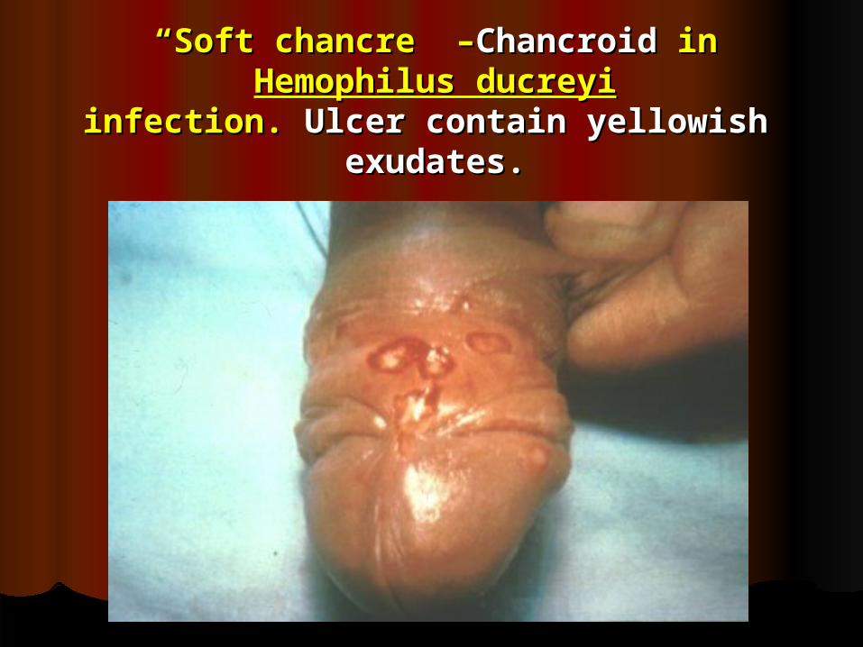
““Soft chancre” –Soft chancre” –ChancroidChancroid in in Hemophilus ducreyiHemophilus ducreyi
infection. infection. Ulcer contain yellowish Ulcer contain yellowish exudates.exudates.
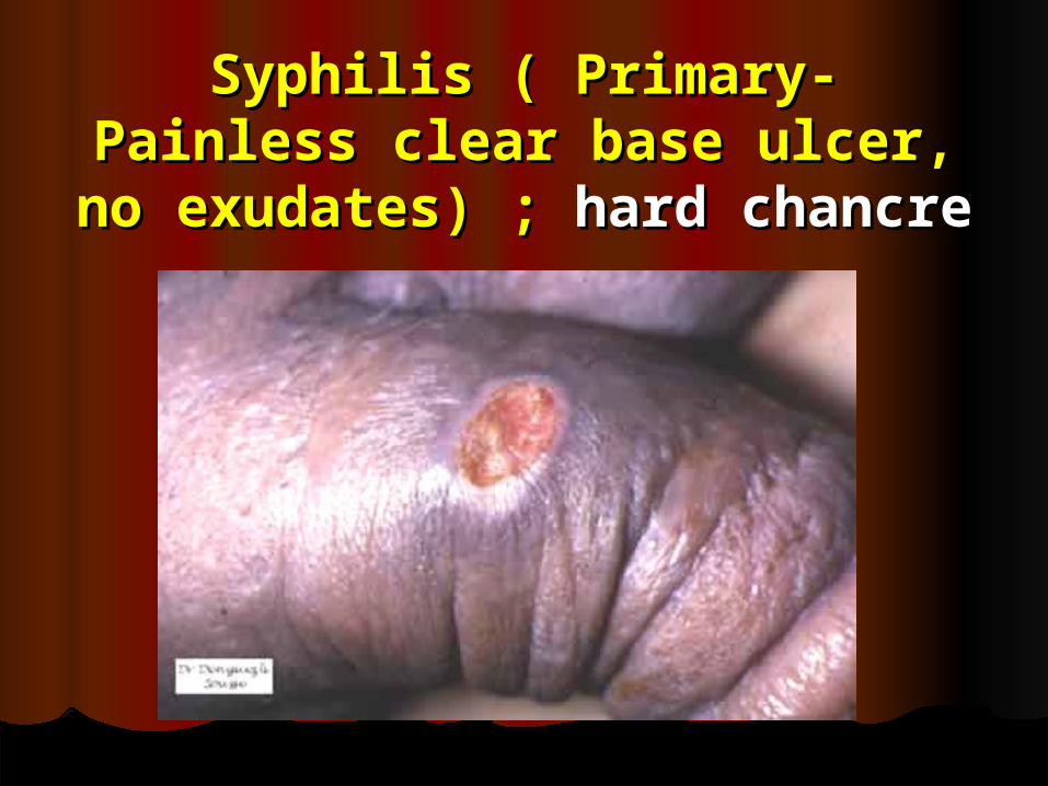
Syphilis ( Primary- Painless Syphilis ( Primary- Painless clear base ulcer, no clear base ulcer, no
exudates) ; exudates) ; hard chancrehard chancre
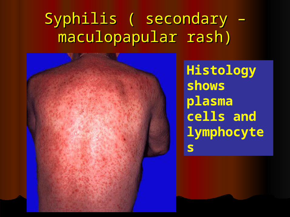
Syphilis ( secondary – Syphilis ( secondary – maculopapular rash)maculopapular rash)
Histology shows plasma cells and lymphocytes
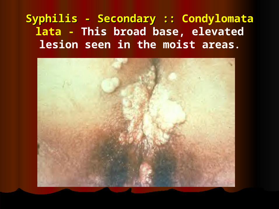
Syphilis - Secondary :: Syphilis - Secondary :: Condylomata lata - This broad base, elevated lesion
seen in the moist areas.
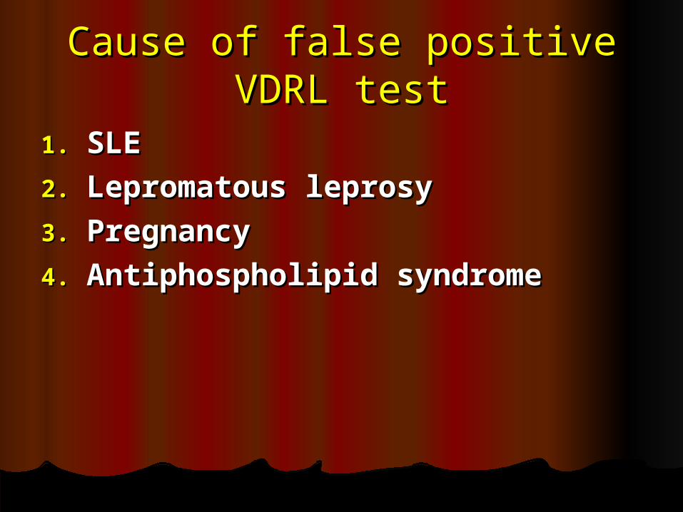
Cause of false positive VDRL Cause of false positive VDRL testtest
1.1. SLESLE
2.2. Lepromatous leprosyLepromatous leprosy
3.3. PregnancyPregnancy
4.4. Antiphospholipid syndromeAntiphospholipid syndrome
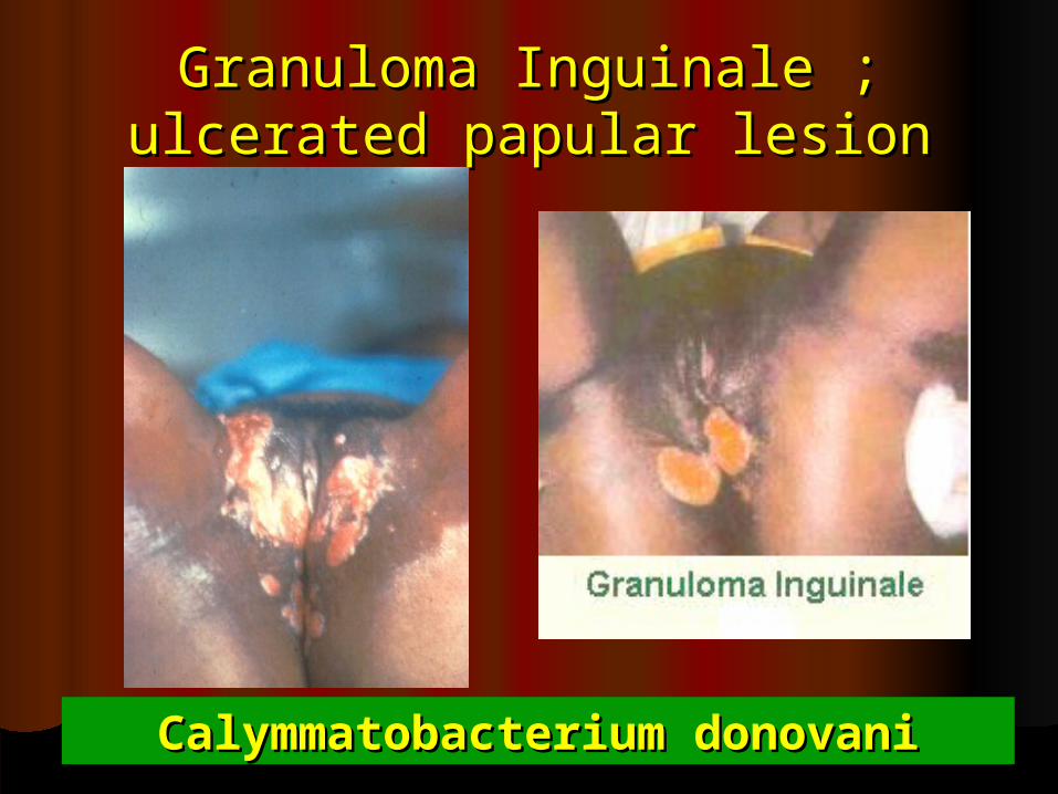
Granuloma Inguinale ; ulcerated Granuloma Inguinale ; ulcerated papular lesionpapular lesion
Calymmatobacterium donovaniCalymmatobacterium donovani
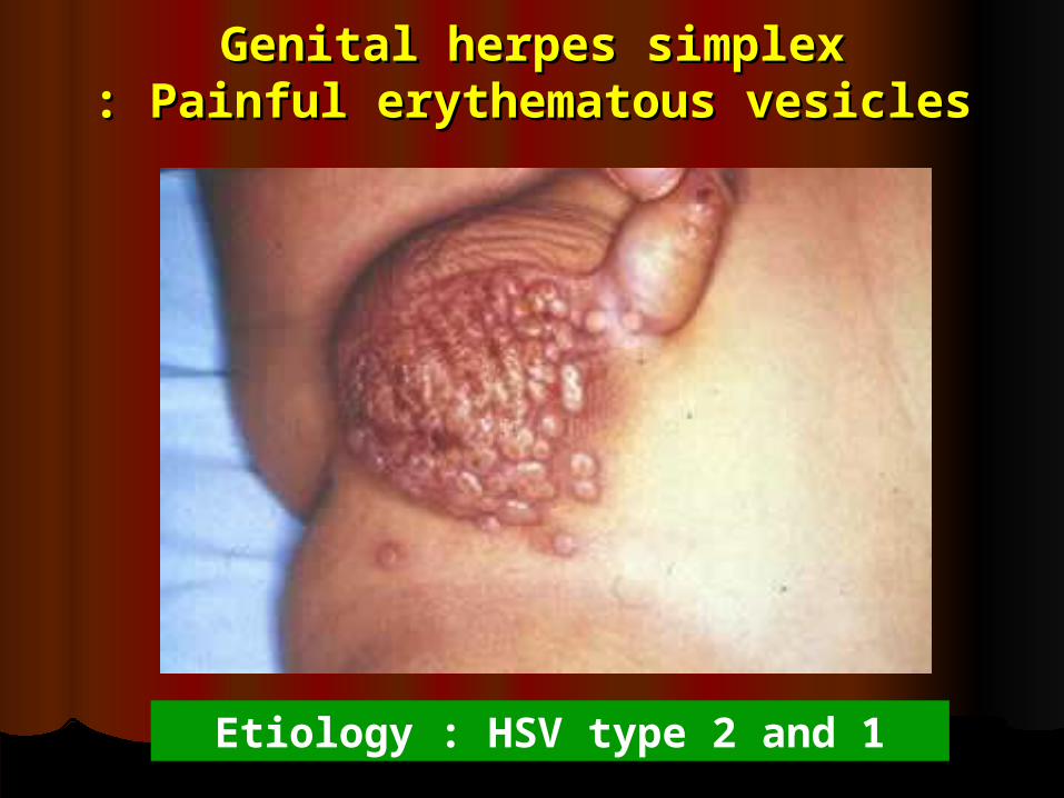
Genital herpes simplexGenital herpes simplex: Painful erythematous vesicles: Painful erythematous vesicles
Etiology : HSV type 2 and 1
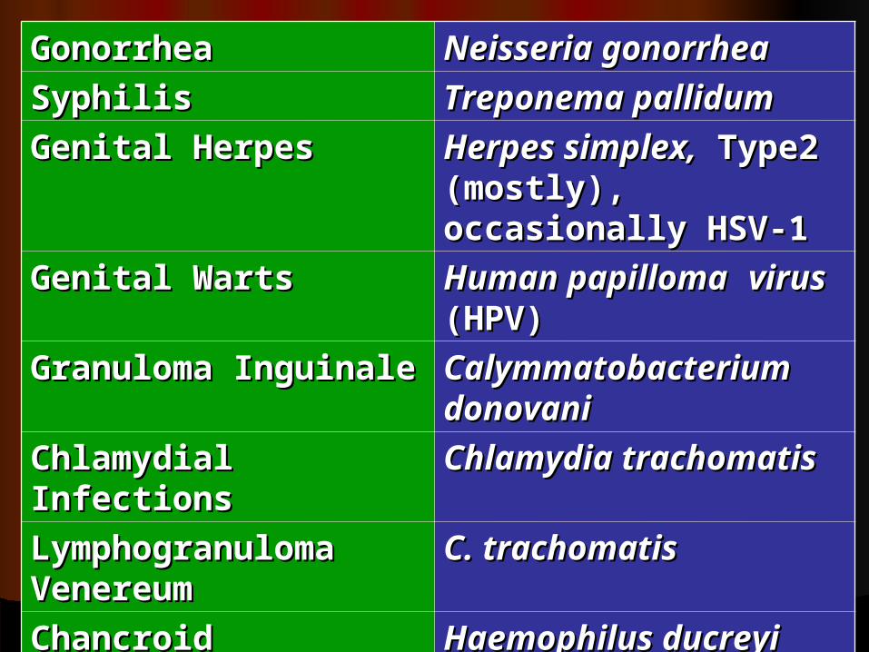
Gonorrhea Gonorrhea Neisseria gonorrheaNeisseria gonorrhea
Syphilis Syphilis Treponema pallidumTreponema pallidum
Genital Herpes Genital Herpes Herpes simplex,Herpes simplex, Type2 (mostly), Type2 (mostly), occasionally HSV-1 occasionally HSV-1
Genital Warts Genital Warts Human papillomaHuman papilloma virusvirus (HPV) (HPV)
Granuloma Granuloma Inguinale Inguinale
CalymmatobacteriuCalymmatobacterium donovanim donovani
Chlamydial Chlamydial Infections Infections
Chlamydia Chlamydia trachomatistrachomatis
Lymphogranuloma Lymphogranuloma Venereum Venereum
C. trachomatisC. trachomatis
Chancroid Chancroid Haemophilus ducreyiHaemophilus ducreyi

Best of luckBest of luck


![Does primary tumor localization has prognostic importance ... · groups; seminoma and non-seminoma. The group called pure seminoma constitutes approximately 60% of the whole GCT [1,2]](https://img.pdfslide.us/doc/110x75/5f3d5bded6321624f4620c6d/does-primary-tumor-localization-has-prognostic-importance-groups-seminoma-and.jpg)