Embed Size (px)
Citation preview
1
JPET-AR-2021-000590
Title page
A novel selective PKR inhibitor restores cognitive deficits and neurodegeneration
in Alzheimer’s disease experimental models
Authors: Matilde Lopez-Grancha, Patrick Bernardelli, Nicolas Moindrot, Elisabeth Genet,
Carine Vincent, Valerie Roudieres, AIain Krick, Jean-François Sabuco1, David Machnik,
Delphine Ibghi, Laurent Pradier, Veronique Taupin2
Affiliations:
Primary Lab of origin: Neurodegeneration Cluster, Rare and Neurologic Disease Research
TA, Sanofi R&D, 1 Av Pierre Brossolette, 91385 Chilly-Mazarin, France
Neurodegeneration Cluster, Rare and Neurologic Disease Research TA (MLG, NM, EG, CV,
VR, DI, LP), Integrated Drug Discovery (PB, JFS, DM), DMPK (AK), Sanofi R&D, 1 Av Pierre
Brossolette, 91385 Chilly-Mazarin, France
1 Current affiliation: Toxicology, Preclinical Safety, Global Operations France, Sanofi R&D,
371 Av Prof. Blayac, 34184 Montpellier, France, [email protected]
2 Current affiliation: Evotec ID, 1541, Av Marcel Merieux, 69280 Marcy l’Etoile, France,
This article has not been copyedited and formatted. The final version may differ from this version.JPET Fast Forward. Published on June 16, 2021 as DOI: 10.1124/jpet.121.000590
at ASPE
T Journals on M
arch 1, 2022jpet.aspetjournals.org
Dow
nloaded from
This article has not been copyedited and formatted. The final version may differ from this version.JPET Fast Forward. Published on June 16, 2021 as DOI: 10.1124/jpet.121.000590
at ASPE
T Journals on M
arch 1, 2022jpet.aspetjournals.org
Dow
nloaded from
This article has not been copyedited and formatted. The final version may differ from this version.JPET Fast Forward. Published on June 16, 2021 as DOI: 10.1124/jpet.121.000590
at ASPE
T Journals on M
arch 1, 2022jpet.aspetjournals.org
Dow
nloaded from
This article has not been copyedited and formatted. The final version may differ from this version.JPET Fast Forward. Published on June 16, 2021 as DOI: 10.1124/jpet.121.000590
at ASPE
T Journals on M
arch 1, 2022jpet.aspetjournals.org
Dow
nloaded from
This article has not been copyedited and formatted. The final version may differ from this version.JPET Fast Forward. Published on June 16, 2021 as DOI: 10.1124/jpet.121.000590
at ASPE
T Journals on M
arch 1, 2022jpet.aspetjournals.org
Dow
nloaded from
This article has not been copyedited and formatted. The final version may differ from this version.JPET Fast Forward. Published on June 16, 2021 as DOI: 10.1124/jpet.121.000590
at ASPE
T Journals on M
arch 1, 2022jpet.aspetjournals.org
Dow
nloaded from
This article has not been copyedited and formatted. The final version may differ from this version.JPET Fast Forward. Published on June 16, 2021 as DOI: 10.1124/jpet.121.000590
at ASPE
T Journals on M
arch 1, 2022jpet.aspetjournals.org
Dow
nloaded from
This article has not been copyedited and formatted. The final version may differ from this version.JPET Fast Forward. Published on June 16, 2021 as DOI: 10.1124/jpet.121.000590
at ASPE
T Journals on M
arch 1, 2022jpet.aspetjournals.org
Dow
nloaded from
This article has not been copyedited and formatted. The final version may differ from this version.JPET Fast Forward. Published on June 16, 2021 as DOI: 10.1124/jpet.121.000590
at ASPE
T Journals on M
arch 1, 2022jpet.aspetjournals.org
Dow
nloaded from
This article has not been copyedited and formatted. The final version may differ from this version.JPET Fast Forward. Published on June 16, 2021 as DOI: 10.1124/jpet.121.000590
at ASPE
T Journals on M
arch 1, 2022jpet.aspetjournals.org
Dow
nloaded from
This article has not been copyedited and formatted. The final version may differ from this version.JPET Fast Forward. Published on June 16, 2021 as DOI: 10.1124/jpet.121.000590
at ASPE
T Journals on M
arch 1, 2022jpet.aspetjournals.org
Dow
nloaded from
This article has not been copyedited and formatted. The final version may differ from this version.JPET Fast Forward. Published on June 16, 2021 as DOI: 10.1124/jpet.121.000590
at ASPE
T Journals on M
arch 1, 2022jpet.aspetjournals.org
Dow
nloaded from
This article has not been copyedited and formatted. The final version may differ from this version.JPET Fast Forward. Published on June 16, 2021 as DOI: 10.1124/jpet.121.000590
at ASPE
T Journals on M
arch 1, 2022jpet.aspetjournals.org
Dow
nloaded from
This article has not been copyedited and formatted. The final version may differ from this version.JPET Fast Forward. Published on June 16, 2021 as DOI: 10.1124/jpet.121.000590
at ASPE
T Journals on M
arch 1, 2022jpet.aspetjournals.org
Dow
nloaded from
This article has not been copyedited and formatted. The final version may differ from this version.JPET Fast Forward. Published on June 16, 2021 as DOI: 10.1124/jpet.121.000590
at ASPE
T Journals on M
arch 1, 2022jpet.aspetjournals.org
Dow
nloaded from
This article has not been copyedited and formatted. The final version may differ from this version.JPET Fast Forward. Published on June 16, 2021 as DOI: 10.1124/jpet.121.000590
at ASPE
T Journals on M
arch 1, 2022jpet.aspetjournals.org
Dow
nloaded from
This article has not been copyedited and formatted. The final version may differ from this version.JPET Fast Forward. Published on June 16, 2021 as DOI: 10.1124/jpet.121.000590
at ASPE
T Journals on M
arch 1, 2022jpet.aspetjournals.org
Dow
nloaded from
This article has not been copyedited and formatted. The final version may differ from this version.JPET Fast Forward. Published on June 16, 2021 as DOI: 10.1124/jpet.121.000590
at ASPE
T Journals on M
arch 1, 2022jpet.aspetjournals.org
Dow
nloaded from
2
Running Title Page
a) Running title: New potent PKR inhibitor for Alzheimer’s disease
b) Corresponding author:
Veronique Taupin, PhD
Sanofi R&D, 371 Av Prof. Blayac, 34184 Montpellier, France
Tel: + 33.4.99.77.40.60
Email: [email protected]
c) Number of text pages: 38
Number of tables: 3 (Supplemental: 3)
Number of figures: 5 (Supplemental: 5)
Number of references: 63
Number of words in abstract: 209
Number of words in introduction: 636
Number of words in discussion: 1302
d) Non-standard abbreviations:
AO: oligomers of amyloid-beta 42
eIF2: eukaryotic initiation factor 2
This article has not been copyedited and formatted. The final version may differ from this version.JPET Fast Forward. Published on June 16, 2021 as DOI: 10.1124/jpet.121.000590
at ASPE
T Journals on M
arch 1, 2022jpet.aspetjournals.org
Dow
nloaded from
3
KI: knock-in
MWM: Morris Water Maze
ORT: object recognition test
RI: recognition index
e) Recommended section: Drug discovery and translational medicine
This article has not been copyedited and formatted. The final version may differ from this version.JPET Fast Forward. Published on June 16, 2021 as DOI: 10.1124/jpet.121.000590
at ASPE
T Journals on M
arch 1, 2022jpet.aspetjournals.org
Dow
nloaded from
4
Abstract
In Alzheimer’s disease (AD), the double-strand RNA-dependent kinase PKR/EIF2AK2 is
activated in brain with increased phosphorylation of its substrate eukaryotic initiation factor
2 (eIF2). AD risk-promoting factors, such as ApoE4 allele or the accumulation of
neurotoxic amyloid-beta oligomers (AO), have been associated with activation of PKR-
dependent signalling. Here, we report the discovery of a novel potent and selective PKR
inhibitor (SAR439883) and demonstrate its neuroprotective pharmacological activity in AD
experimental models. In ApoE4 human replacement male mice, one-week oral treatment
with SAR439883 rescued short-term memory impairment in the spatial object recognition
test and dose-dependently reduced learning and memory deficits in the Barnes maze test.
Moreover, in AβO-injected male mice, a two-week administration of SAR439883 in diet
dose-dependently ameliorated the AβO-induced cognitive impairment in both Y-maze and
Morris Water Maze, prevented loss of synaptic proteins and reduced levels of the pro-
inflammatory cytokine IL-1β. In both mouse models, these effects were associated with a
dose-dependent inhibition of brain PKR activity as measured by both PKR occupancy and
partial lowering of peIF2levels. Our results provide evidence that selective
pharmacological inhibition of PKR by a small selective molecule can rescue memory deficits
and prevent neurodegeneration in animal models of AD-like pathology suggesting that
inhibition of PKR is a potential therapeutic approach for AD.
This article has not been copyedited and formatted. The final version may differ from this version.JPET Fast Forward. Published on June 16, 2021 as DOI: 10.1124/jpet.121.000590
at ASPE
T Journals on M
arch 1, 2022jpet.aspetjournals.org
Dow
nloaded from
5
Significance Statement
We report the identification of a new small molecule potent and selective PKR inhibitor that
can prevent cognitive deficits and neurodegeneration in Alzheimer’s disease (AD)
experimental models including a mouse model expressing the most prevalent AD genetic
risk factor ApoE4. With high potency and selectivity, this PKR inhibitor represents a unique
tool for investigating the physiological role of PKR and a starting point for developing new
drug candidates for AD.
This article has not been copyedited and formatted. The final version may differ from this version.JPET Fast Forward. Published on June 16, 2021 as DOI: 10.1124/jpet.121.000590
at ASPE
T Journals on M
arch 1, 2022jpet.aspetjournals.org
Dow
nloaded from
6
Introduction
The double-strand RNA-dependent kinase PKR (EIF2AK2) is one of the four kinases
phosphorylating the alpha subunit of eukaryotic initiation factor (eIF2α), thereby controlling
protein translation, in a global response known as the integrated stress response. The
phosphorylation of eIF2α at Ser51 is tightly regulated by these four kinases leading to
inhibition of general protein translation while favoring translation of a limited subset of
mRNAs including the transcription factor ATF4 (Lu et al. 2004, Vattem and Wek 2004).
PKR is activated in neurodegenerative diseases including Alzheimer’s disease (AD) as
observed with accumulation of phosphorylated PKR (pPKR), its activated form, in
degenerating neurons of human AD cortex and hippocampus compared to age-matched
controls (Chang et al., 2002a; Peel et al., 2003; Onuki et al., 2004; Page et al., 2006; Paquet
et al., 2012). Furthermore, pPKR is co-localized with phosphorylated eIF2α (peIF2α) in AD
brain and in cultured neurons, Aβ oligomers (AβO) increase levels of peIF2α (Mamada et al.,
2015; Chang et al., 2002b, Morel et al., 2009). In addition, levels of pPKR are increased in
CSF and in peripheral blood cells of AD patients compared to healthy controls (Paccalin et
al., 2006; Mouton-Liger et al., 2012; Hugon et al., 2017). pPKR has therefore been proposed
as potential biomarker for AD with high pPKR levels correlating with phosphorylated Tau as
well as predicting faster cognitive decline in newly diagnosed AD patients (Dumurgier et al.,
2013; Paquet, 2015).
ApoE4 allele, the highest genetic risk factor to develop AD (Kim et al., 2009) has also been
associated with PKR activation in human PBMC (Paccalin et al., 2006; Badia et al., 2013).
PKR expression is increased in lymphocytes from young ApoE4 carriers and associated with
subjective cognitive impairment (Badia et al., 2013). Consistent with these human data,
humanized knock-in (KI) ApoE4 mice, have increased peIF2α levels in hippocampus and
cortex associated with cognitive deficits compared to control ApoE3-KI mice (Segev et al.,
2013).
PKR/eIF2α pathway is involved in both memory formation/encoding and in response to
neuronal and inflammatory stress with ATF4 being critical for memory regulation (Costa-
This article has not been copyedited and formatted. The final version may differ from this version.JPET Fast Forward. Published on June 16, 2021 as DOI: 10.1124/jpet.121.000590
at ASPE
T Journals on M
arch 1, 2022jpet.aspetjournals.org
Dow
nloaded from
7
Mattioli et al., 2007, 2009; Chen et al., 2003, Buffington et al., 2014). In human ApoE4
carriers, ATF4 is overexpressed in post-mortem brains (Baleriola et al., 2014; Segev et al.,
2015). Conversely, enhanced long-term memory storage is observed in PKR-KO mice (Zhu
et al., 2011) and in eIF2-S51A mutant mice (Costa-Mattioli, 2007). PKR-KO mice were also
shown to be resistant to intracerebroventricular (i.c.v) injection of AβO. AβO-induced eIF2
inhibition and amnesic effect (Lourenco et al., 2013). In double mutant 5xFAD/PKR-KO
transgenic mice, spatial memory, synaptic alteration and brain inflammation were
ameliorated compared to 5xFAD mice (Tible et al., 2019).
Pharmacological inhibition or genetic inactivation of another eIF2 kinase, PERK, was
shown to improve cognitive functions (Zhu et al., 2016) but PERK-KO mice develop
pancreatic dysfunction that might represent serious liability for PERK inhibitors (Harding et
al., 2001). Pharmacological inhibition of PKR with the small molecule C16 was also shown to
enhance cognitive performance in wild type mice (Ingrand 2007; Stern 2013; Zhu 2011) and
to reverses deficits in ApoE4-Ki (Segev et al., 2015) but C16 lacks potency and selectivity
versus other eIF2AKs and other kinases (Chen et al., 2008). Our first-generation PKR
inhibitor was previously demonstrated to provide neuroprotection in a short-term model of
thiamine deficiency (Mouton-Liger et al., 2015).
We now report on a novel isoindolinone SAR439883 with improved selectivity as well as
drug-like properties and demonstrate its protective/restorative effects on cognitive deficits
and on synaptic loss in two AD-related animal models. SAR439883 is a potent and orally
bioavailable PKR inhibitor highly selective versus other eIF2AKs and kinases. Oral
administration of SAR439883 restores cognitive function in ApoE4-KI mice and protects from
cognitive deficits and neurodegeneration markers in AO i.c.v. injected mice. Our results
suggest that PKR selective inhibition represents a potential therapeutic treatment for AD.
This article has not been copyedited and formatted. The final version may differ from this version.JPET Fast Forward. Published on June 16, 2021 as DOI: 10.1124/jpet.121.000590
at ASPE
T Journals on M
arch 1, 2022jpet.aspetjournals.org
Dow
nloaded from

8
MATERIALS AND METHODS
Synthesis of SAR439883
SAR439883 synthetized by the medicinal chemistry department of Sanofi R&D following
medicinal chemistry optimization from an isoindolinone chemical series (see structure in Fig.
1A and chemical synthesis in Supplementary information provided on-line). SAR439883 was
tested through a high-throughput screening based on a biochemical assay measuring the
inhibitory effect of compound on human PKR-mediated phosphorylation of its substrate
eIF2α.
Biochemical assay for kinase activity
HTRF technology (Homogeneous Time Resolved Fluorescence, CisBio) was used to
determine compound activity on PKR.
First, PKR or PERK or GCN2, his-tagged eIF2α (Sanofi) as substrate, ds polyIC and ATP
(Sigma) as co-substrates, and MgCl2 (Sigma) are mixed for allowing phosphorylation of
eIF2α. Then, the reaction is stopped by addition of the HTRF reagents (Cisbio) diluted in
HTRF buffer containing EDTA. The phosphorylated form of eIF2α is titrated by the HTRF
reagents. The anti his-XL binds to the his-tag of eIF2α. The antiphospho-eIF2α-cryptate
binds to peIF2α. A signal at 665 nM is generated by energy transfer from the cryptate
(emission at 620 nm following laser excitation) to the XL when the two fluorophores are held
in proximity. The signal at 665 nm is thus proportional to the quantity of peIF2α produced
during the enzymatic reaction. Signals detected by HTRF are the fluorescence intensity at
both 665 nm and 620 nm. The HTRF signal corresponds to the ratio em665 nm/em620 nm.
Background signal corresponds to the ratio from control samples in which the enzyme
activity is fully inhibited by an excess of EDTA or is absent. Total signal corresponds to the
ratio from control samples in which enzyme is incubated with eIF2α substrate and co-
substrates (ATP, ds poly IC) in the absence of inhibitor compound.
Activity on Cdk9 was measured by binding (IMAP technology, Molecular Devices) revealed
by fluorescent polarization.
This article has not been copyedited and formatted. The final version may differ from this version.JPET Fast Forward. Published on June 16, 2021 as DOI: 10.1124/jpet.121.000590
at ASPE
T Journals on M
arch 1, 2022jpet.aspetjournals.org
Dow
nloaded from
9
For PKR compound, the concentration-inhibition curve and IC50 value (concentration giving
50% inhibition of enzymatic activity) are determined by non-linear regression analysis by
using Speed V2.0 software (developed by Sanofi).
Cellular assay for PKR
The assay for PKR activity is based on the use of an inducible HEK-293-FlpIn-TRex cell line
expressing either human or murine PKR. Cells are plated in 96-well plates (Greiner, µclear
Poly-D-lysine) at the density of 10 000cells/well in DMEM (Gibco Invitrogen) +10% FCS
(Gibco Invitrogen) and incubated at 5% CO2, 37°C. After doxycycline induction (50mg/ml, 16
h), the compound was added and incubated for 4 h with the cells that overexpress the
human recombinant PKR. At the end of the incubation, the cells were fixed with
formaldehyde 4% and permeabilized with PBS/0.2% Triton X-100 for immunostaining with
both rabbit anti-peIF2 (Biosource) and goat anti-eIF2 (Santacruz) antibodies. PKR activity
was determined by direct measurement of the phosphorylation of the native substrate eIF2.
PeIF2 level was determined by image analysis with InCell Analyzer 2200 (GE Healthcare).
The intensity of the fluorescence signal at Ex480nm/Em535 is proportional to the quantity of
peIF2 in cells. The intensity of the signal at Ex595/Em620 is proportional to the quantity of
total eIF2. Cell number was measured by counting the nuclei after DAPI staining. The
maximal response corresponds to the percentage of cells in the wells where the recombinant
PKR is maximally induced with doxycycline and without compound.
Cellular assay for PERK: activity on phospho-PERK (pPERK) was evaluated by measuring
the autophosphorylation of PERK in A549 cell line in which pPERK expression was induced
by treatment with thapsigargin.
Cellular assay for GCN2: activity on phospho-GCN2 (pGCN2) was evaluated by measuring
the autophosphorylation of GCN2 in A549 cell line in which pGCN2 expression was induced
by treatment with L-tryptophanol.
This article has not been copyedited and formatted. The final version may differ from this version.JPET Fast Forward. Published on June 16, 2021 as DOI: 10.1124/jpet.121.000590
at ASPE
T Journals on M
arch 1, 2022jpet.aspetjournals.org
Dow
nloaded from

10
For the tested compound, the concentration-inhibition curve and IC50 value (concentration
giving 50% inhibition of enzymatic activity) were determined by non-linear regression
analysis by using Speed V2.0 software (developed by Sanofi).
Primary neuronal cultures
Primary neuronal cultures were prepared from brain of 16-day-old mouse (OF1, Charles
River Laboratories, France) embryos by dissecting and then dissociating cerebral cortices.
Cells were plated in DMEM supplemented with N2 and B27 at a cell density of 4x10⁵
cells/ml in poly-D-lysine-coated wells of 96-well culture microplate. After 6 days in vitro,
neurons were incubated with AβO42 (5 µM) for 48 h. Cell treatment with drug-free medium
but supplemented with the corresponding DMSO concentration (i.e., 0.1%, same as for AβO
treatment) was run in parallel to test substance.
Quantification of caspase 3/7 enzymatic activity
Following experimental treatment, caspase-Glo 3/7 Assay kit solution (Promega Corporation
USA, G7790) were mixed to each well and then incubated for 4 hours at room temperature.
After incubation, the fluorescence of each sample was quantified using a Spectramax
Gemini plate reader. The measurement of fluorescence intensity is proportional to caspase
3/7 enzymatic activity.
Quantification of peIF2 levels in primary neuronal cultures
Phosphorylated eIF2 levels were determined by Western blot analysis of the ratio between
peIF2 and total eIF2 with the following antibodies: anti-phosphoSer51 eIF2 antibody
(3398S, D9G8) and anti-total eIF2 antibody (9722S) from Cell Signaling Technology.
Briefly, aliquots of cell extracts (20 µg of protein) were separated by 4–12% gradients
sodium dodecyl sulphate-polyacrylamide gel electrophoresis (SDS-PAGE) and transferred
onto polyvinylidene fluoride membranes (NP0336, InvitroGen). The membranes were probed
with the following antibodies: rabbit anti-eIF2α and rabbit anti-peIF2α from Cell Signaling;
Horseradish peroxidase-conjugated rabbit or mouse secondary antibodies were used
(Promega) and after extensive washing, the immunoreactive bands were detected by
This article has not been copyedited and formatted. The final version may differ from this version.JPET Fast Forward. Published on June 16, 2021 as DOI: 10.1124/jpet.121.000590
at ASPE
T Journals on M
arch 1, 2022jpet.aspetjournals.org
Dow
nloaded from

11
enhanced chemiluminescence (ECL Select Amersham RPN2235). Image acquisition and
analysis was performed with the Amersham imager S600 and the Multi Gauge software.
Animals
Experiments were performed in our AAALAC-accredited facility or at SynAging (Contract
Research Organisation, France) in full compliance with standards for the care and use of
laboratory animals, according to French and European Community (Directive 2010/63/EU)
legislation. All procedures were approved by the local Animal Ethic Committees and the
French Ministry for Research. All animal experiments were designed with a commitment to
refinement, reduction, and replacement, minimizing the number of mice and suffering via
emphasis on human end points, while using biostatistical advice for optimization of mouse
number.
In order to reduce the variability introduced by sex factors such as the hormonal fluctuation
that occurs during the estrous cycle (Meziane et al 2007) and based on cognitive and
metabolic differences between male and female ApoE3 and ApoE4-KI mice obtained in our
labs (data not shown) only male mice were used in these studies.
Animals (housed 4 to 5 per cage) were kept in a pathogen-free facility at a constant
temperature of 22±2°C and humidity (50±10%) on a 12 h light/dark cycle (lights on at 7 am)
with ad libitum access to food and water except during the tests. All tests were conducted
during the light phase at roughly the same time each day to minimize variability in
performance due to time of day (between 9 am and 4 pm) except otherwise specified. For
the behavioral tests, the mice were brought to the experimental room for at least 30 min
acclimation prior to testing. The animals were randomized to the treatment groups according
to their body weight, the timing of test sessions and the different enclosures used in the
spatial recognition test. For each experiment, the mice were assigned identification numbers
so that the experimenters were blinded to the treatment conditions (genotype, treatment,
A or Vehicle i.c.v. stereotaxic injection) as well as during cognitive testing and scoring.
15 homozygous human ApoE3 and 79 male homozygous human ApoE4 targeted
replacement mice (catalogue numbers 001548 and 001549, Taconic Farm) were used for
This article has not been copyedited and formatted. The final version may differ from this version.JPET Fast Forward. Published on June 16, 2021 as DOI: 10.1124/jpet.121.000590
at ASPE
T Journals on M
arch 1, 2022jpet.aspetjournals.org
Dow
nloaded from
12
evaluating the effect of our PKR inhibitor on short and long-term memory. ApoE4 human
targeted replacement mice (ApoE4-KI) express the human ApoE4 isoform under the control
of endogenous murine ApoE regulatory sequences (Sullivan et al. 1997) while mouse ApoE
has been deleted. C57B6/J mice obtained from Charles River were also used. For the
Barnes maze test, PKR inhibitor SAR439883 (base form) was given BID by gavage at 10
and 30 mg/kg of body weight in 0.6% methylcellulose/0.5% Tween-80 for seven days (first
administration in the mornings 1 h before the training and the second one 6 h later). On day
8, mice received the last administration 1 h before the probe test and were sacrificed
immediately after. For spatial object recognition, 5.5-month-old mice were treated for seven
days with vehicle or SAR439883 incorporated in the diet (Ssniff Spezialdiäten GmbH) at two
concentrations (0.1% and 0.3%). Considering the exposure in the plasma after a single oral
administration (data not shown), 0.1% SAR439883 in the diet was calculated to be
bioequivalent to a 10 mg/kg single oral dose. Testing occurred on day 7.
AO i.c.v. injection model
Amyloid-β1-42 (A42) was obtained from Bachem (H1368, batch number 1052301). The
stable A42 oligomers were prepared according to SynAging protocol (Garcia et al., 2010).
The oligomeric preparation contains a mixture of stable trimers and tetramers ofA42 as
well as monomeric forms of the peptides. Oligomer preparation was previously characterized
in terms of oligomer composition and in vitro neurotoxicity by SynAging and called as AβO.
Global experimental design is illustrated in Fig. 4A. Briefly, 73 C57BI6/J male mice (3-month-
old) mice were fed with the diet, either control diet (vehicle Ssniff Spezialdiäten GmbH) or
diet containing the PKR inhibitor at 3 different concentrations (0.03%, 0.1%, 0.3%), 3 days
before the induction of the disease. At Day 0, mice received, under anaesthesia, a single
unilateral i.c.v. injection of vehicle or AβO (50 pmol/1 µl) into the right lateral ventricle.
Four days after disease induction, spatial working memory was assessed using the Y-maze
test. From days +3 to days +14, learning capacities and long-term memory were investigated
in the MWM assay. Animals were sacrificed at day +15 and tissues were prepared for further
This article has not been copyedited and formatted. The final version may differ from this version.JPET Fast Forward. Published on June 16, 2021 as DOI: 10.1124/jpet.121.000590
at ASPE
T Journals on M
arch 1, 2022jpet.aspetjournals.org
Dow
nloaded from
13
ex-vivo analyses. During the entire protocol, animals were weighted every other day, from
day -10 to day+15.
Behavioral tests
Y-maze and Morris Water Maze
Immediate spatial working memory performance was assessed by recording spontaneous
alternation behavior in a Y-maze. Spontaneous alternation is a natural tendency of the
animals of numerous species including rodents to alternate their response when facing
identical and repeated choices. Its rating, in a Y maze, allows the evaluation of spatial
orientation capabilities (Hughes, 2004). Animal performances in mazes are related to the
integrity of hippocampus and spatial memory function (Means et al., 1971, Roberts et al.,
1962). The maze is made of opaque Plexiglas and each of the three arms is 40 cm long, 16
cm high, 9 cm wide and positioned at equal angles. The apparatus was placed in a
homogeneously lit test room to obtain 15 lux in all arms as well as in the central zone. Mice
are placed in the middle of one arm and are allowed to explore the maze freely during 5 min
sessions. The series of arm entries are video recorded (Smart v3.0 software, Bioseb). An
arm entry is considered complete when the hind paws of the mouse are completely placed in
the arm. Alternation is defined as successive entries into the 3 arms on overlapping triplet
sets. The percentage alternation is calculated as the ratio of actual (total alternations) to
possible alternations (defined as the number of arm entries minus two), multiplied by 100.
Locomotor activity was also recorded and evaluated by monitoring average speed and total
distance. Mice were discarded if they do not perform the minimum of twelve arm entries or if
they exhibit aberrant behaviors (e.g., mice follow the wall or presenting anxious behaviors).
Spatial learning capabilities and long-term memory were further investigated using the MWM
as described previously (Garcia et al., 2010). The experimental apparatus consists of a
circular white opaque plastic water tank (diameter, 90cm; height, 50cm) containing water
(21°C) to a depth of 25 cm. A white artificial colorant (Lytron) is spread over the water
surface to camouflage the escape platform (5 cm x 5 cm) made of white plastic and covered
with a wire mesh to ensure a firm grip. The pool was placed in a test room homogeneously lit
This article has not been copyedited and formatted. The final version may differ from this version.JPET Fast Forward. Published on June 16, 2021 as DOI: 10.1124/jpet.121.000590
at ASPE
T Journals on M
arch 1, 2022jpet.aspetjournals.org
Dow
nloaded from
14
at 100 lux. The swimming paths, swimming distance, swimming speed and thigmotaxis were
recorded using a video tracking system (Smart v3.0 software, Bioseb.) The MWM assay
consists of 3 different steps described as followed: Habituation (visible platform, no visual
cues) - Navigation to a visible platform was carried out before place-navigation to evaluate
visual and motor abilities of mice. Mice were submitted to 4 trials (two trials in the morning
and two trials in the afternoon) of 60 s each per day, during 2 consecutive days, with an
inter-trial interval of at least 1 h. Once mice have found the platform, they were left alone on
the platform for an additional time of 30 s. There were no additional maze cues in the room.
The platform position and starting points were randomly distributed over all 4 quadrants of
the pool. Mice that failed to find the platform after 60 s were guided to its location and placed
on it for 30 s. After removal from the pool, mice were manually dried with a terrycloth towel
and placed in their home cage. Next, memory-acquisition (learning trials with hidden
platform, visual cues) was performed during 5 consecutive days. Several prominent visual
cues on the wall near the rim of the pool were added. The hidden platform was submerged
1cm below the water surface and placed at the midpoint of one quadrant. Mice were
submitted to 4 trials of 60 s per day, with an inter-trial interval of at least 1 h. The mice were
allowed to swim freely for 60 s, left alone for an additional 30 s on the hidden platform and
then returned to the home cage during the inter-trial interval. Start positions (set at the
border between quadrants) were randomly selected for each animal. In each trial, the time
required to escape onto the hidden platform was recorded. Mice failing to find the platform
within 60 s were placed on the platform for 30 s before returning to their home cage.
Memory-retention test (probe trial) was performed three days after the last training
session. The platform was removed, and each animal was allowed a free 60 s swim. During
the probe trial, the time spent in the target quadrant, the number of crossings over the
original platform point, and the time required for the first crossing over were registered and
monitored by video tracking. In habituation, the mean latency and swim speed for the
second day will be calculated for each mouse. Mice with 2 standard deviations above the
group mean will be excluded, as this may be indicative of motor or visual impairments. Mice
This article has not been copyedited and formatted. The final version may differ from this version.JPET Fast Forward. Published on June 16, 2021 as DOI: 10.1124/jpet.121.000590
at ASPE
T Journals on M
arch 1, 2022jpet.aspetjournals.org
Dow
nloaded from
15
exhibiting aberrant behavior, such as cork-screw swimming or floating most of the time will
be also discarded.
Spatial Object Recognition Test (ORT)
This test relies on rodents' natural proactivity for exploring novelty (Ennaceur et al., 1988)
and was adapted for use in mice and performed as previously described (Delay-Goyet et al.,
2016). Accordingly, two weeks before the start of the study, mice were housed individually in
an enriched environment. On day 1 and 2, mice were allowed to become familiar with the
experimental environment twice a day for 10 min. It consisted of 4 PVC enclosures (59 x 59
x 30 cm height) with four black walls, a white floor and a video camera positioned 160 cm
above the bench. The arenas were uniformly lit (30 lux). On day 3, mice were placed in the
test enclosure in the presence of two identical objects placed in diagonal. Time spent
exploring each object during the 10 min was recorded (exploration was defined as the
animal having its head within 2 cm of the object while looking at, sniffing, or touching it).
After a forgetting interval of 1 h, mice were placed back in the enclosure (recall session) for
10 min with one of the objects (A) in the same location as before (familiar), and the other
object (B) in a novel location. Ambient cues in the room served as place references. Time
spent exploring the familiar and novel location (in seconds) was recorded. A recognition
index was calculated as follows: 100 x Time for object B / (Time for object A + Time for
Object B) for the ten minutes of the recall phase. For a short-term forgetting delay, during the
recall session, normal mice spent more time exploring the novel location of the object)
compared to the familiar one. That reflects a remembering of the familiar location.
The different objects were counterbalanced and were used equally as old and novel objects.
Objects were cleaned with 70% ethanol between phases, to eliminate odor cues. During
both, training and testing mice should explore at least 2 s each object. First exploration
should be done before 6 min otherwise, the mice will be excluded. N=12 was the sample
size calculated to detect an absolute difference of at least 10% with 90% or 75% power
when the variability is median and high, respectively.
Barnes Test
This article has not been copyedited and formatted. The final version may differ from this version.JPET Fast Forward. Published on June 16, 2021 as DOI: 10.1124/jpet.121.000590
at ASPE
T Journals on M
arch 1, 2022jpet.aspetjournals.org
Dow
nloaded from
16
The Barnes maze task was employed to test spatial memory as described previously
(Barnes 1979). It consisted of a white circular polyethylene platform 92 cm in diameter with
20 holes measuring 5 cm in diameter evenly spaced around the perimeter (2 cm from the
edge) of an elevated (70 cm above the floor) maze. One of the holes led to a black Plexiglas
escape box (5 x 5 x 11 cm) filled with sawdust. The maze was illuminated by overhead
fluorescent white room lighting (400 lux) and surrounded by white walls, which contained
spatial cues (posters with different figures). Mice were trained to locate the escape box
hidden underneath one of 20 holes. The location of the escape hole remains constant
throughout the training sessions. To familiarize mice with the maze and the existence of the
escape hole, they were subjected to a pretraining session (identical to the training sessions).
At the beginning of the trial to prevent orientation to the target, the mouse was placed in the
middle of the maze under a start chamber (a cylinder black box, 12 cm in diameter), and a
buzzer (80 dB) was turned on. After 10 s, the chamber was lifted and the mouse was gently
guided by the experimenter to the escape hole, the buzzer was turned off, and the mouse
remained into the box for 60 s. During the acquisition trials, the animals were allowed to
freely explore the maze and used the distal cues, to localize and to enter the escape hole.
Mice were given four training trials per day with a 15 min intertrial interval over 5 days. Each
trial takes 3 min long or when the mouse enters the escape box, whatever it comes first. If a
mouse did not enter the escape hole within 3 min, it was gently pulled by the experimenter to
the escape box and allowed to stay there for 60 s. A 70% ethanol solution was used to wipe
clean the platform after every trial and the escape box after each session. Seventy-two
hours later mice were given a 90 s probe trial transfer test without the escape hole. During
this trial all the holes are closed and there is no escape box. Trials were recorded using a
camera mounted above the maze and animals’ movements were tracked and analyzed
using a video tracking system (Viewpoint). Performance was assessed by latency to reach
the virtual escape hole and time spent in the escape hole zone. Mice not moving during the
90 s of the probe test were excluded from the analysis. The sample size N=12 by group of
This article has not been copyedited and formatted. The final version may differ from this version.JPET Fast Forward. Published on June 16, 2021 as DOI: 10.1124/jpet.121.000590
at ASPE
T Journals on M
arch 1, 2022jpet.aspetjournals.org
Dow
nloaded from

17
ApoE4 enables to show a difference at 72h of at least 50% for the parameter Time spent in
the target quadrant with a power of 82%.
Quantification of SAR439883 in blood and brain tissues
At the completion of the experiments, mice were anesthetized using a mixture of 100 mg
ketamine plus 10mg/kg xylazine and blood samples were collected by cardiac puncture into
Sarstedt Lithium-Heparin gel tubes. After centrifugation (1500–2000g for 10 min at 4°C)
plasma samples were frozen in microtubes and stored at −80°C. Then, brains were
removed. Hippocampi, cortex, pons and cerebellum were collected and stored at −80°C until
used for biochemical, RNA, or pharmacokinetics (PK) analyses. For the quantification of
SAR439883 levels, after the addition of the precipitant solution (acetonitrile), SAR439883
was quantified in both plasma and pons/cerebellum samples by liquid chromatography-mass
spectrometry/mass spectrometry.
Preparation of hippocampal and brain cortical homogenates
Hippocampi or brain cortices were homogenized in cold RIPA buffer (Cell Signaling, #9806)
containing an anti-protease cocktail (Roche, #05056489001), 1 mM PMSF and 1 mM
sodium orthovanadate, added freshly, just before use. Samples were vortexed, kept on ice
for 10 min, and exposed to 3 freeze-thaw cycles (liquid nitrogen). Lysates were centrifuged
at 800g for 15 min at 4°C. The supernatant was dispensed into aliquots and stored at -80°C
for later analysis. Total protein content was assessed using the BCA assay. Data were
recorded using a FLUOSTAR-Omega plate reader (BMG-LABTECH) and expressed as mg
protein per ml.
KiNativ™ binding selectivity assay
Target occupancy in cell lysates (PC3 cell line) or in brain homogenates after in vivo
treatment with SAR439883 was performed using KiNativ™ platform to quantitatively profile
responses of our PKR inhibitor against PKR and all kinases detectable in brain extract.
KiNativ™ is based on biotinylated acyl phosphates of ATP and ADP that irreversibly react
with protein kinases on conserved lysine residues in the ATP-binding pocket. The ActivX
This article has not been copyedited and formatted. The final version may differ from this version.JPET Fast Forward. Published on June 16, 2021 as DOI: 10.1124/jpet.121.000590
at ASPE
T Journals on M
arch 1, 2022jpet.aspetjournals.org
Dow
nloaded from
18
unique chemical probes combined with quantitative mass spectrometry of pulled-down
proteins yield relative quantification of kinases detectable in sample. Presence of a catalytic
site kinase inhibitor in a sample prevents ActivX probe binding and can be differentially
quantified compared to samples without inhibitor. For each kinase detected, the occupancy
by the inhibitor is reported as percentage vs sample without inhibitor as described by
KiNativ™. PKR was detectable in brain samples as well as over 200 other ATP/ADP binding
kinases.
RT-PCR on mouse brain tissues
Mouse brain cortical tissues were homogenized using Precellys. Total RNA extraction from
the hippocampus was performed using the RNAeasy Tissue mini kit (QIAGEN) according to
the manufacturer’s recommendations. Reverse transcription was performed using High-
Capacity cDNA Reverse Transcription kit (Applied Biosystems). Real-time PCR was
performed with TaqMan universal PCR master mix (Applied Biosystems) using the cDNA
and gene specific TaqMan reactions (Applied Biosystems). Real-time PCRs were performed
in triplicates using the thermocycler Quant Studio3 (Applied Biosystems) under the following
conditions: 50°C for 2 min, 95°C for 10 min, and 40 cycles of 95°C for 15 s and 60°C for 1
min. Threshold cycle (Ct) values of the general activation transcription factor 4 (ATF4;
Mm00515325_g1) were normalized to the Ct values of the mouse GAPDH (Glyceraldehyde
3 phosphate dehydrogenase) (Mm99999915g-1) and HPRT (hypoxanthine phosphoribosyl
transferase) (Mm0154599m-1).
Quantification of peIF2 levels in brain tissue samples
peIF2α levels were analyzed by measuring the ratio between peIF2 and total eIF2 using
Simple Western Assay (Sally SueTM, ProteinSimple Technology) designed to run an
automated Western Blot-like workflow, and the following antibodies: anti-phosphoSer51
eIF2 antibody (3398S, D9G8) and anti-total eIF2 antibody (9722S) from Cell Signaling
Technology. Target proteins were immunoprobed and detected by chemiluminescence and
automatically detected and analyzed.
This article has not been copyedited and formatted. The final version may differ from this version.JPET Fast Forward. Published on June 16, 2021 as DOI: 10.1124/jpet.121.000590
at ASPE
T Journals on M
arch 1, 2022jpet.aspetjournals.org
Dow
nloaded from
19
Synaptic and neuroinflammation markers
AO-injected animals were sacrificed at day 15 and brain tissues sampled for further ex-vivo
analyses. Brain levels of PSD95, SNAP25, synaptophysin and IL1-β were assessed by
ELISA in hippocampal lysates using commercially available kits using a FLUOSTAR-Omega
plate reader (BMG-LABTECH) and according to manufacturer’s recommendations (Cloud-
Clone Corp.)
Statistical analysis
Everstat V6 based on SAS® 9.2 software was used for statistical analysis. Differences
between control and treated groups were analyzed using one-way analysis of variance
(ANOVA) on raw data followed by a Dunnett’s test (biochemical data; Y-maze) or repeated
two-way ANOVA followed by a post-hoc Student test for each group (MMW). The
significance level was taken to 5%. GraphPad/Prism software was used for figures. In spatial
object recognition test, ApoE3-KI and ApoE4-KI vehicle groups were compared by
performing a two-way Anova with factors Week and Group followed by a Dunnett’s test for
treated mice. In the Barnes test, due to censored values for the parameter “latency to find
the escape hole” in the acquisition phase a time-to-event analysis was performed for data
obtained in the Barnes test. A Gehan test was used for comparing ApoE4-KI and C57B6/J
vehicle groups. Gehan tests were adjusted for multiplicity by Bonferroni-Holm correction to
compare ApoE4-KI treated mice. For the “latency to the escape hole” in probe trial test, a
one-way ANOVA followed by a Dunnett’s test comparing ApoE-KI treated mice vs ApoE4-KI
vehicle mice.
RESULTS
In vitro, SAR439883 is a soluble, potent and selective PKR inhibitor
SAR439883 has been developed by a medicinal chemistry optimization (see structure in Fig.
1A) starting from a chemical series discovered in a high-throughput screening using a
biochemical assay to measure the inhibitory effect of compound on human PKR-mediated
phosphorylation of its substrate eIF2α. In this assay, SAR439883 was demonstrated to be a
This article has not been copyedited and formatted. The final version may differ from this version.JPET Fast Forward. Published on June 16, 2021 as DOI: 10.1124/jpet.121.000590
at ASPE
T Journals on M
arch 1, 2022jpet.aspetjournals.org
Dow
nloaded from

20
potent PKR inhibitor with an IC50 of 30nM (Table 1). In inducible murine and human PKR
expressing HEK cell lines, SAR439883 potently inhibited eIF2α phosphorylation with IC50 of
0.69µM and 0.68µM, respectively (Fig. 1B). In biochemical assays, it displayed some activity
on the other EIF2AK, GCN2 (62nM) but not PERK (1260nM) (Table 1). Otherwise
SAR439883 had a particularly good kinase selectivity with only weak activity on CDK9 (IC50
964nM) and good selectivity in extensive panels of receptors and enzymes (304 kinases and
148 receptors/ion channels tested in Eurofins) (Table 1 and data not shown).
When measuring autophosphorylation SAR439883 was 30-fold more selective for PKR
versus GCN2, i.e., IC50 of 179nM for pPKR and 6.3µM for pGCN2 and showed greater than
500-fold selectivity versus other eiF2AKs PERK and HRI (Table 1 and data not shown).
ActivX/KiNativ™ profiling (that measured occupancy of the ATP-binding site of all detectable
kinases in a sample) confirmed activity and increased selectivity of SAR439883, particularly
versus GCN2, both in cells and in vivo at Cmax of brain exposure (supplemental Table 1 and
Table 2).
In isolated neuronal cultures, we demonstrated both an elevation of peIF2 induced by AO
and its reversion by SAR439883 at 3µM which correlates with its protective effect against
AO neurotoxicity as measured by caspase-3/7 enzymatic activity (supplemental Fig.1 and
supplemental Table 3).
Oral bioavailability and brain penetration after oral gavage (30mg/kg) were documented in
WT (Wild Type) mice under standard procedure (t1/2 = 2.6h, Fu = 10%, Cmax brain =
6.6µM, brain to plasma ratio = 0.34; supplemental Fig.2 and data not shown). In vivo activity
after oral administration was confirmed with inhibition of hippocampal peIF2a levels by -51%
and -61% at tmax at doses of 30 and 100 mg/kg, respectively (supplemental Fig.3). For
subchronic administration in diet and to determine the optimal timepoint for analysing PKR
target engagement in brain, plasma and brain concentration of SAR439883 were analyzed in
C57BL6/J mice (WT), at different timepoints (7pm, 8pm, 2am, 8am, 2pm) between Day 7
and Day 8 of treatment and the corresponding brain peIF2α decrease evaluated for both
This article has not been copyedited and formatted. The final version may differ from this version.JPET Fast Forward. Published on June 16, 2021 as DOI: 10.1124/jpet.121.000590
at ASPE
T Journals on M
arch 1, 2022jpet.aspetjournals.org
Dow
nloaded from
21
0.1% and 0.3% doses (supplemental Table 2). As mice are known to feed more during the
night than daytime, we focused the assessments during the night period to determine the
maximum SAR439883 concentration. A dose-dependent increase in blood and brain total
concentrations was observed together with stronger inhibition of brain peIF2α levels.
Maximum inhibition of peIF2α (-53%) was obtained at 2am corresponding to highest brain
compound exposure in WT mice.
SAR439883 subchronic treatment normalizes PKR overactivation and cognitive
deficits in ApoE4-KI mouse
PKR has been shown to be overactivated and eIF2 phosphorylation increased in brain of
ApoE4-KI compared to ApoE3-KI mice (Segev et al., 2013, 2015). We confirmed that levels
of brain eIF2α phosphorylation were significantly increased in ApoE4-KI mice carrying 2
alleles of the human E4, compared to ApoE3 counterparts (i.e., +27%, p<0.0001; Fig. 2A).
Likewise, the downstream marker of eIF2α pathway, ATF4 was upregulated in ApoE4 vs
ApoE3-KI animals (p<0.0001, Fig. 2B).
The optimal experimental conditions i.e., a 7 day-treatment in diet (0.1 and 0.3%) with
SAR439883 and sampling at 2am, were applied to ApoE4-KI mice. PKR inhibitor decreased
brain peIF2α levels in a dose-dependent manner (-34% and -43%, at 0.1 and 0.3% in diet,
respectively) trending to be lower than levels for ApoE3-KI control mice (Fig. 2A).
Consistently, ATF4 mRNA expression was reversed by 52% and 61% following a 0.1 and
0.3% SAR439883 treatment respectively compared to vehicle in ApoE4-KI mouse (Fig. 2B).
Additional markers linked to ATF4/eIF2α downstream pathway, CHOP, EGR1, Ophn1, and
GADD34 were investigated at the mRNA levels but were not significantly modulated by
either the ApoE4 genotype or after PKR inhibitor treatment (data not shown).
Remarkably, the kinase profiling performed in ex vivo brain samples using the
KiNativ™/ActivX technology revealed a strong and selective binding of SAR439883
compound to PKR (81% and 90% target occupancy for the doses of 0.1 and 0.3% in diet
respectively, Table 2) and high selectivity among a panel of more than 200 kinases detected.
This article has not been copyedited and formatted. The final version may differ from this version.JPET Fast Forward. Published on June 16, 2021 as DOI: 10.1124/jpet.121.000590
at ASPE
T Journals on M
arch 1, 2022jpet.aspetjournals.org
Dow
nloaded from
22
In association to the PKR engagement, the 7-day treatment with SAR439883 reversed
deficits in short-term memory displayed by ApoE4-KI mice. As shown in Figure 2C, the
recognition index (RI) (50.9% +/-1.8) displayed by vehicle ApoE4-KI mice was significantly
lower p<0.0001 than for vehicle ApoE3-KI mice (65.7% +/-2.1), confirming the cognitive
deficits associated to the E4 allele previously reported (Salomon-Zimri et al., 2014). In
animals treated with the PKR inhibitor, mean RI were increased to 60.2 +/-2.4% (p=0.0114)
and 61.6 +/-2.0% (p=0.0035) in the 0.1% and 0.3% SAR439883 dose groups, respectively
(Fig. 2C). Both doses partially reversed the deficit. Two ApoE3-KI mice (vehicle) and one
ApoE4-KI mice (vehicle) were excluded from the analysis due to a lack of exploration, as
specified in the protocol.
Learning and long-term memory improvements were also documented in a separate study
using the Barnes test (Fig. 3A). Here, SAR439883 (10-30 mg/kg BID per os) was
administered from the beginning of the training. Figure 3B shows the latency to find the
escape hole during the acquisition learning. ApoE4-KI mice show a strong deficit in learning
process (p <0.0001 at days 3, 4 and 5) compared to C57B6J mice. ApoE4-KI mice deficits
were partly reversed by SAR439883 treatment at 30 mg/kg (p<0.001) but not with the lower
dose. Seventy-two hours after the last training trial, all the holes were closed, and animals
were tested in a single 90-s probe trial to assess long-term spatial memory. ApoE4-KI mice
displayed a longer latency to the escape hole (p=0.0017, Fig. 3C) and spent less time in the
target hole quadrant (p=0.0049; Fig. 3D) than C57BL6/J vehicle mice. These long-term
memory deficits in ApoE4-KI mice were reversed with the higher dose of SAR439883 (30
mg/kg; p=0.0406 and p=0.0004 for the latency to the escape hole and time spent in the
escape hole quadrant, respectively) (Fig. 3C/D). There only was a non-statistically significant
trend at the lower dose.
SAR439883 prevents acute AβO-induced cognitive impairment
It has been previously reported that i.c.v. injection of soluble Aβ oligomers (AβO) triggers
inflammatory response and cellular stress leading to direct damage to synapses (Ferreira et
al., 2011, 2015; Viola et al., 2015). AβO have been used as neurotoxins in experimental
This article has not been copyedited and formatted. The final version may differ from this version.JPET Fast Forward. Published on June 16, 2021 as DOI: 10.1124/jpet.121.000590
at ASPE
T Journals on M
arch 1, 2022jpet.aspetjournals.org
Dow
nloaded from
23
mouse model (Balducci and Forluni, 2014), and triggered PKR kinase activation, promoting
synapse and memory impairments (Paquet et al., 2012; Bomfim et al., 2012; Lourenco et al.,
2013; Ma et al., 2013).
Therefore, we assessed the effect of our PKR inhibitor SAR439883 on the AβO-induced
cognitive deficit, inflammation, and synapse loss. The full experimental design over 15 days
is described in Fig. 4A. A group of animals receiving humanin was used as positive
treatment control, as it has been shown to reverse inflammation and neurodegeneration in
this model (Yuan et al., 2016). The spatial working memory was investigated using the Y-
maze on day 4 post AO injection. 15 mice were excluded from the analysis as specified in
the protocol (less than 12 arm entries or abnormal behavior). The AβO-injected mice deficit
in spatial working memory (16% decrease in alternation behavior, p=0.0019) compared to
control mice was fully reversed by Humanin (p<0.05) or by the 0.3% SAR439883 diet
(p<0.05; Fig. 4B). Total distance was also analyzed and showed no difference among all the
experimental groups (p=0.1148) indicating that changes in alternation behavior were not due
to generalized exploratory or locomotor effects The wide dispersion at the two lower doses
prevented from reaching statistical significance but trended for improvement.
Spatial learning and long-term memory were further explored in the MWM task (Fig. 4C/D
and supplemental Fig. 4). Performance in the MWM is influenced by sensorimotor function
and motivation, and these parameters were assessed using a visual cue test. Escape
latency during the visual cue test was decreased from day 1 to day 2 in all the groups while
the swim speed remained unchanged over the sessions. On learning phase, no significant
differences were observed in escape latency between Vehicle mice and every group,
globally and at each day (data not shown).
To assess spatial long-term memory, a probe trial was administered 72h after the last
learning session. As expected AβO i.c.v. injection adversely affected performance in the
probe test with an increased escape latency (p=0.0338) and a decreased number of Target
crossings (p=0.001) compared to Vehicle animals. This memory impairment was completely
prevented by humanin and by 0.3% SAR439883 for the latency parameter (p=0.0157 and
This article has not been copyedited and formatted. The final version may differ from this version.JPET Fast Forward. Published on June 16, 2021 as DOI: 10.1124/jpet.121.000590
at ASPE
T Journals on M
arch 1, 2022jpet.aspetjournals.org
Dow
nloaded from
24
p=0.0111 respectively, Fig. 4C), for the number of crossings over the platform, (p=0.0201
and p=0.0200 respectively, Fig. 4D) and for the time spent in target vs. opposite quadrant
(p=0.0153 and p=0.0220 respectively, supplemental Fig. 4). AβO’s injection did not affect
swim speed or total distance. Only mice treated with humanin or with the lower dose of
SAR439883 (0;03%) displayed a reduction in both parameters. Swim speed (p=0.0286;
p=0.0337) and total distance (p=0.0203; p=0.0401) for humanin and 0.03% of SAR439883-
treated mice, respectively, were reduced without affecting the deficit in memory, suggesting
that the effects observed on cognition did not reflect either dysfunction of locomotion, visual
or poor swimming ability.
SAR439883 prevents acute AβO-induced synapto-toxicity
At the end of the treatment, brain analysis revealed that protein levels of the post-synaptic
marker PSD95 and of the pre-synaptic markers synaptophysin and SNAP25 were all
decreased in AβO-injected mice compared to vehicle mice (Fig. 5A). Hippocampal levels of
PSD95 were decreased by 60% (Fig. 5A). Similarly, hippocampal levels of synaptophysin
and SNAP25 were decreased by 59% and 44% respectively (Fig. 5B/C).
SAR439883 treatment dose-dependently reduced the loss of the 3 synaptic markers with
maximal protection similar to the humanin positive control (Fig. 5).
Brain levels of IL1 were also shown to be elevated following AβO injection (+251%) and
almost fully reversed after treatment with all tested doses of PKR inhibitor (i.e., 82%, 77%
and 91% after 0.03%, 0.1% and 0.3% in diet, respectively) as well as with humanin
treatment (Fig. 5D).
As expected, brain peIF2/eIF2α levels were dose-dependently decreased by SAR439883
treatment (-8%, -16% and -40% vs vehicle in 0.03%, 0.1% and 0.3% groups, respectively
supplemental Fig. 5 and Table 3) while humanin control did not impact brain peIF2 levels.
Of note, AβO injection group had the same levels than vehicle injected. Whole brain kinase
occupancy profiling confirmed a dose-dependent and selective binding of SAR439883 to
PKR (33%, 65%, 84% for 0.03, 0.1 and 0.3% respectively), while the other 200 kinases
This article has not been copyedited and formatted. The final version may differ from this version.JPET Fast Forward. Published on June 16, 2021 as DOI: 10.1124/jpet.121.000590
at ASPE
T Journals on M
arch 1, 2022jpet.aspetjournals.org
Dow
nloaded from
25
detected were not occupied by the compound (Table 3 and data not shown). Dose
dependent drug exposure in brain was further confirmed (Table 3).
It is noteworthy that, the robust PKR inhibition at the lowest tested dose (0.1% in diet)
corresponded to free drug brain concentration of 0.15μM, calculated from values of both
unbound fraction (Fu) in brain and brain exposure (data not shown and Table 2), consistent
with SAR439883 cellular activity.
This article has not been copyedited and formatted. The final version may differ from this version.JPET Fast Forward. Published on June 16, 2021 as DOI: 10.1124/jpet.121.000590
at ASPE
T Journals on M
arch 1, 2022jpet.aspetjournals.org
Dow
nloaded from
26
DISCUSSION
In the present study, we demonstrate that in vivo pharmacological inhibition of the dsRNA
activated protein kinase PKR can decrease cognitive deficits in two relevant experimental
models for AD and with synaptoprotective action. SAR439883 is an original isoindolone
generated using high throughput screening and medicinal chemistry optimization. It is highly
potent on both murine and human PKR, selective versus the other EIF2AKs PERK, HRI and
GCN2 and versus a large panel of kinases and receptors.
Of note, SAR439883 is more potent than the non-selective C16 compound PKR inhibitor
largely used in the literature (Ingrand et al., 2007; Chen et al., 2008) and more selective than
our previous PKR inhibitor which was neuroprotective in a thiamine deficiency model
(Mouton-Liger et al., 2015). In vivo SAR439883 is orally bioavailable, brain penetrant and
exhibits a safe overall profile.
In line with data by Segev et al. (2013), we confirmed an increase in peIF2 levels (i.e., ratio
peIFα/total eIF2α) and of the ATF4 downstream marker in the hippocampus of ApoE4-KI
mice. This increase was reversed by our selective PKR inhibitor SAR439883 demonstrating
PKR activation in this model. In addition, SAR439883 reversed cognitive deficit in a 7-day
treatment further supporting that inhibition of PKR/peIF2 pathway leads to enhanced long-
term memory involving ATF4 reduction in mice as previously suggested (Costa-Mattioli et
al., 2007)
Cognitive deficits in ApoE4-KI mice were characterized in the ORT, using experimental
conditions close to human clinical tests (Lueptow, 2017), with a 1-hour inter-trial interval
revealing a deficit in short term memory as previously reported (Segev et al, 2013).
SAR439883 subchronic oral treatment restored short-term memory in ORT as well as
learning and long-term memory in the Barnes test in ApoE4-KI mice expanding on findings
with the less selective C16 PKR inhibitor (Segev et al 2015). Interestingly a single
administration was not sufficient to restore ORT deficit, suggesting that sustained inhibition
is necessary to reverse deficits downstream from peIF2 (data not shown). Testing
This article has not been copyedited and formatted. The final version may differ from this version.JPET Fast Forward. Published on June 16, 2021 as DOI: 10.1124/jpet.121.000590
at ASPE
T Journals on M
arch 1, 2022jpet.aspetjournals.org
Dow
nloaded from
27
SAR439883 in other hippocampus-related behavioral tasks could be useful to further
broaden the spectrum of cognitive functions potentially sensitive to treatment (Segev et al.,
2015; Kornecook et al., 2010; Salomon-Zimri et al., 2014). SAR439883 pro-cognitive effect
in humanized ApoE4-KI model was associated with highly selective, robust, and dose-
dependent inhibition of brain PKR activity. SAR439883 brain to plasma ratio is consistently
in the range of 0.2-0.34% indicating reasonable brain penetration (Hitchcock et al, 2006).
Occupancy of PKR ATP binding site (using KiNativ technology) was almost complete (90%)
demonstrating that PKR was engaged in most cell types in the brain (PKR is largely
distributed across brain cell types: endothelial cells and neurons) at the high dose and
leading to a 43% inhibition of peIF2 levels. The partial inhibition of peIF2 is consistent with
the presence in brain of PERK and GCN2, two other EIF2AKs likely responsible for the
residual eIF2 phosphorylation. PERK has been reported to be the major kinase to
determine levels of eIF2 phosphorylation in brain (Ounallah-Saad et al., 2014; Gal-Ben-Ari
et al., 2019) but, at least in WT and in ApoE4-KI mice, PKR appears to be equally important,
accounting for almost half of brain p-eIF2α levels. We could not generate any reliable data
on the levels of phosphorylated PKR due to the mediocre quality of the anti-pPKR antibodies
when used with mouse brain tissues.
Remarkably, the ex-vivo kinase profiling from brain samples confirmed the potency and
selectivity of SAR439883 for PKR among a panel of more than 200 native kinases detected
in brain extract. SAR439883 showed minimal interaction with the two EIF2AK GCN2 and
PERK, further confirming its in vitro and in cell high selectivity profile.
Elevation of phosphorylated eIF2 may also trigger pro-apoptotic signals through induction
of downstream CHOP (Li et al., 2010). However, CHOP was not significantly modulated in
ApoE4-KI mice nor following SAR439883 treatment (data not shown). In addition, we did not
observe any modulation of the expression of GADD34 which promotes the
dephosphorylation of eIF2α. The absence of modulation of the memory-related transcription
factor EGR1 suggested that PKR compound could enhance memory via additional ATF4-
This article has not been copyedited and formatted. The final version may differ from this version.JPET Fast Forward. Published on June 16, 2021 as DOI: 10.1124/jpet.121.000590
at ASPE
T Journals on M
arch 1, 2022jpet.aspetjournals.org
Dow
nloaded from
28
downstream pathways. Quite similarly, no modulation of downstream markers such as Trb3
and EBR1 as well as ATF4 is observed in the second experimental model used in this study
AO injection (data not shown). This could be explained by the rapid and transitory kinetic of
this downstream response while it was analyzed here 15-day post AO injection.
Our data further underline that partial inhibition of eIF2 via PKR inhibition is sufficient to
fully reverse cognitive deficits at least in ApoE4-KI model, consistent with results in eIF2
S51A heterozygote knock-in mice (Costa-Mattioli, 2007).
The i.c.v. AβO injection model is considered as a useful complement to transgenic mouse
models for the evaluation of therapeutic approaches to AD (Balducci and Forluni 2014;
Ferreira et al. 2015). It recapitulates several features of the disease pathophysiology,
including synaptic degeneration without the potential interference of compensation
phenomenon linked to the transgenicity from the in-utero stage. Here, we used the acute
i.c.v. injection of synthetic human Aβ1-42 oligomers rather than A25-35 peptide that has been
extensively used but miss the important conformational aspect of the full-length A-derived
pathological species (Murphy et al., 2010).
In accordance with previous reports (Garcia et al., 2010; Ali et al., 2015), a single injection of
50 pmol AβO produced a significant reduction in cognitive performance in Y-Maze and
MWM after both 4 days (Y-maze) and 15 days (MWM). Both short-term and long-term
memory deficits were prevented by SAR439883 treatment consistent with previous studies
in PKR-KO mice (Lourenco et al., 2013; Zhu et al., 2011).
In addition, SAR439883 significantly prevented synaptic loss evidenced by the synapse
associated proteins synaptophysin, SNAP25 and PSD95, with similar protection as the
neuroprotective humanin reference peptide (Chai et al., 2014).
SAR439883 inhibited the induced-production of IL-1 in AβO-injected mice which would
certainly contribute to the overall neuroprotective compound profile. Next steps could include
the further documentation of the synapse protective and anti-inflammatory effects of PKR
inhibition by immunohistochemistry and using other markers of microglial/astroglial activation
This article has not been copyedited and formatted. The final version may differ from this version.JPET Fast Forward. Published on June 16, 2021 as DOI: 10.1124/jpet.121.000590
at ASPE
T Journals on M
arch 1, 2022jpet.aspetjournals.org
Dow
nloaded from

29
that are certainly directly regulated by the eIF2/ATF4 pathway (Couturier et al., 2012).
SAR439883 synaptoprotective effect in vivo is in line with its neuroprotective properties in
vitro against AO and with our previous findings that pharmacologic PKR inhibition can
protect against neuronal loss in a thiamine deficiency model characterized by oxidative
stress and neuroinflammation (Mouton-Liger et al., 2015). As expected, SAR439883 could
robustly decrease brain peIF2 and fully engage its target with high selectivity, quite like in
the ApoE4-KI model. Previous studies have demonstrated that i.c.v. AO induces peIF2
protein in hypothalamus and hippocampus (Lourenco et al., 2013; Clarke et al., 2015). We
failed to detect any significant increase in peIF2 in the present conditions. Potential
explanations could be the local increase in peIF2 remained below the detection limit of our
analysis on whole hippocampal tissue and/or the transient nature of peIF2 increase which
we analyzed only 2 weeks after injection. Of note, only a slight increase in peIF2 was
observed right after i.c.v. AO injection in a previous study (Hwang et al., 2017).
Interestingly, it has been recently reported that spatial memory, synaptic alteration and brain
inflammation were ameliorated in double mutant 5xFAD PKR-KO mice at 9 months of age
(Tible et al., 2019) and that ISR inhibition, including via PKR-deletion could improve
behavioral and neurophysiological abnormalities in Down’s syndrome rodent models (Zhu et
al 2019). We provide here convincing evidence that systemic treatment with a highly
selective PKR inhibitor, while leading only to a partial decrease in brain peIF2 levels,
prevented AO-induced synaptic loss, neuroinflammation and subsequent cognitive deficits
and importantly could reverse cognitive deficits in ApoE4-KI mice, two animal models highly
relevant for AD. Downstream from p-eIF2, the ATF4 branch of ISR was downregulated.
These data suggest that PKR could represent a promising target for therapeutic treatment in
both sporadic and familial AD.
This article has not been copyedited and formatted. The final version may differ from this version.JPET Fast Forward. Published on June 16, 2021 as DOI: 10.1124/jpet.121.000590
at ASPE
T Journals on M
arch 1, 2022jpet.aspetjournals.org
Dow
nloaded from
30
Acknowledgments: The authors thank Thierry Pillot (SynAging) for his help with the AbO-
i.c.v. injection model; Martine Latta-Mahieu (Sanofi) for her help with the project; Albane
Courjaud, Irene Mahfouz, Olivier Tempier, Anne Pommeret and Nadine Vaucher (Sanofi) for
their help with the biochemical and cellular studies; Bernard Roisil, Joanna Tsi, Celine
Prevost, Eric Brohan (Sanofi) for their help with the SAR439883 synthesis; Laurent Andrieu
and his team (Sanofi) for their help with statistical analyses.
Authorship contribution:
Participated in research design: Taupin, Lopez-Grancha, Ibghi, Bernardelli, Krick, Pradier,
Sabuco, Machnik
Conducted experiments: Moindrot, Genet, Vincent, Roudieres, Lopez-Grancha, Taupin
Contributed new reagents or analytic tools: Bernardelli, Sabuco, Machnik
Performed data analysis: Lopez-Grancha, Taupin, Ibghi, Bernardelli, Krick, Sabuco, Machnik
Wrote or contributed to the writing of the manuscript: Taupin, Lopez-Grancha, Bernardelli,
Pradier, Ibghi
This article has not been copyedited and formatted. The final version may differ from this version.JPET Fast Forward. Published on June 16, 2021 as DOI: 10.1124/jpet.121.000590
at ASPE
T Journals on M
arch 1, 2022jpet.aspetjournals.org
Dow
nloaded from
31
References
Ali T, Yoon GH, Shah SA, Lee HY and Kim MO (2015) Osmotin attenuates amyloid beta-
induced memory impairment, tau phosphorylation and neurodegeneration in the mouse
hippocampus. Sci Rep 5: 11708.
Badia MC, Lloret A, Giraldo E, Dasí F, Olaso G, Alonso MD and Viña J (2013) Lymphocytes
from young healthy persons carrying the ApoE4 allele overexpress stress-related proteins
involved in the pathophysiology of Alzheimer's disease. J Alzheimers Dis 33: 77-83.
Balducci C and Forloni G (2014) In vivo application of beta amyloid oligomers: a simple tool
to evaluate mechanisms of action and new therapeutic approaches. Curr Pharm 20: 2491-
505.
Baleriola J, Walker CA, Jean YY, Crary JF, Troy CM, Nagy PL and Hengst U (2014)
Axonally synthesized ATF4 transmits a neurodegenerative signal across brain regions. Cell
158: 1159-1172.
Barnes CA (1979) Memory deficits associated with senescence: a neurophysiological and
behavioral study in the rat. J Comp Physiol Psychol 93: 74-104.
Bomfim TR, Forny-Germano L, Sathler LB, Brito-Moreira J, Houzel JC, Decker H, Silverman
MA, Kazi H, Melo HM, McClean PL, Holscher C, Arnold SE, Talbot K, Klein WL, Munoz DP,
Ferreira ST and De Felice FG (2012) An anti-diabetes agent protects the mouse brain from
defective insulin signaling caused by Alzheimer's disease- associated Aβ oligomers. J Clin
Invest 122: 1339-53.
Buffington SA, Huang W and Costa-Mattioli M (2014) Translational control in synaptic
plasticity and cognitive dysfunction. Annu Rev Neurosci 37: 17-38.
This article has not been copyedited and formatted. The final version may differ from this version.JPET Fast Forward. Published on June 16, 2021 as DOI: 10.1124/jpet.121.000590
at ASPE
T Journals on M
arch 1, 2022jpet.aspetjournals.org
Dow
nloaded from
32
Chai GS, Duan DX, Ma RH, Shen JY, Li HL, Ma ZW, Luo Y, Wang L, Qi XH, Wang Q, Wang
JZ, Wei Z, Mousseau DD, Wang L and Liu G (2014) Humanin attenuates Alzheimer-like
cognitive deficits and pathological changes induced by amyloid β-peptide in rats. Neurosci
Bull 30:923-935.
Chang RC, Wong AK, Ng HK and Hugon J (2002a) Phosphorylation of eukaryotic initiation
factor-2alpha (eIF2alpha) is associated with neuronal degeneration in Alzheimer's disease.
Neuroreport 13: 2429-32.
Chang RC, Suen KC, Elyaman W, Ng HK and Hugon J (2002b) Involvement of double-
stranded RNA-dependent protein kinase and phosphorylation of eukaryotic initiation factor-
2alpha in neuronal degeneration. J Neurochem 83: 1215-25.
Chen A, Muzzio IA, Malleret G, Bartsch D, Verbitsky M, Pavlidis P, Yonan AL, Vronskaya S,
Grody MB, Cepeda I, Gilliam TC and Kandel ER (2003) Inducible enhancement of memory
storage and synaptic plasticity in transgenic mice expressing an inhibitor of ATF4 (CREB-2)
and C/EBP proteins. Neuron 39: 655-69.
Chen H-M, Wang L and D’Mello SR (2008) A chemical compound commonly used to inhibit
PKR, {8-(imidazol-4-ylmethylene)-6H-azolidino[5,4-g]benzothiazol-7-one}, protects neurons
by inhibiting cyclin-dependent kinase. Eur J Neurosci 28: 2003-16.
Clarke JR, Lyra E Silva NM, Figueiredo CP, Frozza RL, Ledo JH, Beckman D, Katashima
CK, Razolli D, Carvalho BM, Frazão R, Silveira MA, Ribeiro FC, Bomfim TR, Neves FS,
Klein WL, Medeiros R, LaFerla FM, Carvalheira JB, Saad MJ, Munoz DP, Velloso LA,
Ferreira ST and De Felice FG. (2015) Alzheimer-associated Aβ oligomers impact the central
nervous system to induce peripheral metabolic deregulation. EMBO Mol Med. 7: 190-210.
This article has not been copyedited and formatted. The final version may differ from this version.JPET Fast Forward. Published on June 16, 2021 as DOI: 10.1124/jpet.121.000590
at ASPE
T Journals on M
arch 1, 2022jpet.aspetjournals.org
Dow
nloaded from
33
Costa-Mattioli M, Gobert D, Stern E, Gamache K, Colina R, Cuello C, Sossin W, Kaufman R,
Pelletier J, Rosenblum K, Krnjević K, Lacaille JC, Nader K and Sonenberg N (2007)
eIF2alpha phosphorylation bidirectionally regulates the switch from short- to long-term
synaptic plasticity and memory. Cell 129: 195-206.
Costa-Mattioli M, Sossin W, Klann E and Sonenberg N (2009) Translational control of long-
lasting synaptic plasticity and memory. Neuron 61:10-26.
Couturier J, Paccalin M, Lafay-Chebassier C, Chalon S, Ingrand I, Pinguet J, Pontcharraud
R, Guillard O, Fauconneau B and Page G. (2012) Pharmacological inhibition of PKR in
APPswePS1dE9 mice transiently prevents inflammation at 12 months of age but increases
Abeta42 levels in the late stages of the Alzheimer's disease. Curr Alzheimer Res. 9: 344-60.
Delay-Goyet P, Blanchard V, Schussler N, Lopez-Grancha M, Ménager J, Mary V, Sultan E,
Buzy A, Guillemot JC, Stemmelin J, Bertrand P, Rooney T, Pradier L and Barnéoud P (2016)
SAR110894, a potent histamine H3-receptor antagonist, displays disease-modifying activity
in a transgenic mouse model of tauopathy. Alzheimers Dement (NY) 2: 267-280.
Dumurgier J, Mouton-Liger F, Lapalus P, Prevot M, Laplanche JL, Hugon J and Paquet C
(2013) Cerebrospinal fluid PKR level predicts cognitive decline in Alzheimer's disease. PLoS
One 8: e53587.
Ennaceur A and Delacour J (1988) A new one-trial test for neurobiological studies of
memory in rats. J.Behav Brain Res. 31(1): 47-59.
Ferreira ST and Klein WL (2011) The Aβ oligomer hypothesis for synapse failure and
memory loss in Alzheimer's disease. Neurobiol Learn Mem 96:529-43.
This article has not been copyedited and formatted. The final version may differ from this version.JPET Fast Forward. Published on June 16, 2021 as DOI: 10.1124/jpet.121.000590
at ASPE
T Journals on M
arch 1, 2022jpet.aspetjournals.org
Dow
nloaded from
34
Ferreira ST, Lourenco MV, Oliveira MM and De Felice FG (2015) Soluble amyloid-β
oligomers as synaptotoxins leading to cognitive impairment in Alzheimer's disease. Front
Cell Neurosci 9: 191.
Gal-Ben-Ari S, Barrera I, Ehrlich M and Rosenblum K (2019) PKR: A Kinase to Remember.
Front Mol Neurosci 11: 480.
Garcia P, Youssef I, Utvik JK, Florent-Béchard S, Barthelemy V, Malaplate-Armand C, Kriem
B, Stenger C, Koziel V, Olivier JL, Escanye MC, Hanse M, Allouche A, Desbène C, Yen FT,
Bjerkvig R, Oster T, Niclou SP and Pillot T (2010) Ciliary neurotrophic factor cell-based
delivery prevents synaptic impairment and improves memory in mouse models of
Alzheimer's disease. J Neurosci 30: 7516-27.
Harding H, Zeng H, Zhang Y, Jungries R, Chung P, Plesken H, Sabatini D and Ron D (2001)
Diabetes mellitus and exocrine pancreatic dysfunction in perk-/- mice reveals a role for
translational control in secretory cell survival. Mol Cell 7:1153-63.
Hitchcock SA and Pennington LD (2006) Structure-brain exposure relationships. J Med
Chem 49:7559-83.
Hughes RN (2004) The value of spontaneous alternation behavior (SAB) as a test of
retention in pharmacological investigations of memory. Neuroscience and Biobehavioral
Reviews 28: 497–505.
Hugon J, Mouton-Liger F, Dumurgier J and Paquet C (2017) PKR involvement in
Alzheimer's disease. Alzheimer's Res Ther 9: 83.
This article has not been copyedited and formatted. The final version may differ from this version.JPET Fast Forward. Published on June 16, 2021 as DOI: 10.1124/jpet.121.000590
at ASPE
T Journals on M
arch 1, 2022jpet.aspetjournals.org
Dow
nloaded from
35
Hwang KD, Bak MS, Kim SJ, Rhee S and Lee YS (2017) Restoring synaptic plasticity and
memory in mouse models of Alzheimer's disease by PKR inhibition. Mol Brain 10: 57.
Ingrand S, Barrier L, Lafay-Chebassier C, Fauconneau B, Page G and Hugon J (2007) The
oxindole/imidazole derivative C16 reduces in vivo brain PKR activation. FEBS Lett 581:
4473-8.
Kim J, Basak J and Hoktzman D (2009) The role of apolipoprotein E in Alzheimer's disease.
Neuron 63: 287-303.
Kornecook TJ, McKinney AP, Ferguson MT and Dodart JC (2010) Isoform-specific effects of
apolipoprotein E on cognitive performance in targeted-replacement mice overexpressing
human APP. Genes Brain Behav 9:182-92.
Li G, Scull C, Ozcan L and Tabas I (2010) NADPH oxidase links endoplasmic reticulum
stress, oxidative stress, and PKR activation to induce apoptosis. J Cell Biol 191: 1113-25.
Lourenco MV, Clarke JR, Frozza RL, Bomfim TR, Forny-Germano L, Batista AF, Sathler LB,
Brito-Moreira J, Amaral OB, Silva CA, Freitas-Correa L, Espírito-Santo S, Campello-Costa P,
Houzel JC, Klein WL, Holscher C, Carvalheira JB, Silva AM, Velloso LA, Munoz DP, Ferreira
ST and De Felice FG (2013) TNF-α mediates PKR-dependent memory impairment and brain
IRS-1 inhibition induced by Alzheimer's β-amyloid oligomers in mice and monkeys. Cell
Metab 18: 831-43.
Lu PD, Harding HP and David R (2004) Translation reinitiation at alternative open reading
frames regulates gene expression in an integrated stress response. J Cell Biol 167: 27–33.
This article has not been copyedited and formatted. The final version may differ from this version.JPET Fast Forward. Published on June 16, 2021 as DOI: 10.1124/jpet.121.000590
at ASPE
T Journals on M
arch 1, 2022jpet.aspetjournals.org
Dow
nloaded from
36
Lueptow LM (2017) Novel Object Recognition Test for the Investigation of Learning and
Memory in Mice. J Vis Exp 126: 55718.
Ma T, Trinh MA, Wexler AJ, Bourbon C, Gatti E, Pierre P, Cavener DR and Klann E (2013)
Suppression of eIF2α kinases alleviate Alzheimer's disease-related plasticity and memory
deficits. Nat Neurosci 16: 1299-305.
Mamada N, Tanokashira D, Hosaka A, Kametani F, Tamaoka A and Araki W (2015) Amyloid
β-protein oligomers upregulate the β-secretase, BACE1, through a post-translational
mechanism involving its altered subcellular distribution in neurons. Mol Brain 8: 73.
Means LW, Leander JD and Isaacson RL (1971) The effects of hippocampectomy on
alternation behavior and response of novelty. Physiol Behav 6: 17-22.
Meziane H, Ouagazzal AM, Aubert L, Wietrzych M and Krezel W (2007) Estrous cycle
effects on behavior of C57BL/6J and BALB/cByJ female mice: implications for phenotyping
strategies. Genes Brain Behav 6: 192-200.
Morel M, Couturier J, Lafay-Chebassier C, Paccalin M and Page G (2009) PKR, the double
stranded RNA-dependent protein kinase as a critical target in Alzheimer's disease. J Cell
Mol Med 13: 1476-88.
Mouton-Liger F, Paquet C, Dumurgier J, Lapalus P, Gray F, Laplanche JL and Hugon J
(2012) Increased cerebrospinal fluid levels of double-stranded RNA-dependant protein
kinase in Alzheimer's disease. Biol Psychiatry 71: 829-35.
Mouton-Liger F, Rebillat AS, Gourmaud S, Paquet C, Leguen A, Dumurgier J, Bernadelli P,
Taupin V, Pradier L, Rooney T and Hugon J (2015) PKR downregulation prevents
This article has not been copyedited and formatted. The final version may differ from this version.JPET Fast Forward. Published on June 16, 2021 as DOI: 10.1124/jpet.121.000590
at ASPE
T Journals on M
arch 1, 2022jpet.aspetjournals.org
Dow
nloaded from

37
neurodegeneration and β-amyloid production in a thiamine-deficient model. Cell Death Dis
6: e1594.
Murphy MP and LeVine H (2010) Alzheimer's disease and the amyloid-beta peptide. J
Alzheimers Dis 19: 311-23.
Onuki R, Bando Y, Suyama E, Katayama T, Kawasaki H, Baba T, Tohyama M and Taira K
(2004) An RNA‐ dependent protein kinase is involved in tunicamycin‐ induced apoptosis
and Alzheimer's disease. EMBO J. 23: 959-968.
Ounallah-Saad H, Sharma V, Edry E and Rosenblum K (2014) Genetic or pharmacological
reduction of PERK enhances cortical-dependent taste learning. J Neurosci 34: 14624-32.
Paccalin M, Pain-Barc S, Pluchon C, Paul C, Besson MN, Carret-Rebillat AS, Rioux-Bilan A,
Gil R and Hugon J (2006) Activated mTOR and PKR kinases in lymphocytes correlate with
memory and cognitive decline in Alzheimer's disease. Dement Geriatr Cogn Disord 22: 320-
6.
Page G, Rioux Bilan A, Ingrand S, Lafay Chebassier C, Pain S, Perault, Pochat MC,
C.Bouras C, Bayer T and Hugon J (2006) Activated double-stranded RNA-dependent protein
kinase and neuronal death in models of Alzheimer’s disease. Neuroscience 139: 1343-1354.
Paquet C, Mouton-Liger F, Meurs EF, Mazot P, Bouras C, Pradier L, Gray F and Hugon J
(2012) The PKR activator PACT is induced by Aβ: involvement in Alzheimer's disease. Brain
Pathol 22: 219-29.
Paquet C, Dumurgier J and Hugon J (2015) Pro-Apoptotic Kinase Levels in Cerebrospinal
Fluid as Potential Future Biomarkers in Alzheimer's Disease. Front Neurol 6: 168.
This article has not been copyedited and formatted. The final version may differ from this version.JPET Fast Forward. Published on June 16, 2021 as DOI: 10.1124/jpet.121.000590
at ASPE
T Journals on M
arch 1, 2022jpet.aspetjournals.org
Dow
nloaded from

38
Peel AL and Bredesen DE (2003) Activation of the cell stress kinase PKR in Alzheimer's
disease and human amyloid precursor protein transgenic mice. Neurobiol Dis 14: 52-62.
Roberts WW, Dember WN, and Brodwick M (1962) Alternation and exploration in rats with
hippocampal lesions. J. Comparative Physiological Psychology 55: 695–700.
Salomon-Zimri S, Boehm-Cagan A, Liraz O and Michaelson DM (2014) Hippocampus-
related cognitive impairments in young apoE4 targeted replacement mice. Neurodegener Dis
13: 86-92.
Segev Y, Michaelson DM and Rosenblum K (2013) ApoE ε4 is associated with eIF2α
phosphorylation and impaired learning in young mice. Neurobiol Aging 34: 863-72.
Segev Y, Barrera I, Ounallah-Saad H, Wibrand K, Sporild I, Livne A, Rosenberg T, David O,
Mints M, Bramham CR and Rosenblum K (2015) PKR Inhibition Rescues Memory Deficit
and ATF4 Overexpression in ApoE ε4 Human Replacement Mice. J Neurosci 35: 12986-93.
Stern E, Chinnakkaruppan A, David O, Sonenberg N and Rosenblum K (2013) Blocking the
eIF2α kinase (PKR) enhances positive and negative forms of cortex-dependent taste
memory. J Neurosci 33: 2517-25.
Sullivan PM, Mezdour H, Aratani Y, Knouff C, Najib J, Reddick RL, Quarfordt SH and Maeda
N (1997) Targeted replacement of the mouse apolipoprotein E gene with the common
human APOE3 allele enhances diet-induced hypercholesterolemia and atherosclerosis. J
Biol Chem 272:17972-80.
This article has not been copyedited and formatted. The final version may differ from this version.JPET Fast Forward. Published on June 16, 2021 as DOI: 10.1124/jpet.121.000590
at ASPE
T Journals on M
arch 1, 2022jpet.aspetjournals.org
Dow
nloaded from
39
Tible M, Mouton Liger F, Schmitt J, Giralt A, Farid K, Thomasseau S, Gourmaud S, Paquet
C, Rondi Reig L, Meurs E, Girault JA and Hugon J (2019) PKR knockout in the 5xFAD
model of Alzheimer's disease reveals beneficial effects on spatial memory and brain lesions.
Aging Cell 18: e12887.
Vattem KM and Wek RC (2004) Reinitiation involving upstream ORFs regulates ATF4
mRNA translation in mammalian cells. Proc Natl Acad Sci USA 101: 11269–11274.
Viola KL and Klein WL (2015) Amyloid β oligomers in Alzheimer's disease pathogenesis,
treatment, and diagnosis. Acta Neuropathol 129: 183-206.
Yuan L, Liu XJ, Han WN, Li QS, Wang ZJ, Wu MN, Yang W and Qi JS (2016) [Gly14]-
Humanin Protects Against Amyloid β Peptide-Induced Impairment of Spatial Learning and
Memory in Rats. Neurosci Bull 32: 374-82.
Zhu PJ, Huang W, Kalikulov D, Yoo JW, Placzek AN, Stoica L, Zhou H, Bell JC, Friedlander
MJ, Krnjević K, Noebels JL and Costa-Mattioli M (2011) Suppression of PKR promotes
network excitability and enhanced cognition by interferon-γ-mediated disinhibition. Cell 147:
1384-96.
Zhu S, Henniger K, McGrath B and Cavener D (2016) PERK Regulates Working Memory
and Protein Synthesis-Dependent Memory Flexibility. PLoS one 11: e0162766.
Zhu P, Khatiwada S, Cui Y, Reineke L, Dooling S, Kim J, Li W, Walter P and Costa-Mattioli
M (2019) Activation of the ISR mediates the behavioral and neurophysiological abnormalities
in Down syndrome. Science 366: 843-849.
This article has not been copyedited and formatted. The final version may differ from this version.JPET Fast Forward. Published on June 16, 2021 as DOI: 10.1124/jpet.121.000590
at ASPE
T Journals on M
arch 1, 2022jpet.aspetjournals.org
Dow
nloaded from

40
Foonotes
This study received no external funding.
Matilde Lopez-Grancha, Patrick Bernardelli, Nicolas Moindrot, Elisabeth Genet, Carine
Vincent, Valerie Roudieres, AIain Krick, David Machnik, Delphine Ibghi, Laurent Pradier,
Veronique Taupin are fully employed by Sanofi.
Jean-François Sabuco was fully employed by Sanofi at the time studies were conducted and
is now fully employed by Evotec.
The authors declare that this manuscript is original, has not been published before and is not
currently being considered for publication elsewhere.
This article has not been copyedited and formatted. The final version may differ from this version.JPET Fast Forward. Published on June 16, 2021 as DOI: 10.1124/jpet.121.000590
at ASPE
T Journals on M
arch 1, 2022jpet.aspetjournals.org
Dow
nloaded from
41
Legends for Figures
Fig.1 SAR439883 compound
A. Chemical structure of SAR439883: 6-chloro-5-[[(3R,5R,6R,8R)-3,6-difluoro-1-
azaspiro[4.5]decan-8-yl]oxy]-3,3-dimethyl-isoindolin-1-one - C19H23CIF2N2O2
B. Inhibition profile of phospho-eIF2α levels by SAR439883 in murine and in human PKR
inducible HEK cells. Mouse or human PKR expressing HEK cells are induced overnight with
doxycycline then treated with SAR439883 for 4h. Data are presented as means ± S.D. from
6 replicates per tested concentration and expressed as % peIF2 inhibition of control.
Calculation of the relative IC50 for both human and mouse PKR determined using Speed
V2.0 software.
Fig.2 Effect of subchronic treatment with SAR439883 in ApoE4-KI mice.
SAR439883 was administered 7-day in diet. Phosphorylated eIF2 levels, ATF4 mRNA and
short-term memory were evaluated by western immunoblot and following calculation of
phosphorylated eIF2/total eIF2 ratio (A), using RQ-PCR (B) and spatial ORT (C),
respectively. Data are presented as individual data point from each animal and as means
(panel A and C) and medians (panel B) from 10 to 15 hippocampus samples per group.
Sample size (based on previous experience with these assays (A and C)) and all the
analysis steps have been set prior to the experiment.
(A) p values are from Dunnett's test versus vehicle groups after a one-way ANOVA, #
p<0.0001 versus ApoE3 Vehicle; * p<0.0001 versus ApoE4 Vehicle. (B) p values are from
Dunnett's test following a ranked delta CT and after one-way ANOVA, # p<0.0001 versus
ApoE3 Vehicle; * p<0.0001 versus ApoE4 Vehicle. (C) p values are from two-way Anova on
recognition index # p<0.0001 versus ApoE3 Vehicle; and Dunnett's test following a two-way
ANOVA on recognition index *p= 0.0114; ** p=0.0035 versus ApoE4 Vehicle.
This article has not been copyedited and formatted. The final version may differ from this version.JPET Fast Forward. Published on June 16, 2021 as DOI: 10.1124/jpet.121.000590
at ASPE
T Journals on M
arch 1, 2022jpet.aspetjournals.org
Dow
nloaded from
42
Fig.3 Effect of subchronic treatment of SAR439883 in learning and long-term memory.
SAR439883 was administered at 10 and 30 mg/kg BID p.o. for 8 days to ApoE4-KI mice.
Spatial learning and long-term memory were assessed using the Barnes Test (A). For
latency to the escape hole in the acquisition phase (B), data are presented as medians [Q1;
Q3] by group and by day. *p-values are obtained from Gehan test at each day between
vehicle C57B6/J and vehicle ApoE4 treated mice, ***p<0.0003, p<0.0001 and p<0.0001 for
day 3, 4 and 5, respectively. #p-values obtained with Gehan tests with Bonferroni-Holm
correction of ApoE4 treated mice at 30mg/kg versus ApoE4 vehicle at each day #p<0.0363,
p<0.0191; ##p<0.0015.
Probe test was performed 72h after the last training day: latency to the escape hole (C) and
time in the escape hole quadrant (D). Scatter dot plots show median, [Q1; Q3], maximum
and minimum values. #p-values are from student t-test ApoE4 vehicle mice versus
C57BL6/J for the parameters Latency to target hole (C, ## p=0.0017) and time spent in the
target zone (D, ## p=0.0049); *p-values are obtained from one-way ANOVA followed by a
Dunnett's test comparing ApoE4 treated mice vs ApoE4 vehicle mice for the same both
parameters (C, *p=0.0406; D, ***p=0.0004).
Sample size (N=12) have been calculated after the experiment as we did not have enough
data set.
Fig.4 Effect of SAR439883 on memory impairment in AβO i.c.v. mouse.
SAR439883 was administered at 0.03, 0.1 and 0.3% in diet for 18 days to AβO i.c.v. injected
C57B6/J mice. Schedule of the different tests are described in panel A.
The effect on working memory impairment was evaluated using the spontaneous alternation
in the Y maze test (B). p values were obtained from either a Student test (Vehicle versus
AO; Humanin versus AO) or a one-way ANOVA followed by a Dunnett’s test to compare
compound effect at 0.03%, 0.1% and 0.3% in diet to vehicle AβO group.
This article has not been copyedited and formatted. The final version may differ from this version.JPET Fast Forward. Published on June 16, 2021 as DOI: 10.1124/jpet.121.000590
at ASPE
T Journals on M
arch 1, 2022jpet.aspetjournals.org
Dow
nloaded from

43
# p=0.0019; * p=0.0356; ** p=0.0299. The effect on spatial long-term memory was evaluated
using the MWM (panels C/D). Individual data and boxplot by group present Medians, [Q1;
Q3], maximum and minimum values per group and means +/- S.D. Probe test was
performed 72h after the last training day: latency to target platform (C) A log-rank test was
performed to compare AβO to Vehicle group and Humanin AβO to AβO group. p values are
obtained from one-way ANOVA on ranks followed by multiple comparisons with Dunnett’s
correction. # p=0.0338; * p=0.0157; ** p=0.0111 and number of target crossings (D) p-value
are from a Student t-test for pathology induction (AβO versus Vehicle) and for humanin
effect versus AβO and from Dunnett's test after a one-way ANOVA. # p=0.0010; * p=0.0201;
** p=0.0200.
All analysis steps and the sample size per group have been decided before we performed
the experiments. Sample size was unequal for one group (N=9) at the beginning of the
experiment N=13 for the rest.
Fig.5 Effect of SAR439883 18-day diet treatment on synaptic protein loss and inflammation
in AβO i.c.v. injection model.
SAR439883 was administered at 0.03, 0.1 and 0.3% in diet for 18 days to AβO - i.c.v.
injected mice. At the end of the treatment period, synaptic proteins, i.e. PSD95 (A),
synaptophysin (B), SNAP25 (C) and IL1 (D) were measured in the individual brain
homogenates by ELISA.
Individual data and boxplot by group present Medians, [Q1; Q3], maximum and minimum
values per group, and means + S.D. p values are from Dunnett's test versus AβO after a
one-way ANOVA, #p<0.0001 ***p<0.0001, **p<0.001, * p<0.05.
All analysis steps and the sample size per group have been decided before we performed
the experiment. Sample size was unequal for one group (N=9) at the beginning of the
experiment N=13 for the rest.
This article has not been copyedited and formatted. The final version may differ from this version.JPET Fast Forward. Published on June 16, 2021 as DOI: 10.1124/jpet.121.000590
at ASPE
T Journals on M
arch 1, 2022jpet.aspetjournals.org
Dow
nloaded from
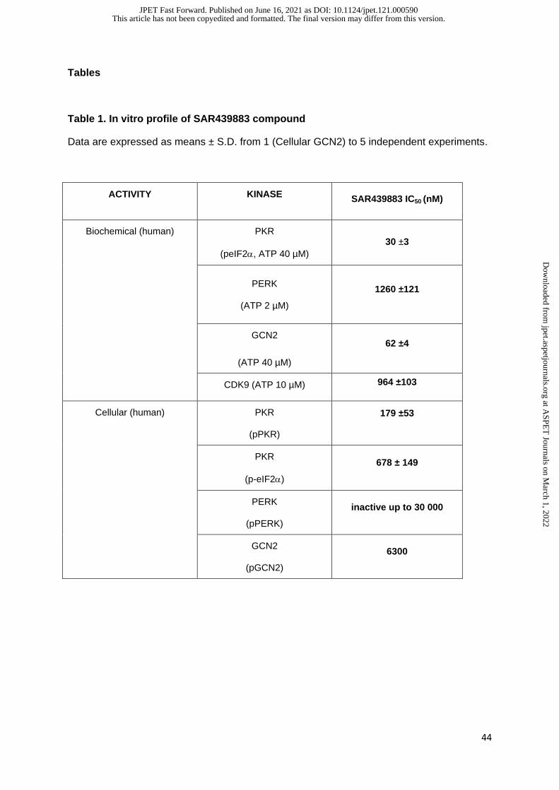
44
Tables
Table 1. In vitro profile of SAR439883 compound
Data are expressed as means ± S.D. from 1 (Cellular GCN2) to 5 independent experiments.
ACTIVITY KINASE SAR439883 IC50 (nM)
Biochemical (human) PKR
(peIF2, ATP 40 µM)
30 ±3
PERK
(ATP 2 µM)
1260 ±121
GCN2
(ATP 40 µM)
62 ±4
CDK9 (ATP 10 µM) 964 ±103
Cellular (human) PKR
(pPKR)
179 ±53
PKR
(p-eIF2)
678 ± 149
PERK
(pPERK)
inactive up to 30 000
GCN2
(pGCN2)
6300
This article has not been copyedited and formatted. The final version may differ from this version.JPET Fast Forward. Published on June 16, 2021 as DOI: 10.1124/jpet.121.000590
at ASPE
T Journals on M
arch 1, 2022jpet.aspetjournals.org
Dow
nloaded from
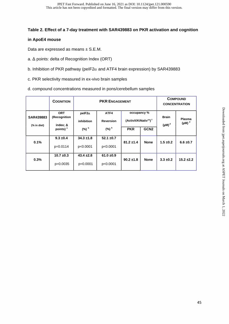
45
Table 2. Effect of a 7-day treatment with SAR439883 on PKR activation and cognition
in ApoE4 mouse
Data are expressed as means ± S.E.M.
a. Δ points: delta of Recognition Index (ORT)
b. Inhibition of PKR pathway (peIF2 and ATF4 brain expression) by SAR439883
c. PKR selectivity measured in ex-vivo brain samples
d. compound concentrations measured in pons/cerebellum samples
COGNITION PKR ENGAGEMENT COMPOUND
CONCENTRATION
SAR439883
(% in diet)
ORT
(Recognition
index; Δ
points) a
peIF2
inhibition
(%) b
ATF4
Reversion
(%) b
occupancy %
(ActivX/KiNativTM) c
Brain
(µM) d
Plasma
(µM) d
PKR GCN2
0.1%
9.3 ±0.4
p=0.0114
34.3 ±1.8
p<0.0001
52.1 ±0.7
p<0.0001
81.2 ±1.4 None 1.5 ±0.2 6.6 ±0.7
0.3%
10.7 ±0.3
p=0.0035
43.4 ±2.8
p=0.0001
61.0 ±0.9
p<0.0001
90.2 ±1.8 None 3.3 ±0.2 15.2 ±2.2
This article has not been copyedited and formatted. The final version may differ from this version.JPET Fast Forward. Published on June 16, 2021 as DOI: 10.1124/jpet.121.000590
at ASPE
T Journals on M
arch 1, 2022jpet.aspetjournals.org
Dow
nloaded from
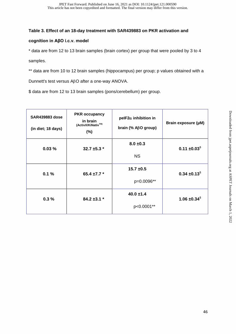
46
Table 3. Effect of an 18-day treatment with SAR439883 on PKR activation and
cognition in AβO i.c.v. model
* data are from 12 to 13 brain samples (brain cortex) per group that were pooled by 3 to 4
samples.
** data are from 10 to 12 brain samples (hippocampus) per group; p values obtained with a
Dunnett's test versus AO after a one-way ANOVA.
$ data are from 12 to 13 brain samples (pons/cerebellum) per group.
SAR439883 dose
(in diet; 18 days)
PKR occupancy
in brain (ActivX/KiNativ
TM)
(%)
peIF2 inhibition in
brain (% AO group)
Brain exposure (µM)
0.03 % 32.7 ±5.3 * 8.0 ±0.3
NS
0.11 ±0.03$
0.1 % 65.4 ±7.7 * 15.7 ±0.5
p=0.0096**
0.34 ±0.13$
0.3 % 84.2 ±3.1 * 40.0 ±1.4
p<0.0001**
1.06 ±0.34$
This article has not been copyedited and formatted. The final version may differ from this version.JPET Fast Forward. Published on June 16, 2021 as DOI: 10.1124/jpet.121.000590
at ASPE
T Journals on M
arch 1, 2022jpet.aspetjournals.org
Dow
nloaded from
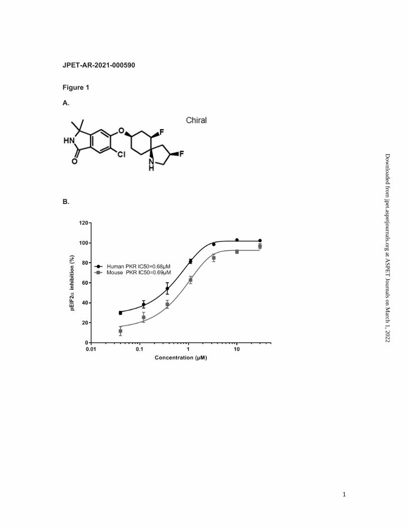
1
JPET-AR-2021-000590
Figure 1
A.
B.
This article has not been copyedited and formatted. The final version may differ from this version.JPET Fast Forward. Published on June 16, 2021 as DOI: 10.1124/jpet.121.000590
at ASPE
T Journals on M
arch 1, 2022jpet.aspetjournals.org
Dow
nloaded from
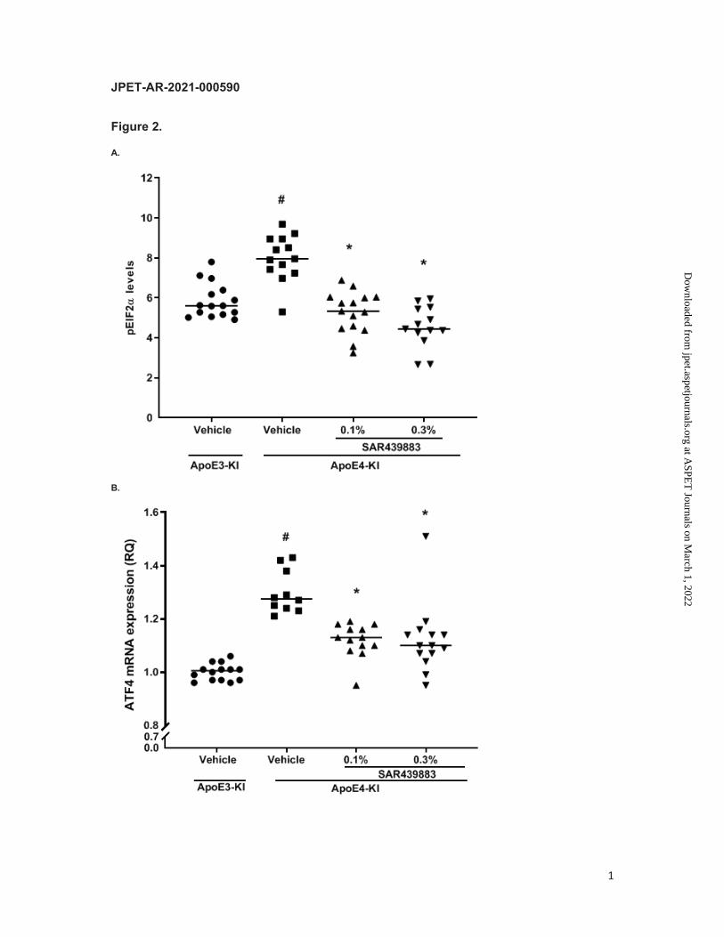
1
JPET-AR-2021-000590
Figure 2. A.
B.
This article has not been copyedited and formatted. The final version may differ from this version.JPET Fast Forward. Published on June 16, 2021 as DOI: 10.1124/jpet.121.000590
at ASPE
T Journals on M
arch 1, 2022jpet.aspetjournals.org
Dow
nloaded from

2
C.
This article has not been copyedited and formatted. The final version may differ from this version.JPET Fast Forward. Published on June 16, 2021 as DOI: 10.1124/jpet.121.000590
at ASPE
T Journals on M
arch 1, 2022jpet.aspetjournals.org
Dow
nloaded from
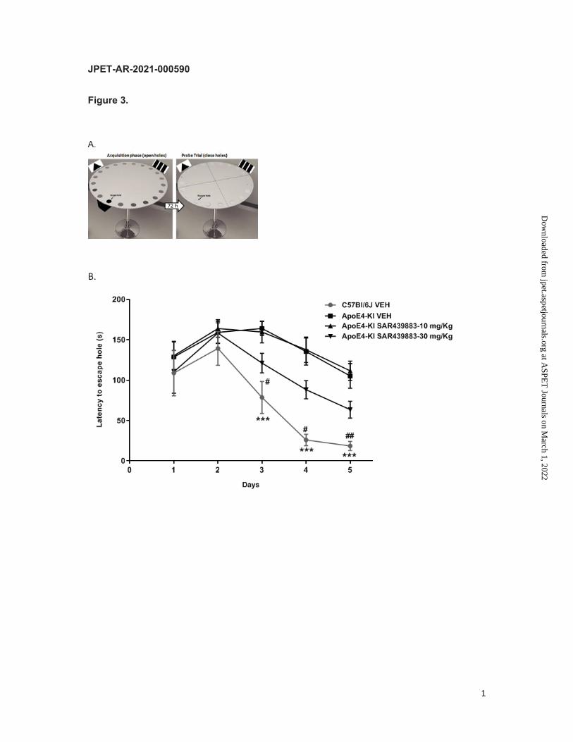
1
JPET-AR-2021-000590
Figure 3. A.
B.
This article has not been copyedited and formatted. The final version may differ from this version.JPET Fast Forward. Published on June 16, 2021 as DOI: 10.1124/jpet.121.000590
at ASPE
T Journals on M
arch 1, 2022jpet.aspetjournals.org
Dow
nloaded from
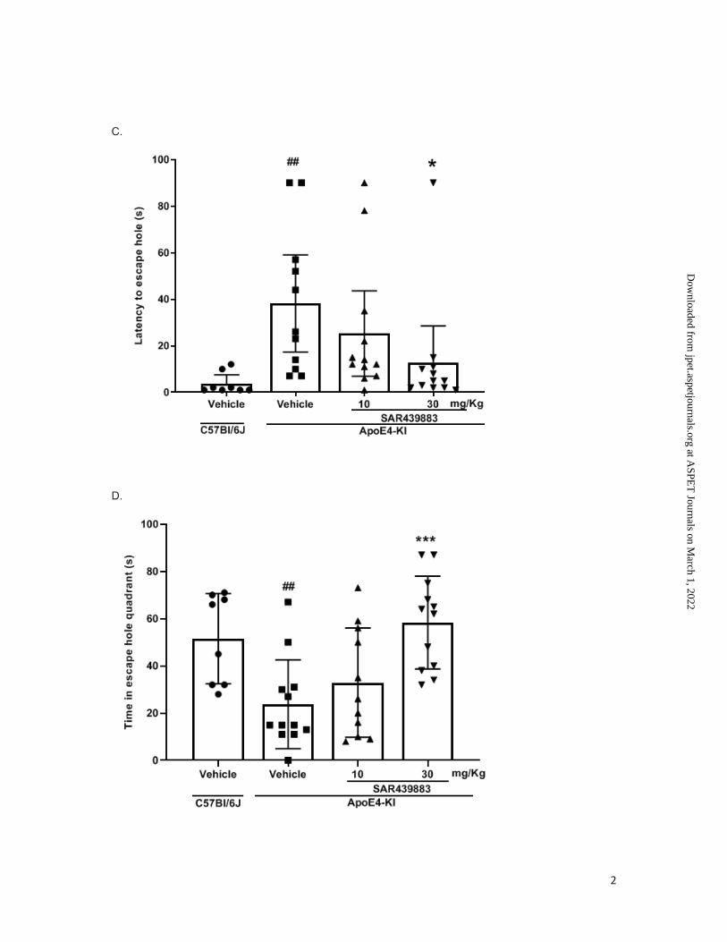
2
C.
D.
This article has not been copyedited and formatted. The final version may differ from this version.JPET Fast Forward. Published on June 16, 2021 as DOI: 10.1124/jpet.121.000590
at ASPE
T Journals on M
arch 1, 2022jpet.aspetjournals.org
Dow
nloaded from
1
JPET-AR-2021-000590
Figure 4.
A
B
This article has not been copyedited and formatted. The final version may differ from this version.JPET Fast Forward. Published on June 16, 2021 as DOI: 10.1124/jpet.121.000590
at ASPE
T Journals on M
arch 1, 2022jpet.aspetjournals.org
Dow
nloaded from

2
C
D
This article has not been copyedited and formatted. The final version may differ from this version.JPET Fast Forward. Published on June 16, 2021 as DOI: 10.1124/jpet.121.000590
at ASPE
T Journals on M
arch 1, 2022jpet.aspetjournals.org
Dow
nloaded from
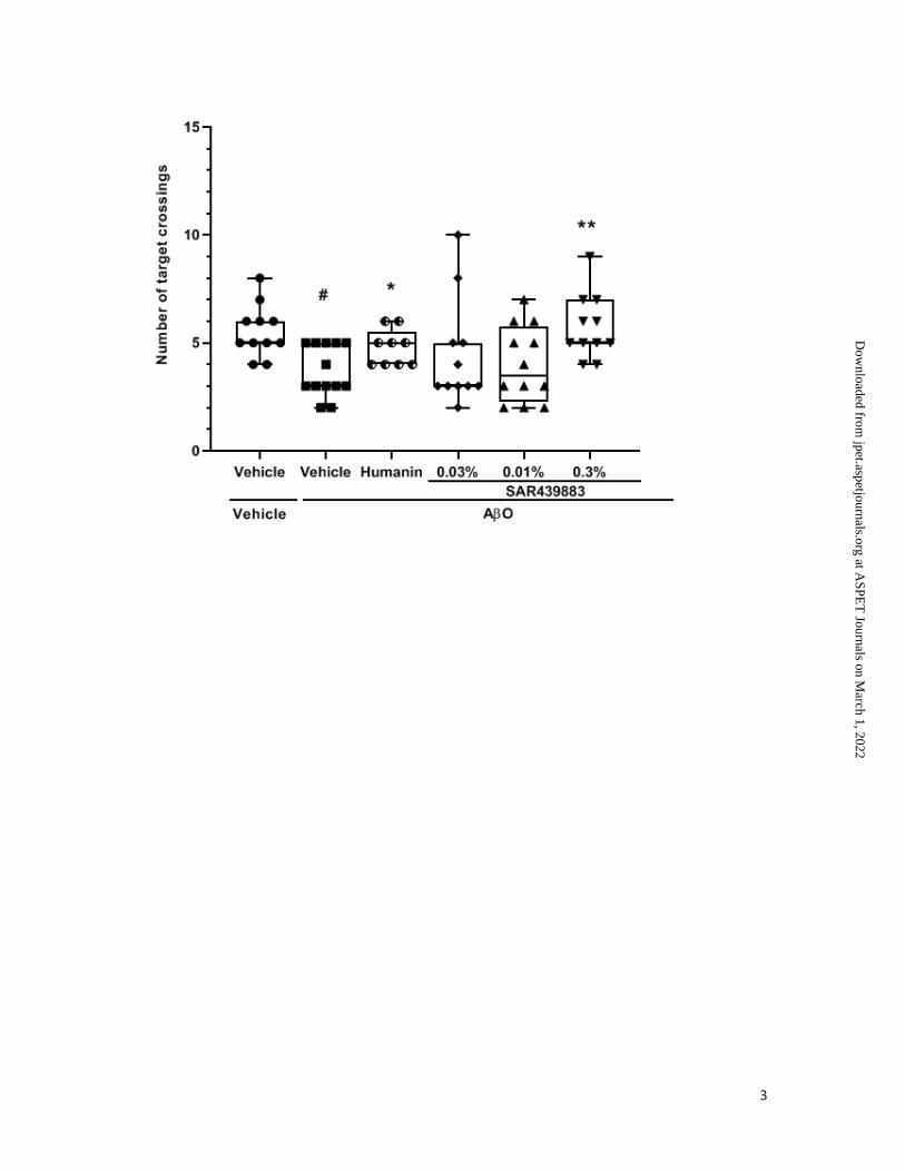
3
This article has not been copyedited and formatted. The final version may differ from this version.JPET Fast Forward. Published on June 16, 2021 as DOI: 10.1124/jpet.121.000590
at ASPE
T Journals on M
arch 1, 2022jpet.aspetjournals.org
Dow
nloaded from
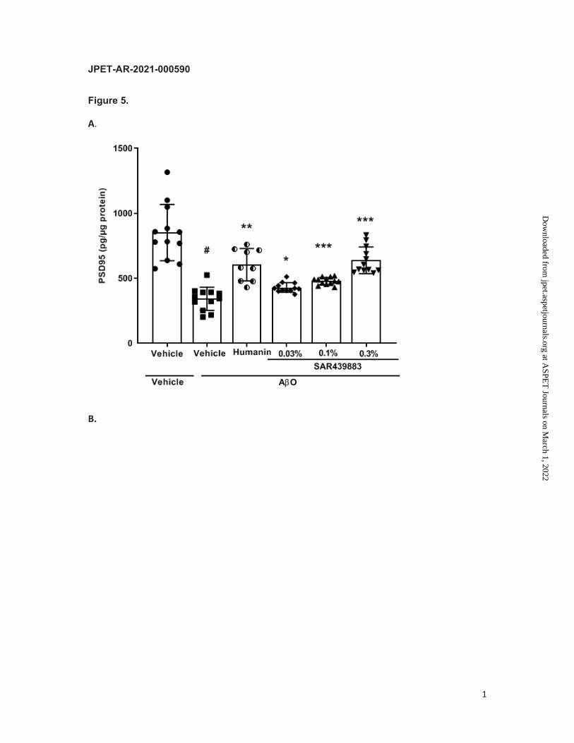
1
JPET-AR-2021-000590
Figure 5.
A.
B.
This article has not been copyedited and formatted. The final version may differ from this version.JPET Fast Forward. Published on June 16, 2021 as DOI: 10.1124/jpet.121.000590
at ASPE
T Journals on M
arch 1, 2022jpet.aspetjournals.org
Dow
nloaded from

2
C.
D.
This article has not been copyedited and formatted. The final version may differ from this version.JPET Fast Forward. Published on June 16, 2021 as DOI: 10.1124/jpet.121.000590
at ASPE
T Journals on M
arch 1, 2022jpet.aspetjournals.org
Dow
nloaded from
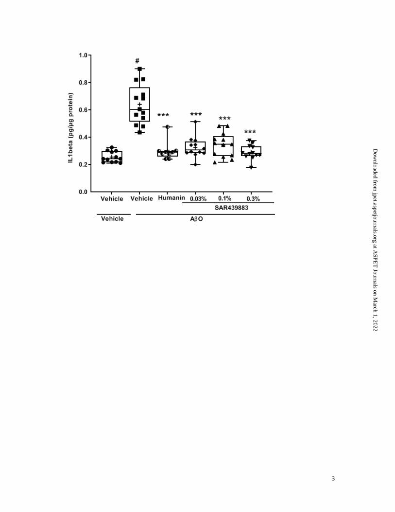
3
This article has not been copyedited and formatted. The final version may differ from this version.JPET Fast Forward. Published on June 16, 2021 as DOI: 10.1124/jpet.121.000590
at ASPE
T Journals on M
arch 1, 2022jpet.aspetjournals.org
Dow
nloaded from
1
JPET-AR-2021-000590
A novel selective PKR inhibitor restores cognitive deficits and neurodegeneration
in Alzheimer’s disease experimental models
Authors: Matilde Lopez-Grancha, Patrick Bernardelli, Nicolas Moindrot, Elisabeth Genet,
Carine Vincent, Valerie Roudieres, AIain Krick, Jean-François Sabuco1, David Machnik,
Delphine Ibghi, Laurent Pradier, Veronique Taupin2
Supplemental Fig. 1
SAR439883 effect on Aβ42 oligomer (AO)-induced neurotoxicity in vitro.
Effect of SAR439883 (3µM) on cell viability was measured by caspase-3/7 enzymatic activity
induced by AβO (5µM) in mouse primary neuronal cultures. Humanin (1µM) was used as
reference neuroprotective compound. Data are expressed as means ± S.D. from 4 biological
replicates. P values are from Dunnett's test after a one-way ANOVA. # p <0.0001 versus
Control; *p <0.0001 versus Control (+) AβO.
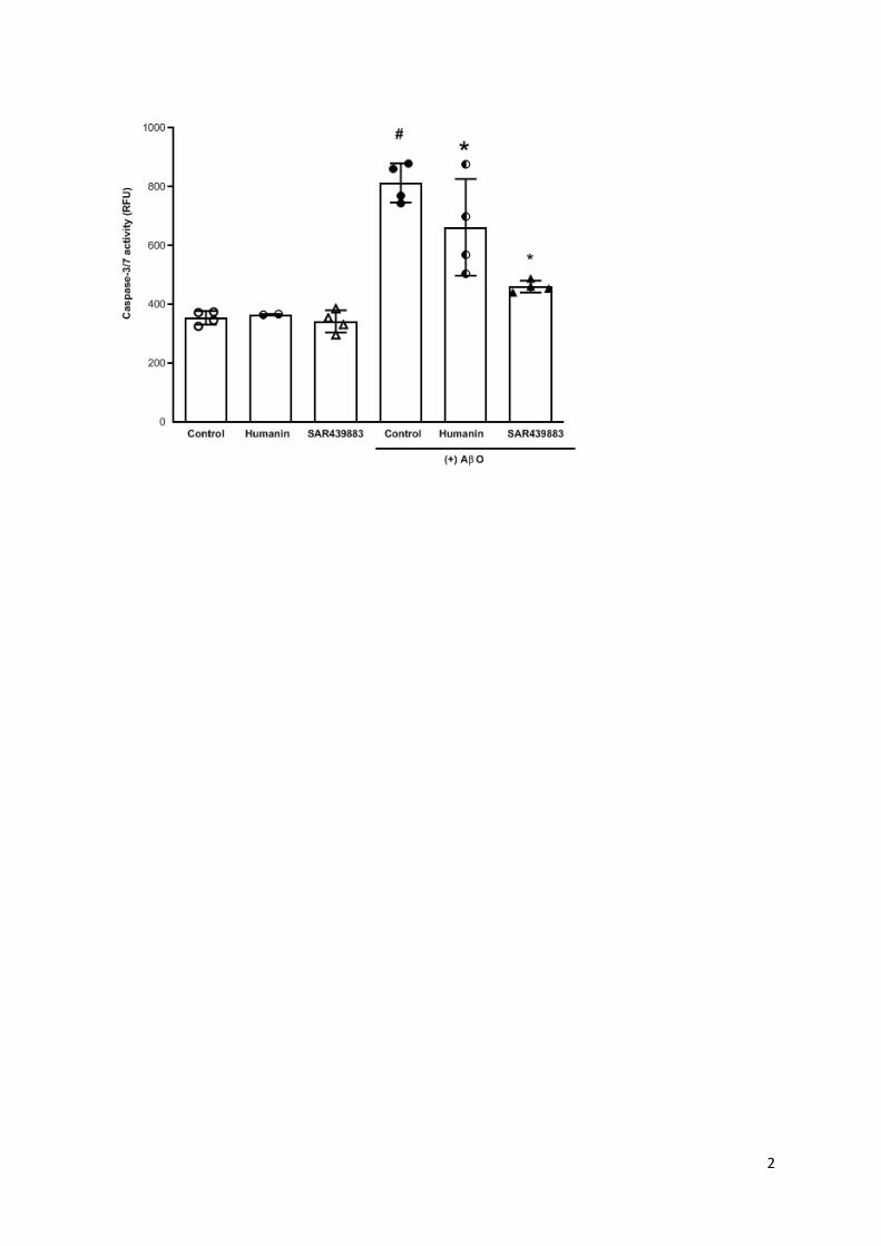
2
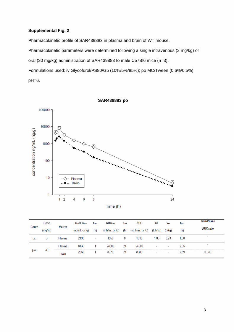
3
Supplemental Fig. 2
Pharmacokinetic profile of SAR439883 in plasma and brain of WT mouse.
Pharmacokinetic parameters were determined following a single intravenous (3 mg/kg) or
oral (30 mg/kg) administration of SAR439883 to male C57Bl6 mice (n=3).
Formulations used: iv Glycofurol/PS80/G5 (10%/5%/85%); po MC/Tween (0.6%/0.5%)
pH=6.
SAR439883 po
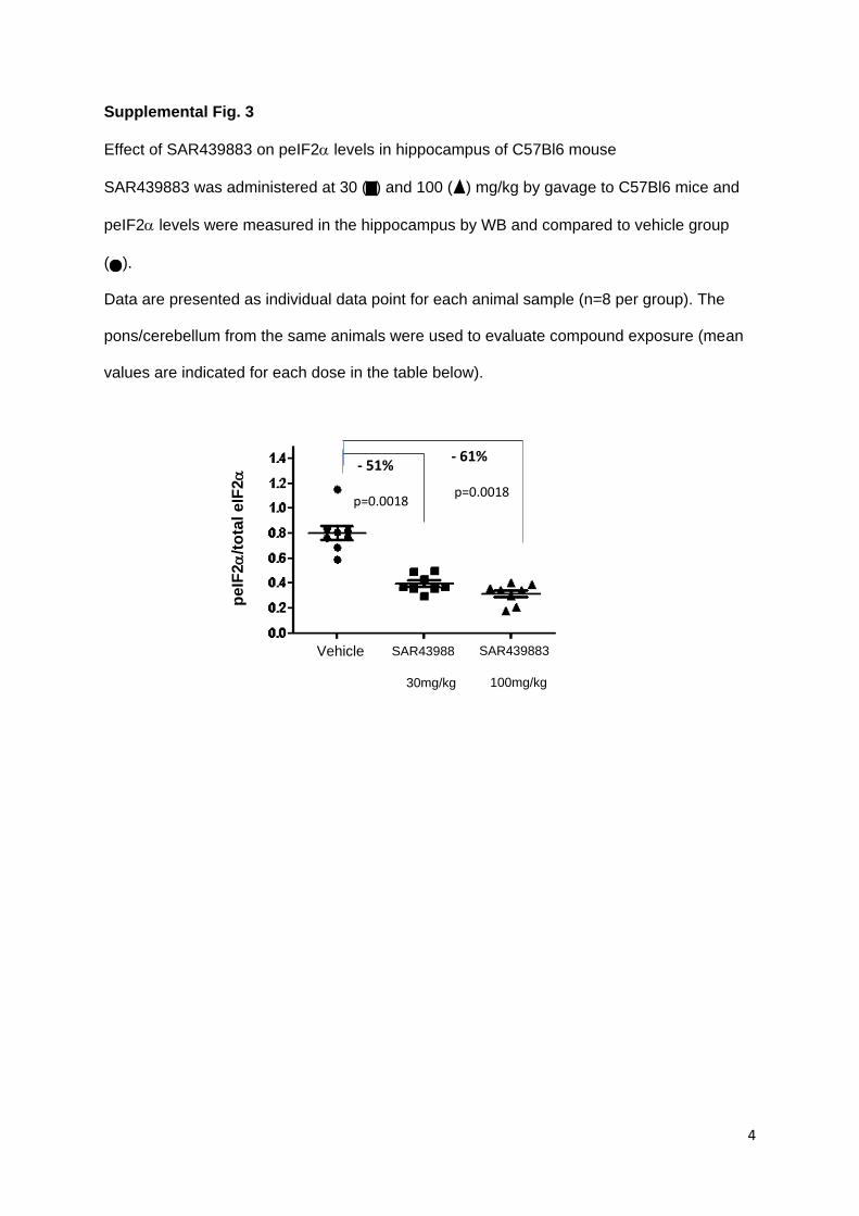
4
Supplemental Fig. 3
Effect of SAR439883 on peIF2 levels in hippocampus of C57Bl6 mouse
SAR439883 was administered at 30 ( ) and 100 ( ) mg/kg by gavage to C57Bl6 mice and
peIF2 levels were measured in the hippocampus by WB and compared to vehicle group
(. ).
Data are presented as individual data point for each animal sample (n=8 per group). The
pons/cerebellum from the same animals were used to evaluate compound exposure (mean
values are indicated for each dose in the table below).
pe
IF2
/to
tal
eIF
2
Vehicle SAR43988
30mg/kg
SAR439883
100mg/kg
- 51%
p=0.0018
- 61%
p=0.0018
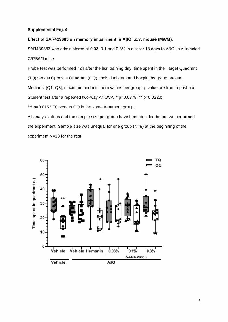
5
Supplemental Fig. 4
Effect of SAR439883 on memory impairment in AβO i.c.v. mouse (MWM).
SAR439883 was administered at 0.03, 0.1 and 0.3% in diet for 18 days to AβO i.c.v. injected
C57B6/J mice.
Probe test was performed 72h after the last training day: time spent in the Target Quadrant
(TQ) versus Opposite Quadrant (OQ). Individual data and boxplot by group present
Medians, [Q1; Q3], maximum and minimum values per group. p-value are from a post hoc
Student test after a repeated two-way ANOVA, * p=0.0378; ** p=0.0220;
*** p=0.0153 TQ versus OQ in the same treatment group,
All analysis steps and the sample size per group have been decided before we performed
the experiment. Sample size was unequal for one group (N=9) at the beginning of the
experiment N=13 for the rest.
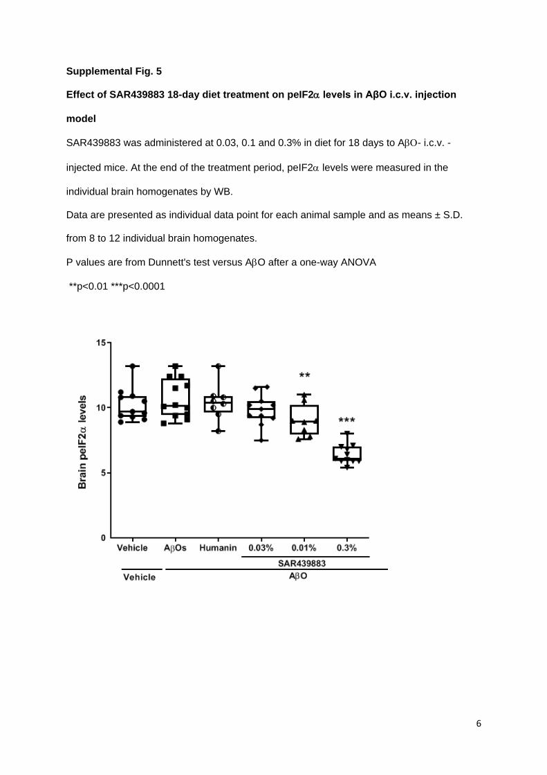
6
Supplemental Fig. 5
Effect of SAR439883 18-day diet treatment on peIF2 levels in AβO i.c.v. injection
model
SAR439883 was administered at 0.03, 0.1 and 0.3% in diet for 18 days to A- i.c.v. -
injected mice. At the end of the treatment period, peIF2 levels were measured in the
individual brain homogenates by WB.
Data are presented as individual data point for each animal sample and as means ± S.D.
from 8 to 12 individual brain homogenates.
P values are from Dunnett's test versus AO after a one-way ANOVA
**p<0.01 ***p<0.0001

7
Supplemental Table 1
SAR439883 selectivity profile using KiNativ™ technology (ActivX Biosciences, San
Diego, CA) in in vitro cells or in vivo 2h after oral treatment in ApoE4 mice.
a.Data are from lysates of PC3 cell treated at 6 concentrations for IC50 determination
b.Data are mean ± S.E.M. from 5 pooled brain homogenates (3 brains per pool) from ApoE4
mice
* NT: not tested
SAR439883
PC3 cells a
Brain tissue
30 mg/kg b
Kinase IC50 (µM) % inhibition
PKR 0.073 74.1 ± 1.8
GCN2 8-12 8.8 ± 5.2
PERK 8.8 NT*
CDC2 NT* 28.8 ± 4.8
SRPK1, SRPK2 >30 28.1 ± 4.3
RSK2 >30 26.4 ± 4.3
RSKL1 NT* 22.4 ± 4.7
ULK3 NT* 20.1 ± 3.3
other 111 kinases >30
other 194 kinases <20
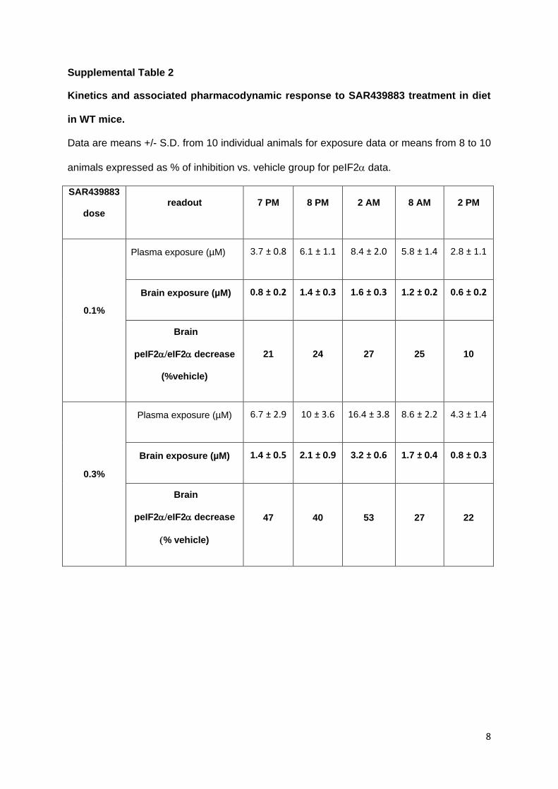
8
Supplemental Table 2
Kinetics and associated pharmacodynamic response to SAR439883 treatment in diet
in WT mice.
Data are means +/- S.D. from 10 individual animals for exposure data or means from 8 to 10
animals expressed as % of inhibition vs. vehicle group for peIF2data.
SAR439883
dose readout 7 PM 8 PM 2 AM 8 AM 2 PM
0.1%
Plasma exposure (µM) 3.7 ± 0.8 6.1 ± 1.1 8.4 ± 2.0 5.8 ± 1.4 2.8 ± 1.1
Brain exposure (µM) 0.8 ± 0.2 1.4 ± 0.3 1.6 ± 0.3 1.2 ± 0.2 0.6 ± 0.2
Brain
peIF2eIF2decrease
(%vehicle)
21 24 27 25 10
0.3%
Plasma exposure (µM) 6.7 ± 2.9 10 ± 3.6 16.4 ± 3.8 8.6 ± 2.2 4.3 ± 1.4
Brain exposure (µM) 1.4 ± 0.5 2.1 ± 0.9 3.2 ± 0.6 1.7 ± 0.4 0.8 ± 0.3
Brain
peIF2eIF2decrease
% vehicle)
47
40
53
27 22

9
Supplemental Table 3
SAR439883 effect on Aβ42 oligomers (AO)-induced neurotoxicity in vitro: Effect on
peIF2 levels.
Data are means from pooled samples from one exploratory study.
Cell treatment
peIF2levels
(% control)
AO
177
AO + SAR439883 3 µM
97
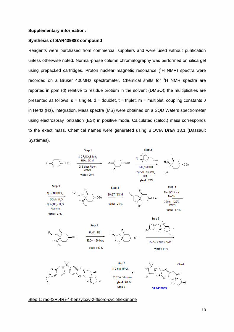
10
Supplementary information:
Synthesis of SAR439883 compound
Reagents were purchased from commercial suppliers and were used without purification
unless otherwise noted. Normal-phase column chromatography was performed on silica gel
using prepacked cartridges. Proton nuclear magnetic resonance (1H NMR) spectra were
recorded on a Bruker 400MHz spectrometer. Chemical shifts for 1H NMR spectra are
reported in ppm (d) relative to residue protium in the solvent (DMSO); the multiplicities are
presented as follows: s = singlet, d = doublet, t = triplet, m = multiplet, coupling constants J
in Hertz (Hz), integration. Mass spectra (MS) were obtained on a SQD Waters spectrometer
using electrospray ionization (ESI) in positive mode. Calculated (calcd.) mass corresponds
to the exact mass. Chemical names were generated using BIOVIA Draw 18.1 (Dassault
Systèmes).
Step 1: rac-(2R,4R)-4-benzyloxy-2-fluoro-cyclohexanone

11
To a mechanically stirred solution of 4-benzyloxycyclohexanone (500 g, 2.45 mol) and
triethylamine (997 mL, 7.17 mol, 2.9 equiv.) in dichloromethane (DCM) (7 L) at -5°C under
nitrogen was dropwise added a solution of trimethylsilyl trifluoromethanesulfonate (491 mL,
2.78 mol, 1.1 equiv.) in DCM (1.5 L) over 3 hours while maintaining the temperature below
0°C. The mixture was mechanically stirred for 15 minutes at 0°C and then a saturated
auqueous NaHCO3 solution (10 L) was added. After stirring for 5 minutes the layers were
separated. The aqueous layer was extracted with DCM (1 L). The combined organic layers
were washed with brine (2.5 L), dried over sodium sulfate and concentrated under reduced
pressure to give the corresponding trimethylsilyl enol ether (816.7 g) as a brown oil. To a
solution of the resulting intermediate (816.7 g, 2.45 mol) in acetonitrile (7.5 L) was
portionwise added Selectfluor (1101 g, 3.11 mol, 1.3 eq) in 1 hour while maintaining the
temperature below 25°C with an ice-bath. The mixture was stirred for 2 hours allowing the
temperature to warm up to room temperature. Diethylether (10 L) was added, and the
precipitate was filtered through Celite and washed with diethylether (3 L). The filtrate was
concentrated under reduced pressure to give 1198 g of a crude mixture of diastereomers as
a brown oil. This mixture was separated by column chromatography on silica gel eluting with
a gradient of 0.05% to 5% methanol in dichloromethane. The second eluting product was
collected and concentrated under reduced pressure then taken into ethyl acetate (220 mL)
followed by heptane (880 mL). After stirring for 45 minutes, the product was filtered, washed
with heptane (100 mL) and dried under vacuum to give 110 g (20%) of rac-(2R,4R)-4-
benzyloxy-2-fluoro-cyclohexanone as a white solid. MS (EI): mass calcd. for C13H15FO2, 222;
m/z found, 222 [M+]. 1H NMR (400 MHz, CDCl3): d 7.32-7.43 (m, 5 H), 4.90 (ddd, J = 48.0,
12.4, 6.8 Hz, 1 H), 4.62 (s, 2 H), 3.83-3.90 (m, 1 H), 2.80-2.83 (m, 1 H), 2.52-2.54 (m, 1 H),
2.28-2.32 (m, 2 H), 1.94-2.00 (m, 1H), 1.75-1.79 (m, 1 H).
Step 2: rac-(1S,2S,4S)-1-allyl-N-benzyl-4-benzyloxy-2-fluoro-cyclohexanamine
To a solution of rac-(2R,4R)-4-benzyloxy-2-fluoro-cyclohexanone (25 g, 112.48 mmol) in 7N
methanolic ammonia solution (170 mL, 1.19 mol, 10 equiv.) was added 2-allyl-4,4,5,5-
tetramethyl-1,3,2-dioxaborolane (27 mL, 143 mmol, 1.27 equiv.). The mixture was stirred for
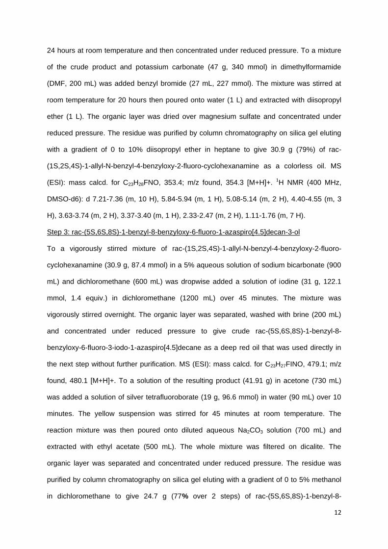
12
24 hours at room temperature and then concentrated under reduced pressure. To a mixture
of the crude product and potassium carbonate (47 g, 340 mmol) in dimethylformamide
(DMF, 200 mL) was added benzyl bromide (27 mL, 227 mmol). The mixture was stirred at
room temperature for 20 hours then poured onto water (1 L) and extracted with diisopropyl
ether (1 L). The organic layer was dried over magnesium sulfate and concentrated under
reduced pressure. The residue was purified by column chromatography on silica gel eluting
with a gradient of 0 to 10% diisopropyl ether in heptane to give 30.9 g (79%) of rac-
(1S,2S,4S)-1-allyl-N-benzyl-4-benzyloxy-2-fluoro-cyclohexanamine as a colorless oil. MS
(ESI): mass calcd. for C23H28FNO, 353.4; m/z found, 354.3 [M+H]+. 1H NMR (400 MHz,
DMSO-d6): d 7.21-7.36 (m, 10 H), 5.84-5.94 (m, 1 H), 5.08-5.14 (m, 2 H), 4.40-4.55 (m, 3
H), 3.63-3.74 (m, 2 H), 3.37-3.40 (m, 1 H), 2.33-2.47 (m, 2 H), 1.11-1.76 (m, 7 H).
Step 3: rac-(5S,6S,8S)-1-benzyl-8-benzyloxy-6-fluoro-1-azaspiro[4.5]decan-3-ol
To a vigorously stirred mixture of rac-(1S,2S,4S)-1-allyl-N-benzyl-4-benzyloxy-2-fluoro-
cyclohexanamine (30.9 g, 87.4 mmol) in a 5% aqueous solution of sodium bicarbonate (900
mL) and dichloromethane (600 mL) was dropwise added a solution of iodine (31 g, 122.1
mmol, 1.4 equiv.) in dichloromethane (1200 mL) over 45 minutes. The mixture was
vigorously stirred overnight. The organic layer was separated, washed with brine (200 mL)
and concentrated under reduced pressure to give crude rac-(5S,6S,8S)-1-benzyl-8-
benzyloxy-6-fluoro-3-iodo-1-azaspiro[4.5]decane as a deep red oil that was used directly in
the next step without further purification. MS (ESI): mass calcd. for C23H27FINO, 479.1; m/z
found, 480.1 [M+H]+. To a solution of the resulting product (41.91 g) in acetone (730 mL)
was added a solution of silver tetrafluoroborate (19 g, 96.6 mmol) in water (90 mL) over 10
minutes. The yellow suspension was stirred for 45 minutes at room temperature. The
reaction mixture was then poured onto diluted aqueous Na2CO3 solution (700 mL) and
extracted with ethyl acetate (500 mL). The whole mixture was filtered on dicalite. The
organic layer was separated and concentrated under reduced pressure. The residue was
purified by column chromatography on silica gel eluting with a gradient of 0 to 5% methanol
in dichloromethane to give 24.7 g (77% over 2 steps) of rac-(5S,6S,8S)-1-benzyl-8-

13
benzyloxy-6-fluoro-1-azaspiro[4.5]decan-3-ol as a deep orange solid. MS (ESI): mass calcd.
for C23H28FNO2, 369.2; m/z found, 370.2 [M+H]+. 1H NMR (400 MHz, DMSO-d6): d 7.06-
7.38 (m, 10 H), 4.39-4.89 (m, 4H), 4.01-4.09 (m, 2 H),vvvv 3.50-3.60 (m, 2 H), 2.60-2.86 (m,
1 H), 2.26-2.37 (m, 2 H), 1.13-1.61 (m, 6 H).
Step 4: rac-(3S,5S,6S,8S)-1-benzyl-8-benzyloxy-3,6-difluoro-1-azaspiro[4.5]decane
To a solution of rac-(5S,6S,8S)-1-benzyl-8-benzyloxy-6-fluoro-1-azaspiro[4.5]decan-3-ol
(18.61 g, 50.37 mmol) in dry dichloromethane (300 mL) at -10°C under a dry argon
atmosphere was dropwise added diethylaminosulfur trifluoride DAST (10.4 mL, 78.71 mmol,
1.5 equiv.). The mixture was stirred for 2 hours allowing the temperature to warm up to room
temperature then poured onto saturated aqueous NaHCO3 solution (500 mL) and
dichloromethane (200 mL). The pH was slowly made basic with careful solid NaHCO3
addition. The layers were separated and the auqeous layer extracted with dichloromethane
(200 mL). The combined organic layers were dried over magnesium sulfate and
concentrated under reduced pressure. The residue was purified by column chromatography
on silica gel eluting with a gradient of 20 to 100% dichloromethane in heptane to give 3.9 g
(21%) of rac-(3S,5S,6S,8S)-1-benzyl-8-benzyloxy-3,6-difluoro-1-azaspiro[4.5]decane. 1H
NMR (400 MHz, DMSO-d6): d 7.11-7.41 (m, 10 H), 4.98 (d, J = 56.0 Hz, 1 H), 4.79 (d, J =
52.0 Hz, 1 H), 4.48 (dd, J = 48.0, 12.0 Hz, 2 H), 3.70-4.18 (m, 2 H), 3.60 (s, 1 H), 2.91-3.02
(m, 2H), 2.42-2.47 (m, 2H), 1.88-2.09 (m, 3H), 1.47-1.70 (m, 2H), 1.14-1.18 (m, 1 H).
Step 5: rac-(3S,5S,6S,8S)-1-benzyl-3,6-difluoro-1-azaspiro[4.5]decan-8-ol
To a solution of rac-(3S,5S,6S,8S)-1-benzyl-8-benzyloxy-3,6-difluoro-1-azaspiro[4.5]decane
(3.82 g, 10.28 mmol) in acetonitrile (39 mL) equally partitioned in three 30 mL microwave
vials (Anton Paar apparatus) was added a suspension from equally partitioned sodium
iodide (7.70 g, 51.37 mmol, 5 equiv.) and trimethylsilyl chloride (6.6 mL, 51.64 mmol, 5
equiv.) in acetonitrile (15 mL). The 3 mixture were placed in the microwave apparatus and
heated to 120°C under microwave irradiation (MW) for 30 minutes. The reaction mixtures
were combined and concentrated not to complete dryness under reduced pressure, taken-up
into ethyl acetate (100 mL), washed with a saturated Na2CO3 aqueous solution then with a
14
2N aqueous sodium thiosulfate solution (100 mL). The organic layer was dried over
magnesium sulfate and concentrated under reduced pressure. The residue was purified by
column chromatography on silica gel eluting with a gradient of 0 to 10% ethyl acetate in
dichloromethane to give 1.94 g (67%) of rac-(3S,5S,6S,8S)-1-benzyl-3,6-difluoro-1-
azaspiro[4.5]decan-8-ol as an off-white solid. 1H NMR (400 MHz, DMSO-d6): d 7.16-7.38
(m, 5 H), 5.09 (dt, J = 55, 5 Hz, 1 H), 4.60 (dt, J = 49, 3Hz, 1 H), 4.19 (d, J = 8 Hz, 1 H), 4.17
(s, 1 H), 3.82 (m, 1 H), 3.52 (d, J = 14 Hz, 1 H), 2.70-2.92 (m, 2 H), 2.31 (td, J = 13, 4 Hz, 1
H), 2.15 (m, 1 H), 1.82-2.10 (m, 3 H), 1.77 (m, 1 H), 1.57 (tt, J = 14, 3 Hz, 1 H), 1.20 (m, 1
H).
Step 6: rac-(3S,5S,6S,8S)-3,6-difluoro-1-azaspiro[4.5]decan-8-ol
A solution of rac-(3S,5S,6S,8S)-1-benzyl-8-benzyloxy-3,6-difluoro-1-azaspiro[4.5]decane
(1.92 g, 6.82 mmol) in ethanol (250 mL) is hydrogenated at 60°C under 30 bars of hydrogen
with a H-Cube Pro apparatus (Thales) using a 10% Pd/C cartridge (140 mg) and a 1
mL/minute flowrate. The solution is concentrated under reduced pressure to give 1.3 g
(99%) of rac-(3S,5S,6S,8S)-3,6-difluoro-1-azaspiro[4.5]decan-8-ol. MS (ESI): mass calcd.
for C9H15F2NO, 191.1; m/z found, 192.1 [M+H]+. 1H NMR (400 MHz, DMSO-d6): d 5.19 (d, J
= 52.0 Hz, 1 H), 4.78 (br.s., 1H), 4.52 (ddd, J = 47.2, 13.2, 4.4 Hz, 1H), 3.49-3.53 (m, 1H),
3.01-3.15 (m, 2H), 1.99-2.07 (m, 3H), 1.48-1.69 (m, 4H), 1.13-1.19 (m, 1H).
Step 7: 6-chloro-2-[(4-methoxyphenyl)methyl]-3,3-dimethyl-5-[[rac-(3R,5R,6R,8R)-3,6-
difluoro-1-azaspiro[4.5]decan-8-yl]oxy]isoindolin-1-one
To a solution of rac-(3S,5S,6S,8S)-3,6-difluoro-1-azaspiro[4.5]decan-8-ol (900 mg, 4.71
mmol) and 6-chloro-5-fluoro-2-(4-methoxybenzyl)-3,3-dimethylisoindolin-1-one (1.7 g, 5.09
mmol, 1.08 equiv.) in N,N-dimethylformamide DMF (25 mL) was added over 10 minutes a
1M solution of potassium tert-butoxide in tetrahydrofuran THF (5.4 mL, 5.40 mmol, 1.1
equiv.). The reaction mixture was stirred for 20 minutes at room temperature then poured
onto a non-saturated KH2PO4 aqueous solution and extracted with ethyl acetate (150 mL).
The organic layer was dried over magnesium sulfate and concentrated under reduced
pressure. The residue was purified by column chromatography on silica gel eluting with a

15
gradient of 50 to 100% ethyl acetate in heptane to give 1.93 g (81%) of 6-chloro-2-[(4-
methoxyphenyl)methyl]-3,3-dimethyl-5-[[rac-(3R,5R,6R,8R)-3,6-difluoro-1-
azaspiro[4.5]decan-8-yl]oxy]isoindolin-1-one as a white foam. MS (ESI): mass calcd. for
C27H31ClF2N2O3, 504.1; m/z found, 505.1 [M+H]+. 1H NMR (400 MHz, DMSO-d6): d 7.69 (s,
1 H), 7.57 (s, 1 H), 7.30 (d, J = 9 Hz, 2 H), 6.87 (d, J = 9 Hz, 2 H), 5.20 (dtd, J = 56, 4, 2 Hz,
1 H), 4.50-4.76 (m, 4 H), 3.73 (s, 3 H), 3.03 (m, 2 H), 2.35 (m, 1 H), 1.65-2.15 (m, 6 H), 1.38
(s, 3 H), 1.36 (s, 3 H), 1.28 (m, 1 H).
Step 8: 6-chloro-5-[[(3R,5R,6R,8R)-3,6-difluoro-1-azaspiro[4.5]decan-8-yl]oxy]-2-[(4-
methoxyphenyl)methyl]-3,3-dimethyl-isoindolin-1-one
The racemic mixture of 6-chloro-2-[(4-methoxyphenyl)methyl]-3,3-dimethyl-5-[[rac-
(3R,5R,6R,8R)-3,6-difluoro-1-azaspiro[4.5]decan-8-yl]oxy]isoindolin-1-one (1.9 g) was
separated by chiral HPLC with a Chiralcel OD-I column 20μm (7.65 x 35cm) using heptane
70%/ ethanol 30%/ triethylamine 0.1% as mobile phase, a flowrate of 350 mL/min and a UV
detector at 254 nm to give 916 mg (48%) of 6-chloro-5-[[(3R,5R,6R,8R)-3,6-difluoro-1-
azaspiro[4.5]decan-8-yl]oxy]-2-[(4-methoxyphenyl)methyl]-3,3-dimethyl-isoindolin-1-one as
second eluting dextrogyre enantiomer. [α]D = +15.2° (c= 0.0043, DMSO, 25°C). MS (ESI):
mass calcd. for C27H31ClF2N2O3, 504.1; m/z found, 505.1 [M+H]+. 1H NMR (400 MHz,
DMSO-d6): d 7.69 (s, 1 H), 7.57 (s, 1 H), 7.30 (d, J = 9 Hz, 2 H), 6.87 (d, J = 9 Hz, 2 H), 5.20
(dtd, J = 56, 4, 2 Hz, 1 H), 4.50-4.76 (m, 4 H), 3.73 (s, 3 H), 3.03 (m, 2 H), 2.35 (m, 1 H),
1.65-2.15 (m, 6 H), 1.38 (s, 3 H), 1.36 (s, 3 H), 1.28 (m, 1 H).
Step 9: 6-chloro-5-[[(3R,5R,6R,8R)-3,6-difluoro-1-azaspiro[4.5]decan-8-yl]oxy]-3,3-dimethyl-
isoindolin-1-one
A solution of 6-chloro-5-[[(3R,5R,6R,8R)-3,6-difluoro-1-azaspiro[4.5]decan-8-yl]oxy]-2-[(4-
methoxyphenyl)methyl]-3,3-dimethyl-isoindolin-1-one (915 mg, 1.81 mmol) and anisole (4
ml, 36.62 mmol) in trifluoroacetic acid (20 mL) was heated to 140°C for 45 minutes under
microwave irradiation (Anton Paar apparatus). The reaction mixture was poured onto
16
saturated Na2CO3 aqueous solution (200 mL) and extracted with ethyl acetate (200 mL). The
organic layer was separated, washed with 1N aqueous sodium hydroxyde solution (100 mL),
dried over magnesium sulfate and concentrated under reduced pressure. The residue was
purified by column chromatography on silica gel eluting with ethyl acetate to give 622 mg
(89%) of 6-chloro-5-[[(3R,5R,6R,8R)-3,6-difluoro-1-azaspiro[4.5]decan-8-yl]oxy]-3,3-
dimethyl-isoindolin-1-one as a white foam. MS (ESI): mass calcd. for C19H23ClF2N2O2, 384.1;
m/z found, 385.2 [M+H]+. 1H NMR (400 MHz, DMSO-d6): d 8.54 (s, 1 H), 7.55 (s, 1 H), 7.50
(s, 1 H), 5.19 (dtd, J = 56, 4, 2 Hz, 1 H), 4.52-4.74 (m, 2 H), 2.82-3.13 (m, 2 H), 2.34 (m, 1
H), 1.63-2.16 (m, 6 H), 1.45 (s, 3 H), 1.44 (s, 3 H), 1.29 (m, 1 H).


![Received: 2016.02.21 The Specific Protein Kinase R (PKR ...shown that PKR participates in neurodegenerative processes with neurotoxicity [12,13]. Peel and Couturier considered PKR](https://img.pdfslide.us/doc/110x75/5e45e3e2e3e94073247c9161/received-20160221-the-specific-protein-kinase-r-pkr-shown-that-pkr-participates.jpg)