Embed Size (px)
Citation preview

Takahisa Nakamura, Alessandro Arduini, Brenna Baccaro, Masato Furuhashi, and Gökhan S. Hotamisligil
Small-Molecule Inhibitors ofPKR Improve GlucoseHomeostasis in Obese DiabeticMice
Obesity and metabolic diseases appear asclusters, often featuring high risk for insulinresistance and type 2 diabetes, and constitutea major global health problem with limitedtreatment options. Previous studies have shownthat double-stranded RNA–dependent kinase,PKR, plays an important role in thenutrient/pathogen-sensing interface, and acts asa key modulator of chronic metabolicinflammation, insulin sensitivity, and glucosehomeostasis in obesity. Recently, pathologicalPKR activation was also demonstrated in obesehumans, strengthening its prospects as a potentialdrug target. Here, we investigate the use oftwo structurally distinct small-molecule inhibitorsof PKR in the treatment of insulin resistance andtype 2 diabetes in cells and in a mouse model ofsevere obesity and insulin resistance. Inhibition ofPKR reduced stress-induced Jun NH2-terminalkinase activation and insulin receptor substrate 1serine phosphorylation in vitro and in vivo. Inaddition, treatment with both PKR inhibitorsreduced adipose tissue inflammation, improvedinsulin sensitivity, and improved glucoseintolerance in mice after the establishment ofobesity and insulin resistance. Our findingssuggest that pharmacologically targeting PKR maybe an effective therapeutic strategy for the
treatment of insulin resistance and type 2diabetes.Diabetes 2014;63:526–534 | DOI: 10.2337/db13-1019
The link between cellular stress signals and chronicmetabolic diseases, including obesity-induced insulinresistance, type 2 diabetes, fatty liver disease, andatherosclerosis, has been well-established (1–3). Dur-ing the course of obesity a broad array of inflammatoryand stress responses are evoked in metabolic tissues,leading to activation of several inflammatorysignaling molecules including Jun NH2-terminal kinase(JNK) and inhibitory kB kinase (IKK). These pathwaysplay an important role in the development of insulinresistance and diabetes by controlling the in-flammatory responses in metabolic tissues, the in-hibition of insulin receptor signaling, and thedisruption of systemic glucose and lipid homeostasis(4–10). Evidence emerging from experimentalmodels has demonstrated that suppression of thesebroad inflammatory networks generally results inprotection against obesity-induced insulin resistanceand diabetes (4–7,11–13). However, the translationof these discoveries to the clinic has been slowed bythe lack of effective therapeutic entities, and itremains to be determined whether these strategiesmay be effective interventions after the establishmentof disease.
Department of Genetics and Complex Diseases, Harvard School of Public Health,Boston, MA
Corresponding author: Gökhan S. Hotamisligil, [email protected].
Received 28 June 2013 and accepted 17 October 2013.
T.N. is currently affiliated with the Divisions of Endocrinology and DevelopmentalBiology, Cincinnati Children’s Hospital Medical Center, Cincinnati, Ohio.
© 2014 by the American Diabetes Association. See http://creativecommons.org/licenses/by-nc-nd/3.0/ for details.
See accompanying commentary, p. 393.
526 Diabetes Volume 63, February 2014
SIG
NALTRANSDUCTIO
N
at FRANCIS A COUNTWAY on January 29, 2014http://diabetes.diabetesjournals.org/Downloaded from

Given that metaflammation—the chronic, low-grade,metabolic inflammation characteristic of obesity—iscritical in the regulation of systemic metabolichomeostasis, there is an emerging emphasis on signalingnodes and molecules that integrate pathogen and stressresponses with metabolic pathways as promising targetsin understanding and eventually treating these de-bilitating diseases. In search of such molecules that in-tegrate endoplasmic reticulum (ER) stress and relatedsignaling pathways with inflammatory output, insulinaction, and metabolic control, we recently identified thedouble-stranded RNA–dependent kinase (PKR) (14). PKRis activated by nutrients such as fatty acids and by ERstress, controls major inflammatory cascades such asJNK, and is required for inflammasome activity (14–16).PKR also directly interacts with insulin receptor signalingcomponents and inhibits insulin action (14). There ismarked activation of PKR in liver and adipose tissue ofmice with dietary and genetic obesity, and two in-dependent lines of PKR-deficient mice have been shownto be protected against obesity-induced insulin resistanceand obesity-induced inflammatory changes (14,17). Fi-nally, the ER stress pathways, JNK, and PKR are signif-icantly activated in human obesity, particularly inadipose and liver tissues, raising the possibility that PKRmay represent a suitable target for drug developmentagainst diabetes (18–20).
Based on these observations, in this study we in-vestigated the potential of pharmacological inhibitors ofPKR activity to ameliorate the inflammation and insulinresistance associated with obesity in an established dis-ease model.
RESEARCH DESIGN AND METHODS
Biochemical Reagents
All biochemical reagents were purchased from Sigma-Aldrich (St. Louis, MO) unless otherwise indicated. Anti-insulin receptor substrate (IRS)-1 and anti-phospho-IRS1(Ser307) were from Upstate Biotechnology (Lake Placid,NY). Antibodies against PKR, JNK1, Akt, phospho-Akt,insulin receptor-b subunit (IRb), and b-tubulin werefrom Santa Cruz Biotechnology (Santa Cruz, CA).Anti-phospho-eukaryotic translation initiation factor 2-a(eIF2a; Ser52) antibody was purchased from Invitrogen(Carlsbad, CA). Anti-phospho-insulin receptor (Tyr1162/1163), PKR inhibitor (C13H8N4OS, imoxin), and a neg-ative control of PKR inhibitor (C15H8Cl3NO2) werepurchased from Calbiochem (Gibbstown, NJ). Anti-phospho-JNK (Thr183/Tyr185) antibody was purchasedfrom Cell Signaling Technology (Danvers, MA).Recombinant IRS1, JNKs, p38, IKKb, IkB, myelin basicprotein, and agarose-conjugated PKR antibody werepurchased from Millipore (Billerica, MA).
Kinase Assays
For in vitro kinase assays, each recombinant protein—ata concentration of 10 ng/mL—was mixed with 16.7 mmol/L
PKR inhibitor or DMSO in kinase buffer (25 mmol/LTris-HCl [pH 7.5], 5 mmol/L b-glycerophosphate,2 mmol/L dithiothreitol, 0.1 mmol/L Na3VO4, 10 mmol/LMgCl2) and was kept on ice for 10 min. Then, themixture was incubated with a substrate for each mea-surement and 10 mCi 32P-gATP at 30°C for 20 minfollowed by SDS-PAGE. For PKR kinase assay with tis-sue or cell lysates containing 100–300 mg protein, thelysates were mixed with agarose-conjugated PKR antibodyor 1 mg PKR antibody and protein G-sepharose beads.The mixture was agitated at 4°C for 3 h, pelleted bycentrifugation, and washed three times with lysis bufferfollowed by two additional washes with PKR kinasebuffer (15 mmol/L HEPES [pH 7.4], 10 mmol/L MgCl2,40 mmol/L KCl, and 2 mmol/L dithiothreitol) forequilibration. After washing with kinase buffer, thebeads were incubated in 20 mL kinase buffer containing10 mCi 32P-gATP (PerkinElmer, Waltham, MA) at 30°Cfor 20 min followed by SDS-PAGE.
Mice
Animal care and experimental procedures were per-formed with approval from animal care committees ofHarvard University. Age-matched lean and obese ob/obmale mice, purchased from The Jackson Laboratory (BarHarbor, ME) were treated by daily subcutaneous in-jection with 0.5 mg/kg (200–250 mL) imoxin or vehicle(DMSO/PBS ratio 1:19) for 4 weeks beginning at 9 weeksof age. After 9 days of treatment, an intraperitonealglucose tolerance test (GTT) was performed (0.5 g/kgglucose) after an overnight food withdrawal. After16 days of treatment, an intraperitoneal insulin tolerancetest (ITT) (2 IU/kg insulin) was performed after 6 h ofdaytime food withdrawal. Vehicle (0.5% carboxymethyl-cellulose) or 2-aminopurine (2-AP) (100 mg/kg/day,100–150 mL) was administered daily to male ob/ob miceby oral gavage for 3 weeks beginning at 7 weeks of age.After 1 week of treatment, intraperitoneal GTTs wereperformed as described above. Intraperitoneal ITTs wereperformed after 2 weeks of 2-AP treatment.
Hyperinsulinemic–Euglycemic Clamp Studies
Hyperinsulinemic–euglycemic clamps were performed inob/ob mice after 18 days of PKR inhibitor treatment asdescribed previously (14) with slight modification. Fourdays before the clamp experiments, the right jugular veinof each mouse was catheterized with a polyethylene tubefilled with heparin solution (100 units/mL). After anovernight food withdrawal, high-performance liquidchromatography–purified 3H-glucose (0.05 mCi/min;PerkinElmer) was infused during the 2-h basal period.After the basal period, a 120-min hyperinsulinemic–euglycemic clamp was conducted with a primed-continuousinfusion of human insulin (Novolin; Novo Nordisk) ata rate of 12.5 mU/kg/min. Blood samples were collectedat 20-min intervals for the immediate measurement ofplasma glucose concentration, and 25% glucose was
diabetes.diabetesjournals.org Nakamura and Associates 527
at FRANCIS A COUNTWAY on January 29, 2014http://diabetes.diabetesjournals.org/Downloaded from

infused at variable rates to maintain plasma glucose atbasal concentrations. Insulin-stimulated whole-bodyglucose disposal was estimated with a continuous in-fusion of 3H-glucose throughout the clamps (0.1 mCi/min). To estimate insulin-stimulated glucose uptake inindividual tissues, 2-14C-deoxyglucose (PerkinElmer) wasadministered as a bolus (10 mCi) 75 min after the start ofclamps. Blood samples were collected for the determinationof plasma 3H-glucose, 3H2O, and 2-14C-deoxyglucoseconcentrations. After euthanasia, gastrocnemius musclesfrom both hindlimbs and epididymal adipose tissue wereharvested and immediately frozen in liquid N2 and storedat 280°C until further analysis. For metabolic cagestudies, mice were placed in an indirect open-circuitcalorimeter (Columbus Instruments). Mice were affordedaccess to food and water, and data collections were madeover 24 h after a 24-h acclimation period.
Portal Vein Insulin Infusion and Protein ExtractionFrom Tissues
After food withdrawal, mice were anesthetized with anintraperitoneal injection of tribromoethanol (250mg/kg), and insulin (2 IU/kg) or PBS was injected intomice through the portal vein. Three minutes after in-jection, tissues were removed, frozen in liquid nitrogen,and kept at 280°C until processing. For protein extrac-tion, tissues were placed in a cold lysis buffer (25 mmol/LTris-HCl [pH 7.4], 1 mmol/L EGTA, 1 mmol/L EDTA,10 mmol/L Na4P2O7, 10 mmol/L NaF, 2 mmol/L Na3VO4,1% NP-40, 1 mmol/L PMSF, and 1% protease inhibitorcocktail). After homogenization on ice, the tissue lysateswere centrifuged, and the supernatants were used forWestern blot analysis.
RESULTS
Imoxin Inhibits PKR Action In Vitro and In Vivo
We previously showed that activated PKR inhibits insulinsignaling, at least in part, through the induction of IRS1serine phosphorylation, in vitro and in vivo. This findingprompted our efforts to determine whether a chemicalinhibitor of PKR activation could ameliorate insulin re-sistance, which is considered one of the principal driversof metabolic syndrome. To this end, we selected animidazolo-oxindole PKR inhibitor (imoxin), which waspreviously reported to inhibit PKR kinase activity andautophosphorylation (21,22). We first examined the abil-ity of PKR to directly phosphorylate IRS1 in the presenceor absence of imoxin in vitro. Addition of imoxininhibited PKR activity and suppressed phosphorylationof IRS1 (Fig. 1A) and eIF2a (Fig. 1B). PKR-inducedphosphorylation of IRS1 and eIF2a was not altered bya derivative of inactive oxindole, which served as a neg-ative control (Fig. 1A and B).
To confirm imoxin action in cells, we treated mouseembryonic fibroblasts (MEFs) with tumor necrosis factor-a(TNF-a), which induces both PKR activation and IRS1serine phosphorylation. In this system, imoxin treatment
effectively reduced TNF-a-induced IRS1 serine phos-phorylation (Fig. 1C). Imoxin treatment also blockedactivation of PKR in response to thapsigargin-induced ERstress (Fig. 1D). We previously showed that PKR acts asan upstream regulator of JNK in response to metabolicor organelle stress, and that genetic disruption of PKRactivity uncouples these signals from JNK activation anddownstream events. Accordingly, we observed here thatchemical inhibition of PKR suppressed JNK activation incells (Fig. 1E). Because imoxin does not directly modifyJNK activity in vitro (Fig. 1F), we conclude that the in-hibition of JNK activation occurs downstream of PKRinactivation in cells. As additional controls, we examinedwhether activity of the closely related kinase PKR-like ERkinase (PERK) and several other functionally relevantenzymes was altered in the presence of imoxin. Althoughimoxin treatment dose-dependently decreased PKRfunction and levels of phospho-JNK, these experimentsdid not reveal any effect of imoxin against PERK (Fig.1D), which is further evidence of selectivity of the in-hibitor at least against these structurally or functionallyrelated molecules. We also did not find any direct in-hibitory activity of imoxin against any one of the JNKisoforms (Fig. 1F), p38 (Fig. 1G), or IKKb (Fig. 1H) in invitro kinase assays.
To determine the efficacy of imoxin treatment in PKRinhibition in vivo, we treated genetically obese (ob/ob)mice with the inhibitor for 30 days (0.5 mg/kg/day s.c.).Imoxin treatment markedly reduced PKR levels and PKRactivity in white adipose tissue (WAT) (Fig. 1E). PKRexpression is induced upon PKR activation; thus, thereduction in total PKR level may be a reflection of de-creased inflammation in imoxin-treated mice, and isfurther support that this chemical targeted PKR and re-duced its activity. Indeed, in accordance with our findingsin cell lines, PKR inhibition also resulted in reduced JNKactivity in the WAT of these mice.
Metabolic Outcomes Upon Inhibition of PKR Activityin Obese and Diabetic Mice
To assess the effect of PKR inhibition on glucose ho-meostasis, we examined body weight and glucose ho-meostasis in wild-type and ob/ob mice after dailysubcutaneous administration of imoxin or vehicle for4 weeks. Imoxin had no effect on body weight in eithergenotype over the course of the experiment (data notshown), but markedly improved glucose tolerance inob/ob mice compared with the vehicle treatment (Fig. 2A).Imoxin-treated ob/ob mice also demonstrated signifi-cantly improved insulin sensitivity in an ITT (Fig. 2B).Treatment with imoxin did not produce adverse effectsin liver or kidney function, as determined by alanineaminotransferase (ALT)/aspartate aminotransferase(AST) ratio and blood urea nitrogen level, respectively(Fig. 2C). PKR inhibition also did not alter glucose tol-erance or insulin tolerance in lean control mice (Fig. 2Dand data not shown).
528 PKR Inhibitors and Glucose Homeostasis Diabetes Volume 63, February 2014
at FRANCIS A COUNTWAY on January 29, 2014http://diabetes.diabetesjournals.org/Downloaded from

Histological examination of the tissue samplesrevealed that imoxin treatment did not markedly alteradipocyte size in ob/ob mice. However, the appearance ofcrown-like structures, indicative of mononuclear cellularinfiltration, was reduced in imoxin-treated animalscompared with the vehicle-treated controls (Fig. 2E).Expression of the inflammatory markers Tnfa and Il6 inWAT was also significantly reduced in ob/ob mice treatedwith imoxin compared with controls (Fig. 2F). We nextexamined whether inhibition of PKR, the alterations seenin inflammatory mediators, and the reduced JNK activity
in WAT culminated in enhanced insulin action. Indeed,insulin-stimulated tyrosine phosphorylation of IRb andserine phosphorylation of AKT were significantly in-creased in WAT of imoxin-treated ob/ob mice comparedwith that of vehicle-treated controls (Fig. 2G). Thus,systemic PKR inhibition by a small molecule reducesadipose tissue inflammation and increases insulin sensi-tivity in WAT of ob/ob mice.
To further explore the therapeutic potential of PKRinhibition and generate more confidence in the chemicalPKR inhibition approach, we also tested a second small
Figure 1—Imoxin inhibits PKR activity in vitro and in vivo. PKR activity was assessed by in vitro kinase assay of PKR with IRS1 (A) or eIF2a(B) as substrates in the presence or absence of 16.7 mmol/L imoxin as indicated. An oxindole compound was used as a negative controlfor PKR inhibitor. PKR, IRS1, and eIF2a protein levels were examined by immunoblotting. C: TNF-a–induced IRS1 phosphorylation in wild-type MEFs. Cells were pretreated with 1 mmol/L imoxin before the addition of 10 ng/mL TNF-a for 3 h. IRS1 immunoprecipitates and celllysates were analyzed by Western blot. D: Effect of PKR inhibitor (imoxin) on ER stress–induced PERK phosphorylation in wild-type MEFs.Cells were pretreated with imoxin (0.2, 0.5, or 1 mmol/L) before adding 300 nmol/L thapsigargin (TG) for 3 h. Cell lysates were analyzed byWestern blot. PKR activity was assessed by the autophosphorylation level of PKR using ATP[g-32P]. E: PKR activity and expression andJNK1 activity in WAT of ob/ob mice after 30 days of treatment with vehicle or PKR inhibitor (imoxin). PKR and JNK activities were ex-amined by kinase assay. In vitro assay of JNK1, JNK2, and JNK3 (F ); p38 (G); and IKKb (H) kinase activity with substrates in the presenceor absence of PKR inhibitor are as indicated. p, phospho; IB, immunoblot; IP, immunoprecipitate; MBP, myelin basic protein; IkB, inhibitorof the nuclear factor kB.
diabetes.diabetesjournals.org Nakamura and Associates 529
at FRANCIS A COUNTWAY on January 29, 2014http://diabetes.diabetesjournals.org/Downloaded from
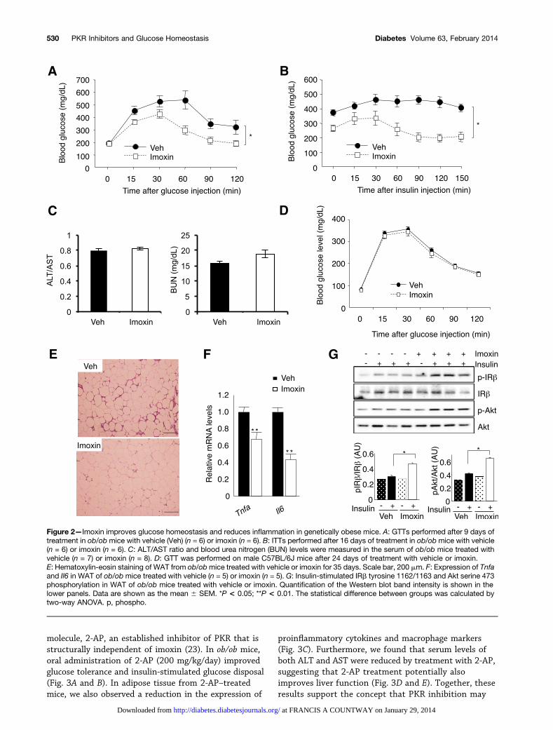
molecule, 2-AP, an established inhibitor of PKR that isstructurally independent of imoxin (23). In ob/ob mice,oral administration of 2-AP (200 mg/kg/day) improvedglucose tolerance and insulin-stimulated glucose disposal(Fig. 3A and B). In adipose tissue from 2-AP–treatedmice, we also observed a reduction in the expression of
proinflammatory cytokines and macrophage markers(Fig. 3C). Furthermore, we found that serum levels ofboth ALT and AST were reduced by treatment with 2-AP,suggesting that 2-AP treatment potentially alsoimproves liver function (Fig. 3D and E). Together, theseresults support the concept that PKR inhibition may
Figure 2—Imoxin improves glucose homeostasis and reduces inflammation in genetically obese mice. A: GTTs performed after 9 days oftreatment in ob/obmice with vehicle (Veh) (n = 6) or imoxin (n = 6). B: ITTs performed after 16 days of treatment in ob/obmice with vehicle(n = 6) or imoxin (n = 6). C: ALT/AST ratio and blood urea nitrogen (BUN) levels were measured in the serum of ob/ob mice treated withvehicle (n = 7) or imoxin (n = 8). D: GTT was performed on male C57BL/6J mice after 24 days of treatment with vehicle or imoxin.E: Hematoxylin-eosin staining of WAT from ob/obmice treated with vehicle or imoxin for 35 days. Scale bar, 200 mm. F: Expression of Tnfaand Il6 in WAT of ob/obmice treated with vehicle (n = 5) or imoxin (n = 5). G: Insulin-stimulated IRb tyrosine 1162/1163 and Akt serine 473phosphorylation in WAT of ob/ob mice treated with vehicle or imoxin. Quantification of the Western blot band intensity is shown in thelower panels. Data are shown as the mean 6 SEM. *P < 0.05; **P < 0.01. The statistical difference between groups was calculated bytwo-way ANOVA. p, phospho.
530 PKR Inhibitors and Glucose Homeostasis Diabetes Volume 63, February 2014
at FRANCIS A COUNTWAY on January 29, 2014http://diabetes.diabetesjournals.org/Downloaded from
generate metabolic benefits in mice with establisheddisease.
Effect of PKR Inhibition on Whole-Body EnergyMetabolism, Glucose Fluxes, and Insulin Sensitivity
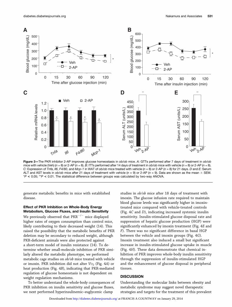
We previously observed that PKR2/2 mice displayedhigher rates of oxygen consumption than control mice,likely contributing to their decreased weight (14). Thisraised the possibility that the metabolic benefits of PKRdeletion may be secondary to reduced weight, althoughPKR-deficient animals were also protected againsta short-term model of insulin resistance (14). To de-termine whether small-molecule inhibition of PKR simi-larly altered the metabolic phenotype, we performedmetabolic cage studies on ob/ob mice treated with vehicleor imoxin. PKR inhibition did not alter VO2 (Fig. 4A) orheat production (Fig. 4B), indicating that PKR-mediatedregulation of glucose homeostasis is not dependent onweight regulation mechanisms.
To better understand the whole-body consequences ofPKR inhibition on insulin sensitivity and glucose fluxes,we next performed hyperinsulinemic–euglycemic clamp
studies in ob/ob mice after 18 days of treatment withimoxin. The glucose infusion rate required to maintainblood glucose levels was significantly higher in imoxin-treated mice compared with vehicle-treated controls(Fig. 4C and D), indicating increased systemic insulinsensitivity. Insulin-stimulated glucose disposal rate andsuppression of hepatic glucose production (HGP) weresignificantly enhanced by imoxin treatment (Fig. 4E andF). There was no significant difference in basal HGPbetween the vehicle and imoxin groups (Fig. 4G).Imoxin treatment also induced a small but significantincrease in insulin-stimulated glucose uptake in muscle(Fig. 4H). These data demonstrate that chemical in-hibition of PKR improves whole-body insulin sensitivitythrough the suppression of insulin-stimulated HGPand the enhancement of glucose disposal in peripheraltissues.
DISCUSSION
Understanding the molecular links between obesity andmetabolic syndrome may suggest novel therapeuticstrategies and targets for the treatment of this prevalent
Figure 3—The PKR inhibitor 2-AP improves glucose homeostasis in ob/ob mice. A: GTTs performed after 7 days of treatment in ob/obmice with vehicle (Veh) (n = 8) or 2-AP (n = 8). B: ITTs performed after 14 days of treatment in ob/obmice with vehicle (n = 8) or 2-AP (n = 8).C: Expression of Tnfa, Il6, F4/80, and Mcp-1 in WAT of ob/ob mice treated with vehicle (n = 8) or 2-AP (n = 8) for 21 days. D and E: SerumALT and AST levels in ob/ob mice after 21 days of treatment with vehicle (n = 9) or 2-AP (n = 9). Data are shown as the mean 6 SEM.*P < 0.05; **P < 0.01. The statistical difference between groups was calculated by two-way ANOVA.
diabetes.diabetesjournals.org Nakamura and Associates 531
at FRANCIS A COUNTWAY on January 29, 2014http://diabetes.diabetesjournals.org/Downloaded from

disease. Our group and others have previously found thatPKR is activated in adipose and other tissues duringobesity in mouse models and in obese humans, and iscritical to the development of metaflammation and in-sulin resistance. Studies in two independent lines of PKRdeficiency in mice showed protection against insulin re-sistance and diabetes (14,17). Hence, we examinedwhether pharmacological inhibition of PKR action couldreduce obesity-induced insulin resistance and metabolicdysfunction. Here, we find that two distinct small-molecule chemical inhibitors of PKR kinase activity
improved glucose homeostasis and insulin sensitivity,and ameliorated adipose inflammation in geneticallyobese mice. Thus, our findings suggest that PKR in-hibition may be a viable interventional strategy in thetreatment of established metabolic disorders.
In this study, PKR inhibition had no effect on glucosetolerance or insulin sensitivity in lean mice, in agreementwith our previous finding of normal glucose homeostasisin lean PKR knock-out animals (14). This suggests that,despite its role as a nutrient and stress sensor, PKR ac-tivity is dispensable for the modulation of insulin
Figure 4—Imoxin treatment improves systemic insulin sensitivity without altering metabolic rate. Metabolic cage studies wereperformed on ob/ob mice treated with vehicle (Veh) (n = 10) or imoxin (n = 10). A: Rate of VO2. B: Heat production. Hyperinsulinemic–euglycemic clamp studies were performed in ob/ob mice after 18 days of treatment with vehicle (n = 8) or imoxin (n = 5). C: Glucoseinfusion rates (GIRs) during the clamp procedure. D: Average (GIR). E: Whole-body glucose disposal rates (Rd). F: HGP duringthe clamp. G: Basal HGP. H: Tissue glucose uptake in gastrocnemius muscle. Data are shown as the mean 6 SEM. *P < 0.05;**P < 0.01.
532 PKR Inhibitors and Glucose Homeostasis Diabetes Volume 63, February 2014
at FRANCIS A COUNTWAY on January 29, 2014http://diabetes.diabetesjournals.org/Downloaded from
responsiveness and adipose inflammation under ho-meostatic conditions. An independent study has sug-gested that PKR deficiency resulting from a deletion inthe RNA-binding domain may increase insulin sensitivityand metabolic health even in lean animals (17). Hence, itis possible that the inhibition achieved through syntheticchemical inhibition is not fully replicating these obser-vations in the lean mice with whole-body genetic ablationof PKR or that there are subtle differences in the deletionmodels. Nevertheless, human studies (18–20) and mul-tiple genetic models (14,17) have provided evidencesupporting the consideration of this enzyme for thera-peutic purposes and should stimulate the development oforally active molecules with enhanced pharmacologicalproperties. Total-body PKR deficiency in mice does notappear to be associated with major immunological com-plications. However, it is worth noting that, in addition tocellular stress signals, PKR can sense pathogens and playsan important role in inflammasome activation (16). Hence,it will be important to examine whether systemic PKRinhibition as used in this paradigm would hinder in-flammatory responses to infection, especially under long-term usage, and to consider alternative targeting strategies.
Our findings here also suggest that blocking PKRactivity in established disease models has the ability toprovide benefits independent of body weight regulation.This raises some interesting possibilities for testing theefficacy of PKR blockade in other diseases. For example,PKR has previously been implicated in mediating b-celldeath as part of the antiviral response that may pre-cipitate the development of type 1 diabetes in somepatients (23). Thus, in addition to our finding that PKRinhibitors can improve glucose homeostasis in a mousemodel of insulin resistance and type 2 diabetes, PKRinhibitors may also have potential as early interven-tional agents in type 1 diabetes models or other chronicmetabolic diseases associated with similar stress, in-flammatory, and/or metabolic underpinnings.
In conclusion, this work further supports the hypoth-esis that PKR plays an important role in integrating nu-trient and stress signaling, and the progression andpersistence of insulin resistance during obesity. As theglobal chronic metabolic disease epidemic continues, it willbe critical to identify new therapeutic entities that can beused to both prevent metabolic disease from developingand to treat individuals after pathologies such as insulinresistance occur. These findings suggest PKR inhibitorsmay offer a strategy to alleviate metaflammation andserve as potential agents to fill this niche.
Acknowledgments. The authors thank Kathryn Claiborn, HarvardSchool of Public Health, for editorial input.
Funding. T.N. was supported by a fellowship from the Human FrontierScience Program and is currently supported by a grant from the AmericanHeart Association. This work is supported in part by a grant from the NationalInstitutes of Health to G.S.H. (DK052539).
Duality of Interest. No potential conflicts of interest relevant to thisarticle were reported.
Author Contributions. T.N. conceived the hypothesis, designed andperformed experiments, analyzed the results, and wrote the manuscript. A.A.,B.B., and M.F. performed experiments and analyzed results. G.S.H. conceivedthe hypothesis, designed the experiments, analyzed the results, and wrote themanuscript. G.S.H. is the guarantor of this work and, as such, had full accessto all the data in the study and takes responsibility for the integrity of the dataand the accuracy of the data analysis.
References1. Hotamisligil GS. Inflammation and metabolic disorders. Nature 2006;444:
860–867
2. Hotamisligil GS, Erbay E. Nutrient sensing and inflammation in metabolicdiseases. Nat Rev Immunol 2008;8:923–934
3. Baker RG, Hayden MS, Ghosh SNF. NF-kB, inflammation, and metabolicdisease. Cell Metab 2011;13:11–22
4. Arkan MC, Hevener AL, Greten FR, et al. IKK-beta links inflammation toobesity-induced insulin resistance. Nat Med 2005;11:191–198
5. Cai D, Yuan M, Frantz DF, et al. Local and systemic insulin resistanceresulting from hepatic activation of IKK-beta and NF-kappaB. Nat Med2005;11:183–190
6. Hirosumi J, Tuncman G, Chang L, et al. A central role for JNK in obesityand insulin resistance. Nature 2002;420:333–336
7. Kaneto H, Nakatani Y, Miyatsuka T, et al. Possible novel therapy for di-abetes with cell-permeable JNK-inhibitory peptide. Nat Med 2004;10:1128–1132
8. Osborn O, Olefsky JM. The cellular and signaling networks linking theimmune system and metabolism in disease. Nat Med 2012;18:363–374
9. Chawla A, Nguyen KD, Goh YPS. Macrophage-mediated inflammation inmetabolic disease. Nat Rev Immunol 2011;11:738–749
10. Shoelson SE, Lee J, Goldfine AB. Inflammation and insulin resistance.J Clin Invest 2006;116:1793–1801
11. Nakatani Y, Kaneto H, Kawamori D, et al. Modulation of the JNK pathway inliver affects insulin resistance status. J Biol Chem 2004;279:45803–45809
12. Wunderlich FT, Luedde T, Singer S, et al. Hepatic NF-kappa B essentialmodulator deficiency prevents obesity-induced insulin resistance butsynergizes with high-fat feeding in tumorigenesis. Proc Natl Acad Sci USA2008;105:1297–1302
13. Reilly SM, Chiang SH, Decker SJ, et al. An inhibitor of the protein kinasesTBK1 and IKK-ɛ improves obesity-related metabolic dysfunctions in mice.Nat Med 2013;19:313–321
14. Nakamura T, Furuhashi M, Li P, et al. Double-stranded RNA-dependentprotein kinase links pathogen sensing with stress and metabolic homeo-stasis. Cell 2010;140:338–348
15. Komiya K, Uchida T, Ueno T, et al. Free fatty acids stimulate autophagy inpancreatic b-cells via JNK pathway. Biochem Biophys Res Commun 2010;401:561–567
16. Lu B, Nakamura T, Inouye K, et al. Novel role of PKR in inflammasomeactivation and HMGB1 release. Nature 2012;488:670–674
17. Carvalho-Filho MA, Carvalho BM, Oliveira AG, et al. Double-stranded RNA-activated protein kinase is a key modulator of insulin sensitivity in physio-logical conditions and in obesity in mice. Endocrinology 2012;153:5261–5274
18. Boden G, Duan X, Homko C, et al. Increase in endoplasmic reticulumstress-related proteins and genes in adipose tissue of obese, insulin-resistant individuals. Diabetes 2008;57:2438–2444
diabetes.diabetesjournals.org Nakamura and Associates 533
at FRANCIS A COUNTWAY on January 29, 2014http://diabetes.diabetesjournals.org/Downloaded from

19. Gregor MF, Yang L, Fabbrini E, et al. Endoplasmic reticulum stress isreduced in tissues of obese subjects after weight loss. Diabetes 2009;58:693–700
20. Carvalho BM, Oliveira AG, Ueno M, et al. Modulation of double-strandedRNA-activated protein kinase in insulin sensitive tissues of obese patients.Obesity 2013;21:2452–2457
21. Eley HL, Russell ST, Tisdale MJ. Attenuation of muscle atrophy in a murinemodel of cachexia by inhibition of the dsRNA-dependent protein kinase.Br J Cancer 2007;96:1216–1222
22. Jammi NV, Whitby LR, Beal PA. Small molecule inhibitors of the RNA-dependent protein kinase. Biochem Biophys Res Commun 2003;308:50–57
23. Hu Y, Conway TW. 2-Aminopurine inhibits the double-stranded RNA-dependent protein kinase both in vitro and in vivo. J Interferon Res 1993;13:323–328
24. Scarim AL, Arnush M, Blair LA, et al. Mechanisms of b-cell death inresponse to double-stranded (ds) RNA and interferon-g: dsRNA-dependentprotein kinase apoptosis and nitric oxide-dependent necrosis. Am J Pathol2001;159:273–283
534 PKR Inhibitors and Glucose Homeostasis Diabetes Volume 63, February 2014
at FRANCIS A COUNTWAY on January 29, 2014http://diabetes.diabetesjournals.org/Downloaded from


![Received: 2016.02.21 The Specific Protein Kinase R (PKR ...shown that PKR participates in neurodegenerative processes with neurotoxicity [12,13]. Peel and Couturier considered PKR](https://img.pdfslide.us/doc/110x75/5e45e3e2e3e94073247c9161/received-20160221-the-specific-protein-kinase-r-pkr-shown-that-pkr-participates.jpg)