Embed Size (px)
Citation preview
Translational Science
A Novel L-Asparaginase with low L-GlutaminaseCoactivity IsHighlyEfficaciousagainstBothT-andB-cell Acute Lymphoblastic Leukemias In VivoHien Anh Nguyen1,2, Ying Su1,2, Jenny Y. Zhang2, Aleksandar Antanasijevic2,Michael Caffrey2, Amanda M. Schalk2, Li Liu3, Damiano Rondelli4, Annie Oh4,Dolores L. Mahmud4, Maarten C. Bosland5, Andre Kajdacsy-Balla5, Sofie Peirs6,7,Tim Lammens7,8, Veerle Mondelaers7,8, Barbara De Moerloose7,8, Steven Goossens6,7,Michael J. Schlicht5, Kasim K. Kabirov9, Alexander V. Lyubimov9, Bradley J. Merrill2,Yogen Saunthararajah10, Pieter Van Vlierberghe6,7, and Arnon Lavie1,2
Abstract
Acute lymphoblastic leukemia (ALL) is the most commontype of pediatric cancer, although about 4 of every 10 casesoccur in adults. The enzyme drug L-asparaginase serves asa cornerstone of ALL therapy and exploits the asparaginedependency of ALL cells. In addition to hydrolyzing theamino acid L-asparagine, all FDA-approved L-asparaginases alsohave significant L-glutaminase coactivity. Since several reportssuggest that L-glutamine depletion correlates with many ofthe side effects of these drugs, enzyme variants with reducedL-glutaminase coactivity might be clinically beneficial if their
antileukemic activity would be preserved. Here we show thatnovel low L-glutaminase variants developed on the backbone ofthe FDA-approved Erwinia chrysanthemi L-asparaginase werehighly efficacious against both T- and B-cell ALL, while dis-playing reduced acute toxicity features. These results supportthe development of a new generation of safer L-asparaginaseswithout L-glutaminase activity for the treatment of human ALL.
Significance:A new L-asparaginase–based therapy is less toxiccompared with FDA-approved high L-glutaminase enzymesCancer Res; 78(6); 1549–60. �2018 AACR.
IntroductionBacterial L-asparaginases are enzymes with dual activities. The
predominant one, the L-asparaginase activity that gives theseenzymes their name, is the ability to hydrolyze the amino acidL-asparagine (Asn) into L-aspartic acid (Asp) and ammonia.
The secondary activity present in L-asparaginases is an L-gluta-minase activity, which drives hydrolysis of L-glutamine (Gln)to L-glutamic acid (Glu) and ammonia. For the FDA-approvedL-asparaginases [Escherichia coli (EcA) and Erwinia chrysanthemi(ErA), approved in 1978 and 2011, respectively], the L-gluta-minase activity ranges from 2% to 10% of their primaryL-asparaginase activity (1). The dual L-asparaginase andL-glutaminase property of EcA and ErA is expected to manifestitself in the depletion of both Asn and Gln in the patient'sblood, a notion that is supported by several studies (2–5).
The anticancer effect of L-asparaginase is believed to be pre-dominantly due to the depletion of Asn from the blood. Indeed,leukemic blasts from acute lymphoblastic leukemia (ALL)patients completely depend on scavenging Asn from the blood,as they lack or display very low levels of the asparagine synthetase(ASNS) enzyme (6–8). In contrast, the clinical importance of theL-glutaminase activity present in all FDA-approved versions of L-asparaginases is still under debate, with conflicting reports in theliterature about its putative antileukemic effect (9, 10). On onehand, pharmacodynamic analyses showed that deamination ofGln is critically required for optimal Asn deamination (2) andother more recent studies indicated contribution of L-glutamin-ase activity to the cytotoxicity of L-asparaginase on leukemiccells (10, 11). In contrast, others have found that the L-gluta-minase activity is not required for the drug's in vitro anticancereffect, as long as the ALL cells lack ASNS (9).
Common side effects in patients treated with L-asparaginases,in addition to an immune response against the bacterialenzymes, include hepatotoxicity, hyperglycemia, dyslipidemia,
1The Jesse Brown VA Medical Center, Chicago, Illinois. 2Department of Bio-chemistry and Molecular Genetics, University of Illinois at Chicago, Chicago,Illinois,. 3Division of Epidemiology and Biostatistics, School of Public Health,University of Illinois at Chicago, Chicago, Illinois. 4Division of Hematology/Oncology, University of Illinois Hospital and Health Sciences System, Chicago,Illinois. 5Department of Pathology, University of Illinois at Chicago, Chicago,Illinois. 6Center for Medical Genetics, Ghent University Hospital, Ghent, Belgium.7Cancer Research Institute Ghent (CRIG), Ghent, Belgium. 8Department ofPediatric Hematology-Oncology and Stem Cell Transplantation, Ghent Univer-sity Hospital, Ghent, Belgium. 9Toxicology Research Laboratory, Department ofPharmacology, University of Illinois at Chicago, Chicago, Illinois. 10Department ofTranslational Hematology & Oncology Research, Cleveland Clinic Foundation,Cleveland, Ohio.
Note: Supplementary data for this article are available at Cancer ResearchOnline (http://cancerres.aacrjournals.org/).
Corresponding Authors: Arnon Lavie, The University of Illinois at Chicago, 900South Ashland Avenue, MBRB Room 1108, Chicago, IL 60607. Phone: 312-355-5029; Fax: 312-355-4535; E-mail: [email protected]; Pieter Van Vlierberghe, GhentUniversity, Center For Medical Genetics Ghent, Medical Research Building 2, 1stFloor, Room 110.006, Corneel Heymanslaan 10, Ghent 9000, Belgium. Phone:329-332-1043; E-mail: [email protected]
doi: 10.1158/0008-5472.CAN-17-2106
�2018 American Association for Cancer Research.
CancerResearch
www.aacrjournals.org 1549
on July 12, 2019. © 2018 American Association for Cancer Research. cancerres.aacrjournals.org Downloaded from
Published OnlineFirst January 17, 2018; DOI: 10.1158/0008-5472.CAN-17-2106
perturbations in blood coagulation factors, and pancreatitis(12–14). Several clinical studies have documented the Glndepletion resulting from the L-glutaminase coactivity of currentL-asparaginase preparations (4, 15, 16), and suggested thatthe aforementioned side effects can, at least in part, be attrib-uted to this property of the drugs. For example, a link betweenthe L-glutaminase activity and the immunosuppressive effectsof these drugs have been reported (17, 18), as well as its role inhepatotoxicity (19), which was proposed to be due to delete-rious effects on Gln homeostasis (3). Likewise, Gln depletioncould likely contribute significantly to the disrupted proteinsynthesis in the liver and spleen that is a cause of the coagulo-pathy aspects of drug toxicity (20). Moreover, hydrolysis ofboth Asn and Gln will produce ammonia as a byproduct of thereaction. However, given that Gln concentrations are muchhigher in the blood as compared with Asn, Gln hydrolysis willhave a more profound effect on the eventual concentration ofammonia in the blood. Indeed, hyperammonemia has beenobserved in patients undergoing L-asparaginase treatment(16, 21–25), which has been associated with neurotoxicity.
Additional information on the putative interplay betweenL-glutaminase activity and drug toxicity came from at least fourclinical trials of L-asparaginases. First, in the early 1980s, a clinicaltrial, which examined an L-asparaginase from Acinetobacter withvery high L-glutaminase activity, was forced to terminate early dueto central nervous system toxicity (26). Second, between2001 and2008, the L-asparaginase from Wolinella succinogenes, which wasinitially thought to be a low L-glutaminase enzyme, was evaluatedclinically through a US National Cancer Institute Rapid Access toIntervention Development (NCI RAID) grant. However, theenzyme produced via this program was found to be toxic inpatients and we recently showed that it actually does containsignificant L-glutaminase activity (27). Third, in 2008, a phase IIclinical trial examining the FDA-approved EcA in ovarian cancerpatients had to be terminated early due to excessive toxicities (28).Interestingly, while weight loss was reported as one of the maindrug-related toxicities in the phase II ovarian cancer study, it isalso a significant L-glutaminase–related toxicity indicator in ouractual preclinical study. Finally and very recently, a clinical trial oferyaspase (red blood cell encapsulated EcA) showed that, for a yetunclear reason, the encapsulation process reduced the L-gluta-minase activity (i.e., increased the selectivity for Asn hydrolysisoverGlnhydrolysis), a factor pointed out as an explanation for thedecrease in adverse events in the eryaspase clinical trial comparedwith naked EcA (29). Hence, these trials support the notion thatcertain side effects observed in patients undergoing L-asparaginasetreatment might be associated with the level of L-glutaminaseactivity. Therefore, reducing the L-glutaminase activity of availableL-asparaginases may be advantageous to lessen toxic side effects,but for now, it is unclearwhether thiswouldbedetrimental for theantileukemic efficacy of these drugs.
Previously, we engineered variants of ErA with decreasedL-glutaminase activity while maintaining near wild-type L-aspar-aginase activity (30). Here we evaluated these novel ErAvariants in vitro and in vivo for their ability to kill ALL cells, andcompared them with their wild-type counterpart. It is importantto appreciate the experimental complexity when comparing dif-ferent L-asparaginases for their efficacy and toxicity, as in additionto the kinetic properties of the enzyme drugs, pharmacokineticsand immunogenicity (when tested in patients) play a major rolein determining the outcome. To simplify the interpretation of the
results, here we present the comparison of L-asparaginases thathave similar L-asparaginase activities and that only differ by 1–3residues, suggesting very similar pharmacokinetic properties, butthat have vastly different L-glutaminase activity. Together, ourresults suggest that high L-glutaminase activity, as present incurrent FDA drugs, is not essential for efficient in vivo eliminationof L-asparaginase–sensitive ALL cells. In addition, reduced toxicitywas observed in the low L-glutaminase variants compared withthe high L-glutaminase enzymes. This sets up the rationale forfurther evaluation of such low L-glutaminase variants, which arepredicted to have fewer side effects, as alternatives to the currentFDA-approved bacterial L-asparaginases for the treatment of ALL.
Materials and MethodsExpression and purification of L-asparaginases
Enzymes used for kinetic, NMR, and cell culture studies wereexpressed and purified as previously reported in Nguyen andcolleagues' study (30, 31) for ErA-WT, ErA-E63Q, ErA-DM, andErA-TM; and as in Schalk and colleagues (32) for EcA-WT.
Kinetic assaysL-asparaginase and L-glutaminase activities were determined
using a continuous spectroscopic enzyme–coupled assay asdescribed previously (32, 33).
Cell cultureThe LOUCY cell line was established from the peripheral blood
of a T-cell ALL patient (34). The luciferase-positive LOUCY cellline was generated as described previously (35). The SUP-B15 celllinewas established fromcells harvested from thebonemarrowofa Philadelphia chromosome–positive B-cell ALL patient (36). Theluciferase-expressing SUP-B15 cell line was a kind gift fromDr. Michael Jensen (University of Washington School of Medi-cine, Seattle, WA). All cell lines were analyzed by STR (shorttandem repeat) and confirmed to match 100% to correspondingSTRprofile data from theGlobal BioresourceCenter ATCC.All celllines were verified to be mycoplasma free. The Alamar Blue assayfor cell viability is described in Supplementary Methods. IC50
values were determined by GraphPad Prism 6.0 using sigmoidalinterpolation model with 95% confidence intervals.
In vivo treatment of cell line xenografts with L-asparaginasesNonobese diabetic/severe combined immunodeficient g
(NSG) mice (The Jackson Laboratory) were intravenouslyinjected at 6 weeks of age with 150-mL DPBS containing5 � 106 luciferase-positive LOUCY or SUP-B15 cells. At regulartime points, the bioluminescence was measured using the IVISLumina II imaging system (PerkinElmer). After evidence ofleukemic cell engraftment, the mice were randomly dividedinto different groups that were administered via intraperito-neal injection at a dose of 50 IU/mouse daily for 14 days witheither ErA-WT, ErA-E63Q, ErA-DM, ErA-TM or the same vol-ume of DPBS. In another experiment, LOUCY-engrafted micewere treated with 25 IU/mouse at days 0, 2, 4, 7, 9, 11, 12, 13,and 14 via intraperitoneal injection. The bioluminescent imag-ing (BLI) signal was measured every two to three days asindicated in Fig. 2 and Supplementary Figs. S1, S2, and S3.During the experiment, the mice were observed and weighedevery day. The ethical committee on animal welfare at Uni-versity of Illinois at Chicago approved this animal experiment.
Nguyen et al.
Cancer Res; 78(6) March 15, 2018 Cancer Research1550
on July 12, 2019. © 2018 American Association for Cancer Research. cancerres.aacrjournals.org Downloaded from
Published OnlineFirst January 17, 2018; DOI: 10.1158/0008-5472.CAN-17-2106
Acute toxicity studyThe experimental design of this study incorporated a blinded
strategy where the toxicologist was providedwith samples labeledas #1 and #2, without knowing the identity of the enzymes(ErA-WT or ErA-TM). In this dose escalation study, 6 animals(3 males, 3 females) per dose group were administered theenzymes intravenously at a starting dose of 40 IU/g, increasingto 80 and finally 160 IU/g. Because of a shortage of the enzymes,ErA-TM group 6was limited to 4 animals (3 females, 1male), anda few animals did not receive the full intended dose (one animalof ErA-WT group 3 received 136 instead of the intended 160 IU/g,one animal of the ErA-TM group 5 received 60 instead of theintended 80 IU/g). The unexpected shortage of enzymes was dueto higher than expected loss during filtration through a 0.22-mmfilter and the adjustment needed for bigger body weight of themice. After enzyme administration, the animals were monitoreddaily and clinical signs (hunched posture, decreased activity,sunken eyes, and rough coat) were noted if observed. None ofthe animals died during the 4-day observation period, and all theanimals were euthanized at the end of day 4.
In vivo asparaginase activity determinationC57BL/6 mice of 7–10 weeks old were intraperitoneally
injected with two batches of 50 IU of ErA-WT or ErA-TM. Twen-ty-four hours after the injection, peripheral blood was collected(5 animals per group) via cardiac puncture under anesthesia (5%isoflurane in oxygen). Shortly after collection, blood was centri-fuged in heparin-coated tubes (2,000 � g, 10 minutes, 4�C)for plasma preparation. Plasma L-asparaginase activity was quan-tified by incubating the samples with an excess amount of L-aspartic acid b-hydroxamate (AHA; Sigma-Aldrich A6508) at37.0�C. L-Asparaginase hydrolyses AHA to L-aspartic acid andhydroxylamine, which was detected at 690 nmwith a SpectraMaxM3 (Molecular Devices) spectrophotometer, after condensationwith 8-hydroxyquinoline (Merck 8.20261) and oxidation toindo-oxine. Detailed procedure can be found in the Supplemen-tary Data. The ethical committee on animal welfare at GhentUniversity Hospital approved the experiment.
In vivo pharmacodynamics of amino acidC57BL/6 mice of 7–10 weeks old were intraperitoneally
injected with 50 IU of ErA-WT or ErA-TM. For the pharamaco-dynamic study, peripheral blood was collected at days 1, 3, 7, and14 (5 animals per group) via cardiac puncture under anesthesia(5% isoflurane in oxygen). In addition, blood of seven untreatedmice was collected to determine the baseline value (day 0).Shortly after collection, blood was centrifuged in heparin-coatedtubes (2,000 � g, 10 minutes, 4�C) for plasma preparation.Plasma was diluted with equal volume of a 10% 5'-sulfosalicylicacid dihydrate solution in water and stored at �80�C for thedetermination of amino acid levels.
For the amino acid analysis, the plasma samples (50 mL) weredeproteinized by adding 100 mL of a 10% sulfosalicylic acidsolution containing 50 mmol/L internal standards mix. Aftervortexing, 50 mL of UPLC-grade water was added. Centrifugationoccurred for 10 minutes at 9,960 � g. Derivatization of 10 mL ofsupernatant was performed according to the manufacturer'sinstructions of the AccQ-Tag kit of Waters. The 4 amino acids(asparagine, aspartic acid, glutamine, and glutamic acid) weremeasured on an Acquity UPLC with QDA detector of Waters andquantified based on a 5-point calibration curve. The ethical
committee on animal welfare at Ghent University Hospitalapproved these experiments.
Patient-derived xenograft experimentA xenograft of a pediatric primary human T-ALL sample was
established in NSGmice. Upon establishment of disease, humanleukemic cells were isolated from the spleen via Ficoll-Paque (GEHealthcare) density gradient centrifugation. Next, these cells wereinjected in the tail vein of 15 female NSG mice at 7 weeks of age.Each mouse received 150-mL PBS containing 1.2 � 106 cells.Engraftment of the cells was followed by measuring the percent-age of human CD45-positive (%huCD45þ) cells in the blood.Upon evidence of leukemic cell engraftment,mice were randomlydivided into 3 groups (day 0), and treated daily via intraperitonealinjection with 50 IU/mouse for 13 days of either ErA-WT, ErA-TMor the same volume of PBS. At day 0, 7, and 13, blood wascollected via the tail vein. At day 13, all mice were sacrificed andthe spleen and bone marrow were collected. The %huCD45þ
cells in the blood, bone marrow, and spleen was analyzed bystaining with a phycoerythrin-labeled antibody for human CD45(130-098-141; Miltenyi Biotec), performing red blood cell lysisand measuring the percentage on a LSRII flow cytometer usingFACSDiva software (BD Biosciences). During the experiment,mice were observed and weighed every day. The experiment wasapproved by the ethical committee on animal welfare at GhentUniversity Hospital.
qPCR experimentsTotal RNA was isolated using the miRNeasy Mini Kit (Qiagen)
and the RNAse-Free DNAse set (Qiagen). cDNA was synthesizedwith the iScript Advanced cDNA synthesis kit (Bio-Rad). TheSsoAdvancedUniversal SYBRGreen Supermix (Bio-Rad)was usedand the PCR reactions were run on the LightCycler 480 (Roche,model LC480). Every sample was analyzed in duplicate.qBasePLUS software (Biogazelle) was used for analysis. Geneexpression was normalized against 3 reference genes (GAPDH,TBP, YWHAZ). ASNS primers: (F) 5'-CCCTGCACGCCCTCTATG-3', (R) 5'-GGATCCTGAGGTTGTTCTTCACA-3'; GAPDH primers:(F) 5'-TGCACCACCAACTGCTTAGC-3', (R) 5'- GGCATGGACTG-TGGTCATGAG-3'; TBP primers: (F) 5'- CACGAACCACGGCACT-GATT -3', (R) 5'- TTTTCTTGCTGCCAGTCTGGAC -3' and YWHAZprimers: (F) 5'-ACTTTTGGTACATTGTGGCTTCAA-3', (R) 5'-CCGCCAGGACAAACCAGTAT-3'.
Statistical analysisSee Supplementary Data file for details on biostatistics.
ResultsDesign and characterization of ErA variants with highL-asparaginase and low L-glutaminase activities
To determine whether L-asparaginase variants with lowL-glutaminase activitymay hold clinical potential, we investigatedseveral L-glutaminase–deficient ErA variants [denoted as ErA-E63Q, ErA-DM (double mutant) and ErA-TM (triple mutant)]that retain most of their wild-type L-asparaginase activity (30).Comparisons of kinetic parameters between these ErA variantsand the FDA-approved wild-type versions of ErA and EcA(denoted as ErA-WT and EcA-WT) are summarized in Table 1.We selected ErA-WT over EcA-WT as the backbone to develop lowL-glutaminase variants because of its superior L-asparaginaseactivity (�2.5-fold higher rate than EcA-WT in hydrolyzing Asn
ASNase Efficacy with Low L-Glutaminase Coactivity
www.aacrjournals.org Cancer Res; 78(6) March 15, 2018 1551
on July 12, 2019. © 2018 American Association for Cancer Research. cancerres.aacrjournals.org Downloaded from
Published OnlineFirst January 17, 2018; DOI: 10.1158/0008-5472.CAN-17-2106
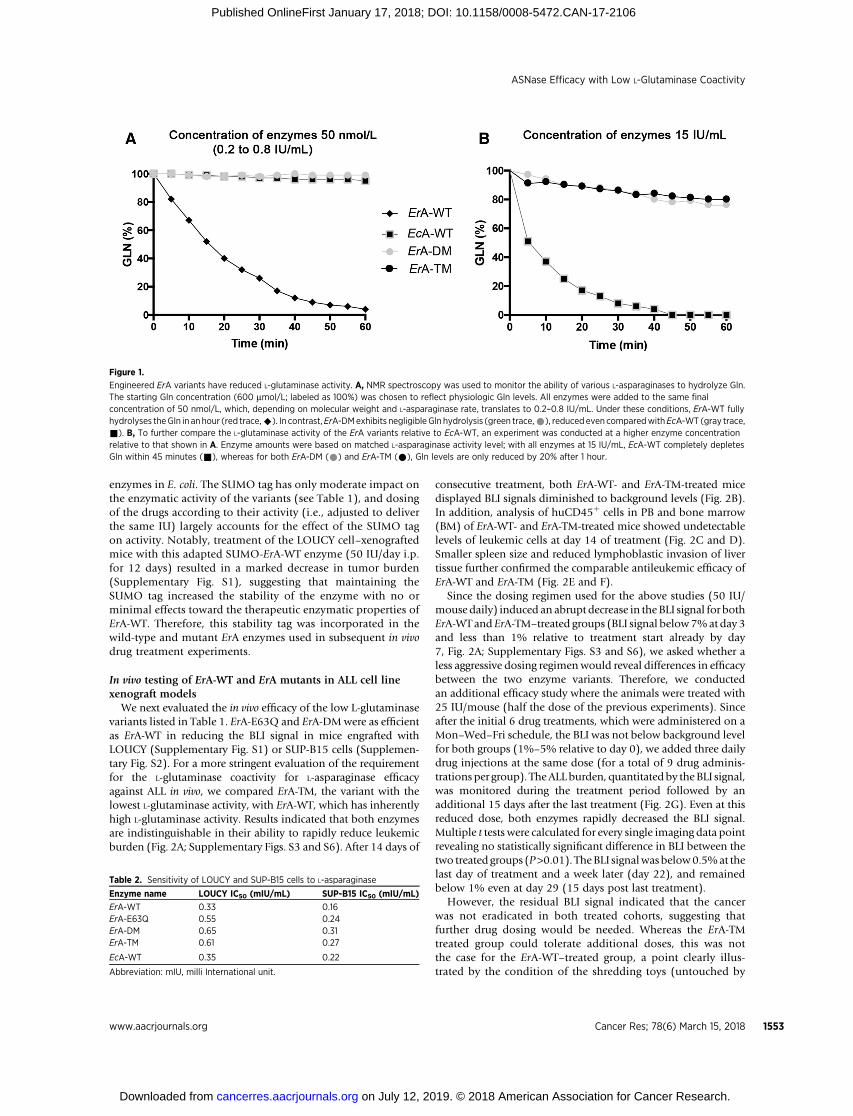
at the physiologic concentration of 50mmol/L).However, ErA-WTalso has >70-fold higher L-glutaminase activity comparedwith EcA-WT at the physiologic concentration of 500 mmol/LGln. As both kcat (i.e., the rate at saturating substrate concentra-tion) and Km influence the turnover of enzyme-catalyzed reac-tions, the kcat/Km ratio is often taken as a measure of enzymeefficiency, and this ratio is shown in Table 1 for the L-asparaginaseand L-glutaminase activities of the examined enzymes. However,to assess the specificities of the enzymes, we also calculatedthe ratio between the kcat/Km of the L-asparaginase reaction tothe kcat/Km of the L-glutaminase reaction (the larger the number,the higher is the specificity for the L-asparaginase reaction). Fromthis calculation, it is clear thatErA-TMandErA-DMaremuchmoreL-asparaginase specific (ratio ¼ 68,750 and 4,842, respectively)compared with the original ErA-WT enzyme (ratio ¼ 58.6), butthat only ErA-TM is significantly more specific than EcA-WT (ratio¼ 4,625). However, this analysis based on the ratios of the L-asparaginase and L-glutaminase kcat/Kmvaluesmaynot best reflectthe physiologic conditions. Therefore, we also calculated speci-ficity ratios based on the observed rates (kobs) at physiologicsubstrate concentrations (50 mmol/L for Asn, 500 mmol/L forGln). These calculations show that our engineered ErA variantshave significantly superior Asn:Gln specificity as compared withErA-WT (Table 1). Using this calculation, even ErA-DM is about 2-fold more L-asparaginase–specific compared with EcA-WT, withErA-TM being 47-fold more specific. The reduced rate of L-gluta-minase activity for each ErA mutant was also demonstratedthrough measuring changes to Gln concentrations over time byNMR spectroscopy (Fig. 1A and B). Notably, while EcA-WTcompletely hydrolyzed Gln in approximately 45 minutes, solu-tions with ErA-DM and ErA-TM contained >80% of the startingGln after 1 hour, demonstrating their exceptionally low L-gluta-minase activities (Fig. 1B).
In vitro testing of ErA-WT and ErA mutants in ALL cell linesThe development of ErA mutants with comparable L-aspar-
aginase but variable L-glutaminase activity (ErA-E63Q > ErA-DM > ErA-TM) allowed us to test whether the high intrinsicL-glutaminase activity of ErA-WT is truly required for its clinical
efficacy. Notwithstanding the limitations of evaluatingL-asparaginase in cell culture, we first validated the antiproli-ferative effect of wild-type and mutant L-asparaginases in vitroon the human leukemic cell lines, LOUCY (T-ALL) and SUP-B15 (B-ALL). Results indicated that both ALL cell lines weresimilarly sensitive to ErA-WT, EcA-WT, and to each of theL-glutaminase–deficient ErA mutants (Table 2; SupplementaryFig. S4). As most cell lines depend on high Gln levels in culture,the slightly lower IC50 values for ErA-WT and EcA-WTcompared with the L-glutaminase-deficient ErA mutants isnot surprising.
The His-SUMO tag acts to stabilize the ErA variants in vivoGiven that in vitro studies cannot unambiguously clarify
whether L-glutaminase activity is required for in vivo effective-ness of L-asparaginases, we subsequently used xenograftmodels of luciferase-positive LOUCY and SUP-B15 cells toperform in vivo drug treatment experiments. Engraftment ofhuman leukemic cells in mice is often considered successfulwhen the percentage of peripheral blood (PB) cells positive forthe human CD45 antigen (%huCD45þ) is �1%–2% (37, 38).Four weeks after NOD-scid IL2Rgammanull mice (NSG) receivedcell line injections, bioluminescence imaging (BLI) flux signalscorresponding to a PB %huCD45þ greater than 8% confirmedsuccessful engraftment and showcased the high level of diseaseburden in the examined animals (Supplementary Figs. S1, S2,S3, and S5 report the calibration between BLI flux and PB%huCD45þ). With this level of engraftment, daily drug treat-ment with ErA-WT (intraperitoneal injection of 50 IU/day for 14 days) was initiated. Surprisingly, this FDA-approvedL-asparaginase failed to reduce tumor cell growth in vivo.Of note, ErA-WT has a half-life of only 0.65 days in humans,compared with 1.24 days for EcA-WT (39). Furthermore, half-lives of these drugs are dramatically shortened in mice (40).Thus, we hypothesized that the short half-life of ErA-WT pre-vented therapeutic efficacy. To evaluate whether drug instabilityindeed hindered the anticancer effect, we retained the N-ter-minal SUMO tag, which was originally incorporated to increasestability and facilitate the heterologous expression of the
Table 1. Enzyme kinetic parameters
Enzyme name kcat (sec�1) Km (mmol/L) kcat/Km (sec�1 mmol�1/L) kobs @50 mmol/La (sec�1) kobs @50 mmol/Lb (sec�1)
ErA-WT 207.5 � 3.6 47.5 � 3.5 4.37 118.9 145.8L-asparaginase ErA-TM 261.2 � 2.8 95.0 � 3.5 2.75 79.6 56.4activity ErA-DM 169.8 � 1.5 185.3 � 5.5 0.92 22.4 23.3
ErA-E63Q 186.8 � 1.7 50.7 � 2.0 3.68 112.7 135.9
EcA-WT 44.4 � 0.3 15.0 � 0.5 2.96 41.3Enzyme name kcat (sec
�1) Km (mmol/L)c kcat/Km (sec�1 mmol�1/L)c kobs @0.5 mmol/La (sec�1) kobs @0.5 mmol/Lb (sec�1)ErA-WT 26.84 � 0.26 360 � 20 74.56 � 10�3 15.87 14.76
L-glutaminase ErA-TM 1.84 � 0.11 47,460 � 695 0.04 � 10�3 0.01 0.04activity ErA-DM 2.93 � 0.03 15,800 � 300 0.19 � 10�3 0.11 0.09
ErA-E63Q 8.33 � 0.16 3,860 � 230 3.68 � 10�3 0.74 1.05
EcA-WT 0.89 � 0.01 1,380 � 90 0.64 � 10�3 0.22Enzyme name kobs[Asnphs]/kobs[Glnphs]
d kcat/Km (Asn)/kcat/Km (Gln)
ErA-WT 6.6 58.6Specificity ErA-TM 8910 68,750
ErA-DM 330 4,842ErA-E63Q 124.6 1,000
EcA-WT 187.7 4,625akobs for enzymes without the SUMO tag.bkobs for enzymes with the SUMO tag.cConcentrations are given in mmol/L to facilitate comparison with the L-asparaginase data.dkobs for Asn@50 mmol/L, kobs for Gln@500 mmol/L.
Nguyen et al.
Cancer Res; 78(6) March 15, 2018 Cancer Research1552
on July 12, 2019. © 2018 American Association for Cancer Research. cancerres.aacrjournals.org Downloaded from
Published OnlineFirst January 17, 2018; DOI: 10.1158/0008-5472.CAN-17-2106
enzymes in E. coli. The SUMO tag has only moderate impact onthe enzymatic activity of the variants (see Table 1), and dosingof the drugs according to their activity (i.e., adjusted to deliverthe same IU) largely accounts for the effect of the SUMO tagon activity. Notably, treatment of the LOUCY cell–xenograftedmice with this adapted SUMO-ErA-WT enzyme (50 IU/day i.p.for 12 days) resulted in a marked decrease in tumor burden(Supplementary Fig. S1), suggesting that maintaining theSUMO tag increased the stability of the enzyme with no orminimal effects toward the therapeutic enzymatic properties ofErA-WT. Therefore, this stability tag was incorporated in thewild-type and mutant ErA enzymes used in subsequent in vivodrug treatment experiments.
In vivo testing of ErA-WT and ErA mutants in ALL cell linexenograft models
We next evaluated the in vivo efficacy of the low L-glutaminasevariants listed in Table 1. ErA-E63Q and ErA-DMwere as efficientas ErA-WT in reducing the BLI signal in mice engrafted withLOUCY (Supplementary Fig. S1) or SUP-B15 cells (Supplemen-tary Fig. S2). For a more stringent evaluation of the requirementfor the L-glutaminase coactivity for L-asparaginase efficacyagainst ALL in vivo, we compared ErA-TM, the variant with thelowest L-glutaminase activity, with ErA-WT, which has inherentlyhigh L-glutaminase activity. Results indicated that both enzymesare indistinguishable in their ability to rapidly reduce leukemicburden (Fig. 2A; Supplementary Figs. S3 and S6). After 14 days of
consecutive treatment, both ErA-WT- and ErA-TM-treated micedisplayed BLI signals diminished to background levels (Fig. 2B).In addition, analysis of huCD45þ cells in PB and bone marrow(BM) of ErA-WT- and ErA-TM-treated mice showed undetectablelevels of leukemic cells at day 14 of treatment (Fig. 2C and D).Smaller spleen size and reduced lymphoblastic invasion of livertissue further confirmed the comparable antileukemic efficacy ofErA-WT and ErA-TM (Fig. 2E and F).
Since the dosing regimen used for the above studies (50 IU/mouse daily) induced an abrupt decrease in the BLI signal for bothErA-WT and ErA-TM–treated groups (BLI signal below7%at day 3and less than 1% relative to treatment start already by day7, Fig. 2A; Supplementary Figs. S3 and S6), we asked whether aless aggressive dosing regimen would reveal differences in efficacybetween the two enzyme variants. Therefore, we conductedan additional efficacy study where the animals were treated with25 IU/mouse (half the dose of the previous experiments). Sinceafter the initial 6 drug treatments, which were administered on aMon–Wed–Fri schedule, the BLI was not below background levelfor both groups (1%–5% relative to day 0), we added three dailydrug injections at the same dose (for a total of 9 drug adminis-trations per group). TheALLburden, quantitatedby the BLI signal,was monitored during the treatment period followed by anadditional 15 days after the last treatment (Fig. 2G). Even at thisreduced dose, both enzymes rapidly decreased the BLI signal.Multiple t tests were calculated for every single imaging data pointrevealing no statistically significant difference in BLI between thetwo treated groups (P>0.01). TheBLI signalwasbelow0.5%at thelast day of treatment and a week later (day 22), and remainedbelow 1% even at day 29 (15 days post last treatment).
However, the residual BLI signal indicated that the cancerwas not eradicated in both treated cohorts, suggesting thatfurther drug dosing would be needed. Whereas the ErA-TMtreated group could tolerate additional doses, this was notthe case for the ErA-WT–treated group, a point clearly illus-trated by the condition of the shredding toys (untouched by
Figure 1.
Engineered ErA variants have reduced L-glutaminase activity. A, NMR spectroscopy was used to monitor the ability of various L-asparaginases to hydrolyze Gln.The starting Gln concentration (600 mmol/L; labeled as 100%) was chosen to reflect physiologic Gln levels. All enzymes were added to the same finalconcentration of 50 nmol/L, which, depending on molecular weight and L-asparaginase rate, translates to 0.2–0.8 IU/mL. Under these conditions, ErA-WT fullyhydrolyses theGln in an hour (red trace,^). In contrast,ErA-DMexhibits negligibleGln hydrolysis (green trace,*), reducedeven comparedwithEcA-WT (gray trace,&). B, To further compare the L-glutaminase activity of the ErA variants relative to EcA-WT, an experiment was conducted at a higher enzyme concentrationrelative to that shown in A. Enzyme amounts were based on matched L-asparaginase activity level; with all enzymes at 15 IU/mL, EcA-WT completely depletesGln within 45 minutes (&), whereas for both ErA-DM (*) and ErA-TM (*), Gln levels are only reduced by 20% after 1 hour.
Table 2. Sensitivity of LOUCY and SUP-B15 cells to L-asparaginase
Enzyme name LOUCY IC50 (mIU/mL) SUP-B15 IC50 (mIU/mL)
ErA-WT 0.33 0.16ErA-E63Q 0.55 0.24ErA-DM 0.65 0.31ErA-TM 0.61 0.27
EcA-WT 0.35 0.22
Abbreviation: mIU, milli International unit.
ASNase Efficacy with Low L-Glutaminase Coactivity
www.aacrjournals.org Cancer Res; 78(6) March 15, 2018 1553
on July 12, 2019. © 2018 American Association for Cancer Research. cancerres.aacrjournals.org Downloaded from
Published OnlineFirst January 17, 2018; DOI: 10.1158/0008-5472.CAN-17-2106
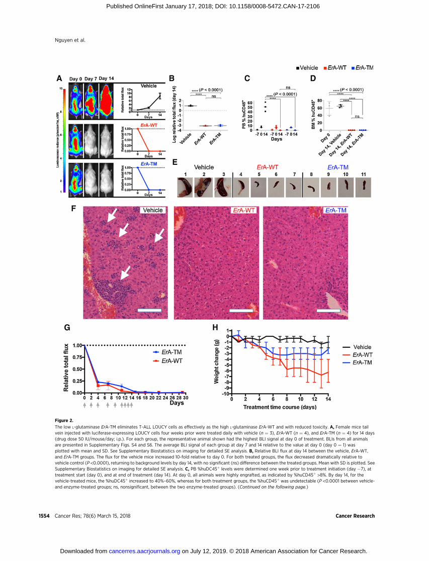
Figure 2.
The low L-glutaminase ErA-TM eliminates T-ALL LOUCY cells as effectively as the high L-glutaminase ErA-WT and with reduced toxicity. A, Female mice tailvein injected with luciferase-expressing LOUCY cells four weeks prior were treated daily with vehicle (n ¼ 3), ErA-WT (n ¼ 4), and ErA-TM (n ¼ 4) for 14 days(drug dose 50 IU/mouse/day; i.p.). For each group, the representative animal shown had the highest BLI signal at day 0 of treatment. BLIs from all animalsare presented in Supplementary Figs. S4 and S6. The average BLI signal of each group at day 7 and 14 relative to the value at day 0 (day 0 ¼ 1) wasplotted with mean and SD. See Supplementary Biostatistics on imaging for detailed SE analysis. B, Relative BLI flux at day 14 between the vehicle, ErA-WT,and ErA-TM groups. The flux for the vehicle mice increased 10-fold relative to day 0. For both treated groups, the flux decreased dramatically relative tovehicle control (P <0.0001), returning to background levels by day 14, with no significant (ns) difference between the treated groups. Mean with SD is plotted. SeeSupplementary Biostatistics on imaging for detailed SE analysis. C, PB %huDC45þ levels were determined one week prior to treatment initiation (day �7), attreatment start (day 0), and at end of treatment (day 14). At day 0, all animals were highly engrafted, as indicated by %huCD45þ >8%. By day 14, for thevehicle-treated mice, the %huDC45þ increased to 40%–60%, whereas for both treatment groups, the %huCD45þ was undetectable (P <0.0001 between vehicle-and enzyme-treated groups; ns, nonsignificant, between the two enzyme-treated groups). (Continued on the following page.)
Nguyen et al.
Cancer Res; 78(6) March 15, 2018 Cancer Research1554
on July 12, 2019. © 2018 American Association for Cancer Research. cancerres.aacrjournals.org Downloaded from
Published OnlineFirst January 17, 2018; DOI: 10.1158/0008-5472.CAN-17-2106
the ErA-WT-treated group, shredded by the ErA-TM–treatedgroup), shown both at the last day of drug treatment and 10days later (Supplementary Fig. S7). Collectively, these resultsconvincingly show that the ultra-low L-glutaminase ErA-TMenzyme maintains a very similar ability to combat ALL cells invivo as compared with the high L-glutaminase ErA-WT enzymebut with an improved tolerability profile.
Indication for reduced toxicity of the low L-glutaminase ErAvariants
In addition to noting the reduced activity of the high L-gluta-minase ErA-WT–treated mice, but not of the low L-glutaminaseErA-TM–treated mice, we also compared the impact of the drugson the animals' body weight. At the end of the experiment, weobserved 27% mean weight loss in ErA-WT–treated mice(Fig. 2H), which is consistent with results from previous studies(41, 42). This effect was largely mitigated in the animals treatedwith ErA-TM, which only experienced 10% mean weight loss.Hence, by treatment day 14, the ErA-WT group lost on average anadditional 4 grams of body weight (7 versus 3) compared withErA-TM (P <0.0001). In addition, the correlation between per-centage weight loss and level of enzyme L-glutaminase activitylevel was reproducible across several independent experiments(Supplementary Fig. S8A and S8B). Finally, we examined theacute toxicity of ErA-WT and ErA-TM in a blinded single dose-escalation study in CD-1 mice, using a concentration range of 40to 160 IU/g. For comparison, the dose used to treat the ALL-bearingmicewas approximately 2.5 IU/g.No fatalities occurred ineither group, but clear physical and behavioral signs of toxicitywere observed in the ErA-WT–treated group (Table 3). These signswere virtually absent in ErA-TM–challenged mice (Table 3).Together, these data show a clear correlation between reducedL-glutaminase activity and reduced drug toxicity. As previously
noted, a common side effect of L-asparaginase treatment is hep-atotoxicity, which presents histopathologically as macrovesicularhepatic steatosis (43), coupled with abnormally elevated serumlevels of the liver enzymes alanine aminotransferase (ALT; ref. 44)and aspartate aminotransferase (AST; ref. 45). However, bloodchemistry analysis did not display a clear difference in ALT or ASTlevels between ErA-WT- and ErA-TM–treatedmice. The absence ofa difference in serum liver enzyme levels in the studied animalsmaynot be surprising since L-asparaginase inducedhepatotoxicityin humans has been linked to fatty liver disease (46), elevatedBMI(12, 47), and increased age (48), whereas themice studied did notpresent histologically with hepatic fat accumulation (Fig. 2F),were lean and young.
The differences in toxicity between ErA-WT and ErA-TM is notdue to a difference in in vivo stability, but correlates with theenzyme's impact on the blood glutamine levels
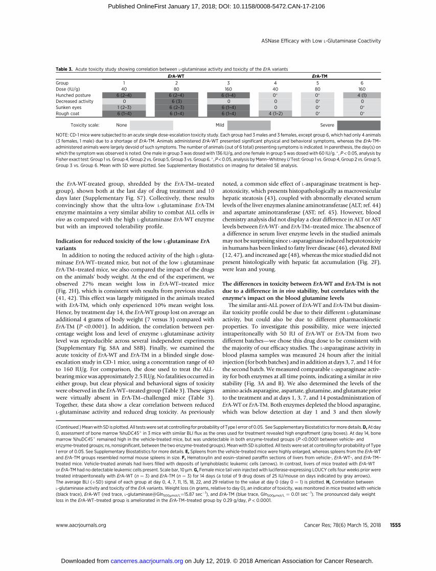
The similar anti-ALL power of ErA-WT and ErA-TM but dissim-ilar toxicity profile could be due to their different L-glutaminaseactivity, but could also be due to different pharmacokineticproperties. To investigate this possibility, mice were injectedintraperitoneally with 50 IU of ErA-WT or ErA-TM from twodifferent batches—we chose this drug dose to be consistent withthe majority of our efficacy studies. The L-asparaginase activity inblood plasma samples was measured 24 hours after the initialinjection (for bothbatches) and in addition at days 3, 7, and14 forthe second batch. We measured comparable L-asparaginase activ-ity for both enzymes at all time points, indicating a similar in vivostability (Fig. 3A and B). We also determined the levels of theamino acids asparagine, aspartate, glutamine, and glutamate priorto the treatment and at days 1, 3, 7, and 14 postadministration ofErA-WT or ErA-TM. Both enzymes depleted the blood asparagine,which was below detection at day 1 and 3 and then slowly
(Continued.)Meanwith SD is plotted. All testswere set at controlling for probability of Type I error of 0.05. See SupplementaryBiostatistics formoredetails.D,Atday0, assessment of bone marrow %huDC45þ in 3 mice with similar BLI flux as the ones used for treatment revealed high engraftment (gray boxes). At day 14, bonemarrow %huDC45þ remained high in the vehicle-treated mice, but was undetectable in both enzyme-treated groups (P <0.0001 between vehicle- andenzyme-treated groups; ns, nonsignificant, between the two enzyme-treated groups). Meanwith SD is plotted. All testswere set at controlling for probability of TypeI error of 0.05. See Supplementary Biostatistics for more details. E, Spleens from the vehicle-treated mice were highly enlarged, whereas spleens from the ErA-WTand ErA-TM groups resembled normal mouse spleens in size. F, Hematoxylin and eosin–stained paraffin sections of livers from vehicle-, ErA-WT-, and ErA-TM–
treated mice. Vehicle-treated animals had livers filled with deposits of lymphoblastic leukemic cells (arrows). In contrast, livers of mice treated with ErA-WTor ErA-TM had no detectable leukemic cells present. Scale bar, 10 mm.G, Female mice tail vein injected with luciferase-expressing LOUCY cells four weeks prior weretreated intraperitoneally with ErA-WT (n ¼ 3) and ErA-TM (n ¼ 3) for 14 days (a total of 9 drug doses of 25 IU/mouse on days indicated by gray arrows).The average BLI (þSD) signal of each group at day 0, 4, 7, 11, 15, 18, 22, and 29 relative to the value at day 0 (day 0 ¼ 1) is plotted. H, Correlation betweenL-glutaminase activity and toxicity of the ErA variants. Weight loss (in grams, relative to day 0), an indicator of toxicity, was monitored in mice treated with vehicle(black trace), ErA-WT (red trace, L-glutaminase@Gln500mmol/L¼15.87 sec�1), and ErA-TM (blue trace, Gln500mmol/L ¼ 0.01 sec�1). The pronounced daily weightloss in the ErA-WT–treated group is ameliorated in the ErA-TM–treated group by 0.29 g/day, P < 0.0001.
Table 3. Acute toxicity study showing correlation between L-glutaminase activity and toxicity of the ErA variants
ErA-WT ErA-TM
Group 1 2 3 4 5 6Dose (IU/g) 40 80 160 40 80 160Hunched posture 6 (2–4) 6 (2–4) 6 (1–4) 0� 0� 4 (1)Decreased activity 0 6 (3) 0 0 0� 0Sunken eyes 1 (2–3) 6 (2–3) 6 (1–4) 0 0� 0�
Rough coat 6 (1–4) 6 (1–4) 6 (1–4) 4 (1–2) 0� 0�
Toxicity scale: None Mild Severe
NOTE: CD-1 mice were subjected to an acute single dose-escalation toxicity study. Each group had 3 males and 3 females, except group 6, which had only 4 animals(3 females, 1 male) due to a shortage of ErA-TM. Animals administered ErA-WT presented significant physical and behavioral symptoms, whereas the ErA-TM–
administered animals were largely devoid of such symptoms. The number of animals (out of 6 total) presenting symptoms is indicated. In parenthesis, the day(s) onwhich the symptomwas observed is noted. One male in group 3 was dosed with 136 IU/g, and one female in group 5 was dosed with 60 IU/g. � , P < 0.05, analysis byFisher exact test: Group 1 vs. Group 4, Group 2 vs. Group 5, Group 3 vs. Group 6. � ,P <0.05, analysis byMann–WhitneyU Test: Group 1 vs. Group 4, Group 2 vs. Group 5,Group 3 vs. Group 6. Mean with SD were plotted. See Supplementary Biostatistics on imaging for detailed SE analysis.
ASNase Efficacy with Low L-Glutaminase Coactivity
www.aacrjournals.org Cancer Res; 78(6) March 15, 2018 1555
on July 12, 2019. © 2018 American Association for Cancer Research. cancerres.aacrjournals.org Downloaded from
Published OnlineFirst January 17, 2018; DOI: 10.1158/0008-5472.CAN-17-2106
recovered (Fig. 3C). Consistent with this, the levels of aspartateincreased at day 1 and 3, and then decreased to the pretreatmentlevel by day 7 (Fig. 3D).
Notably, when we examined the effect of the enzymes onglutamine, we noted that ErA-WT reduced the glutamine levelfrom approximately 600 mmol/L pretreatment to approximately250 mmol/L at day 1, which then promptly recovered by day 3.In contrast, ErA-TM did not reduce the glutamine levels (Fig. 3E).As expected from this, the glutamate levels increased for the ErA-WT–treated mice, but not for the ErA-TM–treated animals(Fig. 3F). These results are consistent with the predictions fromthe kinetic properties of these enzymes (Table 1) and the NMRexperiments (Fig. 1), showing that the in vitro observed similarityin the L-asparaginase activity but differences in the L-glutaminaseactivity translates into the in vivo setting. Moreover, whereas theglutamine levels recovered by day 3, we would expect that thedaily dosing schedule, as used in our efficacy studies, wouldcontinuously impact the glutamine levels.
In vivo evaluationofErA-WTandErA-TMusing apatient-derivedT-ALL xenograft model
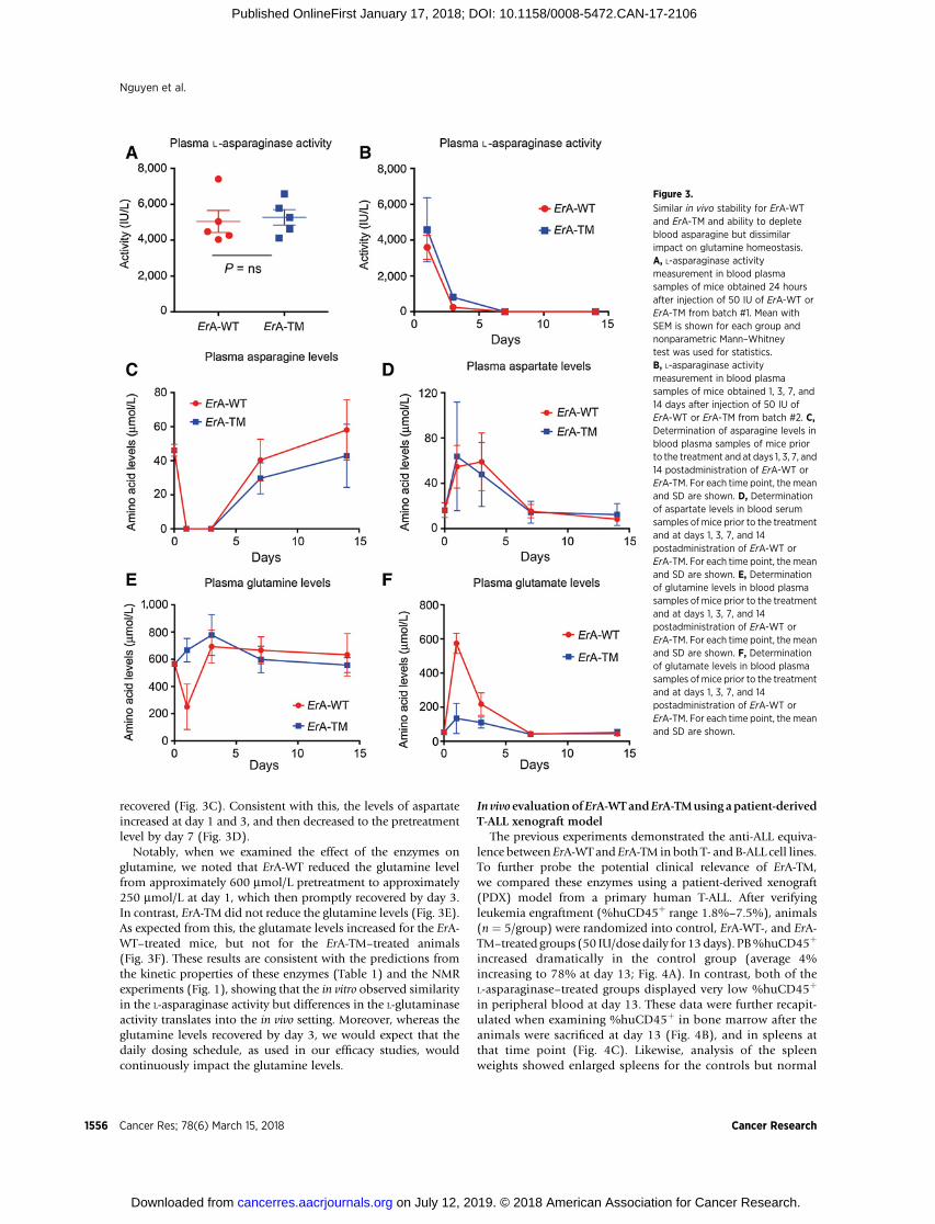
The previous experiments demonstrated the anti-ALL equiva-lence between ErA-WT and ErA-TM in both T- and B-ALL cell lines.To further probe the potential clinical relevance of ErA-TM,we compared these enzymes using a patient-derived xenograft(PDX) model from a primary human T-ALL. After verifyingleukemia engraftment (%huCD45þ range 1.8%–7.5%), animals(n ¼ 5/group) were randomized into control, ErA-WT-, and ErA-TM–treated groups (50 IU/dose daily for 13days). PB%huCD45þ
increased dramatically in the control group (average 4%increasing to 78% at day 13; Fig. 4A). In contrast, both of theL-asparaginase–treated groups displayed very low %huCD45þ
in peripheral blood at day 13. These data were further recapit-ulated when examining %huCD45þ in bone marrow after theanimals were sacrificed at day 13 (Fig. 4B), and in spleens atthat time point (Fig. 4C). Likewise, analysis of the spleenweights showed enlarged spleens for the controls but normal
Figure 3.
Similar in vivo stability for ErA-WTand ErA-TM and ability to depleteblood asparagine but dissimilarimpact on glutamine homeostasis.A, L-asparaginase activitymeasurement in blood plasmasamples of mice obtained 24 hoursafter injection of 50 IU of ErA-WT orErA-TM from batch #1. Mean withSEM is shown for each group andnonparametric Mann–Whitneytest was used for statistics.B, L-asparaginase activitymeasurement in blood plasmasamples of mice obtained 1, 3, 7, and14 days after injection of 50 IU ofErA-WT or ErA-TM from batch #2. C,Determination of asparagine levels inblood plasma samples of mice priorto the treatment and at days 1, 3, 7, and14 postadministration of ErA-WT orErA-TM. For each time point, themeanand SD are shown. D, Determinationof aspartate levels in blood serumsamples of mice prior to the treatmentand at days 1, 3, 7, and 14postadministration of ErA-WT orErA-TM. For each time point, themeanand SD are shown. E, Determinationof glutamine levels in blood plasmasamples of mice prior to the treatmentand at days 1, 3, 7, and 14postadministration of ErA-WT orErA-TM. For each time point, themeanand SD are shown. F, Determinationof glutamate levels in blood plasmasamples of mice prior to the treatmentand at days 1, 3, 7, and 14postadministration of ErA-WT orErA-TM. For each time point, themeanand SD are shown.
Nguyen et al.
Cancer Res; 78(6) March 15, 2018 Cancer Research1556
on July 12, 2019. © 2018 American Association for Cancer Research. cancerres.aacrjournals.org Downloaded from
Published OnlineFirst January 17, 2018; DOI: 10.1158/0008-5472.CAN-17-2106
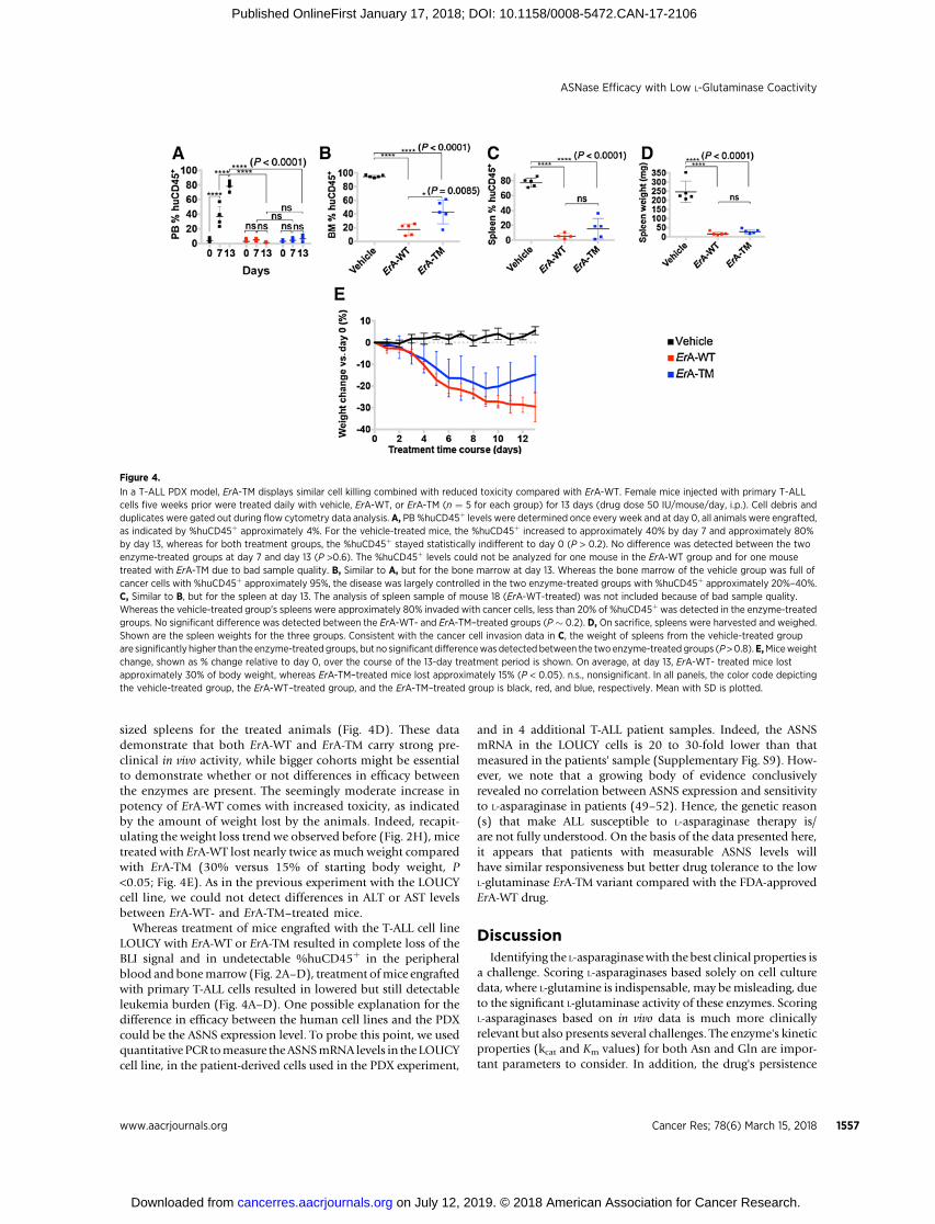
sized spleens for the treated animals (Fig. 4D). These datademonstrate that both ErA-WT and ErA-TM carry strong pre-clinical in vivo activity, while bigger cohorts might be essentialto demonstrate whether or not differences in efficacy betweenthe enzymes are present. The seemingly moderate increase inpotency of ErA-WT comes with increased toxicity, as indicatedby the amount of weight lost by the animals. Indeed, recapit-ulating the weight loss trend we observed before (Fig. 2H), micetreated with ErA-WT lost nearly twice as much weight comparedwith ErA-TM (30% versus 15% of starting body weight, P<0.05; Fig. 4E). As in the previous experiment with the LOUCYcell line, we could not detect differences in ALT or AST levelsbetween ErA-WT- and ErA-TM–treated mice.
Whereas treatment of mice engrafted with the T-ALL cell lineLOUCY with ErA-WT or ErA-TM resulted in complete loss of theBLI signal and in undetectable %huCD45þ in the peripheralblood and bonemarrow (Fig. 2A–D), treatment ofmice engraftedwith primary T-ALL cells resulted in lowered but still detectableleukemia burden (Fig. 4A–D). One possible explanation for thedifference in efficacy between the human cell lines and the PDXcould be the ASNS expression level. To probe this point, we usedquantitative PCR tomeasure theASNSmRNA levels in the LOUCYcell line, in the patient-derived cells used in the PDX experiment,
and in 4 additional T-ALL patient samples. Indeed, the ASNSmRNA in the LOUCY cells is 20 to 30-fold lower than thatmeasured in the patients' sample (Supplementary Fig. S9). How-ever, we note that a growing body of evidence conclusivelyrevealed no correlation between ASNS expression and sensitivityto L-asparaginase in patients (49–52). Hence, the genetic reason(s) that make ALL susceptible to L-asparaginase therapy is/are not fully understood. On the basis of the data presented here,it appears that patients with measurable ASNS levels willhave similar responsiveness but better drug tolerance to the lowL-glutaminase ErA-TM variant compared with the FDA-approvedErA-WT drug.
DiscussionIdentifying the L-asparaginasewith the best clinical properties is
a challenge. Scoring L-asparaginases based solely on cell culturedata, where L-glutamine is indispensable, may bemisleading, dueto the significant L-glutaminase activity of these enzymes. ScoringL-asparaginases based on in vivo data is much more clinicallyrelevant but also presents several challenges. The enzyme's kineticproperties (kcat and Km values) for both Asn and Gln are impor-tant parameters to consider. In addition, the drug's persistence
Figure 4.
In a T-ALL PDX model, ErA-TM displays similar cell killing combined with reduced toxicity compared with ErA-WT. Female mice injected with primary T-ALLcells five weeks prior were treated daily with vehicle, ErA-WT, or ErA-TM (n ¼ 5 for each group) for 13 days (drug dose 50 IU/mouse/day, i.p.). Cell debris andduplicates were gated out during flow cytometry data analysis. A, PB %huCD45þ levels were determined once every week and at day 0, all animals were engrafted,as indicated by %huCD45þ approximately 4%. For the vehicle-treated mice, the %huCD45þ increased to approximately 40% by day 7 and approximately 80%by day 13, whereas for both treatment groups, the %huCD45þ stayed statistically indifferent to day 0 (P > 0.2). No difference was detected between the twoenzyme-treated groups at day 7 and day 13 (P >0.6). The %huCD45þ levels could not be analyzed for one mouse in the ErA-WT group and for one mousetreated with ErA-TM due to bad sample quality. B, Similar to A, but for the bone marrow at day 13. Whereas the bone marrow of the vehicle group was full ofcancer cells with %huCD45þ approximately 95%, the disease was largely controlled in the two enzyme-treated groups with %huCD45þ approximately 20%–40%.C, Similar to B, but for the spleen at day 13. The analysis of spleen sample of mouse 18 (ErA-WT-treated) was not included because of bad sample quality.Whereas the vehicle-treated group's spleens were approximately 80% invaded with cancer cells, less than 20% of %huCD45þ was detected in the enzyme-treatedgroups. No significant difference was detected between the ErA-WT- and ErA-TM–treated groups (P � 0.2). D, On sacrifice, spleens were harvested and weighed.Shown are the spleen weights for the three groups. Consistent with the cancer cell invasion data in C, the weight of spleens from the vehicle-treated groupare significantly higher than the enzyme-treated groups, but no significant differencewas detected between the twoenzyme-treated groups (P>0.8).E,Miceweightchange, shown as % change relative to day 0, over the course of the 13-day treatment period is shown. On average, at day 13, ErA-WT- treated mice lostapproximately 30% of body weight, whereas ErA-TM–treated mice lost approximately 15% (P < 0.05). n.s., nonsignificant. In all panels, the color code depictingthe vehicle-treated group, the ErA-WT–treated group, and the ErA-TM–treated group is black, red, and blue, respectively. Mean with SD is plotted.
ASNase Efficacy with Low L-Glutaminase Coactivity
www.aacrjournals.org Cancer Res; 78(6) March 15, 2018 1557
on July 12, 2019. © 2018 American Association for Cancer Research. cancerres.aacrjournals.org Downloaded from
Published OnlineFirst January 17, 2018; DOI: 10.1158/0008-5472.CAN-17-2106
in circulation (i.e., half-life) will determine the duration ofenzymatic Asn and Gln depletion, a parameter that can be fine-tuned by appropriate dose and frequency of drug administration.Ideally, one would like to combine a prolonged half-life as seenwith the introduction of pegylated EcA, high L-asparaginase activ-ity, and only low or absence of L-glutaminase activity, the latterpredicted to be in part responsible for acute toxicities of the drug.Currently, a pegylated version of ErA-WT is being evaluated (53).While such a version is predicted to solve the problem of shortin vivo persistence of native ErA, we caution that with the con-comitant longer time for Asn depletion, such an enzyme willalso have a longer duration of Gln depletion. Considering thevery high L-glutaminase activity of ErA-WT, this would predictincreased side effects.
Here we present ErA variants with significantly lowerL-glutaminase but comparable L-asparaginase activities relativetoErA-WTand extended circulation time achievedbymaintainingthe SUMO tag. We demonstrated that our engineered lowL-glutaminase Erwinia L-asparaginase variants have preserved cellkilling properties, similar to ErA-WT. Comparable pharmacoki-netic properties of the SUMO-tagged ErA-WT versus ErA-TMenzymes accompanied by similar Asn but remarkably differentGln depletion profiles convincingly discount the possibility thatthe observed anti-leukemic effect in ErA-TMwas due to glutaminedepletion. Moreover, the comparable serum persistence andantileukemic properties of the enzymes indicate that the signif-icant difference in the toxicity profile is linked to the difference inimpact onGln levels, which suggests a superior tolerability for thelow L-glutaminase variant ErA-TM.
Others have also recognized the potential advantages ofL-asparaginases with low L-glutaminase activity. One in particularis the low L-glutaminase S121 variant of the Wolinella succinogen-esL-asparaginase (WoA-S121). For additional details about thisenzyme, see Supplementary Material. The ratio of the L-asparagi-nase to L-glutaminase rates (measured at the physiologic substrateconcentrations of 50mmol/L for Asn and500mmol/L forGln) bestconveys the clinically relevant substrate specificity of thesedrugs (a high ratio describes a more specific L-asparaginase withlow L-glutaminase). We recently investigated the properties ofWoA-S121 and discovered that this ratio is 62 (27), which isactually inferior to that for EcA-WT (ratio¼ 188), but superior toErA-WT (ratio ¼ 6.6; Table 1). However, this ratio for ErA-TM isapproximately 9,000, showcasing the extremely high L-asparagi-nase preference of this variant.
In conclusion, this study convincingly shows that high L-glu-taminase activity, as present in current FDA-approved L-aspara-ginase drugs, is not essential for efficient in vivo elimination of L-asparaginase–sensitive ALL cells. Furthermore, our results suggesta decline in in vivo toxicitywhen thedrug's L-glutaminase activity isreduced. Since the debilitating side effects of current L-asparagi-nases often result in treatment cessation, an event associated withinferior event-free survival (48, 54), L-asparaginases with dimin-ished toxicity are highly pertinent to improving ALL treatmentoutcome. In line with this notion, a recent pilot study (55) thatevaluated an intensified L-asparaginase treatment in a populationof high-risk pediatric ALL patients using pegylated EcA (thecurrent standard of care in the USA with >20-fold higher L-glutaminase activity compared with ErA-TM) had to be aborteddue to an unacceptable frequency of adverse effects. However,since the patients were receiving additional chemotherapeuticdrugs, the causative agent behind the increased toxicity cannot be
directly linked to the more frequent dosing of the L-asparaginase.Notably, the ultra-low L-glutaminase ErA-TMvariant, as presentedin this study, now provides an alternative L-asparaginase that candirectly probe this point.
Disclosure of Potential Conflicts of InterestH.A. Nguyen is a chief scientific officer at Enzyme by Design Inc. and
has ownership interest (including patents) in University of Illinois at Chicago.Y. Su has ownership interest (including patents) in Enzyme by Design, Inc.A.M. Schalk is a chief operating officer and has ownership interest (includingpatents) in Enzyme by Design Inc. T. Lammens provided expert testimony forJazz Pharmaceuticals (support from Jazz Pharmaceuticals to attend the AnnualASH Conference 2015 and 2016). B. De Moerloose provided expert testimonyfor Jazz Pharmaceuticals (travel grant from Jazz Pharmaceuticals for ASHAnnual Meeting in 2015 and in 2016). Y. Saunthararajah is a consultant/advisory board member for EpiDestiny. A. Lavie is a CEO and has ownershipinterest (including patents) at Enzyme by Design, Inc. No potential conflicts ofinterest were disclosed by the other authors.
DisclaimerThe contents do not represent the views of the U.S. Department of Veterans
Affairs or the United States Government.
Authors' ContributionsConception and design:H.A. Nguyen, M. Caffrey, T. Lammens, V. Mondelaers,B. De Moerloose, A.V. Lyubimov, B.J. Merrill, A. LavieDevelopment of methodology: H.A. Nguyen, Y. Su, J.Y. Zhang, A. Antanasi-jevic, D. Rondelli, S. Goossens, A. LavieAcquisition of data (provided animals, acquired and managed patients,provided facilities, etc.): H.A. Nguyen, Y. Su, J.Y. Zhang, A. Antanasijevic,M. Caffrey, A.M. Schalk, D. Rondelli, A. Oh, D.L. Mahmud, A. Kajdacsy-Balla,S. Peirs, T. Lammens, V. Mondelaers, B. De Moerloose, S. Goossens,M.J. Schlicht, K.K. Kabirov, A.V. Lyubimov, P.V. Vlierberghe, A. LavieAnalysis and interpretation of data (e.g., statistical analysis, biostatistics,computational analysis): H.A. Nguyen, Y. Su, A. Antanasijevic, M. Caffrey,L. Liu, D. Rondelli, A. Oh, M.C. Bosland, S. Peirs, S. Goossens, K.K. Kabirov,A.V. Lyubimov, P.V. Vlierberghe, A. LavieWriting, review, and/or revision of the manuscript: H.A. Nguyen, J.Y. Zhang,A. Antanasijevic, M. Caffrey, A.M. Schalk, L. Liu, D. Rondelli, A. Oh,M.C. Bosland, A. Kajdacsy-Balla, S. Peirs, T. Lammens, V. Mondelaers,B. De Moerloose, S. Goossens, A.V. Lyubimov, Y. Saunthararajah,P.V. Vlierberghe, A. LavieAdministrative, technical, or material support (i.e., reporting or organizingdata, constructing databases): H.A. Nguyen, Y. SuStudy supervision: H.A. Nguyen, A.V. Lyubimov, A. Lavie
AcknowledgmentsA. Lavie was supported in part by NIH grant RO1 EB013685 and by Merit
Review Award I01BX001919 from the United States Department of VeteransAffairs Biomedical Laboratory Research and Development Service, and bythe UIC Cancer Center. P. Van Vlierberghe was supported by the Fund forScientific ResearchFlanders (FWO)grant 3G0C4713, by theBelgian FoundationAgainst Cancer grant 365W3415W, and by Kom op tegen Kanker (Stand UpTo Cancer) grant 116000000251, and by the Flemish Cancer Society researchgrant 365Y9115W. S. Peirs was supported by doctoral and postdoc FWO grantFWO17/PDO/111. We thank Dr. Michael Jensen (University of WashingtonSchool ofMedicine) for the generous gift of the SUP-B15-luciferase cell line andB�eatrice Lintermans for excellent technical assistance. The histology work wascarried out by the UIC Research Resources Center's Research Histology andTissue Imaging Core.
The costs of publication of this articlewere defrayed inpart by the payment ofpage charges. This article must therefore be hereby marked advertisement inaccordance with 18 U.S.C. Section 1734 solely to indicate this fact.
Received July 14, 2017; revised December 13, 2017; accepted January 11,2018; published OnlineFirst January 17, 2018.
Nguyen et al.
Cancer Res; 78(6) March 15, 2018 Cancer Research1558
on July 12, 2019. © 2018 American Association for Cancer Research. cancerres.aacrjournals.org Downloaded from
Published OnlineFirst January 17, 2018; DOI: 10.1158/0008-5472.CAN-17-2106
References1. Narta UK, Kanwar SS, Azmi W. Pharmacological and clinical evaluation of
L-asparaginase in the treatment of leukemia. Crit Rev Oncol Hematol2007;61:208–21.
2. Panosyan EH, Grigoryan RS, Avramis IA, Seibel NL, Gaynon PS, Siegel SE,et al. Deamination of glutamine is a prerequisite for optimal asparaginedeamination by asparaginases in vivo (CCG-1961). Anticancer Res2004;24:1121–5.
3. Ollenschlager G, Roth E, Linkesch W, Jansen S, Simmel A, Modder B.Asparaginase-induced derangements of glutamine metabolism: the path-ogenetic basis for some drug-related side-effects. Eur J Clin Invest 1988;18:512–6.
4. Grigoryan RS, PanosyanEH, SeibelNL,GaynonPS, Avramis IA, Avramis VI.Changes of amino acid serum levels in pediatric patients with higher-riskacute lymphoblastic leukemia (CCG-1961). In vivo 2004;18:107–12.
5. Hawkins DS, Park JR, Thomson BG, Felgenhauer JL, Holcenberg JS,Panosyan EH, et al. Asparaginase pharmacokinetics after intensive poly-ethylene glycol-conjugated L-asparaginase therapy for children withrelapsed acute lymphoblastic leukemia. Clin Cancer Res 2004;10:5335–41.
6. Broome JD. Studies on the mechanism of tumor inhibition by L-aspar-aginase. Effects of the enzyme on asparagine levels in the blood, normaltissues, and 6C3HED lymphomas of mice: differences in asparagineformation and utilization in asparaginase-sensitive and -resistant lympho-ma cells. J Exp Med 1968;127:1055–72.
7. Prager MD, Bachynsky N. Asparagine synthetase in normal and malignanttissues: correlation with tumor sensitivity to asparaginase. Arch BiochemBiophys 1968;127:645–54.
8. Prager MD, Bachynsky N. Asparagine synthetase in asparaginase resistantand susceptiblemouse lymphomas. BiochemBiophys Res Commun 1968;31:43–7.
9. ChanWK, Lorenzi PL, Anishkin A, PurwahaP, RogersDM, Sukharev S, et al.The glutaminase activity of L-asparaginase is not required for anticanceractivity against ASNS-negative cells. Blood 2014;123:3596–606.
10. Parmentier JH, Maggi M, Tarasco E, Scotti C, Avramis VI, Mittelman SD.Glutaminase activity determines cytotoxicity of L-asparaginases on mostleukemia cell lines. Leukemia Res 2015;39:757–62.
11. Offman MN, Krol M, Patel N, Krishnan S, Liu J, Saha V, et al. Rationalengineering of L-asparaginase reveals importance of dual activity for cancercell toxicity. Blood 2011;117:1614–21.
12. Aldoss I,DouerD, Behrendt CE, Chaudhary P,Mohrbacher A, Vrona J, et al.Toxicity profile of repeated doses of PEG-asparaginase incorporated into apediatric-type regimen for adult acute lymphoblastic leukemia. Eur JHaematol 2015;96:375–80.
13. HijiyaN, vander Sluis IM. Asparaginase-associated toxicity in childrenwithacute lymphoblastic leukemia. Leuk Lymphoma 2016;57:748–57.
14. Tong WH, Pieters R, de Groot-Kruseman HA, Hop WC, Boos J, Tissing WJ,et al. The toxicity of very prolonged courses of PEGasparaginase or Erwiniaasparaginase in relation to asparaginase activity, with a special focus ondyslipidemia. Haematologica 2014;99:1716–21.
15. Avramis VI, Sencer S, Periclou AP, Sather H, Bostrom BC, Cohen LJ, et al. Arandomized comparison of native Escherichia coli asparaginase and poly-ethylene glycol conjugated asparaginase for treatment of children withnewly diagnosed standard-risk acute lymphoblastic leukemia: a Children'sCancer Group study. Blood 2002;99:1986–94.
16. Heitink-Polle KM, Prinsen BH, de Koning TJ, van Hasselt PM, Bierings MB.High incidence of symptomatic hyperammonemia in children with acutelymphoblastic leukemia receiving pegylated asparaginase. JIMD Rep2013;7:103–8.
17. Durden DL, Distasio JA. Characterization of the effects of asparaginasefrom Escherichia coli and a glutaminase-free asparaginase from Vibriosuccinogenes on specific cell-mediated cytotoxicity. Int J Cancer1981;27:59–65.
18. Kafkewitz D, Bendich A. Enzyme-induced asparagine and glutaminedepletion and immune system function. Am JClinNutr 1983;37:1025–30.
19. Durden DL, Salazar AM, Distasio JA. Kinetic analysis of hepatotoxicityassociated with antineoplastic asparaginases. Cancer Res 1983;43:1602–5.
20. Reinert RB, Oberle LM, Wek SA, Bunpo P, Wang XP, Mileva I, et al. Role ofglutamine depletion in directing tissue-specific nutrient stress responses toL-asparaginase. J Biol Chem 2006;281:31222–33.
21. Leonard JV, Kay JD. Acute encephalopathy and hyperammonaemia com-plicating treatment of acute lymphoblastic leukaemia with asparaginase.Lancet 1986;1:162–3.
22. Alvarez OA, Zimmerman G. Pegaspargase-induced pancreatitis. MedPediatr Oncol 2000;34:200–5.
23. Laterza OF, Gerhardt G, Sokoll LJ. Measurement of plasma ammonia isaffected in patients receiving asparaginase therapy. Clin Chem 2003;49:1710–1.
24. Jorck C, Kiess W, Weigel JF, Mutze U, Bierbach U, Beblo S. Transienthyperammonemia due to L-asparaginase therapy in children with acutelymphoblastic leukemia or non-Hodgkin lymphoma. Pediatr HematolOncol 2011;28:3–9.
25. Nussbaum V, Lubcke N, Findlay R. Hyperammonemia secondary toasparaginase: a case series. J Oncol Pharm Pract 2016;22:161–4.
26. Warrell RP Jr., Arlin ZA, Gee TS, Chou TC, Roberts J, Young CW. Clinicalevaluation of succinylated Acinetobacter glutaminase-asparaginase inadult leukemia. Cancer Treat Rep 1982;66:1479–85.
27. Nguyen HA, Durden DL, Lavie A. The differential ability of asparagine andglutamine in promoting the closed/active enzyme conformation rationa-lizes the Wolinella succinogenes L-asparaginase substrate specificity. SciRep 2017;7:41643.
28. Hays JL, KimG,Walker A, AnnunziataCM, Lee JM, Squires J, et al. A phase IIclinical trial of polyethylene glycol-conjugated L-asparaginase in patientswith advanced ovarian cancer: early closure for safety. Mol Clin Oncol2013;1:565–9.
29. Lorenzi PL, Horvath TD,Martin LA, ChanWK, DuD, Hawke DH, et al. Redblood cell-encapsulation of L-asparaginase favorably modulates targetselectivity and pharmacodynamics American Society of Hematology 58thAnnual Meeting. San Diego, CA; 2016. Available from: https://ash.confex.com/ash/2016/webprogram/Paper97607.html.
30. Nguyen HA, Su Y, Lavie A. Design and characterization of erwinia chry-santhemi l-asparaginase variants with diminished l-glutaminase activity.J Biol Chem 2016;291:17664–76.
31. Nguyen HA, Su Y, Lavie A. Structural insight into substrate selectivity ofErwinia chrysanthemi l-Asparaginase. Biochemistry 2016;55:1246–53.
32. Schalk AM, Nguyen HA, Rigouin C, Lavie A. Identification and structuralanalysis of an L-asparaginase enzyme from guinea pig with putative tumorcell killing properties. J Biol Chem 2014;289:33175–86.
33. Fernandez CA, Cai X, Elozory A, Liu C, Panetta JC, Jeha S, et al. High-throughput asparaginase activity assay in serumof childrenwith leukemia.Int J Clin Exp Med 2013;6:478–87.
34. Ben-Bassat H, Shlomai Z, Kohn G, Prokocimer M. Establishment of ahuman T-acute lymphoblastic leukemia cell line with a (16;20) chromo-some translocation. Cancer Genet Cytogenet 1990;49:241–8.
35. Peirs S, Matthijssens F, Goossens S, Van de Walle I, Ruggero K, deBock CE, et al. ABT-199 mediated inhibition of BCL-2 as a noveltherapeutic strategy in T-cell acute lymphoblastic leukemia. Blood2014;124:3738–47.
36. Fainstein E, Marcelle C, Rosner A, Canaani E, Gale RP, Dreazen O, et al. Anew fused transcript in Philadelphia chromosome positive acute lympho-cytic leukaemia. Nature 1987;330:386–8.
37. Szymanska B, Wilczynska-Kalak U, Kang MH, Liem NL, Carol H, Boehm I,et al. Pharmacokinetic modeling of an induction regimen for in vivocombined testing of novel drugs against pediatric acute lymphoblasticleukemia xenografts. PLoS One 2012;7:e33894.
38. Pan R, Hogdal LJ, Benito JM, Bucci D, Han L, Borthakur G, et al. SelectiveBCL-2 inhibition by ABT-199 causes on-target cell death in acute myeloidleukemia. Cancer Discov 2014;4:362–75.
39. Asselin BL, Whitin JC, Coppola DJ, Rupp IP, Sallan SE, Cohen HJ.Comparative pharmacokinetic studies of three asparaginase preparations.J Clin Oncol 1993;11:1780–6.
40. Reiff A, Zastrow M, Sun BC, Takei S, Mitsuhada H, Bernstein B, et al.Treatment of collagen induced arthritis inDBA/1micewith L-asparaginase.Clin Exp Rheumatol 2001;19:639–46.
41. Viau AT, Abuchowski A,McCoy JR, Kazo GM,Davis FF. Toxicologic studiesof a conjugate of asparaginase and polyethylene glycol in mice, rats, anddogs. Am J Vet Res 1986;47:1398–401.
42. U.S. Food and Drug Administration. Erwinase (L-asparaginase) Phar-amcology Review. FDA Center for Drug Evaluation and Research; 2011.p. 1–44.
www.aacrjournals.org Cancer Res; 78(6) March 15, 2018 1559
ASNase Efficacy with Low L-Glutaminase Coactivity
on July 12, 2019. © 2018 American Association for Cancer Research. cancerres.aacrjournals.org Downloaded from
Published OnlineFirst January 17, 2018; DOI: 10.1158/0008-5472.CAN-17-2106
43. Bodmer M, Sulz M, Stadlmann S, Droll A, Terracciano L, Krahenbuhl S.Fatal liver failure in an adult patient with acute lymphoblastic leukemiafollowing treatment with L-asparaginase. Digestion 2006;74:28–32.
44. Bessho F, Kinumaki H, Yokota S, Hayashi Y, Kobayashi M, Kamoshita S.Liver function studies in children with acute lymphocytic leukemia aftercessation of therapy. Med Pediatr Oncol 1994;23:111–5.
45. Cairo MS. Adverse reactions of L-asparaginase. Am J Pediatr HematolOncol 1982;4:335–9.
46. Roesmann A, Afify M, Panse J, Eisert A, Steitz J, Tolba RH. L-carnitineameliorates L-asparaginase-induced acute liver toxicity in steatotic ratlivers. Chemotherapy 2013;59:167–75.
47. Christ TN, Stock W, Knoebel RW. Incidence of asparaginase-related hep-atotoxicity, pancreatitis, and thrombotic events in adults with acute lym-phoblastic leukemia treated with a pediatric-inspired regimen. J OncolPharm Pract 2017:1078155217701291.
48. Stock W, Douer D, DeAngelo DJ, Arellano M, Advani A, Damon L, et al.Prevention and management of asparaginase/pegasparaginase-associatedtoxicities in adults and older adolescents: recommendations of an expertpanel. Leuk Lymphoma 2011;52:2237–53.
49. Chen SH, YangW, Fan Y, StoccoG, Crews KR, Yang JJ, et al. A genome-wideapproach identifies that the aspartate metabolism pathway contributes toasparaginase sensitivity. Leukemia 2011;25:66–74.
50. Fine BM, Kaspers GJ, HoM, Loonen AH, Boxer LM. A genome-wide view ofthe in vitro response to l-asparaginase in acute lymphoblastic leukemia.Cancer Res 2005;65:291–9.
51. Hermanova I, Zaliova M, Trka J, Starkova J. Low expression of asparaginesynthetase in lymphoid blasts precludes its role in sensitivity to L-aspar-aginase. Exp Hematol 2012;40:657–65.
52. StamsWA, den Boer ML, Beverloo HB, Meijerink JP, Stigter RL, vanWeringER, et al. Sensitivity to L-asparaginase is not associated with expressionlevels of asparagine synthetase in t(12;21)þ pediatric ALL. Blood2003;101:2743–7.
53. Chien WW, Allas S, Rachinel N, Sahakian P, Julien M, Le Beux C, et al.Pharmacology, immunogenicity, and efficacy of a novel pegylated recom-binant Erwinia chrysanthemi-derived L-asparaginase. Invest New Drugs2014;32:795–805.
54. Silverman LB, Gelber RD, Dalton VK, Asselin BL, Barr RD, Clavell LA,et al. Improved outcome for children with acute lymphoblastic leuke-mia: results of Dana-Farber Consortium Protocol 91–01. Blood 2001;97:1211–8.
55. RodriguezV, Kairalla J, SalzerWL, Raetz EA, LohML,Carroll AJ, et al. A pilotstudy of intensified PEG-asparaginase in high-risk acute lymphoblasticleukemia: children's oncology group study AALL08P1. J Pediatr HematolOncol 2016;38:409–17.
Cancer Res; 78(6) March 15, 2018 Cancer Research1560
Nguyen et al.
on July 12, 2019. © 2018 American Association for Cancer Research. cancerres.aacrjournals.org Downloaded from
Published OnlineFirst January 17, 2018; DOI: 10.1158/0008-5472.CAN-17-2106
2018;78:1549-1560. Published OnlineFirst January 17, 2018.Cancer Res Hien Anh Nguyen, Ying Su, Jenny Y. Zhang, et al.
In VivoLeukemias Efficacious against Both T- and B-cell Acute Lymphoblastic A Novel l-Asparaginase with low l-Glutaminase Coactivity Is Highly
Updated version
10.1158/0008-5472.CAN-17-2106doi:
Access the most recent version of this article at:
Material
Supplementary
http://cancerres.aacrjournals.org/content/suppl/2018/01/17/0008-5472.CAN-17-2106.DC1
Access the most recent supplemental material at:
Cited articles
http://cancerres.aacrjournals.org/content/78/6/1549.full#ref-list-1
This article cites 52 articles, 20 of which you can access for free at:
E-mail alerts related to this article or journal.Sign up to receive free email-alerts
Subscriptions
Reprints and
To order reprints of this article or to subscribe to the journal, contact the AACR Publications Department at
Permissions
Rightslink site. Click on "Request Permissions" which will take you to the Copyright Clearance Center's (CCC)
.http://cancerres.aacrjournals.org/content/78/6/1549To request permission to re-use all or part of this article, use this link
on July 12, 2019. © 2018 American Association for Cancer Research. cancerres.aacrjournals.org Downloaded from
Published OnlineFirst January 17, 2018; DOI: 10.1158/0008-5472.CAN-17-2106