Embed Size (px)
Citation preview
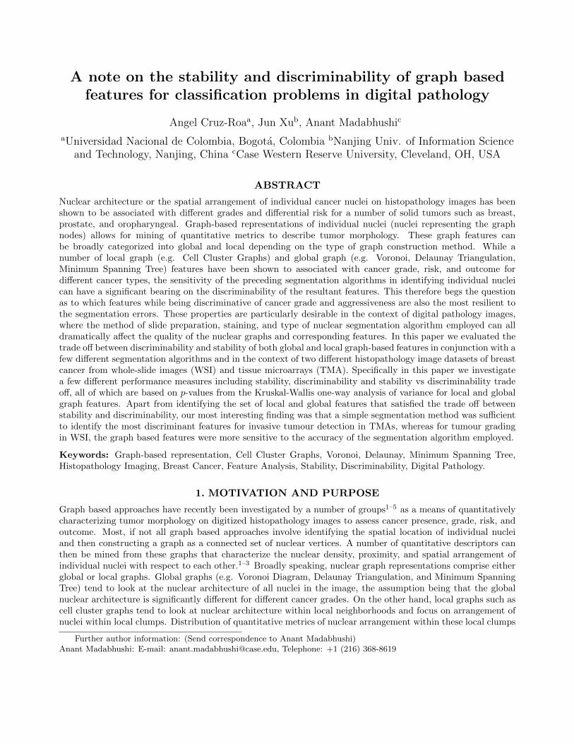
A note on the stability and discriminability of graph basedfeatures for classification problems in digital pathology
Angel Cruz-Roaa, Jun Xub, Anant Madabhushic
aUniversidad Nacional de Colombia, Bogota, Colombia bNanjing Univ. of Information Scienceand Technology, Nanjing, China cCase Western Reserve University, Cleveland, OH, USA
ABSTRACT
Nuclear architecture or the spatial arrangement of individual cancer nuclei on histopathology images has beenshown to be associated with different grades and differential risk for a number of solid tumors such as breast,prostate, and oropharyngeal. Graph-based representations of individual nuclei (nuclei representing the graphnodes) allows for mining of quantitative metrics to describe tumor morphology. These graph features canbe broadly categorized into global and local depending on the type of graph construction method. While anumber of local graph (e.g. Cell Cluster Graphs) and global graph (e.g. Voronoi, Delaunay Triangulation,Minimum Spanning Tree) features have been shown to associated with cancer grade, risk, and outcome fordifferent cancer types, the sensitivity of the preceding segmentation algorithms in identifying individual nucleican have a significant bearing on the discriminability of the resultant features. This therefore begs the questionas to which features while being discriminative of cancer grade and aggressiveness are also the most resilient tothe segmentation errors. These properties are particularly desirable in the context of digital pathology images,where the method of slide preparation, staining, and type of nuclear segmentation algorithm employed can alldramatically affect the quality of the nuclear graphs and corresponding features. In this paper we evaluated thetrade off between discriminability and stability of both global and local graph-based features in conjunction with afew different segmentation algorithms and in the context of two different histopathology image datasets of breastcancer from whole-slide images (WSI) and tissue microarrays (TMA). Specifically in this paper we investigatea few different performance measures including stability, discriminability and stability vs discriminability tradeoff, all of which are based on p-values from the Kruskal-Wallis one-way analysis of variance for local and globalgraph features. Apart from identifying the set of local and global features that satisfied the trade off betweenstability and discriminability, our most interesting finding was that a simple segmentation method was sufficientto identify the most discriminant features for invasive tumour detection in TMAs, whereas for tumour gradingin WSI, the graph based features were more sensitive to the accuracy of the segmentation algorithm employed.
Keywords: Graph-based representation, Cell Cluster Graphs, Voronoi, Delaunay, Minimum Spanning Tree,Histopathology Imaging, Breast Cancer, Feature Analysis, Stability, Discriminability, Digital Pathology.
1. MOTIVATION AND PURPOSE
Graph based approaches have recently been investigated by a number of groups1–5 as a means of quantitativelycharacterizing tumor morphology on digitized histopathology images to assess cancer presence, grade, risk, andoutcome. Most, if not all graph based approaches involve identifying the spatial location of individual nucleiand then constructing a graph as a connected set of nuclear vertices. A number of quantitative descriptors canthen be mined from these graphs that characterize the nuclear density, proximity, and spatial arrangement ofindividual nuclei with respect to each other.1–3 Broadly speaking, nuclear graph representations comprise eitherglobal or local graphs. Global graphs (e.g. Voronoi Diagram, Delaunay Triangulation, and Minimum SpanningTree) tend to look at the nuclear architecture of all nuclei in the image, the assumption being that the globalnuclear architecture is significantly different for different cancer grades. On the other hand, local graphs such ascell cluster graphs tend to look at nuclear architecture within local neighborhoods and focus on arrangement ofnuclei within local clumps. Distribution of quantitative metrics of nuclear arrangement within these local clumps
Further author information: (Send correspondence to Anant Madabhushi)Anant Madabhushi: E-mail: [email protected], Telephone: +1 (216) 368-8619

have been shown to be significantly different between prostate and oropharyngeal cancers which have better andworse outcome.4,5
One important preliminary step for any graph construction method is vertex identification or more preciselynuclear segmentation. While both nuclear segmentation6,7 and graph construction1–5 approaches in the contextof digital pathology are active research areas, a comprehensive study of the influence of segmentation accuracyon the fidelity of the resulting graph features has not been done. This is pertinent since the quality of the vertexidentification step will have important implications on the quality and hence discriminability of the resultantgraph based metrics. In this work we focus on the interplay between nuclear segmentation approaches and itsinfluence on the resulting graph based measures in terms of their ability to predict cancer presence and gradeon digital pathology images. Specifically, we seek to identify for different cancer diagnosis problems as to whichset of global or local graph based features best capture the trade off between discriminability and stability (i.e.resilience to errors in the preceding nuclear segmentation step).
The organization of the rest of this paper is as follows. Section 2 describes the methodology for nucleisegmentation methods and graph construction algorithms. Section 3 defines the performance measures forstability and discriminability and their trade-off for individual graph features. Section 4 presents the experimentaldesign and description of histopathology datasets. Section 5 presents the experimental results and discussion.Section 6 presents concluding remarks.
2. DESCRIPTION OF SEGMENTATION AND FEATURE EXTRACTIONMETHODS
2.1 Nuclei segmentation methods
We evaluated two different segmentation methods based on the blue-ratio colour transformation8 and watershedtransformation.4
Blue-ratio automatic thresholding (S1): This method starts from the blue-ratio colour transformation,8
which assigns higher values to the pixels with a high blue intensity relative to its red and green components.This transformation is able to highlights the nuclei regions successfully (Fig. 1B). A well known automatic globalimage thresholding method is then applied, i.e. Otsu’s method,9 to obtain the binary image (Fig. 1C). Thismethod has been previously successfully applied for nuclei segmentation and mitosis detection.10
Blue-ratio automatic thresholding with watershed (S2): To address the issue of nuclear overlap reso-lution we also considered a variant of the blue-ratio thresholding method that involves a watershed transformationapproach employed on the resulting binary image to split occluding nuclei (Fig. 1D).11
2.2 Global graph-based features
We used three different methods to build global graphs: Voronoi Diagram (VD), Delaunay Triangulation (DT),and Minimum Spanning Tree (MST).1–3 Examples of each graph built into TMA images of Breast cancer areshown in Figures 2 (B,F,C,G,D,H). The VD involves building a polygon around each nucleus such that eachpixel in the image falls into the polygon associated with the nearest nucleus (Fig. 2-B,F). The DT is simplythe dual graph of the VD and it is constructed in a way such that two nuclei are connected by an edge if theirassociated polygons share an edge in the VD (Fig. 2-C,G). The MST involves connecting all nuclei in the imagewhile minimizing the total length of all edges (Fig. 2-D,H).
Once each graph is built, we extracted a set of quantitative features to represent each graph. Table 1 detailsthe set of features per each graph and an additional set of 25 nuclear features calculated from individual nucleito quantify nearest neighbour and global density statistics.
2.3 Local graph-based features
Cell cluster graph (CCG) is one example of a local graph, constructed by defining cell clusters as nodes in orderto extract local topological properties.4,5 Examples of the graphs built into TMA images of BCa are shown inFigures 2 (A and E). The individual nuclei are segmented with different approaches listed in Subsection 2.1.

Figure 1. Nuclear segmentation examples for a region of interest from the DCIS dataset (A), with its blue-ratio colourtransformation (B) that highlights the hematoxylin stain of individual nuclei, and the result obtained via the Otsuthresholding method (C), Panel (D) reveals the result of applying the watershed segmentation scheme on top of the Otsuthresholding result obtained in (C).
Table 1. Global graph-based features from VD, DT, MST and nuclear features (NF).
Type Id Feature Type Id Feature
VD
1 Area Std. Dev.
MST
21 Edge Length Avg.
2 Area Avg. 22 Edge Length Std. Dev.
3 Area Min. - Max. 23 Edge Length Min. - Max.
4 Area Disorder 24 Edge Length Disorder
5 Perimeter Std. Dev.
NF
25 Area of polygons
6 Perimeter Avg. 26 Number of nuclei
7 Perimeter Min. - Max. 27 Density of Nuclei
8 Perimeter Disorder 28-30 Avg. distance to k-NN, k ∈ {3, 5, 7}
9 Chord Std. Dev. 31-33 Std. Dev. distance to k-NN, k ∈ {3, 5, 7}
10 Chord Avg. 34-36 Disorder of distance to k-NN, k ∈ {3, 5, 7}
11 Chord Min. - Max. 37-41 Avg. NN in a k Pixel Radius, k ∈ {10, 20, 30, 40, 50}
12 Chord Disorder 42-46 Std. Dev. NN in a k Pixel Radius, k ∈ {10, 20, 30, 40, 50}
DT
13 Side Length Min. - Max. 47-51 Disorder of NN in a k Pixel Radius, k ∈ {10, 20, 30, 40, 50}
14 Side Length Std. Dev.
15 Side Length Avg. Avg.: Average
16 Side Length Disorder Std. Dev.: Standard Deviation
17 Triangle Area Min. - Max. NN: Nearest Neighbors
18 Triangle Area Std. Dev. Min. :Minimum
19 Triangle Area Avg. Max.: Maximum
20 Triangle Area Disorder

Table 2. Local graph-based features from Cell Cluster Graph (CCG).
Id Feature Id Feature
1 Number of Nodes 14 Number of connected components
2 Number of edges 15 Giant connected component ratio
3 Average Degree 16 Average Connected Component Size
4 Average eccentricity 17 Number of Isolated Nodes
5 Diameter 18 Percentage of Isolated Nodes
6 Radius 19 Number of End points
7 Average Eccentricity 90% 20 Percentage of End points
8 Diameter 90% 21 Number of Central points
9 Radius 90% 22 Percentage of Central points
10 Average Path Length 23-26 Edge length statistics k, k ∈ {1, 2, 3, 4}
11-13 Clustering Coefficient C, D, E
Figure 2. Example of both local graph (A,E) and global graphs (B-D,F-H) built into a TMA image of BCa which waspreviously segmented by automatic thresholding into blue ratio colour space for nuclei detection. Cell Cluster Graphs (Aand E), VD (B and F), DT (C and G), and MST (D and H). First row shows the graphs over TMA image (A-D) andsecond row details only the graphs (E-H). Note that all graphs were built using the same nuclei segmentation methodbased on blue-ratio automatic thresholding with watershed (S2).
Subsequently the segmented nuclei are aggregated into clusters and replaced with a cluster node. These nodalcenters then represent the vertices of the CCG.
CCG construction comprises the following steps. 1) To distinguish nuclei from the background. 2) To identifynuclear clusters for node assignment. 3) The link between nodes is then established, where the pairwise spatialrelation between the nodes is translated to the edges (links) of CCG with a certain probability. 4) A set offeatures (graph metrics) from CCG are then extracted to quantify tumor morphology. These features are listedin Table 2.
3. PERFORMANCE MEASURES
3.1 Kruskal-Wallis one-way analysis of variance
Kruskal-Wallis analysis is a non-parametric method to determine whether two set of samples are generated bythe same probability distribution.12 Note that this is opposed to ANOVA which is a parametric method which

assumes a Gaussian distribution of samples. In our case, each set or group of samples correspond to a particularcategory or class of the dataset, such as invasive cancer or tumour grade. Thus, similar to the t-test we have:
• H0: The populations represented by the k conditions (groups, samples) have the same distribution of scoreson the quantitative response variable.
• To reject H0: The population distributions must be different in some way, center, spread and/or shape.When the forms of the distributions are similar, then rejecting H0 is interpreted to mean that the popula-tions have some pattern of larger and smaller scores (or have some median difference) between them.
In our case, when H0 is rejected, it means that the corresponding graph-based feature has the discriminabilityto distinguish between classes because that feature has significantly separable class distributions. Thus, we cansay whether the null hypothesis is rejected or not and the corresponding feature discriminant capability can bestated as follows:
• Highly discriminant: if p ≤ 0.01 then there is very strong presumption against null hypothesis.
• Discriminant: if 0.01 < p ≤ 0.05, then there is a strong presumption against null hypothesis
• Poor discriminant: if 0.05 < p ≤ 0.1, then there is a low presumption against null hypothesis
• No discrimination: if p > 0.1, then there is not presumption against the null hypothesis
3.2 Discriminability
Discriminability is a desirable property in a feature for purposes of classification. This measure establisheswhether a sample belongs either to a particular class or another. We define discriminability of a feature via itsdiscriminant capability (i.e. p-value) across different N data sets and S different nuclear segmentation algorithms.Formally, we define discriminability (df ) of a graph-based feature (f) as follows:
df = µpf=
1
N + S
N∑n=1
S∑s=1
pf (s, n), (1)
where pf (s, n) is the p-value of the feature f when a nuclei segmentation algorithm s, is used given a datasetn, and µpf
is the mean of p-values for feature f across all S segmentation methods and N datasets.
3.3 Stability
We define the stability of a graph-based feature as the variance in discriminability of the feature across N datasets and S nuclear segmentation algorithms. We formally define the stability (sf ) of a graph-based feature (f)as the standard deviation of p-value coming from a Kruskal–Wallis test of the feature f among different datasets and nuclei segmentation algorithms as follows:
sf = σpf=
√√√√ 1
N + S
N∑n=1
S∑s=1
(pf (s, n) − µpf
)2, (2)
where pf (s, n) is the p-value of the feature f when a nuclei segmentation algorithm s is used given a datasetn, µpf
and σpfare the mean and standard deviation of p-values for feature f across all S segmentation methods
and N datasets respectively.

3.4 Combined stability and discriminability measure
We define stability vs discriminability trade off as a measure that captures the trade off between, stability (sf )and discriminability (df ) of a given feature. The measure is given as,
SDf = 1 − sf + df2
, (3)
Higher values of SDf represent features which are optimized for stability and discriminability compared tolower values.
4. EXPERIMENTAL DESIGN
4.1 Histopathology datasets
Invasive Ductal Carcinoma BCa (D1): This dataset comprises 103 tissue micro arrays (TMA) cores of BCafrom Yale University, 57 presented with invasive ductal carcinoma (IDC) and 46 did not. IDC is the most com-mon phenotypic subtype of BCa which comprises nearly 80% of all BCa. For this particular use case, our goal wasto identify the set of features that could best distinguish between the two categories, i.e. IDC and non-IDC cases.
Ductal Carcinoma In Situ BCa (D2): This dataset comprises 23 whole-slide images (cases) of DuctalCarcinoma In Situ (DCIS) with Oncotype recurrence score (RS) (16 low, 5 intermediate, 2 high). DCIS hasbeen suggested to be a pre-malignant condition for breast cancer and some DCIS are associated with a higherrisk of IDC progression whereas others are not. Oncotype recurrence score is a gene expression panel that hasbeen used to identify the risk of progression of DCIS to IDC. The goal of this use case was to evaluate whetherimage features on WSI images can allow for predicting IDC progression risk of DCIS in a manner analogousto Oncotype DX. The whole-slide images were digitalized by a slide scanner Aperio at 40X magnification. Theground-truth manual annotations were done by expert pathologists who delineated the regions of interest (ROI).This yielded a total of 53 ROIs from 23 cases. Our goal for this use case was to classify each ROI per WSI intothe high versus low/intermediate Oncotype DX risk categories.
4.2 Experimental Setup
In order to evaluate the graph-based features, both global or local, we extracted the same features for eachdataset (D1, D2) described in Tables 1, 2 using different nuclei segmentation methods (S1, S2).
Experiment 1: Evaluating most discriminating global and local graph features. We applied theKruskal-Wallis analysis over each feature per class and across the different segmentation methods and datasets.The quantitative results are presented according the p-value obtained from Kruskal-Wallis analysis where thediscriminative features are categorized as either highly discriminative, medium discrimination, and poorly dis-criminative if p ≤ 0.01, 0.01 < p ≤ 0.05, or 0.05 < p ≤ 0.1, respectively. Non discriminating features are thosewith p > 0.1.
Experiment 2: Identifying graph based features with the best tradeoff between stability anddiscriminability. This experiment uses our new performance measures (see Equations 1-3) that allow forassessing each global or local feature across all combinations of datasets and nuclei segmentation methods. Forthis experiment, we use a scatter plot of global and local graph features where each axis represents the newlypresented discriminability and stability measures (see Figure 4). Finally, the set of features with the best tradeoff between discriminability and stability are identified as those with SDf > 0.9.
5. RESULTS AND DISCUSSION
Figure 1 shows some example images resulting from two different segmentation methods. We can see howdepending on the segmentation method used, the accuracy of nuclear detection can dramatically vary. Ideally,a good segmentation method must be able to isolate only the nuclear regions. However, this is still an openproblem in histopathology image analysis due to the high variability of nuclear appearance in different tissues

Figure 3. (Experiment 2) Discriminant capability comparison between global graph-based features (left) and CCG features(right) for each combination of dataset (D1, D2) and nuclei segmentation method (S1, S2). The green lines are thethreshold where the median (red line into box plot) of p-values rejects the null hypothesis with p ≤ 0.1 (discriminant) ornot with p > 0.1 (not discriminant) for each combination of dataset and segmentation method.
and acquisition processes. Hence, we evaluated the sensitivity of each segmentation method to nuclei detectionand subsequently extracting each graph-based feature to assess changes in discriminability.
Experiment 1: Global features are more discriminant than local features. Table 3 summarizesthe number of discriminant features for each type of graph, each combination of nuclear segmentation method(S1, S2) and each data set (D1, D2). For both global and local graph based features, generated from the S2method, 14/37 from global and 3/23 CCG graph-based features for IDC detection dataset (D1) were identified.This appeares to suggest that for the IDC detection problem, global graph based measures as opposed to localfeatures are probably more relevant.
Experiment 2: Identification and analysis of discriminability and stability for graph features.Figure 3 depicts a summary of the discriminant capability, for both global and local (CCG) graph-based features,for each dataset (D1, D2) and nuclear segmentation method (S1, S2). The green line in Figure 3 enables theidentification of p-value threshold to determine if the median p-value value of the features (red line within theboxplot in Figure 3) are discriminative or not (i.e. below is discriminant, above is not). Figure 3 suggests thatthe watershed (S2) segmentation scheme is important for its nuclear separation capabilities and results in betterdiscriminating global and local graph features for the DCIS dataset. Interestingly the original thresholdingalgorithm (S1) appears to result in more discriminating features for the IDC detection problem compared to S2,for both classes of graph based features. In fact S1 appears to significantly improve the discriminability of theCCG features, even though the stability of the features is greater with segmentation method S1.
These results are actually intuitive. For instance it is well known that nuclear architecture, pleomorphism aredetermining factors in grading DCIS and hence it makes sense that the more careful segmentation approach viawatershed yields more discriminating features for assessing risk of progression to IDC. On the other hand, theblue-ratio Otsu thresholding scheme (S1) yields clumps of nuclei, as opposed to individual nuclear boundarieswhich enables for improved construction of CCG and hence better CCG features. Additionally the CCG featuresare able to use the architectural arrangement of the nuclear clumps to distinguish between true IDC and non-IDC studies (nuclear density and packing being a strong predictor of presence of IDC). Interestingly, the higherstability (i.e. lower standard deviation) is obtained when S2 is applied for nuclei detection in IDC classificationdataset (D1) for both global and local graph features.
Figure 4 shows the visualization of stability vs discriminability for the different features for each global (red)and local CCG (blue) graph-based features. As previously mentioned, lower values represent better performance

Figure 4. (Experiment 2) Scatter plot of graph features visualizing Stability (X-axis) vs Discriminability (Y-axis) trade offof each global (red) and local CCG (blue) graph-based features among both datasets (D1, D2) and nuclear segmentationmethods (S1, S2) (A), and the corresponding detailed plot of the most discriminant graph features identified (i.e. SDf ≥0.9) (B). Most of the discriminant features are global graph-based features (12/51) and only few are local graph-basedfeatures (3/26).

Table 3. (Experiment 1) Number of features for each type of graph algorithm (local and global) across the 2 datasets andsegmentation algorithms that were identified in each of the 5 categories of trade-off between stability and discriminability(very, medium, low, yes, no).
GlobalDiscriminant p-value range D1+S1 D2+S1 D1+S2 D2+S2
High p ≤ 0.01 2 6 0 23Medium 0.01 < p ≤ 0.05 13 11 6 9
Low 0.05 < p ≤ 0.1 12 7 8 3
Yes p ≤ 0.1 27 24 14 35No p > 0.1 24 27 37 16
Local (CCG)Discriminant p-value range D1+S1 D2+S1 D1+S2 D2+S2
High p ≤ 0.01 0 0 0 12Medium 0.01 < p ≤ 0.05 11 4 0 2
Low 0.05 < p ≤ 0.1 6 5 3 1
Yes p ≤ 0.1 17 9 3 15No p > 0.1 9 17 23 11
for the individual features in terms of their trade off between discriminability and stability. Only values smallerthan 0.1 are associated with stable and discriminant features. Figure 4A illustrates the fact that only a smallnumber of features are both stable and discriminant, most of these being global graph-based features. Thedetailed visualization of these relevant features with SDf ≥ 0.9 (Eq. 3) is shown in Figure 4B. Only three (3/26)local CCG features are sufficiently stable and discriminating for both data sets (D1, D2) and nuclear segmentationmethods (S1, S2). As previously mentioned, these features are mainly associated with global nuclear architectureand hence are able to capture differences between IDC and non-IDC tissue. Interestingly, of the 12 global graphfeatures found to be highly discriminating, only 6 were found to optimize the trade-off between stability anddiscriminability (sf ≤ 0.05 and df ≤ 0.05). Note that each type of global graph (VD, DT, MST) and nuclearshape features (NF) contribute at least one discriminating feature. Interestingly, these features are associatedwith nuclear morphology and variance of nuclear distance, features that have been previously well documentedin diagnosing both cancer presence and grade.
6. CONCLUDING REMARKS
In this work we attempted to present a limited, yet rigorous evaluation of graph-based features for diagnosing andgrading of DCIS and IDC from tissue microarray and whole-slide images. Specifically we attempted to evaluatethe sensitivity of the graph-based features (global and local) in terms of the discriminability and stability ofnuclear segmentation methods applied to each of the two datasets.
Our experimental evaluation was done in a systematic way for both graph-based features by applying two dif-ferent nuclear segmentation methods over the two datasets. For the IDC dataset, the objective was to distinguishbetween those slide images that had or did not have IDC on it (diagnosis) while for the DCIS dataset, our ob-jective was to identify the set of features that could distinguish between cases with high versus low/intermediateOncoptype DX scores. In order to evaluate the quality and robustness of global and local graph-based features, weintroduced a set of performance measures: stability (sf ), discriminability (df ) and combined stability vs discrim-inability measure SDf . All these measures were evaluated via p-values from the non-parametric Kruskal-Wallisone-way analysis of variance.
The most interesting findings from this limited study were that (a) a lower quality segmentation algorithm(one that does not involve overlap resolution) is sufficient to produce sufficiently discriminable graph basedfeatures for the problem of cancer detection, while (b) more accurate nuclear segmentation methods are requiredif the goal is disease grading. Future work will involve independently validating the findings from this study in aseparate test cohort and expanding the feature analysis described in this study to involve other types of imagefeatures and use cases.

Acknowledgments
The authors would like to thank Dr. Shridar Ganesan from Rutgers University (New Brunswick NJ, USA) andDr. Sunil Badve from Indiana University (Indianapolis IN, USA) who provided the data sets for this study. Dr.Madabhushi’s work is supported by the National Cancer Institute of the National Institutes of Health underaward numbers R01CA136535-01, R01CA140772-01, R21CA167811-01, R21CA179327-01; the National Instituteof Diabetes and Digestive and Kidney Diseases under award number R01DK098503-02, the DOD Prostate CancerSynergistic Idea Development Award (PC120857); the DOD Lung Cancer Idea Development New InvestigatorAward (LC130463), the Ohio Third Frontier Technology development Grant, the CTSC Coulter Annual PilotGrant, and the Wallace H. Coulter Foundation Program in the Department of Biomedical Engineering at CaseWestern Reserve University. The content is solely the responsibility of the authors and does not necessarilyrepresent the official views of the National Institutes of Health. Mr. Cruz-Roa was supported via a pre-doctoralfellowship grant from the Administrative Department of Science, Technology and Innovation of Colombia (Col-ciencias) 528/2011. Dr. Jun Xu is supported by National Natural Science Foundation of China (No. 61273259),Six Major Talents Summit of Jiangsu Province (No. 2013-XXRJ-019), and Natural Science Foundation of JiangsuProvince of China (No. BK20141482).
REFERENCES
[1] Doyle, S., Feldman, M. D., Shih, N., Tomaszewski, J. E., and Madabhushi, A., “Cascaded discriminationof normal, abnormal, and confounder classes in histopathology: Gleason grading of prostate cancer.,” BMCbioinformatics 13, 282 (2012 2012).
[2] Tabesh, A., Teverovskiy, M., Pang, H.-Y., Kumar, V., Verbel, D., Kotsianti, A., and Saidi, O., “Multifeatureprostate cancer diagnosis and gleason grading of histological images,” Medical Imaging, IEEE Transactionson 26, 1366–1378 (Oct 2007).
[3] Basavanhally, A., Ganesan, S., Feldman, M. D., Shih, N., Mies, C., Tomaszewski, J. E., and Madab-hushi, A., “Multi-field-of-view framework for distinguishing tumor grade in er+ breast cancer from entirehistopathology slides,” IEEE transactions on biomedical engineering 60, 2089–2099 (Aug 2013).
[4] Ali, S., Veltri, R., Epstein, J. I., Christudass, C., and Madabhushi, A., “Cell cluster graph for predictionof biochemical recurrence in prostate cancer patients from tissue microarrays,” in [SPIE Medical Imaging ],8676, 86760H–86760H–11 (2013).
[5] Lewis, J. S., Ali, S., Luo, J., Thorstad, W. L., and Madabhushi, A., “A quantitative histomorphometricclassifier (quhbic) identifies aggressive versus indolent p16-positive oropharyngeal squamous cell carcinoma.,”American Journal of Surgical Pathology 38, 128–37 (2013 Oct 18 2014).
[6] Gurcan, M. N., Boucheron, L. E., Can, A., Madabhushi, A., Rajpoot, N. M., and Yener, B., “Histopatho-logical image analysis: A review,” Biomedical Engineering, IEEE Reviews in 2, 147–171 (2009).
[7] He et al., “Histology image analysis for carcinoma detection and grading,” CMPB 107(3), 538–556 (2012).
[8] Chang, H., Loss, L. A., and Parvin, B., “Nuclear segmentation in h and e sections via multi-referencegraph-cut (mrgc),” International Symposium Biomedical Imaging (2012).
[9] Otsu, N., “A threshold selection method from gray-level histograms,” Systems, Man and Cybernetics, IEEETransactions on 9, 62–66 (Jan 1979).
[10] Wang, H., Cruz-Roa, A., Basavanhally, A., Gilmore, H., Shih, N., Feldman, M., Tomaszewski, J., Gonzalez,F., and Madabhushi, A., “Mitosis detection in breast cancer pathology images by combining handcraftedand convolutional neural network features,” Journal of Medical Imaging 1(3) (2014).
[11] Meyer, F., “Topographic distance and watershed lines,” Signal Process. 38, 113–125 (July 1994).
[12] Kruskal, W. H. and Wallis, W. A., “Use of ranks in one-criterion variance analysis,” Journal of the AmericanStatistical Association 47(260), 583–621 (1952).