Embed Size (px)
Citation preview
A N E W ZOONOSIS OF THE CEREBROSPINAL FLUID OF MAN PROBABLY CAUSED BY MENINGONEMA PERUZZII,
A FILARIA OF THE CENTRAL NERVOUS SYSTEM OF CERCOPITHECIDAE
BOUSSINESQ M*, BAIN O * , CHABAUD A G * , GARDON-WENDEL N.*, KAMGNO J.* & CHIPPAUX J.P.***
Summary :
A female fourth stage larva of Meningonema, probably of M. peruzzii Orihel et Esslinger, 1973, was recovered in Cameroon, from the cerebtospinal fluid of a patient harbouring Loa loa, but without any neurological signs. This observation is the first human case of Meningonema (Filatioidea Splendidofilatiinae) which usually parasitizes the central nervous system of African Cercopithecinae. However, as indicated by Orihel and Esslinger, it seems probable that the perstans- like microfilariae described in cases of cerebral filariasis in Zimbabwe belonged to the same species.
KEY WORDS : zoonosis. Meningonema peruzzii. Splendidofilariinae. cerebrospinal fluid. Cameroon. MOTS CLES : zoonose. Meningonema peruzzii. Splendidofilariinae. LCR. Cameroun.
Résumé : LA MÉNINGONÉMOSE, NOUVELLE ZOONOSE FILARIENNE DU LIQUIDE CÉPHALORACHIDIEN DE L'HOMME, DUE PROBABLEMENT À MENINGONEMA PERUZZI, PARASITE DU SYSTEME NERVEUX CENTRAL DES CERCOPITHECIDAE
Un 4e stade larvaire femelle de Meningonema, probablement de M. petuzzii Orihel et Esslinger, 1973, a été récolté, au Cameroun, dans le liquide céphalorachidien d'un malade, porteur de Loa loa, mais n'ayant aucun trouble neurologique. Cette observation constitue le premier cas humain de Meningonema (Filaire Splendidofilariinae) qui est normalement parasite des méninges des Singes Cercopithecinae. Cependant il est probable, ainsi qu'il a été indiqué par Orihel et Esslinger, que les microfilaires de type perstans décrites dans des cas d'encéphalite cérébrale au Zimbabwe puissent être rattachées à cette espèce.
INTRODUCTION
Studies aiming at assessing the preva lence o f side effects in patients infected with Loa loa and treated with ivermectin have been carried
out in Cameroon for several years (Chippaux et al., 1992) . One such study conducted in Central Hospital, Yaounde, demonstrated that microfilariae (mf) of Loa loa can b e found 3-4 clays after ivermectin treatment in the cerebrospinal fluid (CSF) of patients harbouring initially high Loa loa microfilarial loads in their blood (Chippaux et al., 1994) . These results led us to undertake a study aimed at comparing the Loa loa mf loads before and three days after ivermectin treatment in the CSF of patients with high microfilaraemia of Loa loa b e f o r e treatment . E leven adult volunteers were involved in the study. A nematode macroscopi-cally visible was found in the CSF obtained during the p r e t r e a t m e n t l u m b a r p u n c t u r e o f o n e o f the pat ients . This parasite is descr ibed in the present paper.
* Antenne ORSTOM auprès du Centre Pasteur, BP 1274, Yaoundé, Cameroun. ** Laboratoire de Biologie parasitaire, CNRS URA 114, Museum National d'Histoire Naturelle, 61 rue Buffon, F-75231 Paris cedex. *** CERMES, Niamey. Niger.
CASE REPORT
The patient, Mr ATA..., is a 46 year old farmer, belonging to the Eton ethnic group. He lives in Nkol fep , a vil lage located about 20 km
north of the town of Yaounde in an area of degraded forest. He had not received any filaricide treatment during the five previous years.
The study patients were selected according to age, state of health, and the results of skin and blood mf c o u n t s . O n e s tandardized thick b l o o d s m e a r w a s made b e t w e e n 10 a.m. and 4 p.m., using 30 µl of b lood taken by capillary from a fingerprick, and the blood smear was stained with Giemsa's stain. All mf were counted under a low power microscope. T w o skin snips (one at each iliac crest) were taken with a 1.5mm Holth corneoscleral punch, and immediately placed in saline. After incubation for 24 hours, the emerged mf were counted under a microscope.
Patients el igible for the study and w h o s igned an i n f o r m e d c o n s e n t form, w e r e hospi ta l ized in the Department of Medicine o f the Central Hospital in Yaounde.
Venous blood was obtained before ivermectin treatment from all patients for haematological and biochemical examinations comprising complete b lood cell counts, protein electrophoresis, and determination of plasma electrolytes, glucose, creatinin, proteins, trans-
Parasite, 1995, 2, 173-176 Note de recherche 173
Article available at http://www.parasite-journal.org or http://dx.doi.org/10.1051/parasite/1995022173

BOUSSINESQ M., BAIN O., CHABAUD A.G., GARDON-WENDEL N., KAMGNO J . & CHIPPAUX J . P.
Fig. 2. - Meningonema peruzzii. Adults from the meninges of a talapoin from Gabon. A : male, posterior extremity, ventral view. B-E : head of the female, B: lateral view. C : median view. D : id, optical section. E : en face view. F : transversal sections of the buccal cavity from apex to oesophagus. Scales : A : 100 urn; B,C,D,E : 50 µm; F : 30 µm.
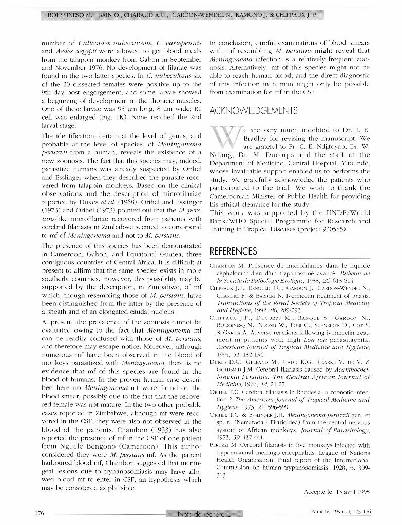
Fig. 1. - Meningonema peruzzii A-H : female, fourth stage larva from cerebrospinal fluid of the patient. A : anterior extremity, lateral view. B : posterior extremity, ventral view. C : tip of the tail, dorsal view. D : posterior extremity, lateral view. E, F, G, head, median, left lateral and right lateral views. H: vulva, lateral view. I-L : larvae from talapoin (Gabon). I : microfilaria from uterus. J : head, dorsal view. K : head, right lateral view. L : first stage larva from the thoracic muscles of Culicoides nubeculosus, 4 days post infestation. Scales : A : 200pm; B, D : 100 µm; C,E,F,G : 20 µm; H : 50 µm; I, L : 10 µm; J, K : hand-drawings.
174 Note de recherche Parasite, 1995, 2, 173-176
MENINGONEMOSIS, A NEW FILARIID ZOONOSES
aminases ( S G O T and SGPT) and C reactive protein (CRP). The pretreatment lumbar puncture was performed after a fundus examination. Biochemical , cytolo-g i c a l , b a c t e r i o l o g i c a l , a n d p a r a s i t o l o g i c a l examinations were carried out on the collected CSF.
Three ml of CSF (one ml in each of three tubes) were col lected from Mr ATA.... T h e CSF was quite clear. Examination of one of the tubes revealed the presence of a mobile nematode measuring 8.7 mm long. T h e two o t h e r tubes w e r e sent to laborator ies o f Centre Pasteur, Yaounde , in order to perform standard examinations. The collected nematode was fixed in hot ethanol (70%) and preserved in the MNHN collection Paris, n° 179 HS. No ivermectin treatment was given to Mr ATA...
Mr ATA. . . did not c o m p l a i n o f any n e u r o l o g i c a l symptoms, nor did the clinical examinations reveal any neurological signs.
The patient harboured 1903 Loa loa mf per 30pl blood, and no Mansonella perstans mf. Only one Onchocerca volvulus mf was recovered from the two skin snips.
Haematological examinations showed only high eosinophil counts (630 per µl, corresponding to 7 .8% of the l e u c o c y t e s ) , low h a e m a t o c r i t (36.8 % ) , a n d a microcytosis (red cell corpuscular volume : 66.9 fl). Blood biochemical parameters were normal, except a slight increase of plasma proteins (89 g/l) and of C reactive protein (9 .9 mg/l).
All biochemical and cytobacteriological examinations of CSF w e r e normal ( 3 0 8 red cel l/mm3; leucocyte counts : <1 per mm3; no bacteria after direct examination and after culture).
No mf was found in the CSF.
IDENTIFICATION OF SPECIMEN
The n e m a t o d e r e c o v e r e d from the CSF was not, as expected, Loa loa. T h e specimen is a female fourth stage larva (vulva closed, geni
tal tracts appearing as a solid cord) and can b e easily identified as belonging to the genus Meningonema Orihel et Esslinger, 1973 (Splendidofilariinae) on the base of the following characters : presence of four uteri, cuticle smooth and thin, muscular body wall very thin, large pseudocoel , tail long, rounded and without lappets. (Fig.1) .
The measurements are the following : body length 8.7 mm, maximum width in the anterior region 180 µm, nervous ring at 150 µm from apex, buccal cavity 6 µm long, oesophagus 600 µm long, vulva at 680 µm from apex, impaired ovijector 600 µm long, tail 300 pm long.
DISCUSSION
M e n i n g o n e m a peruzzii Orihel et Esslinger, 1973, is the only species described at present in the genus. This parasite has been
f o u n d in t h e c e n t r a l n e r v o u s s y s t e m o f t h e Cercopithecidae Cercopithectts (Miopithecus) talapoin in Equatorial Guinea, and was recovered again in the same host in G a b o n ( see b e l o w ) . As suggested by Orihel and Esslinger, this filaria seems to b e the same as that found in 1928 by Peruzzi in a Cercopithecus sp. trapped in Uganda. As the filaria we recovered from the CSF of a Cameroonian individual was a 4th stage larva, it is impossible to warrant the identification at the specific level. However, the similarity of some unusual characters, such as the presence of an oesophago- intest inal torsion, strongly suggests that the filaria found in Cameroon is the same than those previously recovered from monkeys.
O n e Miopithecus talapoin which was maintained at the CNRS Biology Station of Makokou (Gabon) and w a s f o u n d to h a r b o u r Hepatocystis and s h e a t h e d microfilariae, has been splenectomized on 16.07.1976 and shipped by Professor Irene Landau to the MNHN Laboratory in Paris.
The talapoin monkey from Gabon was necropsied on 07.12 .1976, and two adult females and one adult male of M. peruzzii were recovered from the peribulbar meningeal spaces (n° 231 J E MNHN collection. Paris). T h e s p e c i m e n s s h o w e d the s a m e charac ters than those reported in the original description. Additional morphological data on the male's cloacal papillae and the cephalic structures are given in Figure 2 : eight pairs of cloacal papillae can b e seen in ventral view, the left papillae being located more posteriorly. The mouth, the buccal cavity, and the oesophageal lumen are stretched on the lateral plane. The buccal cavity, 5 pm long, is c o n s t r i c t e d hal fway by an internal undulating edge. The cuticle of oesophagus is thick and swollen in the median plane. The four cephalic papillae are only slightly prominent and irregularly arranged.
T w o m i c r o f i l a r i a e ( f r o m b l o o d a n d s t a i n e d with Meldolan b l u e ) w e r e 1 3 5 - 1 3 8 µm long and 4 pm wide; excretory cell , inner body, R1 cell and anal pore respectively at 55-50 µm, 85-82 µm, 98-93 µm and 115-113 µm from apex. Head with a hook on the left side and three cuticular points on the right side.
Among the Splendidofilariinae, Meningonema is one o f the f e w g e n e r a which was found to parasit ize mammals , whereas the other ones are parasites of birds and reptiles. Splendidofilariinae are more often transmitted by Ceratopogonidae than by Culicidae. A
Parasite, 1995, 2, 173-176 Note de recherche 175
B O U S S I N E S Q M., B A I N O . , C H A B A U D A . G . , G A R D O N - W E N D E L N.. K A M G N O J . & C H I P P A U X J . P .
n u m b e r of Culicoides nubeculosus, C. variepennis and Aedes aegypti were allowed to get blood meals from the talapoin monkey from Gabon in September and November 1976. No development of filariae was found in the two latter species. In C. nubeculosus six of the 20 dissected females were positive up to the 9th day post engorgement, and some larvae showed a beginning of development in the thoracic muscles. O n e of these larvae was 95 pm long, 8 pm wide; R l cell was enlarged (Fig. I K ) . None reached the 2nd larval stage.
The identification, certain at the level o f genus, and probable at the level o f spec ies , o f Meningonema peruzzii from a human, reveals the ex is tence of a n e w zoonosis . The fact that this species may, indeed, parasitize humans was already suspected by Orihel and Esslinger when they described the parasite recovered from talapoin monkeys. Based on the clinical o b s e r v a t i o n s a n d the descr ip t ion o f microf i lar iae reported by Dukes et al. (1968) , Orihel and Esslinger (1973) and Orihel (1973) pointed out that the M.pers-tans-like microfilariae recovered from patients with cerebral filariasis in Zimbabwe seemed to correspond to mf of Meningonema and not to M. Persians.
The presence of this species has been demonstrated in Cameroon, Gabon, and Equatorial Guinea, three contiguous countries o f Central Africa. It is difficult at present to affirm that the same species exists in more southerly countries. However, this possibility may b e supported by the description, in Z imbabwe, o f m f which, though resembling those of M. perstans, have been distinguished from the latter by the presence of a sheath and of an elongated caudal nucleus.
At present, the prevalence of the zoonosis cannot b e evaluated owing to the fact that Meningonema m f can b e readily confused with those of M. perstans, and therefore may escape notice. Moreover, although numerous mf have b e e n observed in the b l o o d of monkeys parasitized with Meningonema, there is no e v i d e n c e that m f of this s p e c i e s are found in the blood of humans. In the proven human case described here no Meningonema mf were found on the blood smear, possibly due to the fact that the recovered female was not mature. In the two other probable cases reported in Zimbabwe, although mf were recovered in the CSF, they were also not observed in the b l o o d o f the p a t i e n t s . C h a m b o n ( 1 9 3 3 ) has a lso reported the presence o f m f in the CSF of one patient f r o m N g u e l e B e n g o n o ( C a m e r o o n ) . T h i s a u t h o r considered they were M. perstans mf. As the patient harboured b lood mf, Chambon suggested that meningeal lesions due to trypanosomiasis may have allowed blood mf to enter in CSF, an hypothesis which may be considered as plausible.
In conclusion, careful examinations of b lood smears with m f r e se m bl ing M. perstans might reveal that Meningonema infection is a relatively frequent zoonosis. Alternatively, mf of this species might not b e able to reach human blood, and the direct diagnostic of this infection in human might only b e poss ible from examination for mf in the CSF.
ACKNOWLEDGEMENTS
e are very much indebted to Dr. |. 1.. Bradley for revising the manuscript. We are grateful to Pr. C. E. Ndjitoyap, Dr. W.
N d o n g , D r . M. D u c o r p s a n d t h e s t a f f o f t h e Department of Medicine, Central Hospital, Yaounde, whose invaluable support enabled us to performs the study. W e gratefully acknowledge the patients w h o p a r t i c i p a t e d to t h e t r ia l . W e w i s h to t h a n k t h e Cameroonian Minister o f Public Health for providing his ethical clearance for the study. T h i s w o r k w a s s u p p o r t e d b y t h e U N D P / W o r l d B a n k / W H O S p e c i a l P r o g r a m m e for R e s e a r c h a n d Training in Tropical Diseases (project 930585) .
REFERENCES CHAMBON M. Présence de microfilaires dans le liquide
céphalorachidien d'un trypanosomé avancé. Bulletin de la Société de Pathologie Exotique, 1 9 3 3 , 26, 6 1 3 - 6 1 4 .
CHIPPAUX J.P., ERNOULD J.C., GARDON J. , GARDON-WENDF.L N., CHANDRE F. & BARBF.RI N. Ivermectin treatment of loiasis. Transactions of the Royal Society of Tropical Medicine and Hygiene, 1992, 86, 289 -293 .
CHIPPAUX J . P . , DUCORPS M., RANQUE S., GARDON N., BOUSSINESQ M., NDONG W., FOBI G . , SCHNEIDER D., COT S. & GARCIA A. Adverse reactions following ivermectin treatment in patients with high Loa loa parasi taemia. American Journal of Tropical Medicine and Hygiene, 1 9 9 4 , 51, 1 3 2 - 1 3 4 .
DUKES D.C., GELFAND M. , GADD K.G., CLARKE V . DE V . & GOLDSMID J.M. Cerebral filariasis caused by Acanthochei-lonema perstans. The Central African Journal of Medicine, 1 9 6 6 , 14, 2 1 - 2 7 .
ORIHEL T.C. Cerebral filariasis in Rhodesia- a zoonotic infection ? The American Journal of Tropical Medicine and Hygiene, \91Ъ, 22, 5 9 6 - 5 9 9 .
ORIHEL T.C. & ESSLINGER J.H. Meningonema peruzzii gen. et sp. n. (Nematoda : Filarioidea) from the central nervous system of African monkeys. Journal of Parasitology, 1 9 7 3 , 59, 4 3 7 - 4 4 1 .
PERUZZI M. Cerebral filariasis in five monkeys infected with trypanosomal meningo-encephalitis. League of Nations Health Organisation. Final report of the International Commission On human trypanosomiasis. 1 9 2 8 , p. 3 0 9 -3 1 3 .
Accepté le 1 3 avril 1 9 9 5
1 7 6 Note de recherche Parasite, 1995, 2, 173-176