Embed Size (px)
Citation preview

Rev. bras. paleontol. 16(1):61-76, Janeiro/Abril 2013© 2013 by the Sociedade Brasileira de Paleontologiadoi:10.4072/rbp.2013.1.05
61
ABSTRACT – The record of non-mammaliaformes eucynodonts from the Carnian-Norian Ischigualasto Formation is diverse and abundant, including a medium to large size herbivore and small carnivores. Here is described a new small eucynodont from the Ischigualasto Formation, on the basis of a partial skull. The new taxon is characterized by palatal process of the premaxilla extending posterior to the level of the fi rst postcanine; deep and large maxillary laterodorsal fossa that opens at the level of the root of the upper canine; and postorbital bar diverging posterolaterally at very low angle (35.6°) from the anteroposterior axis of the skull. Results from a phylogenetic analysis supports the new genus placement as a probainognathian eucynodont, more derived than Probainognathus Romer, and more closely related to Ecteninion Martinez, May & Forster and Trucidocynodon Oliveira, Soares & Schultz than to any other eucynodont. Ecteniniidae is proposed as a new clade including the new genus, Ecteninion and Trucidocynodon, and in the phylogenetic hypothesis represents the sister-group of Prozostrodontia (Prozostrodon Bonaparte & Barberena, Tritylodontidae and Mammaliaformes). Additionally, the new taxon from the Ischigualasto Formation shows that the Scaphonyx-Exaeretodon-Herrerasaurus biozone has similar cynodont diversity than the supposedly contemporaneous Hyperodapedon Assemblage Zone of Santa Maria 2 Sequence, in Southern Brazil.
Key words: Triassic, Argentina, Ischigualasto Formation, Cynodontia, Eucynodontia, Ecteniniidae.
RESUMO – O registro dos eucinodontes não-mamaliformes do Carniano-Noriano da Formação Ischigualasto é diversifi cado e abundante, incluindo herbívoros de médio a grande porte e pequenos carnívoros. Descreve-se aqui um pequeno novo eucinodonte da Formação Ischigualasto, com base em um crânio parcial. O novo táxon é caracterizado pelo processo palatal da pré-maxila estendendo-se posteriormente até o nível do primeiro dente pós-canino; uma fossa profunda e ampla abrindo-se látero-dorsalmente na maxila ao nível da raiz do canino superior; e uma barra pós-orbital divergindo póstero-lateralmente em ângulo muito baixo (35,6°) em relação ao eixo ântero-posterior do crânio. Resultado da análise fi logenética sugere que o novo gênero é um eucinodonte Probainognathia, mais derivado que Probainognathus Romer e estreitamente relacionado a Ecteninion Martinez, May & Forster e Trucidocynodon Oliveira, Soares & Schultz do que qualquer outro eucinodonte. O clado novo gênero + Ecteninion + Trucidocynodon é aqui reivindicado como uma nova família chamada Ectininiidae, que é considerada como o clado-irmão de Prozostrodontia (Prozostrodon Bonaparte & Barberena, Tritylodontidae e Mammaliaformes). Além disso, a nova espécie da Formação Ischigualasto mostra que a biozona Scaphonyx-Exaeretodon-Herrerasaurus tem uma diversidade de cinodontes semelhante à da supostamente contemporânea Zona de Associação de Hyperodapedon, da Sequência 2 da Supersequência Santa Maria no sul do Brasil.
Palavras-chave: Triássico, Argentina, Formação Ischigualasto, Cynodontia, Eucynodontia, Ecteniniidae.
A NEW NON-MAMMALIAFORM EUCYNODONT FROM THE CARNIAN-NORIAN ISCHIGUALASTO FORMATION,
NORTHWESTERN ARGENTINA
RICARDO N. MARTÍNEZ, ELIANA FERNANDEZ & OSCAR A. ALCOBERInstituto y Museo de Ciencias Naturales, Universidad Nacional de San Juan, Av. España, 400 (norte), 5400 San Juan, Argentina. [email protected], [email protected], [email protected]
INTRODUCTION
Eucynodontia is defi ned as the clade that includes all cynodonts more derived than Thrinaxodon Seeley, 1894 (Kemp, 1982; Hopson & Barghusen, 1986), or the least inclusive clade including Mammalia and Exaeretodon Cabrera, 1943 (Hopson & Kitching, 2001), and is one of the most stable monophyletic groups within non-mammaliaform cynodonts (Rowe, 1988). The high diversifi cation of eucynodonts during the Middle and Upper Triassic produced several different small to medium size herbivores and carnivores species. The most abundant known record of Upper Triassic eucynodonts is from the Carnian-Norian Santa Maria Formation and
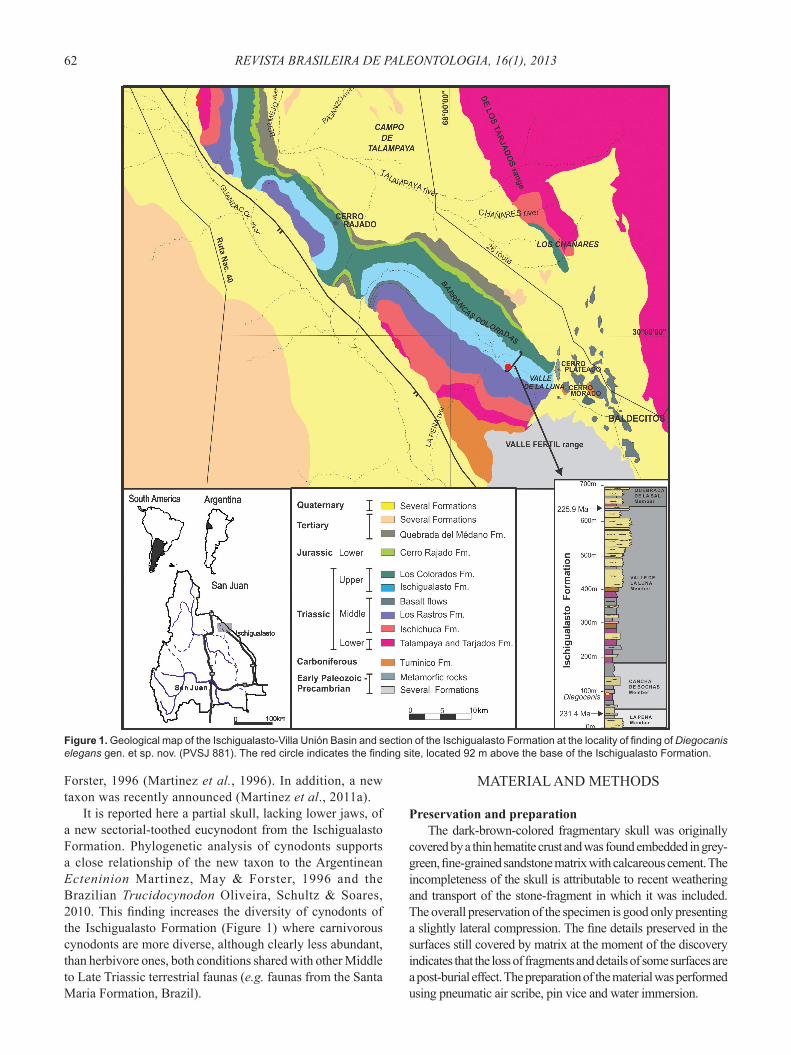
the Norian Caturrita Formation from Brazil, and the Carnian-Norian Ischigualasto Formation from Argentina (Figure 1) (Bonaparte, 1962, 1963, 1966; Barberena, 1981; Bonaparte & Crompton, 1994; Martinez & Forster, 1996; Martinez et al., 1996; Bonaparte & Barberena, 2001; Abdala et al., 2002; Abdala & Ribeiro, 2010; Oliveira et al., 2010). The record from the latter unit includes the medium to large size herbivorous Exaeretodon argentinus Bonaparte, 1962 (=Ischignathus sudamericanus Bonaparte, 1963) (Bonaparte, 1962, 1963; Liu, 2007) and the small to medium size predatory Chiniquodon sanjuanensis (Martinez & Forster, 1996), cf. Probainognathus Romer, 1970 (Bonaparte & Crompton, 1994; see also Fernandez et al., 2011), and Ecteninion lunensis Martinez, May &
REVISTA BRASILEIRA DE PALEONTOLOGIA, 16(1), 201362
Forster, 1996 (Martinez et al., 1996). In addition, a new taxon was recently announced (Martinez et al., 2011a).
It is reported here a partial skull, lacking lower jaws, of a new sectorial-toothed eucynodont from the Ischigualasto Formation. Phylogenetic analysis of cynodonts supports a close relationship of the new taxon to the Argentinean Ecteninion Martinez, May & Forster, 1996 and the Brazilian Trucidocynodon Oliveira, Schultz & Soares, 2010. This fi nding increases the diversity of cynodonts of the Ischigualasto Formation (Figure 1) where carnivorous cynodonts are more diverse, although clearly less abundant, than herbivore ones, both conditions shared with other Middle to Late Triassic terrestrial faunas (e.g. faunas from the Santa Maria Formation, Brazil).
MATERIAL AND METHODS
Preservation and preparationThe dark-brown-colored fragmentary skull was originally
covered by a thin hematite crust and was found embedded in grey-green, fi ne-grained sandstone matrix with calcareous cement. The incompleteness of the skull is attributable to recent weathering and transport of the stone-fragment in which it was included. The overall preservation of the specimen is good only presenting a slightly lateral compression. The fi ne details preserved in the surfaces still covered by matrix at the moment of the discovery indicates that the loss of fragments and details of some surfaces are a post-burial effect. The preparation of the material was performed using pneumatic air scribe, pin vice and water immersion.
Figure 1. Geological map of the Ischigualasto-Villa Unión Basin and section of the Ischigualasto Formation at the locality of fi nding of Diegocanis elegans gen. et sp. nov. (PVSJ 881). The red circle indicates the fi nding site, located 92 m above the base of the Ischigualasto Formation.

63MARTÍNEZ ET AL. – A NEW NON-MAMMALIAFORM EUCYNODONT FROM ISCHIGUALASTO FORMATION
TerminologyIt was employ traditional, or “Romerian” anatomical and
directional terms over veterinarian alternatives (Wilson, 2006). “Anterior” and “posterior”, for example, are used as directional terms rather than the veterinarian alternatives “rostral” or “cranial” and “caudal”.
Institutional abbreviations. PVSJ, Instituto y Museo de Ciencias Naturales, San Juan, Argentina; UFRGS, Universidade Federal do Rio Grande do Sul, Porto Alegre, Brazil.
SYSTEMATIC PALEONTOLOGY
Order THERAPSIDA Broom, 1905Suborder CYNODONTIA Owen, 1861
Infraorder EUCYNODONTIA Kemp, 1982
Ecteniniidae fam. nov.
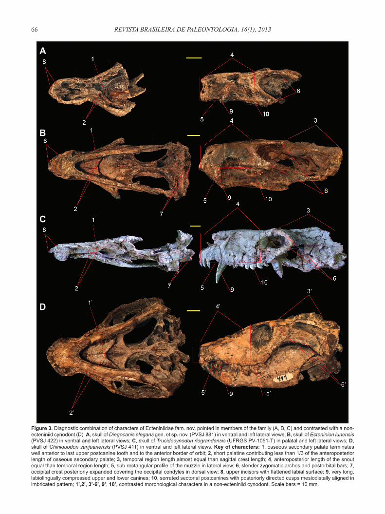
Defi nition. All taxa more closely related to Ecteninion lunensis Martinez, May & Forster, 1996, than to Probainognathus jenseni Romer, 1970, Chiniquodon theotonicus Huene, 1936, Lumkuia fuzzi Hopson & Kitching, 2001, Exaeretodon argentinus Bonaparte, 1962, Prozostrodon brasiliensis Bonaparte & Barberena, 2001, or Homo sapiens Linnaeus, 1758.Type genus. Ecteninion Martinez, May & Forster, 1996.Diagnosis. Eucynodonts presenting the following combination of characters: osseous secondary palate terminates well anterior to the last upper postcanine and to the anterior border of orbit; short palatine contributing less than 1/3 of the anteroposterior length of osseous secondary palate; angular process of dentary close to the craniomandibular joint; elongate parietals relative to basal skull length; snout length equal than temporal region length; sub-rectangular profi le of the snout in lateral view; slender zygomatic arches and postorbital bars; occipital crest posteriorly expanded covering the occipital condyles in dorsal view; incisors with fl attened labial surface; very long, labiolingually compressed canines with serrations; and sectorial postcanines with posteriorly directed cusps mesiodistally aligned in an imbricated pattern.
Diegocanis gen. nov.
Type species. Diegocanis elegans.
Etymology.The epithet honors Diego Abelin, technician of the Instituto y Museo de Ciencias Naturales of the Universidad Nacional de San Juan and discoverer of the type specimen; Canis, dog (Latin). Diagnosis. Same as for species.
Diegocanis elegans sp. nov.(Figures 2-4, Table 1-3)
Etymology. Elegans, elegant (Latin), refers to the sleek look of the snout of the holotype. Holotype. PVSJ 881, partial skull, represented by the snout and the orbital region, with partially preserved upper dentition.
Locality and horizon. Cancha de Bochas, Hoyada de Ischigualasto, Ischigualasto Provincial Park, San Juan Province, Argentina (Figure 1). The specimen was collected from a level located 92 m above the base of the Ischigualasto Formation, 15 m eastern of the location of the type specimen of the basal dinosaur Eoraptor lunensis Sereno et al., 1993. Middle levels of the Cancha de Bochas Member (sensu Currie et al., 2009) at the southern outcrops of the Ischigualasto Formation. Middle Scaphonyx-Exaeretodon-Herrerasaurus biozone (sensu Martinez et al., 2011b).Diagnosis.Eucynodont characterized by possessing the following autapomorphies: long palatal process of the premaxilla, extending backwards at the level of the fi rst postcanine tooth; deep and large fossa opening laterodorsally on the maxilla, at the level of the root of the upper canine; and thin postorbital bar diverging posterolaterally at very low angle (35.6°) from the sagittal axis of the skull.Description. The well preserved anterior fragment of the skull of PVSJ 881 is 53.9 mm long (Table 1), and almost not deformed (Figure 2). The snout is anteroposteriorly long and subrectangular in lateral view, with the anterior section having similar width and height than the preorbital area (Figure 3A), similar to Ecteninion and Trucidocynodon (Figures 3B,C); but different from the sub-trapezoidal snout in lateral view of other eucynodonts (e.g. Chiniquodon sanjuanensis, Probainognathus, Prozostrodon, Massetognathus Romer, 1967; Martinez & Forster, 1996; Romer, 1970; Bonaparte & Barberena, 2001; Barberena, 1981) (Figure 3D). The dorsal surface of the snout is sub-horizontal and straight in lateral view as in Ecteninion and Trucidocynodon (Figures 3A-C), differing from the slightly sinuous and anteriorly descendent dorsal surface of other eucynodonts (Figure 3D). The orbits are large and laterodorsally oriented as in Ecteninion (Figure 3B) and Trucidocynodon (Figure 3C), contrasting with the relative small orbits anteriorly oriented of Chiniquodon sanjuanensis (Figure 3D) and Lumkuia Hopson & Kitching, 2001. The preserved dorsal portion of the postorbital bar is slender as in Ecteninion.
The premaxilla is dorsoventrally high (Figures 2C, D), with the ratio between the height of the anterior portion of the premaxilla and the height of the external naris being 0.61. This value is lower than in Menadon Flynn et al., 2000 (Kammerer et al., 2008) and Cynognathus Seeley, 1895 (Seeley, 1895; Bonaparte, 1969) in which the ratio is 0.8, but higher than in most other Triassic eucynodonts (Table 2). The posterior border of the premaxilla is overlapped by the maxilla at the level of the fourth incisor (Figures 2B,C), as in other cynodonts. The internarial process is high (Figure 2D), whereas the posterolateral ascending process is short reaching the mid-level of the external naris without contacting the nasal (Figures 2C,D). This condition is similar in Ecteninion, but different from the long process of Trucidocynodon which reach the dorsal level of the external naris (Oliveira et al., 2010). In ventral view, the palatal process of the premaxilla is posteromedially very long, extending backwards to the level of the fi rst postcanine tooth, forming the anterior and medial

REVISTA BRASILEIRA DE PALEONTOLOGIA, 16(1), 201364
border of the paracanine fossa (Figure 2B). In contrast, in Trucidocynodon and Ecteninion (PVSJ 422 and PVSJ 693) the premaxilla ends at the anterior border of the fossa (Figure 4A). In other probainognathians the posteromedial process is shorter than in Diegocanis gen. nov., extending to the canine level (e.g. Chiniquodon Huene, 1936, Probainognathus, Therioherpeton Bonaparte & Barberena, 1975; Huene, 1936; Abdala & Giannini, 2002; Romer, 1970; Bonaparte & Barberena, 1975; Oliveira, 2006). The paracanine fossae are located anteromedial to the upper canine (Figure 2B) as in the majority of Triassic cynodonts; in contrast, Massetognathus (Romer, 1967; Liu et al., 2008) and Exaeretodon (Bonaparte, 1962; Abdala et al., 2002, 2006) have the fossae located medially and posteromedially respectively, and in Riograndia Bonaparte, Ferigolo & Ribeiro, 2001 (Bonaparte et al., 2001; Soares et al., 2011) and Pachygenelus Watson, 1913 are absent (Watson, 1913). The paracanine fossae of Diegocanis gen. nov. are dorsally closed, different from Ecteninion (Figures 4C-F) and Trucidocynodon in which the fossae are dorsally opened allowing the lower canines to protrude through the snout roof. The incisive foramina are fully enclosed by the premaxilla (Figures 2B, 4B) as in Ecteninion (Figure 4A), Chiniquodon, Probainognathus, and Prozostrodon; but different from Trucidocynodon, Lumkuia, and Scalenodon Crompton, 1955 (Parrington, 1946; Crompton, 1955), in which the maxilla forms the posterior border of the foramina. The incisive foramina of Diegocanis gen. nov. are small and located posterior to the level of the anterior border of the paracanine fossae, but in Ecteninion they are comparatively larger and located in front to the anterior border of the fossae (Figure 4A).
The facial process of the septomaxilla is long and almost vertical, reaching the level of the dorsal border of the external naris (Figures 2C,D), as in Ecteninion and Trucidocynodon, although in these taxa the processus is dorsoposteriorly inclined. The process forms the posterolateral border of the external naris and separates the anterior margin of the maxilla from the nasal. As in Ecteninion, the septomaxillary foramen lies between the septomaxilla and maxilla at the level of the fourth incisor (Figures 2C,D), although it is smaller in Diegocanis gen. nov. The anteromedial process is ventrally convex and dorsally concave, and extends towards the midline, separated from the fl oor of the nares (Figure 2D), as in Chiniquodon sanjuanensis (Martínez & Forster, 1996),
Trucidocynodon, and in less degree in the larger specimen of Ecteninion (PVSJ 481). In contrast, in most eucynodonts (e.g. Probainognathus) and small specimens of Ecteninion, of similar size than Diegocanis gen. nov., the process lies directly on the premaxilla.
The long and deep maxilla is subtriangular in lateral view (Figure 2C) as in Ecteninion and Trucidocynodon. As in other eucynodonts, the two maxillae are medially nearest each other and reach their greatest height at the level of implantation of the canine roots. The maxilla has a conspicuous constriction behind the canine as in Ecteninion and Trucidocynodon, but different than the pronounced constriction of Chiniquodon and Probainognathus (Romer, 1969, 1970). The jugal overlaps posteriorly the maxilla, except in its ventral part in which the latter extends as a narrow posteroventral process below the jugal (Figure 2C), as in Ecteninion but different from the wide process of Chiniquodon sanjuanesis. Unfortunately, only the anterior roots of both zygomatic arches are preserved. The ventral border of the maxilla forms an incipient protruding platform lateral to the alveolar line (Figure 2C), similar than in Trucidocynodon, but different from Ecteninion, in which the lateral surface of the maxilla is on the same plane than the lateral surface of the postcanine crowns. Diegocanis gen. nov. has a very large, deep, and anteroposteriorly long fossa in both maxillae, at the level of the upper canine root (Figures 2A,C,E). The location (posterior to the paracanine fossa) and the lateral exit (instead dorsal) of these fossae precludes its interpretation as the dorsal opening of the paracanine fossa present in Ecteninion, Trucidocynodon, Pascualgnathus Bonaparte, 1966 (Martinelli, 2010), and Andescynodon Bonaparte, 1967 (Liu & Powell, 2009). The fossae also differ from the nasomaxillary fossae of Cynognathus which are smaller and located on the nasals (Abdala, 1996). The fossae are elliptical in lateral view, measuring 5 mm anteroposteriorly and 3 mm dorsoventrally (Figures 2C,E), with its shape being best observed in the left side (Figure 2E), as the right maxilla is deformed anteriorly (Figure 2C). The inner surface of the fossa is smooth and fl at. The absence of crushes at least in one side of the maxilla, the symmetrical inward projection of both fossae, and the presence of a fl at surface of bone closing internally each fossa, preclude its interpretation as a post-mortem deformation (Figures 2C,E). On the lateral surface of the maxilla, parallel to the alveolar border, Diegocanis gen. nov. has two large anteroposteriorly aligned infraorbital foramina (Figure 2C) as in Ecteninion and Trucidocynodon (Martinez et al., 1996; Oliveira et al., 2010); in contrast some specimens of Chiniquodon have only one large foramen (Romer, 1969). A third, posterior infraorbital foramen opens laterally at the point where lacrimal, jugal and maxilla contact (Figures 2C,E), as in Ecteninion, Probainognathus, and Massetognathus (Martinez et al., 1996; Romer, 1970, 1967).
In palatal view, the maxilla forms 77% of the secondary palate (Figure 2B), shorter than in Trucidocynodon (82%) (Oliveira et al., 2010) (Figure 3B), but longer than in Ecteninion (60%), Probainognathus (61%), and Chiniquodon (48%).
Maximum anteroposterior length of the preserved fragment of skull 53.9
Snout length 35.0Length of secondarypalate 30.9Length of secondary palatal shelf of maxilla 15.1Length of secondary palatal shelf of palatine 3.5Orbit height 15.5Anteroposterior thickness of the postorbital bar 2.3Length of upper tooth row 38.5Length of postcanine upper tooth row 20.6Interorbital width 13.5
Table 1 . Skull measurements (mm) of Diegocanis elegans gen. et sp. nov. (PVSJ 881).

65MARTÍNEZ ET AL. – A NEW NON-MAMMALIAFORM EUCYNODONT FROM ISCHIGUALASTO FORMATION
Figure 2. Skull of Diegocanis elegans gen. et sp. nov. (PVSJ 881) in A, dorsal, B, ventral, C, right lateral, D, anterior and E, left lateral views. F, close-up of the dentition in right lateroventral view. G, close-up of right Pc4 in ventromedial view. Abbreviations: a-d, cusps of Pc4; dmf, dorsomaxillary fossa; ec, ectopterygoid; f, frontal; if, incisive foramen; i1-4, incisors 1 to 4; iof, infraorbital foramen; ipcf, internal paracanine fossa; j, jugal; l, lacrimal; lc, left canine;lf, lacrimal foramen;lr, lacrimal ridge; m, maxilla; mll, maxillar labial lip; n, nasal; p, parietal; pal, palatine; pc1-7, postcanine 1 to 7; pm, premaxilla; po, postorbital; prf, prefrontal; pt, pterygoid; rc, right canine; rec, replacement canine; sm, septomaxilla; smf, septomaxillary foramen; v, vomer. Bones in second plane are in light grey color. Broken surfaces are in dark grey color. Scale bar = 10 mm.
A
C
D
E
F G
B
REVISTA BRASILEIRA DE PALEONTOLOGIA, 16(1), 201366
Figure 3. Diagnostic combination of characters of Ecteniniidae fam. nov. pointed in members of the family (A, B, C) and contrasted with a non-ecteniniid cynodont (D). A, skull of Diegocanis elegans gen. et sp. nov. (PVSJ 881) in ventral and left lateral views; B, skull of Ecteninion lunensis (PVSJ 422) in ventral and left lateral views; C, skull of Trucidocynodon riograndensis (UFRGS PV-1051-T) in palatal and left lateral views; D, skull of Chiniquodon sanjuanensis (PVSJ 411) in ventral and left lateral views. Key of characters: 1, osseous secondary palate terminates well anterior to last upper postcanine tooth and to the anterior border of orbit; 2, short palatine contributing less than 1/3 of the anteroposterior length of osseous secondary palate; 3, temporal region length almost equal than sagittal crest length; 4, anteroposterior length of the snout equal than temporal region length; 5, sub-rectangular profi le of the muzzle in lateral view; 6, slender zygomatic arches and postorbital bars; 7, occipital crest posteriorly expanded covering the occipital condyles in dorsal view; 8, upper incisors with fl attened labial surface; 9, very long, labiolingually compressed upper and lower canines; 10, serrated sectorial postcanines with posteriorly directed cusps mesiodistally aligned in imbricated pattern; 1’,2’, 3’-6’, 9’, 10’, contrasted morphological characters in a non-ecteniniid cynodont. Scale bars = 10 mm.
A
B
C
D

67MARTÍNEZ ET AL. – A NEW NON-MAMMALIAFORM EUCYNODONT FROM ISCHIGUALASTO FORMATION
The maxillae meet at the midline reaching anteromedially the level of the fi rst postcanine where they contact the premaxillae. Anterolaterally the maxilla extends further forming the posterior border of the paracanine fossa (Figure 2B). This fossa is located anteromedially to the canine and do not perforate the dorsal surface of the snout. In contrast, in Ecteninion (Figures 4C,E,F) and Trucidocynodon the fossa perforates the dorsal surface of the snout between the nasal and maxilla. Posteriorly, the maxilla meets the palatine at the level between the fourth and fi fth postcanine, differing from Ecteninion and Trucidocynodon in which the contact is between the second and third postcanines (Martinez et al., 1996; Oliveira et al., 2010), and Chiniquodon sanjuanensis in which it is at the level of the fi fth tooth (Martinez & Forster, 1996).
The nasals are long and narrow (Figures 2A,C). The anterior margin of the nasals forms a medial tip that almost reaches the level of the anterior border of the premaxilla. In dorsal view the nasal has a straight lateral border on the anterior 60% of its length (Figure 2A). This border gently tapers from the anterior end to the level of the postcanine constriction. In other eucynodonts the nasal border is curved (e.g.Chiniquodon, Ecteninion, Trucidocynodon, Probainognathus). Behind this anterior part, the nasals expand posterolaterally close to the level of the anterior border of the orbits, and then taper posteromedially (Figure 2A). The nasal is laterally overlapped by the maxilla from the anterior end to the level of the dorsal fossa from where the nasal overlaps the maxilla until its contact with the lacrimal. Posterolaterally the nasal overlaps the anterior border of the lacrimal and frontal. The nasal of Diegocanis gen. nov. only contacts the anteromedial corner of the prefrontal, different from the wide sutural contact of Ecteninion and Trucidocynodon. The suture with the frontal is not well preserved.
The rostral portion of the lacrimal is anteroposteriorly reduced (Figures 2A,C,E), as in Ecteninion and Trucidocyno-don. The lacrimal forms the anterior and ventral orbital rim and much of the anterior portion of the inner orbital wall. The anterior margin of the bone is rounded, contacting the nasal anterodorsally, the maxilla anteroventrally, and the jugal ventrally as in Ecteninion, but contrasting with the triangular
shape of Trucidocynodon, Chiniquodon, Probainognathus and Therioherpeton (Oliveira et al., 2010; Oliveira, 2006). The lacrimal shows a ventral process directed posteriorly, forming a pronounced ridge along the orbital margin. This process is in contact with the jugal ventrally and extends posteriorly as far as the posterior end of the maxilla (Figure 2C), as in Ecteninion and Riograndia (Bonaparte et al., 2001; Soares et al., 2011), differing from Trucidocynodon that shows a reduced condition and others eucynodonts in which the process is absent (e.g. Probainognathus, Chiniquodon). As in Ecteninion, the lacrimal foramen opens near to the orbital rim, slightly above the level of the suture between nasal, maxilla and lacrimal (Figure 2C), but Trucidocynodon has two small foramina. In the inner orbital wall the lacrimal contacts the frontal posteromedially and the jugal posterolaterally.
The prefrontal is narrow and anteroposteriorly long, forming the greater part of the dorsal orbital rim (Figures 2A,C). The length of the prefrontal in relation to the orbit is similar than in small specimens of Ecteninion, which are of similar size than Diegocanis gen. nov. (PVSJ 693), but shorter than in larger specimens (PVSJ 422, PVSJ 481). The prefrontal has a tiny contact with the nasal anteromedially, and more extended with the lacrimal anteriorly. The prefrontal overlaps the frontal medially, posteriorly is overlapped by the postorbital and anterolaterally forms an interdigitated suture with the lacrimal. The prefrontal forms part of the roof of the orbit, but do not participates in the medial orbital wall. This condition is similar to that of Ecteninion, Trucidocynodon, and chiniquodontids.
The short frontal tapers posteriorly, being overlapped anteriorly by the nasal, anterolaterally by the prefrontal, posterolaterally by the postorbital (Figure 2A). The suture with the parietal is obscured by damage, whereas its contact with the postorbital is longer than with the prefrontals. The frontals seem to be completely fused and they extend ventrally to form part of inner orbital wall, where they contacts laterodorsally the prefrontal, anteriorly the lacrimal, and ventrally the palatine (Figure 2C), as in Ecteninion (Martinez et al., 1996). It also extends posteriorly to contact the parietal ventral to the postorbital.
A small anterior tip of the right parietal, overlapped by the postorbital, is preserved, but the damage precludes seeing details of it (Figure 2A).
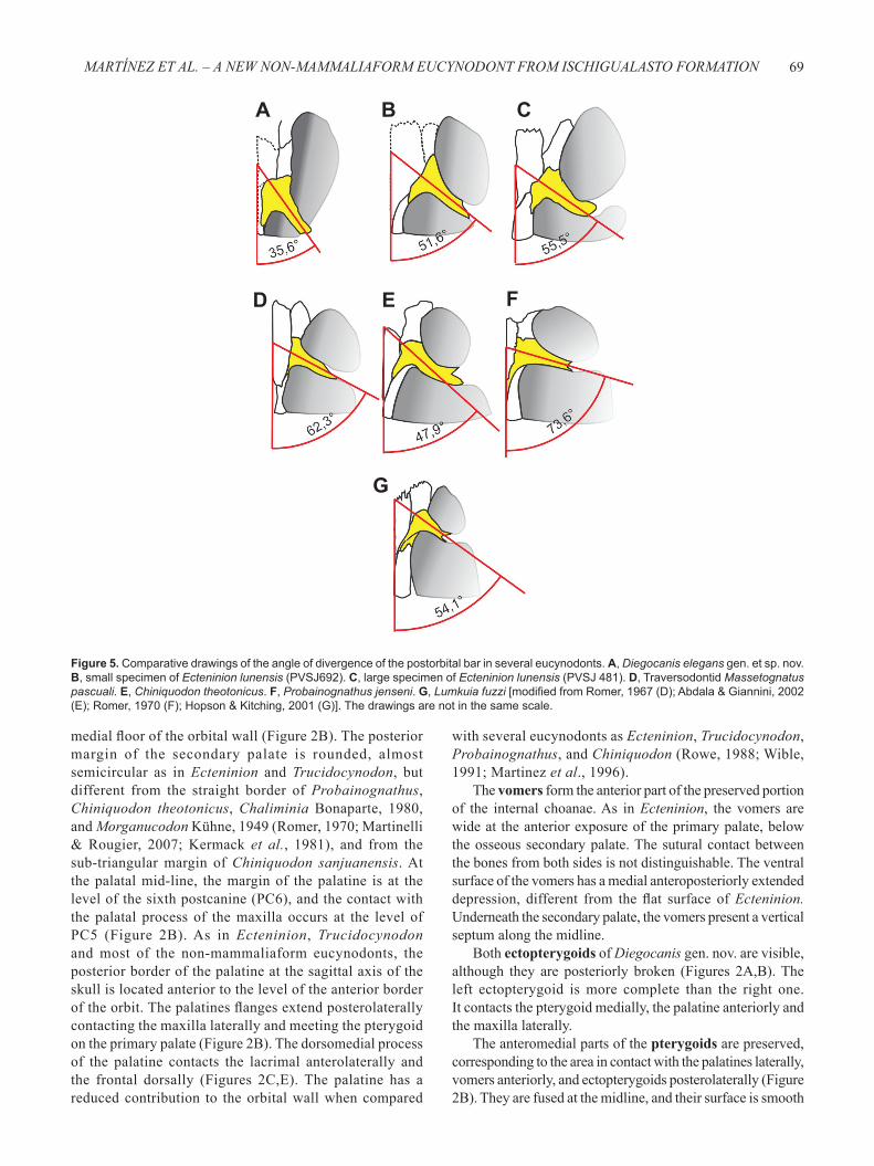
Only the anteromedial portion of the right postorbital is preserved (Figures 2A-C). The postorbital overlaps the posterolateral corner of the frontal, the posterior border of the prefrontal and the anteromedial margin of the parietal, indicating that there is not medial contact with the opposite postorbital. Ventrally it forms a small portion of the dorsal roof of the inner orbital wall. The posterior process overlaps the parietal. The preserved portion of the postorbital bar is very thin and subcircular in cross-section as in Ecteninion. It diverges posterolaterally at 36° from the midline of the skull (Figures 2A, 5A), more posteriorly directed than in other eucynodonts: Ecteninion: 50°-55° (Figures 5B,C); Massetognathus: 62° (Figure 5D); Chiniquodon 48° (Figure 5E); Probainognathus: 74° (Figure 5F); and Lumkuia: 54° (Figure 5G).
Genus Ratio SourceMenadon 0.80 Kammerer et al. (2008)Cynognathus 0.80 Seeley (1895); Bonaparte (1969)Diegocanis 0.61Thrinaxodon 0.50 Seeley (1894b); Fourie (1974)Ecteninion 0.45 Martínez et al. (1996)Chiniquodon 0.43 Romer (1969)
Riograndia 0.40 Bonaparte et al. (2001); Soares et al. (2011)
Diademodon 0.40 Seeley (1894a)
Sinocodon 0.38 Patterson & Olson (1961); Crompton & Luo (1993)
Prozostrodon 0.36 Bonaparte & Barberena (2001)Probainognathus 0.30 Romer (1970)
Table 2. Ratios between the height of the anterior portion of the premaxilla and the height of the external nares in several eucynodonts.

REVISTA BRASILEIRA DE PALEONTOLOGIA, 16(1), 201368
The anterior margin of the jugal expands anteriorly overlapping the maxilla (Figures 2C,E). The jugal contributes 55% of the total height of the suborbital bar; the posteroventral process of lacrimal and maxilla forms the other 45% (Figures 2C,E). The jugal has an anteroventral depression which holds the thin posterior maxillary process (Figure 2C) as in Ecteninion. The jugal has a long contact with the
lacrimal. Judging from the preserved fragment of the jugal, the zygomatic arch of Diegocanis gen. nov. was narrow and slender. The jugal forms the lateral fl oor of the inner orbit, were it contacts the pterygoid medially and lacrimal anterolaterally (Figure 2A).
The palatine forms the rear 23% of the secondary palate, the lateral portion of the primary palate, and the
Figure 4. Comparison of ecteniniids from the Carnian Scaphonyx-Exaeretodon-Herrerasaurus biozone of the Carnian-Norian Ischigualasto Formation. A, horizontal computer tomographic section and interpretative drawing of the anterior part of the skull of the holotype of Ecteninion lunensis (PVSJ 422); B, skull of Diegocanis elegans gen. et sp. nov. in ventral view; C, anterior part of the skull of the holotype of Ecteninion lunensis (PVSJ 422) in right lateral view showing the position of the selected computer tomographic sections; D, Coronal computer tomographic section and interpretative drawing of the anterior part of the snout of the holotype of Ecteninion lunensis (PVSJ 422); E, photography and interpretative drawing of a small specimen of Ecteninion lunensis (PVSJ 693) in anterodorsal view showing the paracanine fossae; F, photography of the skull of a large specimen of Ecteninion lunensis (PVSJ 481) in laterodorsal view and interpretative drawing of the anterior portion of the snout showing the dorsally opened paracanine fossae. Abbreviations: ec, ectopterygoid; f, frontal; if, incisive foramen; i1-4, incisors 1 to 4; ipcf, internal paracanine fossa; j, jugal; lc, lower canine; m, maxilla; n, nasal; pal, palatine; pa, parietal; pc1-8, postcanine 1 to 8: pcfdo, paracanine fossa dorsal opening; pf, prefrontal; pm, premaxilla; po, postorbital; pt, pterygoid; rec, replacement canine; uc, upper canine; v, vomer. Bones in second plane are in light grey color, broken surfaces and matrix are dark grey; hollowed areas are black. Scale bars = 10 mm.
B
C
E F
D
A
69MARTÍNEZ ET AL. – A NEW NON-MAMMALIAFORM EUCYNODONT FROM ISCHIGUALASTO FORMATION
medial fl oor of the orbital wall (Figure 2B). The posterior margin of the secondary palate is rounded, almost semicircular as in Ecteninion and Trucidocynodon, but different from the straight border of Probainognathus, Chiniquodon theotonicus, Chaliminia Bonaparte, 1980, and Morganucodon Kühne, 1949 (Romer, 1970; Martinelli & Rougier, 2007; Kermack et al., 1981), and from the sub-triangular margin of Chiniquodon sanjuanensis. At the palatal mid-line, the margin of the palatine is at the level of the sixth postcanine (PC6), and the contact with the palatal process of the maxilla occurs at the level of PC5 (Figure 2B). As in Ecteninion, Trucidocynodon and most of the non-mammaliaform eucynodonts, the posterior border of the palatine at the sagittal axis of the skull is located anterior to the level of the anterior border of the orbit. The palatines fl anges extend posterolaterally contacting the maxilla laterally and meeting the pterygoid on the primary palate (Figure 2B). The dorsomedial process of the palatine contacts the lacrimal anterolaterally and the frontal dorsally (Figures 2C,E). The palatine has a reduced contribution to the orbital wall when compared
with several eucynodonts as Ecteninion, Trucidocynodon, Probainognathus, and Chiniquodon (Rowe, 1988; Wible, 1991; Martinez et al., 1996).
The vomers form the anterior part of the preserved portion of the internal choanae. As in Ecteninion, the vomers are wide at the anterior exposure of the primary palate, below the osseous secondary palate. The sutural contact between the bones from both sides is not distinguishable. The ventral surface of the vomers has a medial anteroposteriorly extended depression, different from the fl at surface of Ecteninion. Underneath the secondary palate, the vomers present a vertical septum along the midline.
Both ectopterygoids of Diegocanis gen. nov. are visible, although they are posteriorly broken (Figures 2A,B). The left ectopterygoid is more complete than the right one. It contacts the pterygoid medially, the palatine anteriorly and the maxilla laterally.
The anteromedial parts of the pterygoids are preserved, corresponding to the area in contact with the palatines laterally, vomers anteriorly, and ectopterygoids posterolaterally (Figure 2B). They are fused at the midline, and their surface is smooth
Figure 5. Comparative drawings of the angle of divergence of the postorbital bar in several eucynodonts. A, Diegocanis elegans gen. et sp. nov. B, small specimen of Ecteninion lunensis (PVSJ692). C, large specimen of Ecteninion lunensis (PVSJ 481). D, Traversodontid Massetognatus pascuali. E, Chiniquodon theotonicus. F, Probainognathus jenseni. G, Lumkuia fuzzi [modifi ed from Romer, 1967 (D); Abdala & Giannini, 2002 (E); Romer, 1970 (F); Hopson & Kitching, 2001 (G)]. The drawings are not in the same scale.
B CA
D E F
G

REVISTA BRASILEIRA DE PALEONTOLOGIA, 16(1), 201370
and slightly concave, differing from the surface with sharp ridges present in Ecteninion.
Dentition. The upper dental formula of Diegocanis gen. nov. is 4I-1C-7PC, different from the 4I-1C-8PC formula of Ecteninion and Trucidocynodon. The holotype of Ecteninion (PVSJ 422) was originally published presenting seven postcanines, with a diastema between the fi rst and the second tooth (Martinez et al., 1996). Study of coronal computer tomographic slices of this specimen show that it has eight postcanine teeth, with a postcanine root in the place of the supposed diastema (Figure 4A). Additional specimens of Ecteninion, which include smaller (PVSJ 693) and larger individuals (PVSJ 481) than the holotype also, have the same formula. All the functional upper dentition in Diegocanis gen. nov. is broken lacking most of the crowns (Figures 2B-D,F, 4B), right PC4 however, which is a replacement tooth, has an almost complete crown (Figures 2B,F, 4B). The visible parts of the left incisors mainly correspond to the labial surface of the roots, exposed by erosion of part of the left premaxilla (Figure 2E).
The incisors are thin and unevenly located. The sections of the two anterior incisors are semicircular and are in contact each other, but the two posterior teeth are spaced and have subtriangular cross-section (Figure 2B). As in Ecteninion, the labial surface of the incisors is fl at with longitudinal striations which are more developed in the two posterior teeth. The incisors are recumbent as in other eucynodonts, and different from the slightly procumbent incisors of Ecteninion (Martinez et al., 1996). A moderate diastema separates the last incisor from the canine.
The crowns of the upper canines are labiolingually compressed. Although the crowns of both canines are broken but judging by the orientation of the roots they seems to be procumbent, anteroventrally directed at angle of 60° respect to the alveolar margin as in large Ecteninion (PVSJ 481). This condition is different from the almost vertical canines of the smaller specimens of Ecteninion (e.g. PVSJ 693, PVSJ 422), Trucidocynodon, Chiniquodon, and Probainognathus. As in Ecteninion and Trucidocynodon, the canine has a rounded anterior margin and a thin and sharp posterior edge. In the anterolingual side of each canine a replacement tooth of similar size is erupting. The replacement canine is almost straight and slightly tapers distally, giving the impression that they will likely be very longer in the full development stage (Figure 2F). No serrations are visible in the replacement canine, nor in the preserved fragments of the functional ones. Nevertheless, damage of the functional canines and the defi cient preservation of the replacement one preclude certainty in the lack of serration on the canines of Diegocanis gen. nov. A small diastema separates the canine from the fi rst postcanine (Figure 2B).
The seven upper postcanines are in contact each other and forming an imbricated pattern as in Ecteninion. The four anterior postcanines are smaller than the three posteriors as in Ecteninion and Trucidocynodon (Martinez et al., 1996; Oliveira et al., 2010). The fourth postcanine is erupting and is the only presenting a complete crown. It has four anteroposteriorly aligned and slightly posteriorly recurved cusps, one small anterior (a), a larger main second (b) and two
smaller posteriors (c and d) (Figures 2B,F,G). This morphology is similar than that of Ecteninion and Trucidocynodon. As far as is possible to see, due to the damaged crowns, this pattern appears to be similar in the larger crowns.
PHYLOGENETIC ANALYSIS
In order to determine the phylogenetic position of Diegocanis elegans gen. et sp. nov. within Eucynodontia, it was used the data matrix published by Liu & Olsen (2010) because it is the most comprehensive study, including most of the taxa here used for comparisons. Two taxa were added two taxa, Diegocanis elegans gen. et sp. nov. and Trucidocynodon riograndensis Oliveira, Schultz & Soares, 2010, and three characters - character 4: profi le of the anterior region of the snout in lateral view; character 10: temporal region length relative to sagittal crest length, and character 19: orbit height relative to snout height (Appendix 1). Character 3 from Liu & Olsen (2010) was modifi ed as follow: snout anteroposterior length relative to anteroposterior length of the temporal region (measured from the anterior most level of the posterior border of postorbital bar). Our resulting matrix contains 148 characters scored for 33 taxa. Several character scores for Ecteninion by Liu & Olsen (2010) were incorrect and have been changed (Appendix 2).
A heuristic search, using T.N.T. (Goloboff et al., 2003, 2008), has been applied to the data matrix with multistate characters treated as unordered.
The analysis resulted in eight most parsimonious trees of 448 steps (Consistency Index=0.47, Retention Index=0.77) – same number of MPTs and 17 steps longer (429) than in the Liu and Olsen´s (2009) analysis. An implicit enumeration search and support for each branch was estimated by performing 1000 bootstrap replications, and Bremer support of monophyletic groups were also calculated (Figure 6).
The obtained MPTs consistently place Diegocanis gen. nov. within Eucynodontia as more derived than Chiniquodon and Probainognathus, and as a member of a group that includes Ecteninion and Trucidocynodon in an unresolved politomy (Figure 6). This group is referred here as the family Ecteniniidae, representing the sister-group of Prozostrodontia.
Consequently, Ecteniniidae is defi ned here as all taxa more closely related to Ecteninion lunensis, than to Probainognathus jenseni, Chiniquodon theotonicus, Exaeretodon argentinus, Prozostrodon brasiliensis (Barberena, Bonaparte & Teixeira, 1987) or Homo sapiens (see Systematic Paleontology). Bremer support for Ecteniniidae fam. nov. is 3, whereas bootstrap frequencies support is 70% (Figure 6). Nine synapomorphies are supporting the monophyly of Ecteniniidae fam. nov.: 1) anteroposterior snout length equal than temporal region length (3.1); 2) sub-rectangular profi le of the anterior region of snout in lateral view (4.0); 3) temporal region length almost equal than sagittal crest length (10.1); 4) snout tip height greater or equal than the orbit height (19.1); 5) constant width of temporal fossa along its length (26.1); 6) osseous secondary palate terminates well anterior to last upper postcanine tooth (37.0); 7) osseous secondary palate terminates anterior to
71MARTÍNEZ ET AL. – A NEW NON-MAMMALIAFORM EUCYNODONT FROM ISCHIGUALASTO FORMATION
anterior border of orbit (38.0); 8) short palatine contributing less than 1/3 of the anteroposterior length of osseous secondary palate (40.1); and 9) posteroventral angle of dentary close to jaw joint (87.1). Four of these characters (characters 3, 10, 26 and 87) are unknown in Diegocanis gen. nov.
Ecteniniidae fam. nov. is recovered as sister group of Prozostrodontia, and in a more derived placement than Probainognathus, a difference to that obtained by Liu & Olsen (2010), in which Ecteninion was basal to Probainognathus. Seven unambiguous synapomorphies support the sister group relationship of Ecteniniidae fam. nov. and Prozostrodontia: 1) sagittal crest extending posteriorly to reach the posteriormost part of the lamboidal crest (14.1); 2) slender zygomatic arch (20.2); 3) maximum dorsal extent of zygomatic arch below middle of orbit (23.0); 4) pterygoparoccipital foramen as a notch (61.2); 5) strongly developed dens (125.1); 6) well ossifi ed olecranon process of ulna (136.1); and 7) posterior iliac spine has a small nub that lies entirely anterior to the acetabulum (142.1).
Phylogenetic placement of Ecteninion within Eucynodontia has been conflictive in hypotheses proposed by different scholars. Initially, Ecteninion formed a politomy with Probainognathus that represented the sister-group of chiniquodontids, Tritylodontidae, Tritheledontidae and Morganucodon (Martinez et al., 1996). For others, Ecteninion is a basal probainognathian (Hopson & Kitching, 2001; Martinelli
& Rougier, 2007; Liu & Olsen, 2010); or a basal Cynognathia, sister taxon of Cynognathus plus gomphodont cynodonts (Abdala, 2007). This uncertainty is more likely refl ecting the incompleteness of the specimen and the unusual combination of characters (e.g. short secondary palate, slender zygomatic arches, reduced postdentary bones, presence of serrations on the canine margin). The present result showing ecteniniids as more derived than Probainognathus is the consequences of the change in the scoring of several character states for Ecteninion in the data matrix of Liu & Olsen (2010) (Appendix 2), the codifi cation of several previously unknown characters using new specimens of Ecteninion (Appendix 2), and the inclusion of Diegocanis gen. nov. and Trucidocynodon in the matrix. It is important to highlight however, that only two additional steps enforce Ecteniniidae fam. nov. to a placement basal to Probainognathus in our analysis. This difference is non-signifi cant under the Templeton test (p = 0.1967).
DISCUSSION
The holotype and only known specimen of Diegocanis elegans gen. et sp. nov. exhibit several features that allow its distinction from sectorial toothed cynodonts from the Ischigualasto Formation as Ecteninion lunensis, Chiniquodon sanjuanensis (=Probelesodon sanjuanensis),
Figure 6. Strict consensus three resulting from the present phylogenetic analysis of Eucynodontia. Tree length 448 steps; consistency index = 0.47; retention index = 0.77. Analysis was based on the modifi ed data set of Liu & Olsen (2010) including Diegocanis elegans gen. et sp. nov. and Trucidocynodon riograndensis (see Appendix 2). Bremer supports greater than 1 are listed above nodes and Bootstrap values greater than 50% are listed below nodes.

REVISTA BRASILEIRA DE PALEONTOLOGIA, 16(1), 201372
and cf. Probainognathus; as well as from all other known eucynodonts. The autapomorphic features of Diegocanis gen. nov. are:
(i) long palatal process of premaxilla, extending backwards until the level of the fi rst postcanine tooth. In other eucynodonts this process is shorter, ending at the anterior level of the paracanine fossa (e.g.Trucidocynodon, Ecteninion) or in front of the canine level (e.g. Chiniquodon, Probainognathus, Therioherpeton);
(ii) deep and large fossa on the dorsolateral surface of the maxilla. Diegocanis gen. nov. has conspicuous fossae at the level of the canine roots, dorsally roofed by the nasals. The fossa exhibits the similar position, size, and contour in both sides of the snout, and the absence of crushing indicates that they are an original feature. The function of this fossa is unknown;
(iii) postorbital bar diverging posterolaterally at very low angle from the anteroposterior central axis of the skull. The postorbital bar diverges posterolaterally at 36° from the midline of the skull, a clearly smaller angle than in other sectorial eucynodonts. Although the skull of Diegocanis gen. nov. is somewhat laterally compressed, the distinct orientation of its postorbital bar is lesser than that of Ecteninion: PVSJ 692, having a practically non deformed skull, has an angle of 52°; PVSJ 481, highly deformed by lateral compression, has an angle of 55° (Figure 5).
Furthermore, although the overall morphology of Diegocanis gen. nov. and Ecteninion are similar, the new taxon can be distinguished from the latter by several other characters:
(i) the premaxilla of Diegocanis gen. nov. is dorsoventrally deep with the ratio between anterior portion of the premaxilla height: external naris height equals 0.61, contrasting with Ecteninion, in which the ratio is 0.45. To obtain this ratio the premaxilla was measured in the area below the external naris. In Diegocanis gen. nov. was measured the right premaxilla which is complete and well preserved. In Ecteninion was measured the specimen PVSJ 481 because the other specimens have this area damaged;
(ii) in Diegocanis gen. nov. the incisive foramina are small and located posterior to the level of the anterior border of the paracanine fossae (Figure 4B), but in Ecteninion they are very large and located in front of the anterior border of the fossae (Figure 4A);
(iii) the ventral border of the maxilla of Diegocanis gen. nov. forms an incipi ent protruding lip lateral to the alveolar line (Figure 2C), contrasting with Ecteninion in which the lateral surface of the maxilla is on the same plane than the lateral surface of the postcanine crowns;
(iv) in Diegocanis gen. nov. there is no dorsal opening of the paracanine fossae on the snout (Figure 2A), whereas in Ecteninion the dorsal opening allow the lower canines to protrude through the snout roof (Figures 4C,E,F);
(v) the maxilla of Diegocanis gen. nov. forms 77% of the secondary palate (Figures 2B, 4B), longer than Ecteninion in which the maxillary contribution to secondary palate is 60%;
(vi) the maxilla of Diegocanis gen. nov. contacts the palatine at the level of the fi fth postcanine (Figures 2B; 4B),
different from Ecteninion in which the contact is at the level of the third teeth (Martinez et al., 1996);
(vii) in dorsal view, the nasals of Diegocanis gen. nov. have straight lateral borders of the anterior 60% of its entire length (Figure 2A) differing from the curved lateral border of the nasals of Ecteninion (Martinez et al., 1996, fi g. 3A);
(viii) Diegocanis gen. nov. has seven postcanine teeth, differing from the eight postcanines present in all specimens of Ecteninion (Figure 4A), including PVSJ 693 which is of similar size than Diegocanis gen. nov.
TAXONOMIC AND PHYLOGENETIC CONCLUSIONS
Diegocanis elegans gen. et sp. nov. is diagnosed by three autapomorphies: long palatal process of the premaxilla, extending backwards at the level of the fi rst postcanine tooth; deep and large fossa opening laterally on the dorsal border of the maxilla, at the level of the root of the upper canine; and postorbital bar diverging posterolaterally at very low angle (36°) from the anteroposterior central axis of the skull. Additionally, Diegocanis gen. nov. has a different suite of traits that distinguish it from other eucynodonts from the Ischigualasto Formation, as Chiniquodon, Ecteninion, and cf. Probainognathus.
Nine synapomorphies support the inclusion of Diegocanis gen. nov., Ecteninion, and Trucidocynodon in a new family proposed here: Ecteniniidae. Notwithstanding, the phylogenetic robustness of the new family is low and the relationships between its members remain unsolved. Several ambiguous characters suggest more affi nity of Diegocanis gen. nov. to Ecteninion than to Trucidocynodon (e.g. slender postorbital bars and zygomatic arches, large orbits, short posterolateral ascending process of premaxilla, narrow posteroventral process of maxilla, anteroposteriorly reduced lacrimal).
Finally, the novel location of Ecteniniidae within Probainognathia, in a more derived position than Chiniquodon and Probainognathus is weakly supported here. Only two additionally steps are necessary to enforce Ecteniniidae to a position basal to Probainognathus. This situation probably derives from the incompleteness of some ecteniniids and from the characters selected in the matrix used in the analysis. Future fi nds and new character selection will hopefully help to clarify the phylogenetic position of Ecteniniidae within Eucynodontia.
CONSIDERATIONS ABOUT ISCHIGUALASTO CYNODONTS
The new eucynodont reported here, Diegocanis elegans gen. et sp. nov., improves our knowledge of the Carnian-Norian Ischigualasto Formation faunal diversity and distribution. The Ischigualasto fauna is characterized by the presence of cynodont and dicynodont therapsids, diverse pseudosuchian and ornithodiran archosaurs (including several of the most complete known basal dinosaurs), archosauromorphs and amphibians (e.g. Rogers et al., 1993; Martinez et al., 2011b).
73MARTÍNEZ ET AL. – A NEW NON-MAMMALIAFORM EUCYNODONT FROM ISCHIGUALASTO FORMATION
Taxon ReferencesPseudosuchia
Trialestes romeri Reig (1963)Venaticosuchus rusconii Bonaparte (1971)Ornithodira
New lagerpetontid dinosauromorph RNM pers obsEodromaeus murphi Martinez et al. (2011b)Panphagia protos Martinez & Alcober (2009)Eoraptor lunensis Sereno et al. (1993)CynodontiaEcteninion lunensis Martinez et al. (1996)Chiniquodon sanjuanensis Martínez & Forster (1996)
cf. Probainognathus sp. Bonaparte & Crompton (1994)Diegocanis elegans gen. nov. et sp. nov.
Unnamed eucynodont Martínez et al. (2011a)
Table 3. Diversity of small to medium-size non-aquatic predatory vertebrates in the Ischigualasto Formation.
Although the Ischigualasto Formation was originally known by its faunal arrangement mainly composed of medium to large vertebrates, in the last two decades several small vertebrates (<15kg) were reported (Sereno et al., 1993; Bonaparte & Crompton, 1994; Martinez et al., 1996; Martinez & Forster, 1996; Martinez & Alcober, 2010; Ezcurra, 2010; Martinez et al., 2011b). Considering the known record of small to medium-size non-aquatic predatory vertebrates of the Ischigualasto Formation (Martinez et al., 2011b), Cynodontia is the most diverse group, including fi ve genera: cf. Probainognathus, Chiniquodon, Ecteninion, Diegocanis gen. nov., and a new advanced eucynodont (Bonaparte & Crompton, 1994; Martinez & Forster, 1996; Abdala & Giannini, 2002; Martinez et al., 2011a). The other small predators are pseudosuchians such as the ornithosuchid Venaticosuchus Bonaparte, 1971 and the sphenosuchid Trialestes Bonaparte, 1982 (Reig, 1963; Bonaparte, 1971, 1982) and ornithodirans including a new lagerpetontid dinosauromorph (RNM, pers. obs.) and the theropod dinosaur Eodromaeus Martinez et al., 2011. Even considering as predators the ‘omnivorous’sauropodomorph dinosaurs Eoraptor Sereno et al., 1993 (Sereno et al., 1993; Martinez et al., 2011b) and Panphagia Martinez & Alcober, 2010 (Martinez & Alcober, 2010), the diversity of small predatory ornithodirans is lesser than that of cynodonts (Table 3).
Locally, the presence of fi ve different predatory cynodonts of similar size in the Scaphonyx-Exaeretodon-Herrerasaurus biozone (sensu Martinez et al., 2011b) of the Ischigualasto Formation shows the high generic diversity reached by this group at the end of the Carnian, contrasting with the diversity found in the Ladinian Chañares Formation (only two genera: Chiniquodon and Probainognathus). The diverse and abundant record of small vertebrates in the Chañares Formation suggests that the lower diversity of predatory cynodonts is not a taphonomic bias. On the other hand, the data in the Ischigualasto Basin shows that while the predatory cynodonts increase its diversity from the Ladinian to the Carnian, they become less abundant. Nevertheless, this depauperate condition can be the result of taphonomic bias against the preservation of small vertebrates in the Ischigualasto Formation.
Until now, the record of Carnian cynodonts showed a rich taxonomic representation in the Santa Maria Formation from Brazil (Abdala & Ribeiro, 2010; Soares et al., 2011), contrasting with the lower diversity in the Ischigualasto Formation from Argentina (Martinez et al., 2011b). This is true considering the record of the whole formations, but the scenario changes when contrasting two supposedly contemporary faunal zones with equivalent fossil richness. With the addition of two new genera to the record of cynodonts, Diegocanis gen. nov. and the advanced eucynodont (Martinez et al., 2011b), the diversity in the Scaphonyx-Exaeretodon-Herrerasaurus biozone of the Ischigualasto Formation (fi ve carnivores and one herbivore) is similar to that recorded in the Hyperodapedon Assemblage Zone of Santa Maria 2 Sequence, in Southern Brazil (Abdala
& Ribeiro, 2010). Contrasting with other biozones is not clearly informative, because in the Ischigualasto Formation the scarcity of fossil record and possible taphonomic bias against the preservation of small vertebrates after the Scaphonyx-Exaeretodon-Herrerasaurus biozone precludes obtaining confi dent results.
Finally, the presence in the Ischigualasto Formation of Ecteninion lunensis (one of the most abundant predatory cynodont) and Diegocanis elegans gen. et sp. nov. show that the ecteniniinid cynodonts were more diverse than previously thought (Martinez et al., 1996).This suggestion is strengthen by the presence of Trucidocynodon riograndensis, the other member of the family Ecteniniidae, in the nearby Santa Maria Formation from Brazil.
ACKNOWLEDGMENTS
The authors thank to Earthwatch Institute and its volunteers for supporting the fi eld works of the Instituto y Museo de Ciencias Naturales . Fine preparation was done by D. Abelin who also found the specimen. Thanks also the fi eld crew that participates in the expedition of the 2007 – Instituto y Museo de Ciencias Naturales Ischigualasto campaign. Special thanks to T. Rowe by permission to use the computer tomographic sections from Digimorph. The reviewers A. Martinelli and T. Oliveira and the editor F. Abdala provided invaluable help and suggestions that greatly improved this work.
REFERENCES
Abdala, F. 1996. Redescripción del cráneo y reconsideración de la validez de Cynognathusminor (Eucynodontia-Cynognathidae) del Triásico Inferior de Mendoza. Ameghiniana, 33:115-126.
Abdala, F. 2007. Redescription of Platycraniellus elegans (Therapsida, Cynodontia) from the Early Triassic of South Africa, and the cladistics relationships of eutheriodonts. Palaeontology, 50:591-618. doi:10.1111/j.1475-4983.2007.00646.x
Abdala, F.; Barberena, M.C. & Dornelles, J. 2002. A new species of the traversodontid cynodont Exaeretodon from the Santa Maria

REVISTA BRASILEIRA DE PALEONTOLOGIA, 16(1), 201374
Formation (Middle/Late Triassic) of southern Brazil. Journal of Vertebrate Paleontology, 22:313-325.
Abdala, F. & Giannini P. 2002. Chiniquodontid cynodonts: systematic and morphometric considerations. Palaeontology, 45:1151-1170.
Abdala, F.; Neveling, J. &Welman, J. 2006. A new trirachodontid cynodont from the lower levels of the Burgersdorp Formation (Lower Triassic) of the Beaufort Group, South Africa and the cladistics relationships of Gondwanan gomphodonts. Zoological Journal of the Linnean Society, 147:383-413.
Abdala, F. & Ribeiro A.M. 2010. Distribution and diversity patterns of Triassic cynodonts (Therapsida, Cynodontia) in Gondwana. Palaeogeography, Palaeoclimatology, Palaeoecology, 286:202-217. doi:10.1016/j.palaeo.2010.01.011
Barberena, M.C. 1981. Uma nova espécie de Massetognathus (Massetognathus ochagaviae sp. nov.) da Formação Santa Maria, Triássico do Rio Grande do Sul. Pesquisas, 14:181-195.
Bonaparte, J.F. 1962. Descripción del cráneo y mandíbula de Exaeretodon frenguellii, Cabrera y su comparación con Diade-modontidae, Tritylodontidae y los cinodontes sudamericanos. Publicaciones del Museo Municipal de Ciencias Naturales y Tradicional de Mar del Plata, 1:135-202.
Bonaparte, J.F. 1963. Descripción de Ischignathus sudamericanus n. gen., n. sp., nuevo cinodonte gonfodonte del Triásico Medio superior de San Juan, Argentina. Acta Geológica Lilloana, 4:111-118.
Bonaparte, J. F. 1966. Chiniquodon Huene (Therapsida - Cynodontia) en el Triásico de Ischigualasto, Argentina. Acta Geológica Lilloana, 8:157-169.
Bonaparte, J.F. 1969. Cynognathus minor n. sp. (Therapsida-Cynodontia). Nueva evidencia de vinculación faunística Afro-Sudamericana a principios del Triásico. In: COLOQUIO GONDWANA STRATIGRAPHY I.U.G.S., 1967. Actas, Mar del Plata, p. 273-281.
Bonaparte, J.F. 1971. Annotated list of the South American Triassic tetrapods. In: S.H. Haughton (ed.) Council of Scientifi c and Industrial Research [Pretoria], 2:665-682.
Bonaparte, J.F. 1982. Faunal replacement in the Triassic of South America. Journal of Vertebrate Paleontology, 2:362-371
Bonaparte, J.F. & Barberena, M.C. 1975. A possible mammalian ancestor from the Middle Triassic of Brazil (Therapsida, Cynodontia). Journal of Paleontology, 49:931-936.
Bonaparte, J.F. & Barberena, M.C. 2001. On two advanced carnivorous cynodonts from the Late Triassic of Southern Brazil. Bulletin of the Museum of Comparative Zoology, 156:59-80.
Bonaparte, J.F. & Crompton, A.W. 1994. A juvenile probainognathid cynodont skull from the Ischigualasto Formation and the origin of mammals. Revista del Museo Argentino Ciencias Naturales, 5:1-12.
Bonaparte, J.F.; Ferigolo, J. & Ribeiro, A.M. 2001. A primitive Late Triassic Ictidosaur from Rio Grande do Sul, Brazil. Palaeontology, 44:623-635.
Broom, R. 1905. Preliminary notice of some new fossil reptiles collected by Mr. Alfred Brown at Aliwal North, South Africa. Records of the Albany Museum, 1:269-275.
Crompton, A.W. 1955. On some Triassic cynodonts from Tanganyika. Proceedings of the Zoological Society of London, 125:617-669.
Crompton, A.W. & Luo, Z. 1993. Relationships of the Liassic mammals Sinoconodon , Morganucodon oehleri and Dinnetherium. In: F.S. Szalay; M.J. Novacek & M.C. McKenna (eds.) Mammal phylogeny: Mesozoic differentiation, multituberculates, monotremes, Early therians, and marsupials, Springer-Verlag, p. 30-44.
Currie, B.S.; Colombi, C.E.; Tabor, N.A.; Shipman, T.C. & Montañez, I.P. 2009. Stratigraphy and architecture of the Upper Triassic Ischigualasto Formation, Ischigualasto Provincial Park, San Juan, Argentina. Journal of South American Earth Science, 27:74-87. doi: 10.1016/j.jsames.2008.10.004
Ezcurra, M.D. 2010. A new early dinosaur (Saurischia: Sauropodomorpha) from the Late Triassic of Argentina: a reassessment of dinosaur origin and phylogeny. Journal of Systematic Palaeontology, 8:371-425. doi:10.1080/14772019.2010.484650
Fernandez, E.; Martinelli, A.; Martínez, R.N. & Abdala F. 2011. Redescription and taxonomic reinterpretation of cf. Probainognathus (Cynodontia, Probainognathia) from the Upper Triassic Ischigualasto Formation (San Juan, Argentina). Ameghiniana, 48:R107.
Fourie, S. 1974. The cranial morphology of Thrinaxodon liorhinus Seeley. Annals of the South African Museum, 65:337-400.
Goloboff , P.A.; Farris, J.S. & Nixon, K. 2003. TNT: Tree analysis using new technology, vers. 1.1 (WilliHennig Society Edition). Available at:http://www.zmuc.dk/public/phylogeny/tnt.
Goloboff , P.A.; Farris, J.S. & Nixon, K. 2008.TNT, a free program for phylogenetic analysis. Cladistics, 24:774-786.
Hopson, J.A. & Barghusen, H.R. 1986. An analysis of therapsid relationships. In: N. III Hotton; P.D. MacLean & E.C. Roth (eds.) The Ecology and Biology of Mammal-like Reptiles, Smithsonian Institution Press, p. 83-106.
Hopson, J.A. & Kitching, J.W. 2001. A probainognathian cynodont from South Africa and the phylogeny of non-mammalian cynodonts. Bulletin Museum of Comparative Zoology, 156:5-35.
Huene, F. von. 1936. Lieferung 2. Cynodontia. In: F. von Huene. Die fossilen Reptilien des Südamerikanischen Gondwanalandes: Ergebnisse der Sauriergrabungen inSüdbrasilien 1928/29. Munich, Beck’sche Verlagsbuchhandlung, p. 83-160.
Kammerer, C.F.; Flynn, J.J.; Ranivoharimanana, L. & Wyss, A.R. 2008. New material of Menadon besairiei (Cynodontia: Traversodontidae) from the Triassic of Madagascar. Journal of Vertebrate Paleontology, 28:445-462.
Kemp, T.S. 1982. Mammal-like Reptiles and the Origin of Mammals. New York, Academic Press, 362 p.
Kermack, K.A.; Mussett, F. & Rigney, H.W. 1981.The skull of Morganucodon. Zoological Journal of the Linnean Society, 71:1-158.
Liu, J. 2007.The taxonomy of the traversodontid cynodonts Exaeretodon and Ischignathus. Revista Brasileira de Paleontologia, 10:133-136.
Liu, J. & Olsen, P. 2010. The phylogenetic relationships of Eucynodontia (Amniota: Synapsida). Journal of Mammal Evolution, 17:151-176. doi:10.1007/s10914-010-9136-8
Liu, J. & Powell, J. 2009. Osteology of Andescynodon (Cynodontia: Traversodontidae) from the Middle Triassic of Argentina. American Museum Novitates, 3674:1-19.
Liu, J.; Soares, M.B. & Reichel, M. 2008. Massetognathus (Cynodontia, Traversodontidae) from the Santa Maria Formation of Brazil. Revista Brasileira de Paleontologia, 11:27-36.
Martinelli, A.G. 2010. On the postcanine dentition of Pascualnathus polanskii Bonaparte (Cynodontia, Traversodontidae) from the Middle Triassic of Argentina. Geobios, 43:629-638. doi.org/10.1016/j.geobios.2010.03.006
Martinelli, A.G. & Rougier, G.W. 2007. On Chaliminia musteloides (Eucynodontia: Tritheledontidae) from the Late Triassic of Argentina, and a phylogeny of Ictidosauria. Journal of Vertebrate Paleontology, 27:442-460.

75MARTÍNEZ ET AL. – A NEW NON-MAMMALIAFORM EUCYNODONT FROM ISCHIGUALASTO FORMATION
Martínez, R.N. & Alcober, O.A. 2010. A basal Sauropodomorph (Dinosauria: Saurischia) from the Ischigualasto Formation (Triassic, Carnian) and the Early Evolution of Sauropodomorpha. PLoS ONE, 4:e4397. doi:10.1371/journal.pone.0004397.
Martinez, R.N.; Fernandez E. & Alcober, O.A. 2011a. A new advanced eucynodont (Synapsida, Cynodontia) from the Carnian-Norian Ischigualasto Formation, northwestern Argentina. Ameghiniana, 48:R109.
Martínez, R.N. & Forster, C.A. 1996. The skull of Probelesodon sanjuanensis sp. nov., from the Late Triassic Ischigualasto Formation of Argentina. Journal of Vertebrate Paleontology, 16:285-291.
Martínez, R.N.; May, C.L. & Forster, C.A. 1996. A new carnivorous cynodonts from the Ischigualasto Formation (Late Triassic, Argentina), with comment on eucynodont phylogeny. Journal of Vertebrate Paleontology, 16:271-284.
Martinez, R.N.; Sereno, P.C.; Alcober, O.A.; Colombi, C.E.; Renne, P.R.; Montañez, I.P. & Currie, B. 2011b. A basal dinosaur from the dawn of the dinosaur era in Southwestern Pangaea. Science, 331:206-210. doi:10.1126/science.1198467
Oliveira, E.V. 2006. Reevaluation of Therioherpeton cargnini Bona-parte and Barberena, 1975 (Probainognathia, Therioherpetidae) from the Upper Triassic of Brazil. Geodiversitas, 28:447-465.
Oliveira, T.V.; Soares, M.B. & Schultz, C.L. 2010. Trucidocynodon riograndensis gen. nov. et sp. nov. (Eucynodontia), a new cynodont from the Brazilian Upper Triassic (Santa Maria Formation). Zootaxa, 2382:1-71.
Owen, R. 1861. Palaeontology, or a systematic summary of extinct animals and their geological relations. Edinburgh, Adam and Black, 463 p.
Parrington, F.R. 1946.On the cranial anatomy of cynodonts. Proceedings of the Zoological Society of London, 116:181-197.
Patterson, B. & Olson, E.O. 1961. A triconodontid mammal from the Triassic of Yunnan. In: INTERNATIONAL COLLOQUIUM IN THE EVOLUTION OF LOWER AND NON-SPECIALIZED MAMMALS, 1961. Brussels, Koninklijke Vlaamse Academiie voor Wetenschapen, Letteren en Schone Kunsten van Belgie, p. 129-191.
Reig, O.A. 1963. La presencia de dinosaurios saurisquios en los “Estratos de Ischigualasto” (Mesotriásico superior) de las provincias de San Juan y La Rioja (República Argentina). Ameghiniana, 3:3-20.
Rogers, R.R.; Swisher III, C.C.; Sereno, P.C.; Monetta, A.M.; Forster, C.A. & Martinez, R.N. 1993. The Ischigualasto tetrapod
assemblage, Late Triassic, Argentina, and 40Ar/39Ar dating of dinosaurs origins. Science, 260:794-797.
Romer, A.S. 1967.The Chañares (Argentina) Triassic reptile fauna. III. Two new gomphodonts, Massetognathus pascuali and M. terugii. Breviora, 264:1-25.
Romer, A.S. 1969.The Brazilian Triassic cynodonts reptiles Belesodon and Chiniquodon. Breviora, 332:1-16.
Romer, A.S. 1970. The Chañares (Argentina) Triassic reptile fauna. VI. A chiniquodontid cynodont with incipient squamosal-dentary jaw articulation. Breviora, 344:1-18.
Rowe, T. 1988. Defi nition, diagnosis and origin of Mammalia. Journal of Vertebrate Paleontology, 8:241-264.
Seeley, H.G. 1894a. Researches on the structure, organization, and classifi cation of the fossil Reptilia. Part IX, Section 3. On Diademodon. Philosophical Transactions of the Royal Society of London, 185:1029-1041.
Seeley, H.G. 1894b. Researches on the structure, organization, and classifi cation of the fossil Reptilia. Part IX, Section 1. On the Therosuchia. Philosophical Transactions of the Royal Society of London, 183:311-370.
Seeley, H.G. 1895. Researches on the structure, organization, and classifi cation of the fossil Reptilia. Part IX, Section 5. On the skeleton in new Cynodontia from the Karoo rocks. Philosophical Transactions of the Royal Society of London, 186:59-148.
Sereno, P.C.; Forster, C.A.; Rogers, R.R. & Monetta, A.M. 1993.Primitive dinosaur skeleton from Argentina and the early evolution of the Dinosauria. Nature, 361:64-66.
Soares, M.B.; Schultz, C.L. & Horn, B.L.D. 2011. New information on Riograndia guaibensis Bonaparte, Ferigolo and Ribeiro, 2001 (Eucynodontia, Tritheledontidae) from the Late Triassic of southern Brazil: anatomical and biostratigraphic implications. Anais da Academia Brasileira de Ciências, 83:329-354.
Watson, D.M.S. 1913. On a new cynodont from the Stormberg. Geological Magazine, 10:145-148.
Wible, J.R. 1991. Origin of Mammalia: the craniodental evidence reexamined. Journal of Vertebrate Paleontology, 11:1-28.
Wilson, J.A. 2006. Anatomical nomenclature of fossil vertebrates: standardized terms or lingua franca? Journal of Vertebrate Paleontology, 26:511-518.
Received in November, 2012; accepted in February, 2013.
Appendix 1 . Morphological characters modifi ed in the data matrix of Liu & Olsen (2010). In character 3 a new state (three) was added. Characters 4, 10, and 19 are new.
3. Muzzle anteroposterior length relative to anteroposterior length of the temporal region (measured from the anteriormost level of the posterior border of postorbital bar): less than 0.95 (0); greater than 0.95 and smaller than 1.05 (1); greater than 1.05 (2); without postorbital bar (3).
4. Profi le of the anterior region of muzzle in lateral view: sub-rectangular (0); subtrapezoidal, anteriorly cuneiform-shaped (1).
10. Temporal region length relative to sagittal crest length: longer (0); almost equal (1).
19. Orbit height relative to snout height (measured al the middle of the external naris): greater than 1 (0); less than or equal to 1 (1).

REVISTA BRASILEIRA DE PALEONTOLOGIA, 16(1), 201376
App endix 2. Scoring for Diegocanis elegans gen. et sp. nov., Trucidocynodon riograndensis and Ecteninion lunensis in the data matrix of Liu & Olsen (2010), with modifi cations presented in Appendix 1. Dash line (-) represents non-applicable and question mark (?) is missing data. Several characters that were unknown for Liu & Olsen (2010) were scored after new specimens of Ecteninion: characters 6: state 1; 31:1; 123:0; 128:0; 129:2; 130:1; 133:1; 134:0; 135:1; 139:1; 140:1; 141:0; 142:1; 144:1. In addition, coding of several others character states for Ecteninionin data matrix by Liu &Olsen (2010) were changed: characters 9:2 to 0; 14:0 to 1; 17:2 to 1; 18:0 to ?; 20:0 to 2; 29:0 to 1; 54:1 to 0; and 87:0 to 1.
Ecteninion lunensis00100 10001 10010 01?12 00000 10010 11?11 10000 20111 00010 00001 00010 10000 00000 01100 11021 11011 01101 1??00 01100 00010 00100 00--0 0-100 000?? ??0?2 1?101 ???11 01?1? ???
Diegocanis elegans00?00 10??? ????0 01?1? 00??? ????0 11??1 100?0 ????? ????? ????? ????? ????? ????? ????? ????? ????? ????? ???0? 01??? 00??0 00?0? 00--0 0-1?0 00??? ????? ????? ????? ????? ???
Trucidocynodon riograndensis10100 00001 10010 01?12 000?0 10010 11?11 10000 ??111 0001? 10001 000?0 20000 00001 0???? ????? ?0001 0110? 1??00 01101 00010 00100 00--0 0-100 0?0?1 ??0?2 10?01 1??11 01?11 ?00


![POESIA AINDA QUE TARDIA - operamea.weebly.comoperamea.weebly.com/uploads/4/8/8/3/4883419/poesia_ainda_que... · é como se te beijasse ... — Ah tá. — Pois é. — [silêncio]](https://img.pdfslide.us/doc/110x75/5be7352e09d3f26f698c353f/poesia-ainda-que-tardia-e-como-se-te-beijasse-ah-ta-pois-e.jpg)