Embed Size (px)
Citation preview
Instructions for use
Title A New Late Miocene Odobenid (Mammalia: Carnivora) from Hokkaido, Japan Suggests Rapid Diversification of BasalMiocene Odobenids
Author(s) Tanaka, Yoshihiro; Kohno, Naoki
Citation PLOS ONE, 10(8), e0131856https://doi.org/10.1371/journal.pone.0131856
Issue Date 2015-08-05
Doc URL http://hdl.handle.net/2115/60359
Rights(URL) http://creativecommons.org/licenses/by/4.0/
Type article
File Information Pone10-8 0131856.pdf
Hokkaido University Collection of Scholarly and Academic Papers : HUSCAP
RESEARCH ARTICLE
A New Late Miocene Odobenid (Mammalia:Carnivora) from Hokkaido, Japan SuggestsRapid Diversification of Basal MioceneOdobenidsYoshihiro Tanaka1,2,3*, Naoki Kohno4,5
1 Hokkaido University Museum, Kita 8 Nishi 5, Kita-ku, Sapporo, Hokkaido, 060–0808, Japan, 2 Departmentof Geology, University of Otago, 360 Leith walk, PO Box 56, Dunedin, 9054, New Zealand, 3 Numata FossilMuseum, 2-7-49, 1 Minami, Numata Town, Hokkaido, 078–2202, Japan, 4 Department of Geology andPaleontology, National Museum of Nature and Science, 4-1-1 Amakubo, Tsukuba, 305–0005, Japan,5 Graduate School of Life and Environmental Sciences, University of Tsukuba, 1-1-1 Tennodai, Tsukuba,305–8752, Japan
AbstractThe modern walrus,Odobenus rosmarus, is specialized and only extant member of the fam-
ily Odobenidae. They were much more diversified in the past, and at least 16 genera and 20
species of fossil walruses have been known. Although their diversity increased in the late
Miocene and Pliocene (around 8–2 Million years ago), older records are poorly known. A
new genus and species of archaic odobenid, Archaeodobenus akamatsui, gen. et sp. nov.from the late Miocene (ca. 10.0–9.5 Ma) top of the Ichibangawa Formation, Hokkaido, north-
ern Japan, suggests rapid diversification of basal Miocene walruses. Archaeodobenus aka-matsui is the contemporaneous Pseudotaria muramotoi from the same formation, but they
are distinguishable from each other in size and shape of the occipital condyle, foramen
magnum and mastoid process of the cranium, and other postcranial features. Based on our
phylogenetic analysis, A. akamatsuimight have split from P.muramotoi at the late Miocene
in the western North Pacific. This rapid diversification of the archaic odobenids occurred
with a combination of marine regression and transgression, which provided geological isola-
tion among the common ancestors of extinct odobenids.
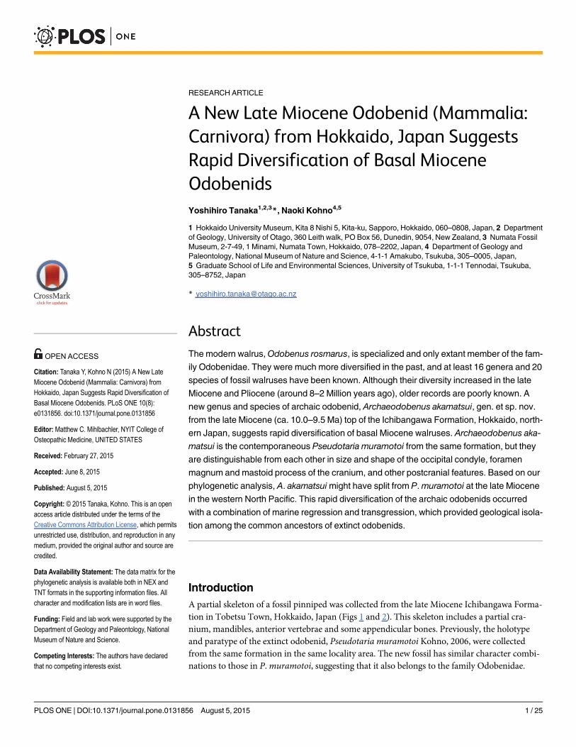
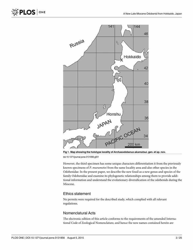
IntroductionA partial skeleton of a fossil pinniped was collected from the late Miocene Ichibangawa Forma-tion in Tobetsu Town, Hokkaido, Japan (Figs 1 and 2). This skeleton includes a partial cra-nium, mandibles, anterior vertebrae and some appendicular bones. Previously, the holotypeand paratype of the extinct odobenid, Pseudotaria muramotoi Kohno, 2006, were collectedfrom the same formation in the same locality area. The new fossil has similar character combi-nations to those in P.muramotoi, suggesting that it also belongs to the family Odobenidae.
PLOSONE | DOI:10.1371/journal.pone.0131856 August 5, 2015 1 / 25
OPEN ACCESS
Citation: Tanaka Y, Kohno N (2015) A New LateMiocene Odobenid (Mammalia: Carnivora) fromHokkaido, Japan Suggests Rapid Diversification ofBasal Miocene Odobenids. PLoS ONE 10(8):e0131856. doi:10.1371/journal.pone.0131856
Editor: Matthew C. Mihlbachler, NYIT College ofOsteopathic Medicine, UNITED STATES
Received: February 27, 2015
Accepted: June 8, 2015
Published: August 5, 2015
Copyright: © 2015 Tanaka, Kohno. This is an openaccess article distributed under the terms of theCreative Commons Attribution License, which permitsunrestricted use, distribution, and reproduction in anymedium, provided the original author and source arecredited.
Data Availability Statement: The data matrix for thephylogenetic analysis is available both in NEX andTNT formats in the supporting information files. Allcharacter and modification lists are in word files.
Funding: Field and lab work were supported by theDepartment of Geology and Paleontology, NationalMuseum of Nature and Science.
Competing Interests: The authors have declaredthat no competing interests exist.
However, the third specimen has some unique characters differentiation it from the previouslyknown specimens of P.muramotoi from the same locality area and also other species in theOdobenidae. In the present paper, we describe the new fossil as a new genus and species of thefamily Odobenidae and examine its phylogenetic relationships among them to provide addi-tional information and understand the evolutionary diversification of the odobenids during theMiocene.
Ethics statementNo permits were required for the described study, which complied with all relevantregulations.
Nomenclatural ActsThe electronic edition of this article conforms to the requirements of the amended Interna-tional Code of Zoological Nomenclature, and hence the new names contained herein are
Fig 1. Map showing the holotype locality of Archaeodobenus akamatsui, gen. et sp. nov.
doi:10.1371/journal.pone.0131856.g001
A New Late Miocene Odobenid from Hokkaido, Japan
PLOS ONE | DOI:10.1371/journal.pone.0131856 August 5, 2015 2 / 25
available under that Code from the electronic edition of this article. This published work andthe nomenclatural acts it contains have been registered in ZooBank, the online registrationsystem for the ICZN. The ZooBank LSIDs (Life Science Identifiers) can be identified and theassociated information viewed through any standard web browser by appending the LSID tothe prefix "http://zoobank.org/". The LSID for this publication is: urn:lsid:zoobank.org:author:E500AA1E-C47F-48C5-9A9D-9B8C0959E8FE. The electronic edition of this work was pub-lished in a journal with an ISSN, and has been archived and is available from the following digi-tal repositories: PubMed Central, LOCKSS.
Systematic paleontologyMAMMALIA Linnaeus, 1758
CARNIVORA Bowdich, 1821ODOBENIDAE Allen, 1880Archaeodobenus gen. nov.urn:lsid:zoobank.org:act:F509D05A-CE42-472F-9311-83405695F5F6Type species. Archaeodobenus akamatsui sp. nov.Diagnosis. As for the only species.Etymology. The generic name, Archaeodobenus, is named in Greek archaios meaning
ancient, and the type genus name of the family Odobenidae.Distribution. Known from the late Miocene (ca. 10.0–9.5 Ma), Japan.Archaeodobenus akamatsui sp. nov.urn:lsid:zoobank.org:act:3ED6F1B3-24C7-4461-B32D-AE3096FB1C62(Figs 3–10, Tables 1–3)Diagnosis. Archaeodobenus akamatsui is an archaic odobenid with a slender and small
cranium and non-tusked, moderate-sized upper canine. Distinguished from other archaic odo-benids (Prototaria primigena, P. planicephala, Proneotherium repenningi, Neotherium mirum,
Fig 2. Locality map and stratigraphic sections of the Archaeodobenus akamatsui holotype locality, based on Takano et al. (1996).
doi:10.1371/journal.pone.0131856.g002
A New Late Miocene Odobenid from Hokkaido, Japan
PLOS ONE | DOI:10.1371/journal.pone.0131856 August 5, 2015 3 / 25
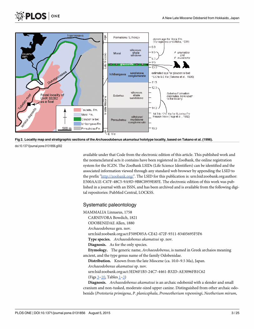
Fig 3. The holotype cranium of Archaeodobenus akamatsui. (A) dorsal view, (B) left lateral view, (C) ventral view.
doi:10.1371/journal.pone.0131856.g003
A New Late Miocene Odobenid from Hokkaido, Japan
PLOS ONE | DOI:10.1371/journal.pone.0131856 August 5, 2015 4 / 25
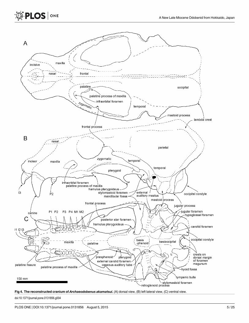
Fig 4. The reconstructed cranium of Archaeodobenus akamatsui. (A) dorsal view, (B) left lateral view, (C) ventral view.
doi:10.1371/journal.pone.0131856.g004
A New Late Miocene Odobenid from Hokkaido, Japan
PLOS ONE | DOI:10.1371/journal.pone.0131856 August 5, 2015 5 / 25
Kamtschatarctos sinelnikovae and Pseudotaria muramotoi) by the following derived characters:pentagonal basioccipital (character 30), blade-like spinous process of axis (character 91). Dis-tinguished from later diverging odobenids (Imagotaria downsi, Pontolis magnus, dusignathinesand odobenines) by retention of the following primitive characters: deep mandibular fossa(character 26); small mastoid process (character 33); two distinct roots on premolars (expectP1) (character 74 and 76).
Holotype. UHR 33282: Hokkaido University Museum, associated partial skeleton includ-ing a partial cranium (mostly left half) with left and right I3, right C, right P2, a left mandiblewith canine, a partial right mandible with incomplete canine, a basihyoid, a left thyrohyoid,ceratohyoids, nearly complete atlas, axis, cervical vertebrae 3–7, incomplete thoracic vertebrae1–8, a left rib, three right ribs, an incomplete sternebra, partial scapulae, and nearly completeright humerus.
Type Locality. UHR 33282 was collected as a float near the Rutaka Bridge on the TobetsuRiver by staff of the Tobetsu Town Board of Education. It is 1.5 km south and also down streamfrom Aoyama dam in Tobetsu-cho, Ishikari-gun, Hokkaido, Japan (43°27’34” north latitudeand 141°35’35” east longitude) (Fig 2).
Formation and Age. UHR 33282 was enclosed in a weathered, dark green fine-grained,massive consolidated glauconitic sand stone block as a float, originally from the glauconite bedfrom the top of the Ichibangawa Formation. Although the Lower Pliocene Tobetsu Formationis exposed at the locality [1], it is very different from the matrix adhering to the new fossil, sug-gesting that the block was transported from the upstream of the river. Three formations (theMorai, Ichibangawa and Ponsubetsu Formations) are exposed further upstream the TobetsuRiver (Fig 2) [1]. But these formations are silt, except the Ichibangawa Formation that is sand-stone. In addition, the rich-occurrence of glauconitic sandstone with marine fossils and car-bonates is known only in the upper part of the Ichibangawa Formation in this area (Kakimiand Uemura [2]), as shown by the gray band in Fig 2. Thus, the origin of UHR 33282 is theglauconite bed on the top of the Ichibangawa Formation.
Recognition of the glauconite bed is disputed. Kakimi and Uemura [2] described the bed asthe uppermost of the Ichibangawa Formation. The Kabato Collaborative Research Group [1]and Takano et al. [3] recognized the bed as the lowermost part of the Morai Formation. On the
Fig 5. The holotype cranium of Archaeodobenus akamatsui in posterior view.
doi:10.1371/journal.pone.0131856.g005
A New Late Miocene Odobenid from Hokkaido, Japan
PLOS ONE | DOI:10.1371/journal.pone.0131856 August 5, 2015 6 / 25
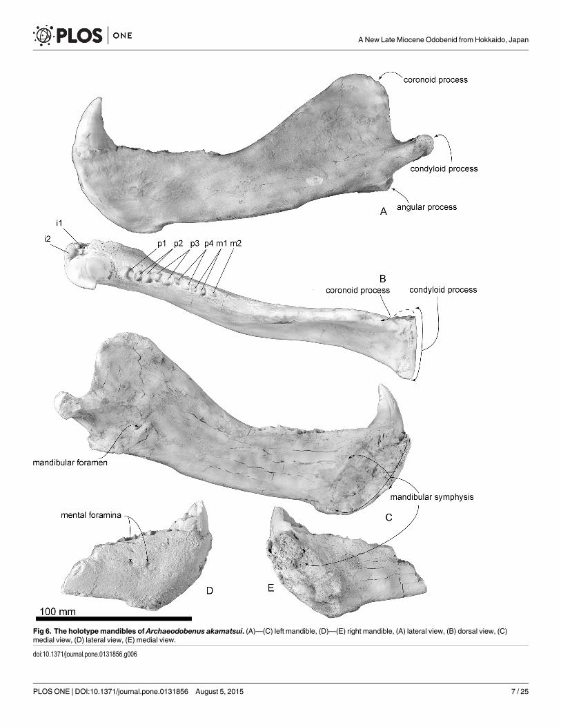
Fig 6. The holotypemandibles of Archaeodobenus akamatsui. (A)—(C) left mandible, (D)—(E) right mandible, (A) lateral view, (B) dorsal view, (C)medial view, (D) lateral view, (E) medial view.
doi:10.1371/journal.pone.0131856.g006
A New Late Miocene Odobenid from Hokkaido, Japan
PLOS ONE | DOI:10.1371/journal.pone.0131856 August 5, 2015 7 / 25
other hand, Takano et al. [4] recognized as a separate unit and did not include it in both theIchibangawa and Morai Formations. The lithology of the matrix, fine-grained, massive hardsand stone is similar to the Ichibangawa Formation (sandstone), rather than the Morai Forma-tion (shale siltstone). In short, here, we follow the oldest study; i.e., Kakimi and Uemura [2], torecognize the fossil odobenid’s original formation, and also follow the estimated age of theglauconite bed of Takano et al. [4].
As Fig 2 shows the age of the Ichibangawa Formation is lacking. However, the overlyingMorai Formation includes the Denticulopsis katayamae diatom zone, 9.2–8.5 Ma [5]. Also, theunderlying Subetsu Formation has a fission track age of 13±1 Ma by Tsuji [6], but it is notexposed at the locality [1]. Based on these chronological information, Takano et al. [3] esti-mated the age of the Ichibangawa Formation (except the glauconite bed) to be approximately11.5–10 Ma, and they also considered the uppermost glauconite bed of the Ichibangawa For-mation to be around 10–9.5 Ma based on previous studies.
Etymology. The species is named in honor of Dr. Morio Akamatsu, a curator emeritus ofthe Hokkaido Museum, for his longstanding contributions to geology and paleontology ofHokkaido, and in gratitude for his encouragement and assistance to both of us throughout thisstudy.
DescriptionMorphological terms follow Mead and Fordyce [7] for postcrania. Measurements are in Tables1–3. A life reconstruction is presented in Fig 11.
Gender and ontogenetic ageThe nine cranial sutures that Sivertsen [8] considered to be useful to evaluate relative ontoge-netic ages of otariid pinnipeds, at least six are nearly closed. This indicates that the new fossilbelongs in Sivertsen’s Group II age class (i.e., subadult). In addition, well developed lambdoidalcrests and relatively large canines suggest that the animal represents a male.
Estimated body size. The estimated body size of the holotype individual is 2.8–3 m intotal length and 390–473 kg in total weight. Holotype of A. akamatsui is an intermediate sizebetween modern male Eumetopias jubatus (3.3m, 1000kg) and Otaria flavescens (2.6 m, 300–350 kg) (Jefferson et al. [9]).
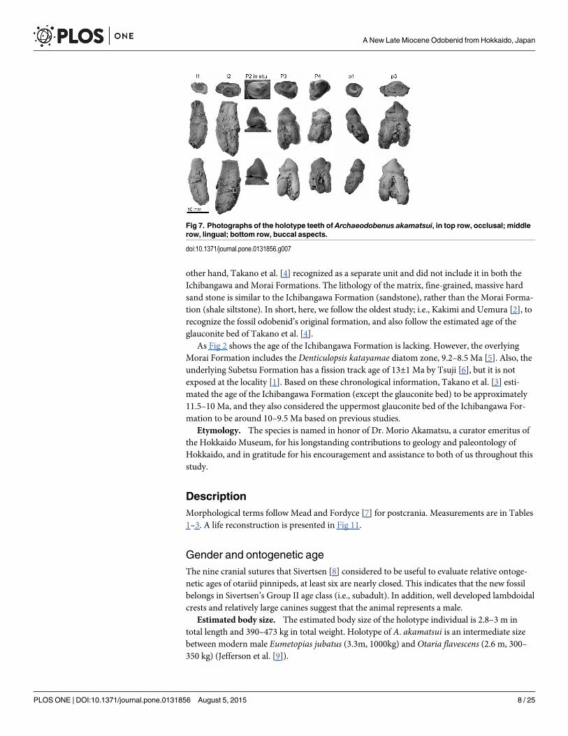
Fig 7. Photographs of the holotype teeth of Archaeodobenus akamatsui, in top row, occlusal; middlerow, lingual; bottom row, buccal aspects.
doi:10.1371/journal.pone.0131856.g007
A New Late Miocene Odobenid from Hokkaido, Japan
PLOS ONE | DOI:10.1371/journal.pone.0131856 August 5, 2015 8 / 25
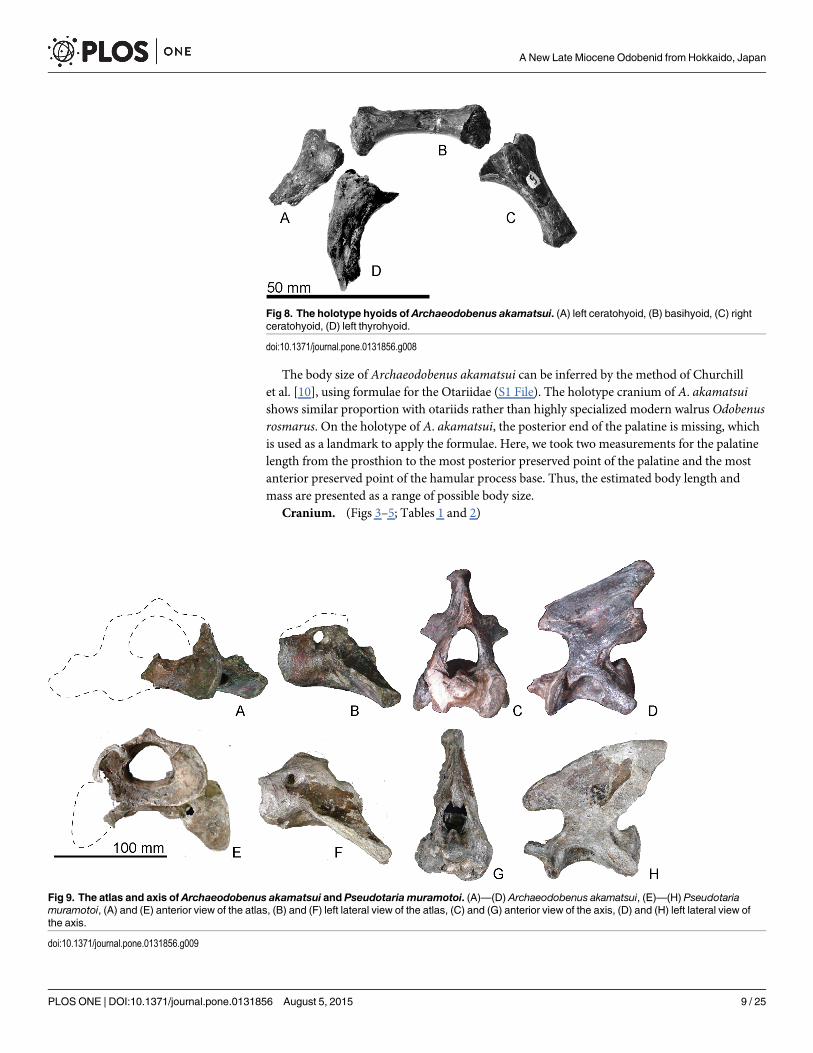
The body size of Archaeodobenus akamatsui can be inferred by the method of Churchillet al. [10], using formulae for the Otariidae (S1 File). The holotype cranium of A. akamatsuishows similar proportion with otariids rather than highly specialized modern walrus Odobenusrosmarus. On the holotype of A. akamatsui, the posterior end of the palatine is missing, whichis used as a landmark to apply the formulae. Here, we took two measurements for the palatinelength from the prosthion to the most posterior preserved point of the palatine and the mostanterior preserved point of the hamular process base. Thus, the estimated body length andmass are presented as a range of possible body size.
Cranium. (Figs 3–5; Tables 1 and 2)
Fig 9. The atlas and axis of Archaeodobenus akamatsui and Pseudotaria muramotoi. (A)—(D) Archaeodobenus akamatsui, (E)—(H) Pseudotariamuramotoi, (A) and (E) anterior view of the atlas, (B) and (F) left lateral view of the atlas, (C) and (G) anterior view of the axis, (D) and (H) left lateral view ofthe axis.
doi:10.1371/journal.pone.0131856.g009
Fig 8. The holotype hyoids of Archaeodobenus akamatsui. (A) left ceratohyoid, (B) basihyoid, (C) rightceratohyoid, (D) left thyrohyoid.
doi:10.1371/journal.pone.0131856.g008
A New Late Miocene Odobenid from Hokkaido, Japan
PLOS ONE | DOI:10.1371/journal.pone.0131856 August 5, 2015 9 / 25
A New Late Miocene Odobenid from Hokkaido, Japan
PLOS ONE | DOI:10.1371/journal.pone.0131856 August 5, 2015 10 / 25
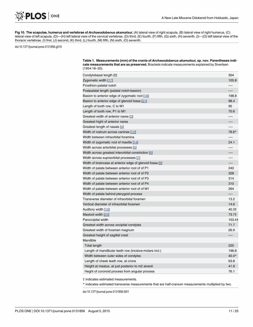
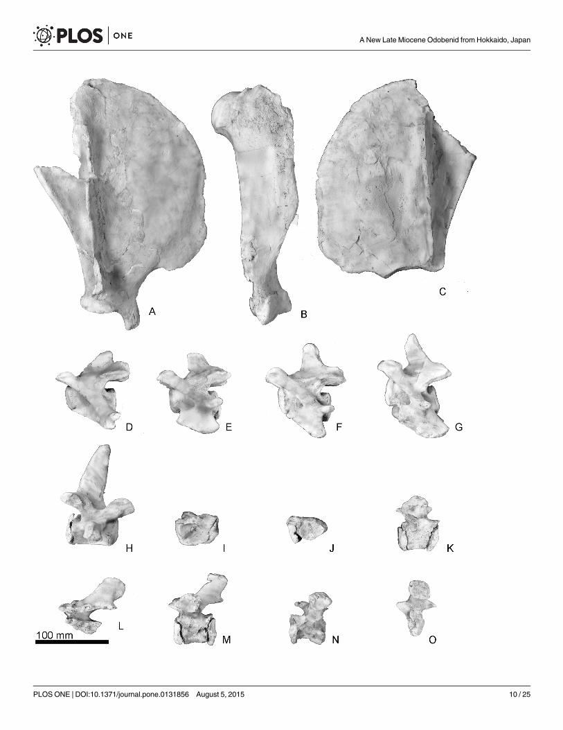
Fig 10. The scapulae, humerus and vertebrae of Archaeodobenus akamatsui. (A) lateral view of right scapula, (B) lateral view of right humerus, (C)lateral view of left scapula, (D)—(H) left lateral view of the cervical vertebrae, (D) third, (E) fourth, (F) fifth, (G) sixth, (H) seventh, (I)—(O) left lateral view of thethoracic vertebrae, (I) first, (J) second, (K) third, (L) fourth, (M) fifth, (N) sixth, (O) seventh.
doi:10.1371/journal.pone.0131856.g010
Table 1. Measurements (mm) of the crania of Archaeodobenus akamatsui, sp. nov. Parentheses indi-cate measurements that are as preserved. Brackets indicate measurements explained by Sivertsen(1954:18–20).
Condylobasal length [0] 304
Zygomatic width [17] 105.8
Prosthion-palatal notch —-
Postpalatal length (palatal notch-basion) —-
Basion to anterior edge of zygomatic root [18] 198.8
Basion to anterior edge of glenoid fossa [21] 98.4
Length of tooth row, C to M1 95
Length of tooth row, P1 to M1 70.6
Greatest width of anterior nares [3] —-
Greatest hight of anterior nares —-
Greatest length of nasals [4] —-
Width of rostrum across canines [12] 78.6*
Width between infraorbital foramina —-
Width of zygomatic root of maxilla [14] 24.1
Width across antorbital processes [5] —-
Width across greatest interorbital constriction [6] —-
Width across supraorbital processes [7] —-
Width of braincase at anterior edge of glenoid fossa [8] —-
Width of palate between anterior root of of P1 240
Width of palate between anterior root of of P2 328
Width of palate between anterior root of of P3 314
Width of palate between anterior root of of P4 310
Width of palate between anterior root of of M1 264
Width of palate behind pterygoid process —-
Transverse diameter of infraorbital foramen 13.2
Vertical diameter of infraorbital foramen 14.6
Auditory width [19] 40.3†
Mastoid width [20] 73.7†
Paroccipital width 103.4†
Greatest width across occipital condyles 71.7
Greatest width of foramen magnum 26.9
Greatest height of sagittal crest —-
Mandible
Total length 220
Length of mandibular teeth row (incisive-molars incl.) 196.8
Width between outer sides of condyles 40.4*
Length of cheek teeth row, at crone 63.8
Height at meatus, at just posterior to m2 alveoli 41.9
Heght of coronoid process from angular process 76.1
† indicates estimated measurements.
* indicates estimated transverse measurements that are half-cranium measurements multiplied by two.
doi:10.1371/journal.pone.0131856.t001
A New Late Miocene Odobenid from Hokkaido, Japan
PLOS ONE | DOI:10.1371/journal.pone.0131856 August 5, 2015 11 / 25
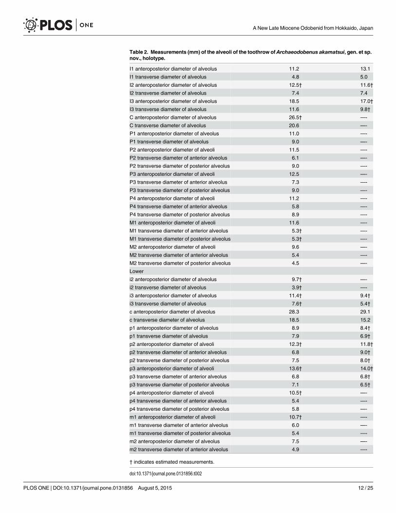
Table 2. Measurements (mm) of the alveoli of the toothrow of Archaeodobenus akamatsui, gen. et sp.nov., holotype.
I1 anteroposterior diameter of alveolus 11.2 13.1
I1 transverse diameter of alveolus 4.8 5.0
I2 anteroposterior diameter of alveolus 12.5† 11.6†
I2 transverse diameter of alveolus 7.4 7.4
I3 anteroposterior diameter of alveolus 18.5 17.0†
I3 transverse diameter of alveolus 11.6 9.8†
C anteroposterior diameter of alveolus 26.5† —-
C transverse diameter of alveolus 20.6 —-
P1 anteroposterior diameter of alveolus 11.0 —-
P1 transverse diameter of alveolus 9.0 —-
P2 anteroposterior diameter of alveoli 11.5 —-
P2 transverse diameter of anterior alveolus 6.1 —-
P2 transverse diameter of posterior alveolus 9.0 —-
P3 anteroposterior diameter of alveoli 12.5 —-
P3 transverse diameter of anterior alveolus 7.3 —-
P3 transverse diameter of posterior alveolus 9.0 —-
P4 anteroposterior diameter of alveoli 11.2 —-
P4 transverse diameter of anterior alveolus 5.8 —-
P4 transverse diameter of posterior alveolus 8.9 —-
M1 anteroposterior diameter of alveoli 11.6 —-
M1 transverse diameter of anterior alveolus 5.3† —-
M1 transverse diameter of posterior alveolus 5.3† —-
M2 anteroposterior diameter of alveoli 9.6 —-
M2 transverse diameter of anterior alveolus 5.4 —-
M2 transverse diameter of posterior alveolus 4.5 —-
Lower
i2 anteroposterior diameter of alveolus 9.7† —-
i2 transverse diameter of alveolus 3.9† —-
i3 anteroposterior diameter of alveolus 11.4† 9.4†
i3 transverse diameter of alveolus 7.6† 5.4†
c anteroposterior diameter of alveolus 28.3 29.1
c transverse diameter of alveolus 18.5 15.2
p1 anteroposterior diameter of alveolus 8.9 8.4†
p1 transverse diameter of alveolus 7.9 6.9†
p2 anteroposterior diameter of alveoli 12.3† 11.8†
p2 transverse diameter of anterior alveolus 6.8 9.0†
p2 transverse diameter of posterior alveolus 7.5 8.0†
p3 anteroposterior diameter of alveoli 13.6† 14.0†
p3 transverse diameter of anterior alveolus 6.8 6.8†
p3 transverse diameter of posterior alveolus 7.1 6.5†
p4 anteroposterior diameter of alveoli 10.5† —-
p4 transverse diameter of anterior alveolus 5.4 —-
p4 transverse diameter of posterior alveolus 5.8 —-
m1 anteroposterior diameter of alveoli 10.7† —-
m1 transverse diameter of anterior alveolus 6.0 —-
m1 transverse diameter of posterior alveolus 5.4 —-
m2 anteroposterior diameter of alveolus 7.5 —-
m2 transverse diameter of anterior alveolus 4.9 —-
† indicates estimated measurements.
doi:10.1371/journal.pone.0131856.t002
A New Late Miocene Odobenid from Hokkaido, Japan
PLOS ONE | DOI:10.1371/journal.pone.0131856 August 5, 2015 12 / 25
Occipitals. The occipitals are preserved mainly on the left side of the cranium. The basioc-cipital is broad posteriorly and trapezoidal in shape. The muscular tubercle for the origin of thelongus and rectus capitis muscles is well developed anterolaterally on the midportion of thebasioccipital. The portion posterior to this tubercle bears a deep and anteroposteriorly longhemispherical fossa. The jugular (posterior lacerate) foramen is large (12.0 mm long and 11.2width) and crescentic in shape, but this might be a result of deformation, a weak slide to themedial. The hypoglossal foramen is relatively large, and its diameter is 4.6 mm. The occipitalcondyles are transversely narrow and dorsoventrally high, and are projected weakly posteriorly.The medial margins of the occipital condyles from posterior view are nearly vertical. The fora-men magnum is dorsoventrally deep and elliptical in shape. The dorsal and ventral condyloidfossae are deep. A pair of crests (around 20 mm length) rise up from the medial side of theoccipital condyles to the middle, on the dorsal margin of the foramen magnum uniquely(Fig 25). The jugular process is short, mediolaterally thin, and plate-like in structure. Thesuture between the jugular process and mastoid process is not fused. The nuchal tubercle isvery weak. The occipito-squamosal suture is almost fused. The lambdoidal crest is thick andposterodorsally projected.
Squamosal. The left squamosal is nearly complete, but missing the anterior part of theslender zygomatic process. The pseudosylvian sulcus is weakly present, and it is located justabove the temporal fossa. The temporal fossa, between the braincase and the zygomatic pro-cess, is relatively wide, smooth and is continued posteriorly to the dorsal surface of the laterallyprojected mastoid process, so that the temporal fossa has an anteroposteriorly extended flatfloor of 38.3 mm in length. The mandibular fossa is subcylindrical, and it makes a deep groovedorsally. The retroglenoid process is a transversely wide and anteroposteriorly thin plate,which projects strongly anteroventrally and forms deep mandibular fossa. The mastoid processis relatively small and projected laterally. Its lateral surface for the origins of the sternomastoi-deus and cleidomastoideus muscles is smooth probably because of the young age of the holo-type individual.
Tympanic Bulla. The ventral surface of the tympanic bulla is inflated and smooth. Thetympanic bulla is comprised mostly of ectotympanic and delimited anterolaterally by the pos-teromedial side of the retroglenoid process and posterolaterally by the anteromedial side of themastoid process. An anterior part of the bulla has a mediolaterally elongated shallow depres-sion, just posterior to the retroglenoid process. The external acoustic meatus is relatively largeand circular, approximately 7.8 mm in diameter. The stylomastoid foramen opens ventrally at
Table 3. Measurements (mm) of preserved teeth of Archaeodobenus akamatsui, gen. et sp. nov.,holotype.
I1 height/width (crown)/length(crown) 21.9/3.6/6.0
I2 height/width (crown)/length(crown) 25.8*/5.9/10.4
I3 (in situ) height (without root)/width (crown)/length(crown) 24.6/11.8/16.2
C (in situ) height (without root)/width (crown)/length(crown) 41.1/19.3/23.8
P2 (in situ) height (without roots)/ width (crown)/length(crown) 8.9/10.0/8.0
P3 height/width (crown)/length(crown) 19.8/8.0/7.2
P4 height/width (crown)/length(crown) 15.6/7.6/8.5
C (in situ) height (without root)/width (crown)/length(crown) 32.0/24.5/15.9
p1 height/width (crown)/length(crown) 17.2*/6.2/7.0
p3 height/width (crown)/length(crown) 21.5/7.2/10.6
* indicates estimated measurements.
doi:10.1371/journal.pone.0131856.t003
A New Late Miocene Odobenid from Hokkaido, Japan
PLOS ONE | DOI:10.1371/journal.pone.0131856 August 5, 2015 13 / 25
the point medial to the mastoid process. The hyoid fossa opens at the posterolateral end of thetympanic bulla and it is 7.5 mm long and 5.0 mm wide. Slightly medial to the hyoid fossa, thecarotid canal opens as size is 6.7 mm long and 4.9 mm wide. The anterior opening of thecarotid canal is circular, moderate (4.5 mm diameter) and located at the anteromedial marginof the tympanic bulla.
Petrosal. The middle ear region of a long hexagonal petrosal is visible on the ventral floorof the braincase. Its anterior portion is positioned ventrally and is wider than its posterior por-tion. The petrosal crest is observed on the anterior part of the petrosal. A deep transverse sulcuslies on dorsal border of the petrosal, just below to the bony tentorium. The cerebellar fossa isdorsoventrally elliptical and has a small slit on the ventral part of the fossa. There is a knob-liketubercle on the medial margin of the cerebellar fossa. The internal acoustic meatus (internalauditory meatus) is anteroposteriorly elliptical, and the entrances for the cranial nerves VIIand VIII are separated within the meatus.
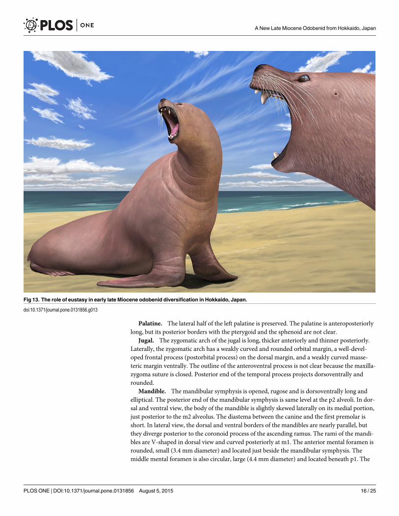
Fig 11. Reconstruction of Archaeodobenus akamatsui by Tatsuya Shinmura (Ashoro Museum of Paleontology).
doi:10.1371/journal.pone.0131856.g011
A New Late Miocene Odobenid from Hokkaido, Japan
PLOS ONE | DOI:10.1371/journal.pone.0131856 August 5, 2015 14 / 25
Sphenoids. The left side of the sphenoids are partially preserved, but the presphenoid andthe dorsal half of the alisphenoid are missing. The ventral half of the alisphenoid forms the pos-terior wall of the choanal trough. The anterior opening of the alisphenoid canal is observed onthe medial wall of the alisphenoid at the position of the orbital fissure. The posterior openingof the alisphenoid canal and the foramen ovale are joined in a common recess, and it is locatedjust posterior to the laterally expanded pterygoid strut at the medial side of the mandibularfossa.
Pterygoid. The pterygoid strut is dorsoventrally and transversely broad. There is a sharplyedged ventromedial ridge present on the ventral surface of the pterygoid that extends from theposterior end of the palate to the long and slender, posteroventrally projected pterygoidhamulus.
Maxilla. The medial part of the palatal portion of the maxilla is broken away, but enoughof that portion remains to show a transversely arched palate. The palatal margin of the maxillais laterally divergent at the portion of M1. The infraorbital foramen is relatively small anddorsoventrally elliptical. The infraorbital canal runs through rather thick bone at the maxillaryforamen. The dorsal roof of the infraorbital foramen is dorsoventrally thick. The orbit is large,however the medial wall of the orbit is broken away. On the ventral surface of the zygomaticroot, there is a shallow fossa and a weakly developed ventral tuberosity.
Premaxilla. The dorsal part of the premaxilla is not preserved. The rostral process is largeand blunt. The palatine fissures (premaxilla foramina) are separated by a thin septum at themid-portion, and each foramen is anteroposteriorly elongated (14.4 mm long and 5.6 wide onthe right side).
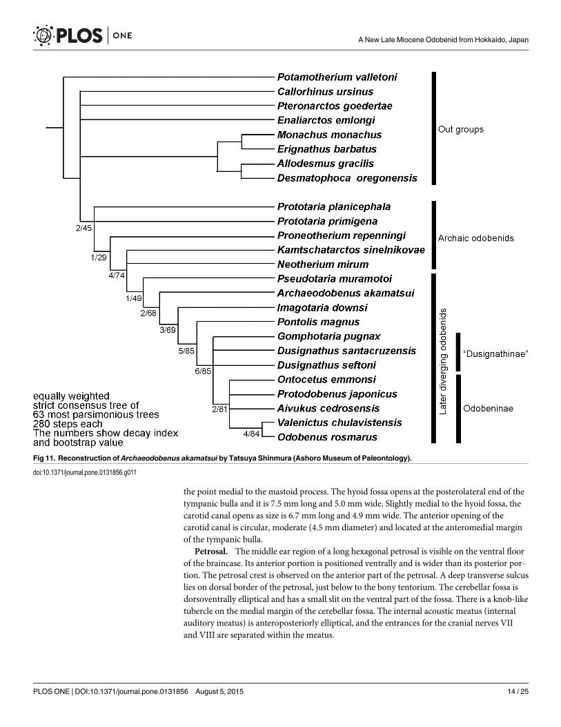
Fig 12. The strict consensus tree of equally weighted analysis of Archaeodobenus akamatsui and the Odobenidae, with Bremer support at nodes.
doi:10.1371/journal.pone.0131856.g012
A New Late Miocene Odobenid from Hokkaido, Japan
PLOS ONE | DOI:10.1371/journal.pone.0131856 August 5, 2015 15 / 25
Palatine. The lateral half of the left palatine is preserved. The palatine is anteroposteriorlylong, but its posterior borders with the pterygoid and the sphenoid are not clear.
Jugal. The zygomatic arch of the jugal is long, thicker anteriorly and thinner posteriorly.Laterally, the zygomatic arch has a weakly curved and rounded orbital margin, a well-devel-oped frontal process (postorbital process) on the dorsal margin, and a weakly curved masse-teric margin ventrally. The outline of the anteroventral process is not clear because the maxilla-zygoma suture is closed. Posterior end of the temporal process projects dorsoventrally androunded.
Mandible. The mandibular symphysis is opened, rugose and is dorsoventrally long andelliptical. The posterior end of the mandibular symphysis is same level at the p2 alveoli. In dor-sal and ventral view, the body of the mandible is slightly skewed laterally on its medial portion,just posterior to the m2 alveolus. The diastema between the canine and the first premolar isshort. In lateral view, the dorsal and ventral borders of the mandibles are nearly parallel, butthey diverge posterior to the coronoid process of the ascending ramus. The rami of the mandi-bles are V-shaped in dorsal view and curved posteriorly at m1. The anterior mental foramen isrounded, small (3.4 mm diameter) and located just beside the mandibular symphysis. Themiddle mental foramen is also circular, large (4.4 mm diameter) and located beneath p1. The
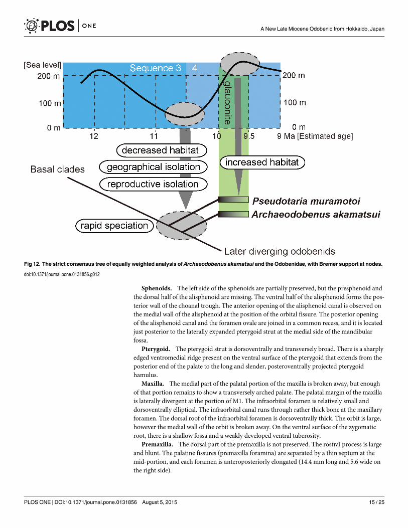
Fig 13. The role of eustasy in early late Miocene odobenid diversification in Hokkaido, Japan.
doi:10.1371/journal.pone.0131856.g013
A New Late Miocene Odobenid from Hokkaido, Japan
PLOS ONE | DOI:10.1371/journal.pone.0131856 August 5, 2015 16 / 25
posterior mental foramen is smaller (3.1 mm diameter) than both the anterior and middlemental foramina and located beneath p2. At the level of p2 and p3, a well-developed genialtuberosity is present on the ventral margin of the body. A narrow coronoid process is thinnerdorsally, rounded and anterodorsally projected. A wide (39.2+ mm) and smooth condyloidprocess is present at the level of the tooth row. The mandibular notch opens wide anterodor-sally. The angular process is weakly projected posteriorly and forms a small medial shelf. Themasseteric fossa is shallow and its borders are unclear, with a weak horizontal crest at the dor-sal part of the fossa. Medially, the mandibular foramen is small, dorsoventrally elliptical anddirected anterodorsally. The mandibular canal, which is at the center of the broken surface ofthe right mandible, is also elliptical.
Dentition. The dentition is represented by upper and lower alveoli on the left maxilla andboth dentaries as I1-3, C, P1-4, M1-2, i2-3, c, p1-4 and m1-2. In addition, both I3s, right C andP2, and the lower canines are preserved in place. Isolated left I2 and P3, and the right P4 arealso preserved.
The alveoli for I1 and I2 are transversely narrow but anteroposteriorly wide. The crown ofthe I2 consists of an anteriorly located large cusp and posteriorly located small cusplet. The I2root is laterally thin and anteroposteriorly wide, and it is gently curved posteriorly. There is adistinct constriction between the crown and the root. A much larger isolated left I2 has a shal-low groove on the lateral and proximal surface and is weakly curved posteriorly.
The strongly and anteriorly curved I3s have worn ends and are oval shape. The anterior sur-face of the crown is smooth, but the posterior surface makes a vertical crest.
The canine is not tusk-like, but large and conical, and it has a strong posterior crista. Theroot has a broken surface of the maxilla, which is very long and large (86.3 mm long in total ofthe canine), and transversely narrow. The proximal end of the root is visible from the brokencranium (see Fig 3A), and has an open pulp cavity.
The premolars are double rooted except for the P1. The P1 has a relatively large single root.The P2 has a narrow anterior and a wide posterior alveolus, and its crown has one small maincusp with a blunt lingual cingulum with several incipient cusplets. The P2 to M2 have narrowsubcircular anterior alveoli and wide oval posterior alveoli.
Isolated left P3 and right P4, crowns have a posteromedially placed protocone shelf. Theyare similar in shape, but the P3 is slightly higher in crown height. The P3 has a posterolingualcingulum with one distinct cusp (protocone shelf) at the posteromedial margin of the cingu-lum. The P3 has a small cusp at the posterior end of the crown. The P4 has a more expandedprotocone shelf and relatively large and high cusp also at the posterior end of the crown. It isanteroposteriorly slightly enlarged, forming a small blade-like ridge that is separated from themain cusp by a distinct notch.
The M1 is double rooted, and the posterior alveolus lies outside the general alignment of thecheek teeth. The alveoli of i2 and i3 are mediolaterally thin, anteroposteriorly long, and theyare single rooted. The i2 alveolus is smaller than i3.
The lower canine is nearly equal size, but slightly shorter dorsoventrally and narrower trans-versely than the upper canine, and has a well developed posterior crista. Its root is almost ovalwith a weak longitudinal groove on the buccal side in cross section and is oriented dorsally.
The p2—m1 are double rooted, while p1 and m2 are single rooted. The p1 to p3 have bul-bous crowns with well-developed enamel. An isolated left p3 is preserved, and possesses threedistinct cusps; an anteriorly located developed cusp (paraconid), a main cusp (protoconid),and a small but distinct posterior cusp just behind the main cusp. The anterior cusp is smallerthan the posterior cusp. The lingual cingulum is well developed and its surface is slightly crenu-lated. Its root is wider than the crown. The tooth row is short (63.6 mm).
A New Late Miocene Odobenid from Hokkaido, Japan
PLOS ONE | DOI:10.1371/journal.pone.0131856 August 5, 2015 17 / 25
Hyoid. The basihyoid, the left thyrohyoid and the ceratohyoids are preserved (Fig 8). Thebasihyoid is thickened toward both the proximal and distal extremities It has two small jointson the posterior margin for the thyrohyoid The thyrohyoid is missing part of the distal extrem-ity, but its proximal extremity is thickened. The ceratohyoid is slender and it is thickenedtoward both the proximal and distal extremities. The proximal extremity of the ceratohyoid isabout twice as wide as the distal extremity, and it has a flat edge and a nearly perpendiculararticulation.
Vertebral columnAtlas. The wing process is large and extends ventrolaterally. The ventral tubercle of the
ventral arch is weakly inflated ventrally. The lateral vertebral foramen is small and circular, andit is visible only in lateral view. The transverse foramen is also small; it is anteriorly circular andposteriorly triangular. The atlantal fossae are relatively weak, forming depressions ventral tothe base of the wing. Each intraosseous canal opens posteriorly into the anterior transverseforamen. The transverse foramen is relatively large, and the odontoid fovea is also large anddeep. Two shallow grooves extend transversely on the posterior surface of the ventral arch. Asmall and deep foramen is present on the mid-line of the posterior articular surface.
Axis. The axis is nearly complete, missing parts of the right anterior articular surface andanterior spinous process. The dens is large and thick. The blade-like spinous process extendsposterodorsally. The dorsal margin of the spinous process is straight and anteroposteriorlylong. The posterior edge of the spinous process expands laterally. The vertebral foramen islarge and elliptical. The transverse process is large, and it is projected posterolaterally and ven-trally. The transverse foramen is large and circular, and they are located at the base of the trans-verse process. On the ventral surface of the vertebral body, a pair of deep fossae are divided bya median crest. The anterior end of the median crest is developed as an anteriorly angled trian-gular tubercle.
Third to seventh cervical vertebrae. The third to seventh cervical vertebrae are preserved(Fig 10D–10H). The transverse processes are long and knob-like in the third cervical, trans-versely thin and plate-like in the forth to sixth cervicals, and very weak knob-like in the seventhcervical vertebra. The spinous processes are projected weakly in the third to sixth cervical andstrongly ventrally in the seventh cervical vertebra.
Thoracic vertebrae. Seven thoracic vertebrae are preserved although their exact positionsare not clear (Fig 10I–10O). The transverse processes are strongly projected in the “third, fifthto seventh” thoracics; squire shaped costal fovea are present on the transverse process. The spi-nous processes are projected posteroventrally in the “fourth and fifth” thoracics, but otherthoracics are not well preserved.
Rib. Seven ribs are preserved. The left first to third ribs are nearly complete, but other ribsare preserved only proximal parts. The first rib is the shortest. The head and tubercle are dis-tinct and well separated in each rib. Each rib is gently curved. The body of each rib is somewhatsquare in cross section, and therefore the anterior and posterior surfaces are relatively flat. Thecostochondral junction is semicircular and elliptical in cross section.
Sternum. Two intermediary sternebrae are preserved. The sternebrae are short and thick,forming a square pole. Lateral thickness is wider than dorsoventral. It is thickened toward theanterior and posterior extremity. Both extremities are display a rugose articulation.
Appendicular skeletonScapula. Both scapulae are well preserved (Fig 10A and 10C); the right scapula is nearly
complete, missing only a part of the posterior angle, lateral margin of the spine, and the
A New Late Miocene Odobenid from Hokkaido, Japan
PLOS ONE | DOI:10.1371/journal.pone.0131856 August 5, 2015 18 / 25
acromion. The dorsal part of the spine is thick and high compared to its ventral part. Thebase of the acromion extends ventral to the scapular notch. The scapular spine is positionedposteriorly; so that the supraspinous fossa is wider than the infraspinous fossa. Within thesupraspinous fossa are two weak ridges parallel to the spine; the anterior one is weakly curvedanteriorly, and posterior one is nearly straight. The scapular notch is deep. The anterior borderis gradually curved. The posterior border is nearly straight. The scapular tuberosity is large. Onthe medial surface of the scapula, four muscule attachment ridges are present. Two most poste-rior lines are merged ventrally, forming a Y-shape.
Humerus. The right humerus (Fig 10B) is nearly complete, but missing part of the ventralmargin of the lateral epicondyle. The head is transversely small and anteroposteriorly long andelliptical. The greatest length of the head is around 20% of the total length of the humerus. Anepiphyseal suture is still persistent at the anterior border of the head. The neck of the humeralhead is indistinct. The shaft is relatively straight and slender. The greater tubercle is robust andelevated slightly above the head, and its dorsal surface is rugose. An elliptically shaped surfaceis present at the apex of the greater tubercle. The lesser tubercle is medially projected andlocated distinctly below the head. The intertubercular groove is narrow and deep between thegreater and lesser tubercles. The tricipital line is present. The deltoid tuberosity is well devel-oped on the pectoral crest. The pectoral crest is straight and directed distally toward the mediallip of the trochlea. The brachial groove is deep. The lateral epicondyle is conspicuously reducedand scarcely projected from the lateral margin of the distal radial capitulum. There is a deepgroove on the lateral surface of the lateral epicondyle. The supinator ridge is weak and blunt.The medial epicondyle is knob-like and quadrangular in outline. The epicondylar foramen isabsent. The capitulum of the humerus is low proximodistally. The trochlea is mediolaterallyshort. Both the olecranon and radial fossae are shallow. The supratrochlear foramen is absent.Although the distal tip of the trochlea and the posterodistal part of the capitulum are somewhatbroken, the former is much greater in anteroposterior diameter than the latter as inferred fromits width.
Phylogenetic Analysis
Relationships of the new fossil to other odobenidsThe new fossil belongs to the Family Odobenidae in having a laterally expanded pterygoidstrut (Character 13), a bony tentorium that is closely appressed to the petrosal (Character 29)and well-developed P1 and P2 with lingual cingula (Character 71). Those characters are recog-nized as unequivocal synapomorphies for the family [11–13]. Thus, the phylogenetic analysisfor the new fossil was focused on members of the Odobenidae with member of other pinnipedclades as outgroups. Previously Deméré [14] and Kohno [11, 12] presented cladistic analysesfor the family Odobenidae mainly on the basis of cranial, mandibular and dental morphologies.Recently, Boessenecker and Churchill [13] analyzed additional characters of both crania andmandibles with cheek teeth of the odobenids. Although most of these characters were samecharacters used by Deméré [14], Kohno [11, 12], Kohno et al. [15], and Deméré and Berta [16],they defined all the characters used in their analyses more clearly and thoroughly illustrated onMorphobank (http://morphobank.org/index.php/Projects/ProjectOverview/project_id/530).Accordingly our data matrix is the one by Boessenecker and Churchill [13] with minor changesand an additional axial character (Character 91) for our analysis (S2–S5 Files). We included 17odobenids and eight outgroup taxa that were used by Deméré [14], Kohno [11, 12], and Boes-senecker and Churchill [3], and we also added extinct and early diverging arctoid, Potamother-ium valletoni Geoffroy, 1833 (following Rybczynski et al. [17]) as an outgroup in this analysis.Among the taxa within Odobenidae, Pelagiarctos thomasi Barnes, 1988 and an indeterminate
A New Late Miocene Odobenid from Hokkaido, Japan
PLOS ONE | DOI:10.1371/journal.pone.0131856 August 5, 2015 19 / 25
species belonging in the same genus reported by Boessenecker and Churchill [13] from themiddle Miocene of California were excluded from our analysis because of the lack of any cra-nial material for this taxon at present. In total, our analysis used 17 ingroup and nine outgrouptaxa coded for 91 morphological and morphometric characters (S2–S5 Files). Both characterand tree data were manipulated using Mesquite 2.75 [18]. A cladistic analysis was performedwith TNT, 1.1 [19]. All characters were unweighted and unordered. A heuristic search of10,000 replicates was conducted, using tree bisection reconnection (TBR) and saving 10 treesper replication. To measure node stability, we calculated a bootstrap value using symmetricresampling with 2000 replicates and the decay index [20] for the strict consensus tree.
Our analysis resulted in 63 most parsimonious trees with 280 steps. The variations amongthe 63 trees are represented by differences in branching patterns among outgroup taxa andalso ingroup taxa (mostly “dusignathines”) within the later diverging odobenids (see a strictconsensus tree in Fig 12). Phylogenetic analysis revealed that the new fossil is recognized as anindependent linage. The new fossil is distinguished from such earlier diverging odobenids asPrototaria spp, Proneotherium repenningi, Kamtschatarctos sinelnikovae, Neotherium mirumand Pseudotaria muramotoi in having two unequivocal and equivocal synapomorphies withlater diverging odobenids such as the pentagonal basioccipital (Character 30) and straightdorsal margin of the spinous process of the axis (Character 91) respectively. However, the newfossil lacks synapomorphies of later diverging odobenids (i.e., Imagotaria downsi, Pontolismagnus, the “dusignathines” and the Odobeninae) such as the presence of ventral tuberosity ofthe zygomatic root of the maxilla (Character 7), shallow glenoid fossa (Character 26), largemastoid process of the squamosal (Character 33), absence of the metaconid on the lower cheekteeth (Character 70) and double rooted upper P3 roots (Character 74). Although not includedwithin the computer-based phylogenetic analyses, Pelagiarctos thomasi and P. sp. of Boesse-necker and Churchill [13] are clearly distinguishable from the new fossil by their very large sizeand robustness and by having extremely large cheek teeth. Therefore, the new fossil is differen-tiated from all previously known taxa within the Odobenidae. In addition, the new fossil hasautapomorphic characters such as sharply divergent mandibular arch (Character 44) andslightly concave talonid basin of the lower cheek teeth (Character 69).
These distinctive features of the new fossil among taxa within the Odobenidae indicate thatthis taxon is monophyletic (Fig 12). Therefore, the generic level assignment as a new taxon iswarranted for the new fossil described here. Thus, we propose Archaeodobenus akamatsui as anew genus and new species within the family Odobenidae.
Discussion
Disparity of Archaeodobenus akamatsui and Pseudotaria muramotoiArchaeodobenus akamatsui gen. et sp. nov. is presently known only from the holotype, which isfrom the top of the Ichibangawa Formation as described above. The same formation has alsoproduced another odobenid Pseudotaria muramotoi Kohno, 2006. Therefore, both species aresympatric at least geochronologically. These two species of fossil odobenids are closely relatedto each other based on the phylogenetic analysis, but are not monophyletic. Indeed, the basioc-cipital in A. akamatsui is pentagonal in shape as a derived condition of all the later divergingodobenids, but the same portion in P.muramotoi is primitively parallel sided as in earlierdiverging odobenids as well as the late Oligocene and early Miocene pinnipedimorphs. In addi-tion, the mastoid process is small in A. akamatsui among the Odobenidae, but is stronger incomparison with the sister P.muramotoi. Also, the preserved cervicals in this new taxon showmorphological differences between them (Fig 9). For example, the blade-like dorsal margin ofthe spinous process of the axis (Character 91) in A. akamatsui is recognized as a derived
A New Late Miocene Odobenid from Hokkaido, Japan
PLOS ONE | DOI:10.1371/journal.pone.0131856 August 5, 2015 20 / 25
condition that unites this taxon with later diverging odobenids such as the modern walrus,Odobenus rosmarus and the Pliocene extinct walrus, Valenictus chulavistensis (SDSNH 36786:San Diego Natural History Museum) of the subfamily Odobeninae as the potential synapomor-phy for all the later diverging odobenids such as Imagotaria, Pontolis and “dusignathines” aswell as the monophyletic odobenines.
Our analysis also indicates that P.muramotoi does not have its own character (autapomor-phy) and therefore the least modified condition in the transformation series of characters tothe later diversing odobenids as suggested also by Kohno [12]. In this sense, these differencesbetween the two taxa within the same family in the same geologic age and same geographicrange suggest that A. akamatsuimight have split from P.muramotoi as a different species, withthe rapid structural divergences during a short time period in the late Miocene in the westernNorth Pacific.
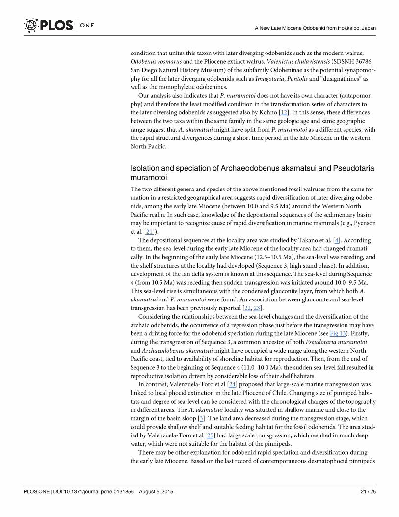
Isolation and speciation of Archaeodobenus akamatsui and PseudotariamuramotoiThe two different genera and species of the above mentioned fossil walruses from the same for-mation in a restricted geographical area suggests rapid diversification of later diverging odobe-nids, among the early late Miocene (between 10.0 and 9.5 Ma) around the Western NorthPacific realm. In such case, knowledge of the depositional sequences of the sedimentary basinmay be important to recognize cause of rapid diversification in marine mammals (e.g., Pyensonet al. [21]).
The depositional sequences at the locality area was studied by Takano et al, [4]. Accordingto them, the sea-level during the early late Miocene of the locality area had changed dramati-cally. In the beginning of the early late Miocene (12.5–10.5 Ma), the sea-level was receding, andthe shelf structures at the locality had developed (Sequence 3, high stand phase). In addition,development of the fan delta system is known at this sequence. The sea-level during Sequence4 (from 10.5 Ma) was receding then sudden transgression was initiated around 10.0–9.5 Ma.This sea-level rise is simultaneous with the condensed glauconite layer, from which both A.akamatsui and P.muramotoi were found. An association between glauconite and sea-leveltransgression has been previously reported [22, 23].
Considering the relationships between the sea-level changes and the diversification of thearchaic odobenids, the occurrence of a regression phase just before the transgression may havebeen a driving force for the odobenid speciation during the late Miocene (see Fig 13). Firstly,during the transgression of Sequence 3, a common ancestor of both Pseudotaria muramotoiand Archaeodobenus akamatsuimight have occupied a wide range along the western NorthPacific coast, tied to availability of shoreline habitat for reproduction. Then, from the end ofSequence 3 to the beginning of Sequence 4 (11.0–10.0 Ma), the sudden sea-level fall resulted inreproductive isolation driven by considerable loss of their shelf habitats.
In contrast, Valenzuela-Toro et al [24] proposed that large-scale marine transgression waslinked to local phocid extinction in the late Pliocene of Chile. Changing size of pinniped habi-tats and degree of sea-level can be considered with the chronological changes of the topographyin different areas. The A. akamatsui locality was situated in shallow marine and close to themargin of the basin sloop [3]. The land area decreased during the transgression stage, whichcould provide shallow shelf and suitable feeding habitat for the fossil odobenids. The area stud-ied by Valenzuela-Toro et al [25] had large scale transgression, which resulted in much deepwater, which were not suitable for the habitat of the pinnipeds.
There may be other explanation for odobenid rapid speciation and diversification duringthe early late Miocene. Based on the last record of contemporaneous desmatophocid pinnipeds
A New Late Miocene Odobenid from Hokkaido, Japan
PLOS ONE | DOI:10.1371/journal.pone.0131856 August 5, 2015 21 / 25
from the early late Miocene Montesano Formation [25], these enigmatic pinnipeds might havereduced in population size and became extinct in the North Pacific [26] in the same period ofthe possible divergence time of Pseudotaria and Archaeodobenus. The estimation of the rapidspeciation and diversification of these odobenids during the same time period might have beenthe evidence of the exchange of niche between the two families in the North Pacific. This mayalso be plausible but is not corroborated by sufficient evidence at present.
Also, Suto et al. [27] has discussed correlations between seqcuences of ocean productivitiesand evolution of marine organisms during the Neogene and Pleistocene based on the abun-dances of Chaetoceros resting spores (diatoms). According to their results, one of the rich pro-ductivities was recognized as the Pacific Chaetoceros Explosion Event (PACE-1) in the LateMiocene (i.e., Messinian, ca. 8.5–6.4 Ma), and its incipient increase was also recognized around10 Ma in the North Pacific. In this regard, Churchill et al. [28] pointed out the productivity andclimate change during the Messinian to have strong influence on otariid diversification andrange. Although the synchronicity between the increase/decrease of ocean productivities andthe marine transgression/regression cycles have not been clear, this may also have related tothe fragmentation of the ancestral population and concecutive speciation of A. akamatsui andP.muramotoi, and the subsequent increase of the ocean productivity that promoted increase ofboth populations of two walruses in the locality area.
Newly speciated odobenids diverged allopatrically and migrated or invaded into the”localityarea” around 10.0–9.5 Ma during the transgression period. Expansion of their habitats duringthe transgression just after the regression period allowed the cross over of their existences inthe restricted area.
The geographical fragmentation of odobenid population during the period just before thesedimentation of glauconitic layer might have resulted in speciation of the Archaeodobenus-like odobenids from common ancestors with the Pseudotaria-like odobenids and subsequentincrease in size of each population, which may resulted in the diversification of two separatespecies in the same locality area during the period between 10.0 and 9.5 Ma.
Pseudotaria and Archaeodobenus among the odobenids can, therefore, be recognized as thebeginning of diversification among middle/late Miocene later diverging odobenids that are thetwo subfamilies Odobeninae+Dusignathinae+Imagotaria+Pontolis. As discussed above, thedriving force of their speciation can be thought as is controlled by the rapid regression, whichpushed both geographical and reproductive isolation. The two fossil odobenids from the sameformation might show such case of the causal relationship between the sea-level fall and geo-graphical isolation or segmentation.
Enigmatic odobenid PelagiarctosWe did not include Pelagiarctos thomasi from the middle Miocene of California in our phylo-genetic analysis because of the lack of its cranial material, but this enigmatic species might havebeen a member of the “later diverging odobenids” as has suggested by Boessenecker and Chur-chill [13]. According to them, Pelagiarctos shares such derived characters as lateral lower inci-sors greater in size than medial ones (Character 54) and bulbous postcanine crowns (Character63) with later diverging odobenids. However, these tooth characters are recognized not only inthe odobenids but also in other pinnipeds such as allodesmine desmatophocids [29–31]. Onthe contrary, Pelagiarctos has distinctively double rooted cheek teeth like earlier divergingpinnipeds in spite of having aforementioned derived characters. In this regard, those derivedcharacters may be altered as homoplastic when the cranial material for all the species can beincluded in the phylogenetic analysis. Therefore, we prefer to reserve judgment temporarily on
A New Late Miocene Odobenid from Hokkaido, Japan
PLOS ONE | DOI:10.1371/journal.pone.0131856 August 5, 2015 22 / 25
the phylogenetic position of this enigmatic taxon based only upon the cheek tooth morpholo-gies until the taxon is better understood by cranial material.
ConclusionsArchaeodobenus akamatsui gen. et sp. nov. represents the second taxon of the archaicodobenids from the early late Miocene (ca. 10.0–9.5 Ma) Ichibangawa Formation, Hokkaido,northern Japan. The same formation has also produced the archaic odobenid Pseudotaria mur-amotoi Kohno, 2006. These two odobenids are distinguishable from another in size and shapeof the occipital condyle, foramen magnum and mastoid process of the cranium, and some post-cranial features. Phylogenetic analysis reveals that A. akamatsui has two synapomorphies thatunites the new species with later diverging odobenids (i.e., Imagotaria downsi, Pontolis magnus,the “dusignathines” and the Odobeninae). Therefore, two closely similar odobenids having rel-atively ancestral characters (P.muramotoi) and relatively derived characters (A. akamatsui)both lived in the same area during the same age. Perhaps the coexistence of two morphologi-cally similar archaic odobenids as represented by A. akamatsui and P.muramotoi in the earlylate Miocene suggest their rapid speciations by geographical and reproductive isolation duringa regressive phase and subsequent transgressions.
Supporting InformationS1 Appendix.(DOCX)
S2 Appendix.(DOC)
S3 Appendix.(ZIP)
S4 Appendix.(ZIP)
S1 File. Data of the body estimation of Archaeodobenus akamatsui.(DOCX)
S2 File. Specimen list.(DOCX)
S3 File. Character list.(DOC)
S4 File. Data matrix in nexus format.(ZIP)
S5 File. Data matrix in TNT format.(ZIP)
AcknowledgmentsWe are grateful to Dr. Morio Akamatsu (formerly Historical Museum of Hokkaido, now Hok-kaido Museum) for his encouragement and assistance throughout this study. Our thanks alsogo to the staff of the Tobetsu Town Board of Education who collected and deposited the holo-type specimen of the new odobenid described here to the Hokkaido University Museum. We
A New Late Miocene Odobenid from Hokkaido, Japan
PLOS ONE | DOI:10.1371/journal.pone.0131856 August 5, 2015 23 / 25
thank Yoshitsugu Kobayashi (Hokkaido University Museum) who received the specimen intothe university museum collection and permitted us to study it. We are grateful to YoshinoriHikida (Nakagawa Museum of Natural History) and Yuji Soeda (Hokkaido Museum) for theircontinuous assistance during our fieldwork and organization of the specimen deposition. Thehuge block of the nodule that contained the odobenid fossil described here was skillfully cut offand reduced by Shotaro Fujita, Satoru Itaya and Shigejiro Kanazawa (all Tobetsu Town), andthe specimen was further prepared by Takako Furuya, Michiyo Ida, Sadakazu Kihara, YoshikoMaguchi, Kousuke Nishikawa and Jun-ichi Nukazuka (then Chiba University) which aregreatly acknowledged. We are also grateful to David J. Bohaska (United States NationalMuseum of Natural History, Smithsonian Institution), Tomas A. Deméré (SDSNH), TadasuYamada and Yuko Tajima (National Museum of Nature and Science), Hitoshi Furusawa (Sap-poro Museum Activity Center), and Sinji Isaji (Natural History Museum and Institute, Chiba,)for their permissions to examine comparative specimens. We are deeply indebted to MorganChurchill (University of Wyoming) and an anonymous reviewer for constructive comments,and R. Ewan Fordyce and Robert W. Boessenecker (both University of Otago) for their pre-submission review. Morgan Churchill also helped to estimate the body size the new species.We are also indebted to Chieko Shimada (then Hokkaido University, now Akita University),Yoshihiro Tanimura (National Museum of Nature and Science) and Yukio Yanagisawa (Geo-logical Survey of Japan) who have been generously shared for many years their extensiveknowledge of collecting biostratigraphic information of the locality area. Finally, the seniorauthor would like to express his deep appreciation to Yoshitsugu Kobayashi (HUM) and Hir-oshi Nishi (Tohoku University Museum) for their encouragement and advice during the courseof this research.
Author ContributionsConceived and designed the experiments: YT NK. Performed the experiments: YT NK. Ana-lyzed the data: YT NK. Contributed reagents/materials/analysis tools: NK YT. Wrote thepaper: YT NK.
References1. The Kabato Collaborative Research Group (1995) Neogene stratigraphy of the Aoyama area, Tobetsu
Town in the southern part of the Kabato Mountains, Hokkaido, Japan. Earth Science (Chikyu Kagaku)49: 363–378.
2. Kakimi T, Uemura T (1958) Explanatory text of the geological map of Japan. Tsukigata 1:50000. GeolSurv Jpn Cruise Rep: 1–54.
3. Takano O, Hoyanagi K, Noto M, Ota K, Yahata M, Sedimentary facies group of the Kabato Collabora-tive Research Group (1995) Slope, shelf, fan delta and gravelly fluvial systems of the Neogene Systemin the southern part of the Kabato Mountains, Hokkaido, Japan. Earth Science (Chikyu Kagaku) 49:253–270.
4. Takano O, Hoyanagi K, Noto M, Ota K, Yahata M, Sedimentary facies group of the Kabato Collabora-tive Research Group (1996) Formation processes of depositional sequences of the Neogene system inthe southern part of the Kabato Mountains, Hokkaido, Japan. Earth Science (Chikyu Kagaku) 50: 9–28.
5. Yanagisawa Y, Akiba F (1998) Refined Neogene diatom biostratigraphy for the northwest Pacificaround Japan, with an introduction of code numbers for selected diatom biohorizons. Journal of Geo-logical Society of Japan: 395–414.
6. Tsuji T, Saito Y, Ikuji Y, Ichinoseki T, Obuse A, Kai K (1992) Sedimentary environments and sequencesof the Ponsubetsu and Subetsu Formations of the Minenobu Gas Field, northern Ishikari plain, centralHokkaido. Research Reports of the Japan Petroleum Exploration Research Center: 1–26.
7. Mead JG, Fordyce RE (2009) The therian skull: a lexicon with emphasis on the odontocetes. SmithsonContrib Zool 627: 1–248.
A New Late Miocene Odobenid from Hokkaido, Japan
PLOS ONE | DOI:10.1371/journal.pone.0131856 August 5, 2015 24 / 25
8. Sivertsen E (1954) A survey of the eared seals (family Otariidae) with remarks on the Antarctic sealscollected by M/K "Norvegia" in 1928–1929. Norske Vidensk-Akad Oslo (Sci Res Norweg Antarct Exped1927–1928 No. 36: : 1–76.
9. Jefferson TA, Webber MA, Pitman RL (2008) Marine mammals of the world: a comprehensive guide totheir identification. Oxford, UK: Academic Press.
10. Churchill M, Clementz MT, Kohno N (2014) Predictive equations for the estimation of body size in sealsand sea lions (Carnivora: Pinnipedia). J Anat 225: 232–245. doi: 10.1111/joa.12199 PMID: 24916814
11. Kohno N (1994) A new Miocene pinniped in the genus Prototaria (Carnivora: Odobenidae) from theMoniwa Formation, Miyagi, Japan. J Vertebr Paleontol 14: 414–426.
12. Kohno N (2006) A new Miocene odobenid (Mammalia: Carnivora) from Hokkaido, Japan, and its impli-cations for odobenid phylogeny. J Vertebr Paleontol 26: 411–421.
13. Boessenecker RW, Churchill M (2013) A reevaluation of the morphology, paleoecology, and phyloge-netic relationships of the enigmatic walrus Pelagiarctos. PLoS ONE 8: e54311. doi: 10.1371/journal.pone.0054311 PMID: 23342129
14. Deméré TA (1994) The family Odobenidae: a phylogenetic analysis of fossil and living taxa. Proc SanDiego Soc Nat Hist 29: 99–123.
15. Kohno N, Barnes LG, Hirota K (1995) Miocene fossil pinnipeds of the genera Prototaria andNeother-ium (Carnivora; Otariidae; Imagotariinae) in the North Pacific Ocean: evolution, relationships and distri-bution. Isl Arc 3: 285–308.
16. Deméré TA, Berta A (2001) A reevaluation of Proneotherium repenningi from the Miocene Astoria For-mation of Oregon and its position as a basal odobenid (Pinnipedia: Mammalia). J Vertebr Paleontol 21:279–310.
17. Rybczynski N, DawsonMR, Tedford RH (2009) A semi-aquatic Arctic mammalian carnivore from theMiocene epoch and origin of Pinnipedia. Nature 458: 1021–1024. doi: 10.1038/nature07985 PMID:19396145
18. MaddisonWP, Maddison DR (2011) Mesquite: a modular system for evolutionary analysis. Available athttp://mesquiteproject.org.
19. Goloboff PA, Farris JS, Nixon KC (2008) TNT, a free program for phylogenetic analysis. Cladistics 24:774–786.
20. Bremer K (1994) Branch support and tree stability. Cladistics 10: 295–304.
21. Pyenson ND, Irmis RB, Lipps JH, Barnes LG, Mitchell ED, McLeod SA (2009) Origin of a widespreadmarine bonebed deposited during the middle Miocene climatic optimum. Geology 37: 519–522.
22. Huggett J, Gale A (1997) Petrology and palaeoenvironmental significance of glaucony in the Eocenesuccession at Whitecliff Bay, Hampshire Basin, UK. Journal of the Geological Society 154: 897–912.
23. Obasi CC, Terry DO Jr, Myer GH, Grandstaff DE (2011) Glauconite composition and morphology,shocked quartz, and the origin of the Cretaceous (?) Main Fossiliferous Layer (MFL) in Southern NewJersey, USA. Journal of Sedimentary Research 81: 479–494.
24. Valenzuela-Toro AM, Gutstein CS, Varas-Malca RM, Suarez ME, Pyenson ND (2013) Pinniped turn-over in the South Pacific Ocean: new evidence from the Plio-Pleistocene of the Atacama Desert, Chile.J Vertebr Paleontol 33: 216–223.
25. Bigelow PK (1994) Occurrence of a squaloid shark (Chondrichthyes: Squaliformes) with the pinnipedAllodesmus from the upper Miocene of Washington. J Paleontol 68: 680–684.
26. Kohno N (2005) Climatic change and the phylogenetic diversification with feeding adaptation of the pin-nipeds. Fossil 77: 34–40.
27. Suto I, Kawamura K, Hagimoto S, Teraishi A, Tanaka Y (2012) Changes in upwelling mechanismsdrove the evolution of marine organisms. Palaeogeography, Palaeoclimatology, Palaeoecology 339:39–51.
28. Churchill M, Clementz MT, Kohno N (2015) Cope's rule and the evolution of body size in Pinnipedimor-pha (Mammalia: Carnivora). Evolution: n/a-n/a.
29. Barnes LG, Hirota K (1995) Miocene pinnipeds of the otariid subfamily Allodesminae in the NorthPacific Ocean: Systematics and Relationships. Isl Arc 3(4 for 1994): 329–360.
30. Kohno N (1996) Miocene pinniped Allodesmus (Mammalia: Carnivora); with special reference to the"Mito seal" from Ibaraki Prefecture, Central Japan. Transactions and proceedings of the Palaeontologi-cal Society of Japan New series: 388–404.
31. Deméré TA, Berta A (2002) The Miocene pinniped Desmatophoca oregonensis Condon, 1906 (Mam-malia: Carnivora) from the Astoria Formation, Oregon. Later Cenozoic mammals of land and sea: trib-utes to the career of Clayton E Ray Smithsonian Contributions to Paleobiology 93: 113–147.
A New Late Miocene Odobenid from Hokkaido, Japan
PLOS ONE | DOI:10.1371/journal.pone.0131856 August 5, 2015 25 / 25