Embed Size (px)
Citation preview

A New Clade of Insect-SpecificFlaviviruses from Australian AnophelesMosquitoes Displays Species-SpecificHost Restriction
Agathe M. G. Colmant,a,b Jody Hobson-Peters,a,b Helle Bielefeldt-Ohmann,a,b
Andrew F. van den Hurk,c Sonja Hall-Mendelin,c Weng Kong Chow,d
Cheryl A. Johansen,e,f Jelke Fros,g Peter Simmonds,g Daniel Watterson,a,b
Chris Cazier,h Kayvan Etebari,b,i Sassan Asgari,b,i Benjamin L. Schulz,a,b
Nigel Beebe,i,j Laura J. Vet,a,b Thisun B. H. Piyasena,a,b Hong-Duyen Nguyen,a,b
Ross T. Barnard,a,b Roy A. Halla,b
School of Chemistry and Molecular Biosciences, The University of Queensland, St. Lucia, Queensland,Australiaa; Australian Infectious Diseases Research Centre (AIDRC), The University of Queensland, St. Lucia,Queensland, Australiab; Public Health Virology, Forensic and Scientific Services, Department of Health, CoopersPlains, Queensland, Australiac; Australian Army Malaria Institute, Gallipoli Barracks, Enoggera, Queensland,Australiad; School of Pathology and Laboratory Medicine, The University of Western Australia, Nedlands,Western Australia, Australiae; PathWest Laboratory Medicine WA, Nedlands, Western Australia, Australiaf;Nuffield Department of Medicine, University of Oxford, Oxford, England, United Kingdomg; Technical Services,Biosciences Division, Faculty of Health, Queensland University of Technology, Gardens Point Campus,Brisbane, Queensland, Australiah; School of the Biological Sciences, The University of Queensland, St. Lucia,Queensland, Australiai; CSIRO, Dutton Park, Queensland, Australiaj
ABSTRACT Flaviviruses are arthropod-borne viruses found worldwide and are re-sponsible for significant human and veterinary diseases, including dengue, Zika, andWest Nile fever. Some flaviviruses are insect specific and replicate only in mosqui-toes. We report a genetically divergent group of insect-specific flaviviruses fromAnopheles mosquitoes that do not replicate in arthropod cell lines or heterologousAnopheles species, exhibiting unprecedented specialization for their host species. De-termination of the complete sequences of the RNA genomes of three of these vi-ruses, Karumba virus (KRBV), Haslams Creek virus, and Mac Peak virus (McPV), thatare found in high prevalence in some Anopheles mosquito populations and detec-tion of virus-specific proteins, replicative double-stranded RNA, and small interferingRNA responses in the host mosquito species provided strong evidence of a func-tional replicating virus in the mosquito midgut. Analysis of nucleotide compositionin the KRBV and McPV sequences also revealed a pattern consistent with the virusevolving to replicate only in insects. These findings represent a significant advancein our knowledge of mosquito-borne flavivirus ecology, host restriction, and evolu-tion.
IMPORTANCE Flaviviruses like dengue, Zika, or West Nile virus infect millions ofpeople each year and are transmitted to humans via infected-mosquito bites. A sub-set of flaviviruses can only replicate in the mosquito host, and recent studies haveshown that some can interfere with pathogenic flaviviruses in mosquitoes and limitthe replication and transmission of the latter. The insect-specific flaviviruses (ISFs) re-ported here form a new Anopheles mosquito-associated clade separate from theAedes- and Culex-associated ISF clades. The identification of distinct clades for eachmosquito genus provides new insights into the evolution and ecology of flaviviruses.One of these viruses was shown to replicate in the midgut of the mosquito hostand exhibit the most specialized host restriction reported to date for ISFs. Under-standing this unprecedented host restriction in ISFs could help identify the mecha-
Received 9 June 2017 Accepted 13 June2017 Published 12 July 2017
Citation Colmant AMG, Hobson-Peters J,Bielefeldt-Ohmann H, van den Hurk AF, Hall-Mendelin S, Chow WK, Johansen CA, Fros J,Simmonds P, Watterson D, Cazier C, Etebari K,Asgari S, Schulz BL, Beebe N, Vet LJ, PiyasenaTBH, Nguyen H-D, Barnard RT, Hall RA. 2017. Anew clade of insect-specific flaviviruses fromAustralian Anopheles mosquitoes displaysspecies-specific host restriction. mSphere 2:e00262-17. https://doi.org/10.1128/mSphere.00262-17.
Editor James M. Pipas, University of Pittsburgh
Copyright © 2017 Colmant et al. This is anopen-access article distributed under the termsof the Creative Commons Attribution 4.0International license.
Address correspondence to Roy A. Hall,[email protected].
RESEARCH ARTICLEHost-Microbe Biology
crossm
July/August 2017 Volume 2 Issue 4 e00262-17 msphere.asm.org 1
on Septem
ber 18, 2018 by guesthttp://m
sphere.asm.org/
Dow
nloaded from
nisms involved in the evolution of flaviviruses and their emergence as mosquito-borne pathogens.
KEYWORDS Anopheles, insect-specific flavivirus, coevolution, dinucleotide analysis,host restriction, immunohistochemistry, monoclonal antibodies, mosquito midgut,recombinant NS1, siRNA
The genus Flavivirus (family Flaviviridae) comprises enveloped, positive-sense single-stranded RNA viruses with a genome of approximately 10 to 11 kb contained in an
icosahedral nucleocapsid (1–3). The viral RNA has 5= and 3= untranslated regions (UTRs)flanking a single open reading frame (ORF) coding for a single polyprotein composedof three structural proteins (capsid, membrane precursor/membrane, and envelope)and seven nonstructural proteins (NS1, NS2A, NS2B, NS3, NS4A, NS4B, and NS5) (4).
Most flaviviruses are arthropod borne (arboviruses) and cycle between an arthropodvector and a vertebrate host. They include medically significant viruses such as the Zikaand dengue viruses. These viruses are usually transmitted horizontally during thefeeding of a mosquito vector on a vertebrate host (5, 6). However, some flaviviruseshave no known vector and have been isolated only from vertebrates, while others aretermed insect-specific flaviviruses (ISFs), as they have been isolated only from mosqui-toes, are unable to replicate in vertebrate cells, and cannot be transmitted via classicalhorizontal transmission (7). All evidence to date indicates that they are maintained innature through vertical transmission (8–10).
ISFs form two distinct genetic lineages within the Flavivirus genus; classical ISFs(cISFs) cluster separately from vertebrate-infecting flaviviruses (VIFs) and include mostof the species identified to date. Another group, divergent ISFs or dual-host-affiliatedISFs (dISFs), clusters more closely with VIFs (7). So far, cISFs have been isolatedpredominantly from Aedes or Culex mosquitoes and separate into two phylogeneticclusters according to the genus of their host. Only limited sequencing data have beenreported for Anopheles-associated ISFs— one virus from Liberia with two full genomesequences (Anopheles flavivirus [AnFV]) and four partial sequences from Senegal andKenya (Anopheles squamosus flavivirus [AnsFV] and Anopheles gambiae flavivirus[AngFV])—and none have been isolated or characterized (11, 12).
While all ISFs fail to replicate in vertebrate cells, in vitro and in vivo growthexperiments have revealed variable host restriction for different mosquito genera.Indeed, Palm Creek virus (PCV), originally isolated from Coquillettidia xanthogaster,replicates effectively in Culex and Aedes species in vitro and in vivo (13, 14), whileParramatta River virus (PaRV), detected exclusively in A. vigilax mosquitoes, replicatesonly in cells of Aedes origin (15). This suggests that some ISFs have evolved to replicateefficiently in their mosquito host species but have either lost or never gained the abilityto infect more distantly related species.
Most arbovirus families are thought to have evolved from insect-only to vertebrate-infecting life cycles (16–18). Similarly, recent evolutionary studies of flaviviruses suggestthat cISFs constitute the ancestral forms from which the VIFs have evolved (19). Tostrengthen this theory, a broader collection of ISFs is required to allow a morecomprehensive analysis. In addition to clarifying the evolutionary origins of the Flavi-virus genus, such studies may also identify viral genetic elements associated with thetransition from an arthropod to a vertebrate host.
Here we report the discovery of highly divergent flaviviruses specific to Anopheleshosts, likely forming the most ancient clade of ISFs detected to date. Our thoroughcharacterization of one of these viruses, found in very high prevalence in Anophelesmeraukensis, demonstrates that its replication is tightly restricted to this species.
RESULTSDiscovery of novel ISFs at high prevalence in Anopheles mosquitoes. As part of
a study of insect-specific virus biodiversity in Australian mosquitoes, archival and recentmosquito homogenates were screened for ISFs. A 640-bp amplicon was produced by
Colmant et al.
July/August 2017 Volume 2 Issue 4 e00262-17 msphere.asm.org 2
on Septem
ber 18, 2018 by guesthttp://m
sphere.asm.org/
Dow
nloaded from

reverse transcription-PCR (RT-PCR) with a pair of pan-flavivirus primers (Flav100F/Flav200R) and RNA extracted from an A. meraukensis pool collected from the town ofKarumba, Queensland, Australia (Fig. 1). Sequence analysis of this product identified anew ISF-like sequence from which specific primers were designed. Of 97 pools ofA. meraukensis from various regions of Australia, 48 (49.5%) were positive for the virussequence (Fig. 1), tentatively named Karumba virus (KRBV), with KRBV-specific primersand RNA extracted directly from the homogenate or from supernatant of inoculatedC6/36 cells. When we assessed A. meraukensis mosquitoes collected from Wide Bay andKarumba in Queensland, the KRBV sequence prevalence was 100% (n � 16 pools), withsome of the pools containing a single mosquito. In comparison, the prevalence of theKRBV sequences in mosquito pools from Western Australia (Wyndham) was 91.7% (n �
24) but it was substantially lower in mosquito populations of the Northern Territory,with detection in 53% of the mosquitoes collected in Bradshaw and no detection inA. meraukensis from Mount Bundey. It should be noted that 20 of the mosquito poolsfrom Mount Bundey contained only a single mosquito (Fig. 1A).
All of the other Anopheles species tested (Fig. 1B) were negative for KRBV. The initialamplification of a KRBV sequence with pan-flavivirus primers could not be repeatedwith this primer pair (Flav100F/Flav200R) or a range of additional pan-flavivirus primersbinding to the NS5 region or the 3= UTR, i.e., FU2/cFD3, PF1S/2Rbis, and EMF1/VD8(Table 1) (20–22), suggesting that KRBV is a very divergent flavivirus.
Following the discovery of KRBV in A. meraukensis mosquito pools, other Anophelesmosquito species were tested for the presence of ISF RNA by RT-PCR with a newlydesigned cISF group-reactive primer pair (ISF F1/ISF R1). This resulted in the discoveryof three additional novel partial NS5 sequences from Anopheles annulipes, Anophelesbancrofti, and Anopheles farauti mosquito pools. These viruses, tentatively namedHaslams Creek virus (HaCV), Dairy Swamp virus (DSwV), and Mac Peak virus (McPV),respectively, were distinct from one another and most closely related to KRBV and otherAnopheles ISFs.
NGS of Anopheles mosquitoes reveals the presence of four new flavivirusspecies. The entire genome sequence of KRBV was obtained by Illumina next-generation sequencing (NGS) for prototype sample 1892 (GenBank accession numberKY460522). The KRBV ORF encodes a single polyprotein that consists of 3,342 aminoacids (aa). The KRBV UTR lengths were typical for a flavivirus (5= UTR, 115 nucleotides[nt] determined by NGS, of which 75 were confirmed by RT-PCR and Sanger sequenc-ing; 3= UTR, 505 nt determined by NGS, of which 500 were confirmed by RT-PCR andSanger sequencing). Sequence alignments were performed over the ORF, revealing thatthe KRBV genome was most closely related to the only other published Anopheles ISFfull-length genome, that of Anopheles flavivirus (AnFV), yielding 72.1% nucleotidesequence identity and 80.7% amino acid sequence identity. The closest non-Anopheles-associated relative of KRBV is Quang Binh virus, with 47.0% nucleotide sequenceidentity and 38.6% amino acid sequence identity.
Subsequently, full ORF sequences for McPV, HaCV, and a second strain of KRBV(Kim1, from the Kimberleys region of Western Australia) were obtained via IlluminaNGS, as well as 5 kb of sequence forming the second half of the DSwV genome(GenBank accession number MF352618). Kim1 has 87.8% nucleotide sequence identitywith the prototype strain of KRBV (isolate 1892) over the whole ORF nucleotidesequence and 95.4% similarity over the amino acid ORF sequence, indicating that it isa divergent strain of KRBV (Fig. 1A). The nucleotide and amino acid sequence identitiesof the new virus sequences with reference flaviviruses are summarized in Table 2 andclearly demonstrate that the four viruses found are distinct and novel flavivirus species.
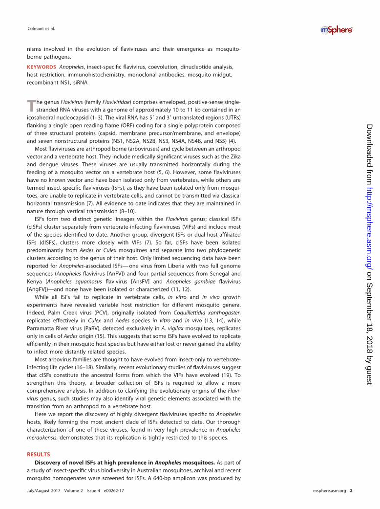
The maximum-likelihood (ML) phylogeny of the ORF revealed that Anopheles ISFscluster with cISFs and branch at a basal position within the clade, indicating theancestral position of the common ancestor (Fig. 2A). KRBV also appears to form adistinct lineage with AnFV, HaCV, and McPV, separate from Culex- and Aedes-associatedcISFs. The partial ISF nucleotide sequences obtained for all HaCV (GenBank accessionno. KY460534 and KY460535), DSwV (KY460536 and KY460537), and McPV (KY460531
Unique Host Restriction of New Australian Flaviviruses
July/August 2017 Volume 2 Issue 4 e00262-17 msphere.asm.org 3
on Septem
ber 18, 2018 by guesthttp://m
sphere.asm.org/
Dow
nloaded from

FIG 1 Detection of ISFs from Anopheles mosquitoes. Summary of A. meraukensis (A) and other Anopheles (B) pools tested. (C) Map ofAustralia showing the locations of mosquito trapping sites and the viruses detected.
Colmant et al.
July/August 2017 Volume 2 Issue 4 e00262-17 msphere.asm.org 4
on Septem
ber 18, 2018 by guesthttp://m
sphere.asm.org/
Dow
nloaded from

to KY460533) virus sequences were included in a partial NS5 gene alignment (~350 nt)along with several KRBV sequences from various locations (KY460524 to KY460530), andan ML tree further showed that the Anopheles ISFs cluster together, forming a single,separate clade (Fig. 2B).
Putative cleavage sites in the predicted KRBV, McPV, and HaCV polyprotein se-quences revealed that their ORFs code for viral proteins according to the standardflavivirus genome organization (Table 3). The pr-M furin cleavage site for KRBV, McPV,and HaCV occurs between amino acids 172 and 173 and follows the consensusestablished for other flaviviruses (23), suggesting that the cleavage is efficient, unlikefor some cISFs (e.g., PaRV) that exhibit inefficient pr-M cleavage (15).
In addition to the main ORF coding for the three structural and seven nonstructuralproteins, sequence analysis uncovered the presence of a predicted 879-bp codingsequence in the �1/�2 frame for KRBV, McPV, and HaCV, known to be present in ISFsas the fairly interesting flavivirus ORF (fifo) (24). The sequence starts with the conserved“slippery” RNA motif GGAUUUU for all three viruses, 53 nt downstream of the predictedcleavage between NS1 and NS2A. The motif is followed by six spacing nucleotidesdirectly followed by a 34-bp stable RNA stem-loop (20 paired nucleotides for KRBV, 22paired nucleotides for McPV, and 22 paired nucleotides for HaCV). These features are
TABLE 1 KRBV sequences at pan-flavivirus primer binding sites
Sequence Primer name, sequencea
Primer FU2, GCTGATGACACCGCCGGCTGGGACAC cFD3, AGCATGTCTTCCGTGGTCATCCAKRBV GCCGATGATGTGGCGGGATGGGATAC AGCATGTCAACAGTTGTCATCCA
Primer PF1S, TGYRTBTAYAACATGATGGG PF2Rbis, GTGTCCCAICCNGCNGTRTCKRBV TGTGTCTACAACACCATGGG GTATCCCATCCCGCCACATC
Primer EMF1, TGGATGACSACKGARGAYATG VD8, GGGTCTCCTCTAACCTCTAGKRBV TGGATGACAACTGTTGACATG Unidentified
Primer Flav100F, AAYTCNACNCANGARATGTAY Flav200R, CCIARCCACATRWACCAKRBV AATTCTACTGCGGAGATGTAT CCGAGCCACATATACCA
aBoldface letters indicate differences between the primer and corresponding KRBV sequences.
TABLE 2 Nucleotide and amino acid sequence identities of novel Anopheles ISFs with reference flavivirusesa
Virus
% Identity with:
1892 WBay2 Kim1 McPV HaCV DSwVb AnFV PaRV CFAV CxFV QBV PCV BgV WNV ZIKV DENV-2 LAMV CHAOV NOUV BJV
1892 99.5 95.4 82.1 81.8 78.8 80.7 35.7 36.0 37.7 38.6 38.0 24.1 25.0 24.9 24.0 24.0 24.2 23.9 24.7WBay2 99.4 95.5 82.1 81.8 78.9 80.7 35.7 36.1 37.7 38.6 38.0 24.2 24.9 24.9 23.9 24.0 24.2 23.9 24.7Kim1 87.8 87.9 82.1 82.1 78.9 80.9 35.9 35.9 37.7 38.5 37.9 24.4 24.9 24.8 24.0 24.0 24.2 23.8 24.6McPV 73.3 73.2 73.4 81.0 79.3 80.2 35.7 36.0 37.6 37.9 37.5 24.1 24.9 24.7 24.0 24.0 23.9 23.3 23.7HaCV 72.6 72.6 73.1 73.7 79.6 79.2 33.4 33.3 36.0 36.2 35.5 23.2 23.9 23.8 22.9 22.9 22.8 22.5 23.3DSwVa 69.7 69.8 69.2 71.8 72.9 77.7 39.7 40.7 43.5 43.4 43.4 29.9 29.8 30.0 30.4 29.4 29.3 29.8 29.9AnFV 72.1 72.1 72.9 72.7 72.2 70.8 35.1 35.6 37.2 37.8 37.5 23.9 24.9 25.0 24.4 23.8 23.8 23.5 24.4PaRV 44.8 44.9 44.9 44.3 43.7 47.0 44.7 41.8 38.7 38.0 38.3 25.1 24.8 24.7 24.4 24.4 24.5 24.5 24.9CFAV 44.8 44.8 44.5 44.5 43.5 48.3 45.0 45.1 43.4 44.0 40.6 24.3 24.3 24.0 23.5 24.0 24.0 23.6 24.0CxFV 46.8 46.8 46.3 46.3 45.4 50.1 46.5 45.0 48.4 64.4 51.9 23.7 24.3 24.4 23.9 23.2 23.2 22.8 23.7QBV 47.0 46.9 46.8 46.8 44.8 49.2 46.5 44.4 48.7 61.9 53.1 23.9 23.9 24,4 23.7 23.5 23.8 23.5 23.9PCV 47.0 47.0 46.6 46.6 45.3 49.6 46.8 44.1 46.3 52.4 52.8 23.9 24.2 23.9 23.8 23.8 23.9 23.0 23.5BgV 37.3 37.3 37.4 36.9 36.5 39.6 37.1 35.8 35.2 35.1 35.6 35.9 44.3 45.2 43.3 44.8 44.9 43.2 44.5WNV 37.0 37.0 37.3 37.1 36.2 39.9 36.9 36.0 36.1 35.9 35.8 36.0 49.1 56.9 51.6 49.5 49.8 46.9 49.2ZIKV 38.1 38.1 37.9 37.7 37.4 41.0 38.3 35.9 36.0 36.0 35.6 36.1 48.2 55.3 54.9 49.8 49.7 48.1 49.6DENV-2 37.6 37.6 37.9 37.0 36.6 40.3 37.3 35.5 35.4 35.0 35.2 34.7 48.8 52.1 55.4 46.7 46.8 44.5 46.8LAMV 36.7 36.7 36.4 36.8 35.4 39.8 36.5 35.9 35.4 35.2 35.1 35.6 48.9 51.8 51.8 50.2 85.5 46.3 48.3CHAOV 37.4 37.4 37.3 37.2 36.3 40.2 37.0 35.7 35.6 35.3 35.1 35.9 48.9 51.8 51.6 49.8 71.8 46.6 48.3NOUV 36.9 36.9 36.8 36.9 36.1 40.4 37.1 35.5 35.4 35.5 35.1 35.7 47.9 50.0 51.5 49.4 49.9 50.1 53.2BJV 37.8 37.9 38.0 38.0 37.2 39.7 38.0 36.1 36.4 36.1 35.8 36.0 48.4 51.2 50.5 49.3 50.2 50.2 53.7aThe top right half is amino acid sequence identity, and bottom left half is nucleotide sequence identity. 1892, KRBV prototype strain; WBay2, KRBV strain; Kim1, KRBVstrain; CFAV, cell fusing agent virus; CxFV, Culex flavivirus; QBV, Quang Binh virus; ZIKV, Zika virus; DENV-2, dengue virus serotype 2; CHAOV, Chaoyang virus; NOUV,Nounane virus.
bAll sequence similarities are for the ORF, except for DSwV, as only the last 5 kb of the genome were available.
Unique Host Restriction of New Australian Flaviviruses
July/August 2017 Volume 2 Issue 4 e00262-17 msphere.asm.org 5
on Septem
ber 18, 2018 by guesthttp://m
sphere.asm.org/
Dow
nloaded from
thought to stimulate a ribosomal frameshift that results in the translation of a 293-aa-long protein. fifo seems to be specific to cISFs and is present in all of the cISF sequencesinvestigated to date. With two predicted transmembrane domains, McPV, HaCV, andKRBV (Kim1 strain) follow the consensus established for other ISFs, while KRBV (proto-type strain) fifo appears to have only one transmembrane domain (11, 24).
The cISFs genomes differ from VIF genomes by conserved insertions and deletionsthat may potentially be associated with ISF host restriction or enhanced verticaltransmission (15). Similar conserved deletions and insertions were observed in bothstructural and nonstructural KRBV proteins (Fig. 3), supporting the affiliation of KRBVwith the cISFs. McPV, HaCV, and DSwV also had these conserved deletions andinsertions (data not shown).
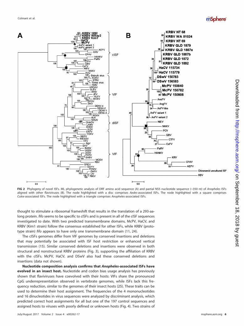
Nucleotide composition analysis confirms that Anopheles-associated ISFs haveevolved in an insect host. Nucleotide and codon bias usage analysis has previouslyshown that flaviviruses have coevolved with their hosts: VIFs share the pronouncedCpG underrepresentation observed in vertebrate genomes, while ISFs lack this fre-quency reduction, similar to the genomes of their insect hosts (25). These traits can beused to determine their host assignment. The frequencies of the 4 mononucleotidesand 16 dinucleotides in virus sequences were analyzed by discriminant analysis, whichpredicted correct host assignments for all but one of the 197 control sequences andassigned hosts to viruses with poorly defined or unknown hosts (Fig. 4). Two strains of
FIG 2 Phylogeny of novel ISFs. ML phylogenetic analysis of ORF amino acid sequence (A) and partial NS5 nucleotide sequence (~350 nt) of Anopheles ISFsaligned with other flaviviruses (B). The node highlighted with a disc comprises Aedes-associated ISFs. The node highlighted with a square comprisesCulex-associated ISFs. The node highlighted with a triangle comprises Anopheles-associated ISFs.
Colmant et al.
July/August 2017 Volume 2 Issue 4 e00262-17 msphere.asm.org 6
on Septem
ber 18, 2018 by guesthttp://m
sphere.asm.org/
Dow
nloaded from

KRBV, HaCV, and other Australian cISFs, PCV and PaRV, grouped with the insect-onlyviruses. Assignments for dISFs varied between insect only and vector borne, with thosephylogenetically related to Lammi virus (LAMV) being assigned to the insect-onlygroup, and those related to Barkedji virus (BJV) assigned to the vector-borne group.Bamaga virus (BgV), another recently discovered Australian flavivirus with limitedreplication in vertebrates, grouped closely with VIFs, consistent with its phylogeneticposition (23). It was noteworthy that the flaviviruses with no known vector generallygrouped closely with vector-borne viruses, with the exception of the highly divergentTamana bat virus sequence, which was predicted to be associated only with a verte-brate host in this analysis.
Anopheles ISFs do not replicate in cells derived from heterologous species.KRBV RNA was initially detected in the supernatant of C6/36 cells that had beeninoculated with mosquito homogenate and incubated for 5 to 7 days at 28°C withoutremoving the inoculum. This suggested that either the amplified sequence was from avirus replicating in the culture or the RNA was residual from the inoculum and likelyprotected from degradation in cell culture by encapsidation in a particle. No viral RNAcould be reliably detected in subsequent passage of these samples on C6/36 cells(Table 4), suggesting that the initial detection was likely due to virion-protected RNA inresidual inoculum and that the virus was unable to infect and replicate in C6/36 cells.In comparison, other mosquito samples collected at the same time and stored in thesame way yielded numerous virus isolates by the same protocol, including PaRV from
TABLE 3 Predicted cleavage sites in the polyproteins of KRBV, McPV, HaCV, and DSwV
Protein cleavagea
Sequenceb
KRBV McPV HaCV DSwV
C/AnchC TRQR 2 TGNN ARQR 2 TGGN TRQR 2 TGGN UnknownAnchC/pr-M LACA 2 KTMN FGCA 2 KTMN YGCA 2 KTMT Unknownpr-M/M RVKR 2 GEPG RVKR 2 DQEG RVKR 2 DSAE UnknownM/E VVQA 2 SLAD VVKA 2 SLAD IVQA 2 SLAD UnknownE/NS1 YVRA 2 DVGC YVRA 2 DVGC YVRA 2 DVGC UnknownNS1/NS2A ESVA 2 QPVT ESNA 2 QAVE ESEV 2 KPIT UnknownNS2A/NS2B NWRR 2 APAP NWRR 2 VPVP NWRK 2 VPVP UnknownNS2B/NS3 SCFR 2 SDDG SCFR 2 SDDD SCFR 2 SDDG UnknownNS3/NS4A LRMR 2 AHIN LRMR 2 TSIN LRMR 2 ASVN LRMR 2 ASVNNS4A/2K SATR 2 SYVD SSTR 2 SYVD STTR 2 SYVD SSTR 2 SYVD2K/NS4B GLVA 2 FELD GLVA 2 FELD GIVA 2 FELD GIVA 2 FELDNS4B/NS5 NSYR 2 SSNK SSTK 2 GDAL SSNK 2 GDAL SSNK 2 GDALaC, capsid; AnchC, capsid anchor; pr-M, premembrane; M, membrane; E, envelope; NS, nonstructural protein;2K, 2K peptide.
bCleavage sites are indicated by downward arrows.
FIG 3 Alignment of KRBV and other flavivirus amino acid sequences displaying conserved deletions and insertions in the structural and nonstructural genes.
Unique Host Restriction of New Australian Flaviviruses
July/August 2017 Volume 2 Issue 4 e00262-17 msphere.asm.org 7
on Septem
ber 18, 2018 by guesthttp://m
sphere.asm.org/
Dow
nloaded from
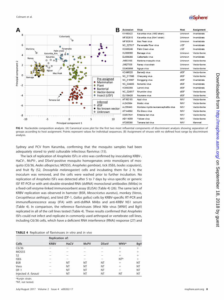
Sydney and PCV from Karumba, confirming that the mosquito samples had beenadequately stored to yield culturable infectious flavivirus (15).
The lack of replication of Anopheles ISFs in vitro was confirmed by inoculating KRBV-,HaCV-, McPV-, and DSwV-positive mosquito homogenates onto monolayers of mos-quito (C6/36, Aedes albopictus; MOS55, Anopheles gambiae), tick (ISE6, Ixodes scapularis),and fruit fly (S2, Drosophila melanogaster) cells and incubating them for 2 h; theinoculum was removed, and the cells were washed prior to further incubation. Noreplication of Anopheles ISFs was detected after 5 to 7 days by virus-specific or genericISF RT-PCR or with anti-double-stranded RNA (dsRNA) monoclonal antibodies (MAbs) ina fixed-cell enzyme-linked immunosorbent assay (ELISA) (Table 4) (26). The same lack ofKRBV replication was observed in hamster (BSR, Mesocricetus auratus), monkey (Veros,Cercopithecus aethiops), and bird (DF-1, Gallus gallus) cells by KRBV-specific RT-PCR andimmunofluorescence assay (IFA) with anti-dsRNA MAbs and anti-KRBV NS1 serum(Table 4). In comparison, the reference flaviviruses (West Nile virus [WNV] and BgV)replicated in all of the cell lines tested (Table 4). These results confirmed that AnophelesISFs could not infect and replicate in commonly used arthropod or vertebrate cell lines,including C6/36 cells, which have a deficient RNA interference (RNAi) response (27) and
FIG 4 Nucleotide composition analysis. (A) Canonical score plot for the first two most influential components of discriminant analysis showing separation ofgroups according to host assignment. Points represent values for individual sequences. (B) Assignment of viruses with no defined host range by discriminantanalysis.
TABLE 4 Replication of flaviviruses in vitro and in vivo
Cells
Replication of:
KRBV HaCV McPV DSwV WNVa BgV
C6/36 � � � � � �MOS55 � � � � � �S2 � � � � � �ISE6 � � � � NTb �BSR � NT NT NT � NTVero � NT NT NT � NTDF-1 � NT NT NT � NTInjected A. farauti � NT NT NT NT NTaKunjin strain.bNT, not tested.
Colmant et al.
July/August 2017 Volume 2 Issue 4 e00262-17 msphere.asm.org 8
on Septem
ber 18, 2018 by guesthttp://m
sphere.asm.org/
Dow
nloaded from

have been shown to support the replication of all of the other mosquito-borneflaviviruses tested to date by the same method of isolation.
To assess whether KRBV could replicate in vivo, RT-PCR-positive A. meraukensishomogenates were inoculated intrathoracically into colonized A. farauti sensu strictomosquitoes shown to be free of ISF RNA and tested for KRBV RNA at 3 h or 5 dayspostinjection in pools of three. KRBV sequence could be detected in 4/6 pools sampledwithin 3 h of inoculation, confirming the presence of detectable KRBV in the inoculum.However, no viral sequence was detected in six pools harvested 5 days postinjection,suggesting a lack of KRBV replication (Table 4).
KRBV sequence was derived from an RNA template, not from integration intomosquito DNA. To confirm that the KRBV sequences amplified by RT-PCR indeedoriginated from RNA, we performed simultaneous RT-PCR and PCR with KRBV-specificprimers on the same RNA extract, as the RNA isolation kit used allows for DNA to beeluted along with purified RNA. KRBV sequence was amplified from mosquito homog-enates only when an RT step was performed before the PCR, indicating that theamplification was from an RNA template (Fig. 5A). We also found that the KRBVtemplate was still amplified by RT-PCR after RQ1 DNase digestion of KRBV RNA,confirming these findings (Fig. 5B).
Some ISF sequences have been shown to be integrated into their mosquito hostgenomes as the virus and host coevolve and are identified as endogenous viralelements (EVEs) (28–30). To assess whether the detected KRBV RNA template foramplification was similarly derived from transcripts of viral genes integrated into thegenome of A. meraukensis, mosquito genomic DNA (gDNA) was tested for KRBVsequence by PCR with various combinations of virus-specific primers. Despite success-ful amplification of the conserved internal transcribed spacer 2 (ITS2) region in therepeated ribosomal DNA genes of the Anopheles punctulatus complex from gDNA ofboth A. farauti controls and A. meraukensis, no KRBV sequence was amplified with anyof the KRBV-specific primer combinations used (data not shown). This implies that theKRBV sequence was amplified from a viral genomic RNA template in the mosquito andnot from mRNA transcripts of viral genes integrated into host chromosomes. In
FIG 5 Detection of positive and negative KRBV genomic RNA strands as an indication of viral replication. (A) Amplification ofKRBV sequence with and without RT with KRBV-specific primers. The first lane has KRBV RNA subjected to RT-PCR, the secondlane has KRBV RNA subjected to PCR only, the third lane has KRBV cDNA subjected to PCR, the fourth lane shows theunamplified input cDNA, and the last lane shows the nontemplate control subjected to RT-PCR. (B) RT-PCR of DNase-treatedKRBV RNA. T, DNase treated; NT, not treated. (C) RT-PCR amplification of Anopheles ISF sequences with forward only (F), reverseonly (R), or both virus-specific primers during RT. Top gel, KRBV; bottom gel, from left to right, DSwV, McPV, and HaCV. (D) PCRsof cDNA generated with forward primer KRBV41F and with downstream (KRBV42R and KRBV3UTR1F/1R pair) and upstream(KRBV20F/R pair) primers and control RT-PCR of KRBV RNA for upstream primers.
Unique Host Restriction of New Australian Flaviviruses
July/August 2017 Volume 2 Issue 4 e00262-17 msphere.asm.org 9
on Septem
ber 18, 2018 by guesthttp://m
sphere.asm.org/
Dow
nloaded from

addition, several A. meraukensis homogenates were found to be negative for the KRBVsequence by RT-PCR, suggesting that the viral sequence was unlikely to be integratedinto the genome of this mosquito species.
Detection of KRBV RNA replicative intermediates and proteins. To detect viralproteins in sections of naturally infected mosquitoes, we produced recombinant KRBVNS1 (rNS1) and generated mouse antiserum and MAbs to this protein. rNS1 displayeddimers and oligomers typical of a flavivirus NS1 protein by Western blotting (Fig. 6A),and its identity was confirmed by mass spectrometry (five peptides identified; data notshown). rNS1 elicited an antibody response in mice sufficient for its detection byWestern blotting and in IFA (Fig. 6). The antiserum displayed cross-reactivity with otherISF NS1 proteins, reacting to both Aedes- and Culex-associated Australian ISF proteins(PaRV and PCV) in an IFA, supporting the classification of KRBV as a cISF (Fig. 6). MAbsderived from the immunized mice detected rNS1 in an ELISA and recognized the nativeviral protein in sections of 3 out of 10 A. meraukensis mosquitoes by immunohisto-chemistry (IHC) analysis, thereby showing expression of KRBV NS1 in these mosquitoes(Fig. 7). KRBV-NS1 expression was restricted to epithelial cells of the midgut in theabsence of overt cytopathology, and the protein colocalized with viral dsRNA detectedwith anti-dsRNA MAbs, suggesting KRBV replication in the mosquitoes (Fig. 7).
To provide further evidence of the presence of viral dsRNA and therefore replicationof KRBV in A. meraukensis tissues, KRBV sequence was amplified from cDNA transcribedfrom both positive and negative RNA strands (Fig. 5C). A similar experiment wasperformed with McPV-, HaCV-, and DSwV-positive homogenate RNA and virus-specificprimers, and it provided evidence of the presence of dsRNA replicative intermediates inthese Anopheles homogenates as well (Fig. 5C; Table 5). To confirm that the PCRproduct was generated from cDNA transcribed from the negative strand and to
FIG 6 Production of recombinant KRBV NS1 and antiserum reactivity. SDS-PAGE and Western blotanalysis of unboiled and unreduced untransfected COS-7L cell lysate (lanes 1); KRBV NS1-transfectedCOS-7L cell lysate (lanes 2), and concentrated supernatant (lanes 3); HisTrap column-purified KRBV NS1(lanes 4) show NS1 monomers at approximately 50 kDa and oligomers at 100 and 150 kDa and highermolecular masses. Lanes L contain the molecular weight marker Precision Plus Protein Kaleidoscope(Bio-Rad). Panel A, with anti-V5 epitope MAb (lanes rearranged for figure clarity); panel B1, withimmunized mouse serum (mouse 2); panel B2, with negative mouse serum. (C) IFA with anti-KRBV NS1mouse serum of COS-7L cells (mock transfected and transfected with KRBV NS1) and C6/36 cells infectedwith cISFs (mouse 2 antiserum).
Colmant et al.
July/August 2017 Volume 2 Issue 4 e00262-17 msphere.asm.org 10
on Septem
ber 18, 2018 by guesthttp://m
sphere.asm.org/
Dow
nloaded from

eliminate the possibility that the cDNA was generated from mispriming of KRBV41F onthe positive strand, downstream of the KRBV41F/42R binding site, we designed primersupstream of the KRBV41F/42R site (KRBV20F/R) and downstream, at the very end of theKRBV genome (KRBV3UTR1F/KRBV3UTR1R). We showed that the upstream primersfailed to amplify a product while the downstream primers successfully amplified a PCRproduct from the cDNA generated with KRBV41F from the negative strand (Fig. 5D).
Detection of Anopheles ISF-specific siRNA provides evidence of the mosquitoimmune response to viral infection. Small RNA (sRNA) reads generated by NGS of
FIG 7 Detection of KRBV protein and replicative intermediates in A. meraukensis mosquitoes. Detection of KRBV NS1 (red signal) with MAbs 5G2(A1) and 6F8 (B1) in midgut epithelial cells (MG). C1, faint signal for dsRNA with MAb 3G1 (arrows) in MG. Negative MG in mosquitoes on sameslide as the KRBV-positive specimens labeled with 5G2 (A2), 6F8 (B2), and 3G1 (C2). Follicles (Fo) are negative for viral NS1 protein. It is importantto note that NS1 accumulates in flavivirus-infected cells and is thus more abundant than the replicative intermediates. In addition, MAb 3G1 isan IgM, which can further explain the faint signal obtained.
TABLE 5 Anopheles ISF primers used in this study
Primera Sequence Usedb with:
KRBV2F ACATTGCCGACAGGGACACG 2RKRBV2R CCAACAGCTGCATCTGAACG 2FKRBV41F GGTCTTGTTTGCGCCTTCATGTGC 42KRBV42R CGCGTTTGTTATTCTTGGCTTCC 41KRBV7F CCAAACTCGTACCGGTCATCAAAC 7R, 8RKRBV7R GCCATAAGTCATGAACGCCTCG 7F, 8FKRBV8F CGGAATATCAACCAGGGGATTGTG 8R, 7R, 9RKRBV8R GCTAACCAATTTTCCAACAGGGTG 8F, 7F, 9FKRBV9F CCAATTAACCTTCGTCACAGCTGC 9R, 8R, 10RKRBV9R GCAGTGGAATTTCTACTTAAGCGC 9F, 8F, 10FKRBV10F CGGATCGACCATCCCTAGAAAGAG 10R, 9RKRBV10R CGTTAGAGCTCTCAATGTTGC 10F, 9FKRBV20F TCATGGAGCATATGCATTCG NAc
KRBV20R TCAGTGACTTCAGATCCTCC NAKRBV3UTR1F CGACGTGTCTTGGACAAACACG NAKRBV3UTR1R CCTGCCTGTGTTTTCTTGG NADSwV1F CGACGTGAATATGGCAAAGG NADSwV1R CACACTAGCTCTCATCCTGAGC NAMcPV3502F CCCTGACGTTGTATTGGTACC NAMCPV3875R GCTAGCTGCGAGATATGTGC NAHaCV2833F CACATGCCTGGGTATCACACG NAHaCV3393R CGACACCAATAAGGACTGTCC NAISF F1 GGGCAAGTARBMACTTATGCVTTGAACAC NAISF R1 GCCCACATCTGGGCRTRNGCCTTNGC NAaF, forward; R, reverse.bCombination(s) used for gDNA detection.cNA, not applicable.
Unique Host Restriction of New Australian Flaviviruses
July/August 2017 Volume 2 Issue 4 e00262-17 msphere.asm.org 11
on Septem
ber 18, 2018 by guesthttp://m
sphere.asm.org/
Dow
nloaded from

mosquito homogenates positive for KRBV, HaCV, or PaRV were aligned with thecorresponding reference ORF to identify virus-specific siRNA (vsiRNA) produced inresponse to replicating virus. Reads that mapped to the virus genomes displayed a peakat 21 nt for all samples, which corresponds to the size of siRNAs generated via thecleavage of viral dsRNA by dicer-2 as part of the mosquito RNAi response to areplicating virus (Fig. 8A) (31, 32). The 21-nt reads mapped to both the sense andantisense strands of the KRBV and HaCV genomes, with a majority mapping to thesense strand, consistent with the expected response for replicating viruses (Fig. 8B toD). The assembly of vsiRNA reads produced an ORF sequence for a third KRBV strain(WBay2) in addition to the prototype strain and KRBV strain Kim1, and it was includedin the phylogenetic analysis (Fig. 2A). WBay2 has 99.4% nucleotide sequence and 99.5%amino acid sequence identity with the prototype strain of KRBV (1892) over the wholeORF sequence.
DISCUSSION
We report here the discovery of four novel ISFs exclusive to an Anopheles host. Theseviruses are highly prevalent in several Anopheles populations, with infection rates of upto 100% in mosquito pools from some cohorts and frequent detection in homogenatesof single mosquitoes. In a cohort of 10 single mosquitoes collected from Bradshaw, 3were shown to be positive for KRBV RNA. Similarly, KRBV NS1 protein was detected in3 out of 10 mosquitoes by IHC analysis. We detected KRBV in A. meraukensis pools frommultiple years in traps located as far as 2,800 km apart. This study is the first todemonstrate the presence of an Anopheles ISF with a widespread geographical andtemporal distribution at a high prevalence. The findings suggest that the virus becameestablished in A. meraukensis populations a long time ago and was maintained via ahighly efficient mode of vertical transmission.
According to the criteria for species demarcation within the Flavivirus genus,as proposed in the 2016 report by the International Committee for the Taxonomyof Viruses (https://talk.ictvonline.org/ictv-reports/ictv_online_report/positive-sense-rna-viruses/w/flaviviridae/360/genus-flavivirus), which includes nucleotide and amino acidsequence identities, host and vector associations, and geographical distributions, KRBV,HaCV, McPV, and DSwV should be considered new species. These new virus sequencesalso group phylogenetically with other cISFs but form a separate Anopheles-associatedclade distinct from Culex- and Aedes-associated cISFs. We propose a new designationfor ISFs, with cISFs becoming lineage I (IA for Aedes-associated ISFs, IB for Anopheles-associated ISFs, and IC for Culex-associated ISFs) and dISFs becoming lineage II, asoutlined by Hall et al. (33). The Anopheles clade (or lineage IB) appears to be moreclosely related to other lineage I ISFs than to other flaviviruses, including an ISF-likepartial sequence discovered in chironomid species (34), demonstrating that the Anoph-eles viruses most likely replicate in mosquitoes, rather than being derived from hostmicrobiota or parasites. In addition, the recent discovery of EVEs in Anopheles minimusand A. sinensis supports our conclusion that Anopheles mosquitoes are natural hosts ofISFs (30). Since most EVEs appear to be integrated into the host genome early duringthe host-virus evolutionary relationship, it is likely that some KRBV sequences havebeen integrated into the A. meraukensis genome at some time in the past, despite ourinability to detect them (30). Sequencing of the A. meraukensis genome could shed lighton the presence of ISF EVEs in this mosquito genome.
Nucleotide motif usage analysis has previously shown that flaviviruses have co-evolved with their hosts: VIFs share the pronounced CpG underrepresentation ob-served for vertebrate genomes, while ISFs lack this frequency reduction, similar to theirinsect hosts’ genomes (25). This reflects the avoidance of CpG in vertebrates to reducethe chance of C-to-T mutations due to spontaneous deamination of cytosine bymethylation at these sites, a process that does not occur in insects (35). Like other ISFs,KRBV and McPV sequences display an insect-like nucleotide usage bias. The fact thatISFs are not constrained to mimic the dinucleotide bias of vertebrates provides furtherevidence of their adaption to a mosquito-only transmission cycle.
Colmant et al.
July/August 2017 Volume 2 Issue 4 e00262-17 msphere.asm.org 12
on Septem
ber 18, 2018 by guesthttp://m
sphere.asm.org/
Dow
nloaded from

Separate clustering of viruses according to mosquito genera would suggest coevo-lution of the viruses and their hosts, at least to some extent. The basal phylogeneticposition of the Anopheles ISF clade supports this theory, as Anopheles is considered theoldest mosquito genus, being the first to have diverged from other mosquito genera
FIG 8 Analysis of siRNA in mosquito homogenates. Size distribution of mapped reads: PaRV-positive A. vigilax homogenate (A), KRBV-positive A. meraukensishomogenates (B1 and C1), and HaCV-positive A. annulipes homogenate (D1). Distribution of 21-nt reads over the genome sequences of KRBV-positiveA. meraukensis homogenates (B2 and C2) and HaCV-positive A. annulipes homogenate (D2) with top reads mapping to the sense strand of the genome andbottom reads mapping to the antisense strand of the genome.
Unique Host Restriction of New Australian Flaviviruses
July/August 2017 Volume 2 Issue 4 e00262-17 msphere.asm.org 13
on Septem
ber 18, 2018 by guesthttp://m
sphere.asm.org/
Dow
nloaded from
(36, 37). Coevolution of the Anopheles ISFs with their host species provides an expla-nation for the narrow host restriction of these viruses and is, to some degree, corrob-orated by our data, as each virus was detected in a single Anopheles species. This doesnot preclude the possibility of several viruses being found in the same species in othermosquito populations; two different ISF sequences were detected in A. gambiae fromKenya and Senegal, approximately 8,000 km apart (11, 12).
Although the new Anopheles ISFs group phylogenetically with other lineage I ISFs,they seem to be genetically and phylogenetically divergent from other flaviviruses.None of these viral sequences were amplified by generic flavivirus primers, whichcollectively recognize all of the other members of the genus tested to date, includingother ISFs. The Anopheles ISFs could be detected only by a set of generic primers basedon lineage I ISF sequences. KRBV has only ~40% sequence identity with its closestnon-Anopheles-associated relative, Quang Binh virus, a lineage IC ISF from Vietnam.Furthermore, although we showed that KRBV replicates in its natural host, we wereunable to find a laboratory model to support KRBV replication, including A. albopictusand A. gambiae cell lines, despite the former being particularly permissive to infectionwith flaviviruses and the latter being derived from the same genus as the natural hostof the viruses. Viruses from various families, including flaviviruses, were isolated fromthe same cohorts of samples as part of separate studies, showing that the homogenateswere appropriately stored and processed for virus isolation (15, 33, 38). Moreover,attempts to infect colonized A. farauti sensu stricto mosquitoes with KRBV were unsuc-cessful. These findings contrast with other studies performed with ISFs. PCV and CxFVreplicated efficiently after intrathoracic inoculation into mosquitoes of different speciesand genera (13, 39). A key experimental difference was the use of KRBV-positivehomogenates as an inoculum in this study, since a pure virus stock could not begenerated. The viral dose may have been suboptimal, or nonviral constituents of thehomogenate may have stimulated an innate immune response, abrogating replicationof the virus. We were not able to assess the replication of KRBV in A. meraukensismosquitoes, as we did not have access to a laboratory colony of this species, andacquisition of wild-caught A. meraukensis from its natural habitats in remote regions ofAustralia was not logistically possible. In any case, on the basis of our current data,wild-caught A. meraukensis would likely have a naturally high prevalence of KRBVinfection. Nevertheless, together, our in vitro and in vivo findings suggest that Anoph-eles ISFs may be uniquely specific to their natural host species.
This unprecedented host restriction may be due to the virus’s reliance on hostcomponents absent from both cell culture and other mosquito species, originatingeither from the host itself or from a coinfecting parasite or virus, which would renderin vitro and in vivo models unsuitable. One explanation may link host restriction withinteractions during virus attachment and entry into the cell, with a potentially strongadaptation of the virus to a cell receptor specific to A. meraukensis.
Recent studies in other virus-insect host systems have demonstrated that while theviruses could suppress the RNAi defenses of their host species and allow efficientreplication in the insect, they were unable to suppress the corresponding RNAi re-sponse in related host species and consequently failed to replicate (40). This evidenceof species-dependent RNAi suppressors in insect viruses suggests that the species-specific tropism exhibited by KRBV may be due to its inability to sufficiently suppressthe RNAi response in other Anopheles species. However, the presence of 21-nt-longsiRNAs in A. meraukensis homogenates that map to the KRBV sequence shows thatKRBV does not completely suppress the A. meraukensis RNAi response. In addition tothis, no KRBV replication was detected, even in dicer-2-deficient C6/36 cells, which havean impaired siRNA response. Nevertheless, the RNAi response in insects comprises threepathways mediated by sRNAs, the siRNA, microRNA (miRNA), and Piwi-interacting RNA(piRNA) pathways (41). Consequently, while siRNA is thought to be the main cellularantiviral innate immune response in arthropods (42), the restricted tropism of KRBVmay be explained by its inability to suppress other RNAi pathways, such as piRNA ormiRNA pathways, in mosquito species other than its main host species. In any case, this
Colmant et al.
July/August 2017 Volume 2 Issue 4 e00262-17 msphere.asm.org 14
on Septem
ber 18, 2018 by guesthttp://m
sphere.asm.org/
Dow
nloaded from

unique host restriction limits the possibilities of virus characterization and is likely onereason why only sequence data for Anopheles ISFs have been published to date.
We attempted to detect KRBV proteins in mosquito homogenates by mass spec-trometry but were unsuccessful, even in those from our control mosquitoes, A. aegyptiinjected with PaRV, suggesting that the limit of detection of the assay was too low todetect ISF proteins. However, we were able to detect KRBV NS1 in naturally infectedmosquitoes by IHC analysis, indicative of viral replication, and this was corroborated bycolocalization of anti-dsRNA antibodies, also by IHC analysis. In addition to provingnatural infection of mosquitoes by KRBV, these data show that the virus appears to beconfined to the epithelial cells of the mosquito midgut, a narrow host cell tropismshared with other ISFs (13), which suggests that this characteristic is not linked to theunique host restriction of KRBV. Moreover, this localization is consistent with a mode oftransmission from mother to progeny that does not necessarily involve replication inmosquito reproductive organs. Rather, it could involve indirect transmission of the virusto the egg prior to egg deposition, as shown by Jia et al. for rice dwarf virus inNephotettix cincticeps leafhoppers (43).
Despite our inability to culture the Anopheles ISFs in either in vitro or in vivo systems,we have shown that KRBV, McPV, and HaCV possess a complete viral genome with afunctional ORF. Furthermore, according to the dinucleotide usage pattern of theirgenomes, KRBV and McPV have evolved to replicate only in insects. We have alsoprovided compelling evidence that Anopheles ISFs replicate in their host mosquitoes bydemonstrating the presence of double-stranded replicative RNA forms, vsiRNA gener-ated by the mosquito RNAi response, and KRBV-specific protein in mosquito tissues,specifically, midgut epithelial cells. Finally, our inability to amplify KRBV sequences fromthe A. meraukensis genome and the existence of KRBV-negative samples of this specieseffectively show that the KRBV sequence detected were not derived from EVEs presentin the mosquito host’s genome. The characterization of these new Anopheles flavivi-ruses necessitated the use of a range of novel techniques and research tools that willenhance our understanding of the ecology, biology, and evolution of this unique groupof flaviviruses.
MATERIALS AND METHODSTrapping and processing of mosquitoes. All of the mosquito samples tested in this study were
archival and recent mosquito pools of the Anopheles genus collected for various studies (University ofWestern Australia, Army Malaria Institute; details are shown in Fig. 1) (38, 44). All mosquito homogenateswere stored at �80°C to ensure proper conservation of potential viruses and RNA, quickly thawed at37°C, and subsequently kept on ice before cell inoculation (see below).
Screening of homogenates for novel flaviviruses. Two hundred microliters of mosquito homog-enate was used to inoculate C6/36 cell monolayers, maintained as described below, in 4 wells of a 96-wellplate. Cultures were incubated for 5 to 7 days at 28°C, supernatant was harvested, and RNA was extractedfrom this supernatant with the NucleoSpin Viral RNA isolation kit from Macherey-Nagel. The originaldetection of the partial KRBV genome was performed by RT-PCR (Superscript III One-Step RT-PCR Systemwith Platinum Taq DNA polymerase; Invitrogen) with generic NS5 flavivirus primers Flav100F/Flav200R(45). Subsequent screening was performed by RT-PCR with generic ISF primers from an alignment of theflavivirus NS5-encoding gene, including ISF sequences (ISF F1/ISF R1; Table 5) or virus-specific primersbased on the short KRBV sequence available. When samples yielded an RT-PCR product of the expectedsize, its sequence was obtained by Sanger sequencing at the Australian Genome Research Facility (AGRF;Brisbane, Queensland, Australia) after amplicons were purified by agarose gel electrophoresis andextracted with the NucleoSpin Gel and PCR cleanup kit (Macherey-Nagel). Species-specific primers weresubsequently designed by hand or with Primer3 in Geneious 8.0.5.
The mosquito species of positive pools were confirmed by sequencing of PCR products obtained withITS2A/ITS2B primers (46). The RNA isolation kit allows for elution of DNA along with RNA, so theseextracts were used as templates that were amplified by PCR with the One-Step RT-PCR System(Invitrogen).
Sequence analysis and phylogenetics. The complete sequence of KRBV was obtained from theprototype isolate (1892) RNA by Illumina NGS at the Roslin Institute, University of Edinburgh (UnitedKingdom), on a HiSeq 2000 platform. The raw sequencing data were analyzed with Geneious 8.0.5 withdefault settings for mapping to a reference. Paired reads were first mapped to the short KRBV sequenceavailable, and the consensus sequence of this assembly was used as a mapping reference for subsequentassembly. The partial KRBV sequence and other partial ISFs sequences were obtained and confirmed bySanger sequencing at AGRF. Whole-genome sequences for McPV, HaCV, and KRBV (Kim1), as well as 5 kbof DSwV sequence, were obtained by Illumina NGS on a HiSeq platform at AGRF (Melbourne). The raw
Unique Host Restriction of New Australian Flaviviruses
July/August 2017 Volume 2 Issue 4 e00262-17 msphere.asm.org 15
on Septem
ber 18, 2018 by guesthttp://m
sphere.asm.org/
Dow
nloaded from

sequencing data were also analyzed with Geneious, and any gaps in the sequences were confirmed byusing virus-specific primers in a Superscript III one-step RT-PCR with Platinum Taq DNA Polymerase HighFidelity (Thermo, Fisher Scientific) and Sanger sequencing at AGRF (Brisbane). Alignments were per-formed with MUSCLE. Phylogenetic trees were constructed by ML with a GTR substitution model for 33partial NS5 nucleotide sequences and an LG�G substitution model for 62 ORF amino acid sequences,100 bootstrap replicates, and midpoint rooting (47). Putative cleavage sites in the virus polyprotein werepredicted in accordance with previously described guidelines (4, 48). The furin cleavage site wasdetermined with ProP 1.0 (http://www.cbs.dtu.dk/services/ProP/), and transmembrane domains weredetermined with the transmembrane domain prediction tool in Geneious 8.0.5.
Nucleotide composition analysis. Complete genome sequences from a set of 197 RNA viruses ofsupergroup II (Flaviviridae, Tombusviridae, Umbravirus, Leviviridae) with a defined host range (vertebrateonly, invertebrate only [cISF], vector borne, plant, and bacterial) were used for nucleotide compositionanalysis. The mononucleotide and dinucleotide frequencies of each sequence were determined with theComposition Scan program in SSE version 1.1 (49). Dinucleotide biases were determined as the ratio ofthe observed frequencies of each of the 16 dinucleotides to the expected frequencies determined bymultiplying the frequencies of each of the two constituent mononucleotides. The frequencies of eachmononucleotide and dinucleotide (20 parameters) were used as predictive factors to infer the hostranges of unknown virus sequences by discriminant analysis as implemented in the statistical packageSystat with default parameters as previously described (35). The two mononucleotide or dinucleotidefrequencies chosen as parameters to plot the result in Fig. 4A (x and y axes) were the two frequenciesthat influenced the result the most.
Replication in vitro. The cell lines used were C6/36 (A. albopictus, RPMI 1640 medium, 2 to 5% fetalbovine serum [FBS], 28°C), MOS55 (A. gambiae, Schneider’s Drosophila medium, 2 to 10% FBS, 28°C), ISE6(I. scapularis, supplemented L-15, 10% FBS, 34°C), S2 (D. melanogaster, Schneider’s Drosophila medium,5% FBS, 28°C), BSR (M. auratus, Dulbecco’s modified Eagle medium [DMEM], 2 to 5% FBS, 37°C), Vero (C.aethiops, DMEM, 2 to 5% FBS, 37°C), and DF-1 (G. gallus, DMEM, 2 to 10% FBS, 37°C) (50, 51). Cell mediawere supplemented with 50 U/ml penicillin, 50 �g/ml streptomycin, and 2 mM L-glutamine.
Monolayers were inoculated with RT-PCR-positive Anopheles homogenates and incubated for 2 h at28 or 37°C. The inoculum was then removed, and the cells were washed three times with sterilephosphate-buffered saline (PBS) and topped up with fresh medium. After incubation for 5 to 7 days, cellswere fixed in 20% acetone in PBS with 0.02% BSA or 100% ice-cold acetone, RNA was extracted from thecell culture supernatants as described above and tested by RT-PCR with ISF generic or virus-specificprimers. The fixed monolayers were tested for viral replication by fixed-cell ELISA or IFA with anti-dsRNAMAbs (MAVRIC) or KRBV NS1 antiserum in accordance with a previously described protocol (26).
Replication in vivo. A total of 201 female A. farauti sensu stricto mosquitoes from a colonyestablished at the Australian Army Malaria Institute (AAMI) with mosquitoes from Rabaul, Papua NewGuinea, were inoculated with approximately 220 nl of one of nine KRBV RT-PCR-positive A. meraukensishomogenates with a Nanoject II (Drummond Scientific, Broomall, PA) microinjector. A subset of eachgroup was collected on the day of inoculation. The remaining mosquitoes were maintained at 28°C inhigh humidity with a 12-h dark–12-h light cycle in an environmental growth cabinet (Sanyo Electric,Gunma, Japan) and provided 15% honey water as a nutrient source. Pools of three mosquitoes harvestedat 5 days postinjection were homogenized in 500 �l of RPMI medium supplemented with 2% FBS witha glass bead in a Tissue Lyser III (Qiagen) for 3 min at 30 Hz. RNA was extracted and tested by RT-PCRwith virus-specific primers in accordance with the procedures described earlier.
Absence of an ISF persistently infecting the colony was tested for by RT-PCR with the generic ISFprimers and five homogenates of 21 to 25 pooled larvae and one homogenate each of 14 female and34 male 1- to 2-day-old adults.
Detection of viral RNA and replicative forms. KRBV-positive RNA was used in simultaneous RT-PCRand PCR with virus-specific primers. All of the conditions and buffers used were the same for the PCR asfor the RT-PCR, except that the template was added after the RT step of the cycle for the PCR sample.A DNA template was used as a positive control for the PCR under these conditions. This template wasviewed alongside the RT-PCR and PCR products to confirm that the sequence was indeed amplified bythe PCR.
KRBV RNA was treated with RQ1 DNase (Promega) in accordance with the manufacturer’s protocol,at 37°C for 1 h. The DNase was then inactivated with stop solution at 65°C for 10 min, and the RNA wasused as the template in an RT-PCR with KRBV-specific primers.
Replicative intermediates were detected in KRBV, McPV, HaCV, and DSwV homogenate RNA byRT-PCR with either a forward or a reverse virus-specific primer for the RT step and addition of the secondprimer only after the RT step of the cycle. The primers used are detailed in Table 5. They wereKRBV41F/KRBV42R (559 bp in NS4), DSwV1F/DSwV1R (640 bp in NS3), McPV3502F/MCPV3875R (373 bpin NS2), and HaCV2833F/HaCV3393R (561 bp in NS1/NS2).
We confirmed that KRBV41F was not mispriming on the positive strand by generating cDNA with thisprimer as described above and by using both upstream and downstream primers to amplify the templateby PCR. The downstream primers used were KRBV42R, as described above, and KRBV3UTR1F/KRBV3UTR1R (534 bp in the 3= UTR). The upstream primer pair used was KRBV20F/KRBV20R (748 bp inNS3), and it was confirmed to bind the KRBV template by RNA amplification by RT-PCR.
Testing of mosquito gDNA. gDNA was extracted from single A. meraukensis mosquito bodies bytwo methods, the Invitrogen PureLink gDNA kit (used in accordance with the manufacturer’s protocol)and a previously published method (52). Single A. farauti mosquitoes were similarly processed ascontrols. The gDNA extracts were used in PCRs with the ITS2 primers as quality control and subsequently
Colmant et al.
July/August 2017 Volume 2 Issue 4 e00262-17 msphere.asm.org 16
on Septem
ber 18, 2018 by guesthttp://m
sphere.asm.org/
Dow
nloaded from

tested for integrated virus sequence with a range of primers based on KRBV NS4 and NS5 sequences. Sixprimer sets were used in 12 combinations to prevent false-negative results due to potential introns in theprimer binding regions (Table 5). PCRs were performed with Taq DNA polymerase and Thermopol buffer(New England Biolabs) in accordance with the manufacturer’s protocol.
Expression of rNS1 in mammalian cells. The NS1-encoding gene was amplified by high-fidelityRT-PCR from RNA extracted from KRBV prototype sample 1892 and cloned into a mammalian expressionvector by infusion cloning (Clontech). The 3= end of the gene was fused to the V5 epitope sequencefollowed by a polyhistidine sequence. Sequencing of the expression plasmid confirmed that therecombinant gene fragment was authentic and in frame for correct translation. COS-7L cells weretransfected with Lipofectamine 3000 (Invitrogen) in accordance with the manufacturer’s protocol. After3 days, the cells were harvested with a cell scraper into PBS with protease inhibitors. rNS1 was purifiedon a HisTrap column (GE), buffer exchanged into PBS, and concentrated on an Amicon Ultra centrifugalfilter with a 10-kDa cutoff (Merck Millipore). The presence of rNS1 was confirmed by Western blotting ofcell lysate, cell supernatant, and purified protein and IFA of transfected cells with an anti-V5 MAb(Invitrogen) as described in reference 14. The identity of KRBV NS1 was confirmed by mass spectrometryin accordance with previously published protocols (53).
Production of polyclonal serum and MAbs against KRBV NS1 in mice. Two mice were injectedintradermally with two doses of expression plasmid (20 �l per earlobe at 892 ng/�l) 2 weeks apart.Four weeks after the second plasmid injection, the mice received a subcutaneous injection of rNS1 withADVAX adjuvant (54). This was followed by an additional two rNS1-plus-adjuvant injections 3 to 4 weeksapart. Mouse blood was obtained from the tail vein, and seroconversion was tested by Western blotassays of transfected cell lysate and purified rNS1 and by IFA of transfected cells. When seroconversionwas deemed sufficient, 7 weeks following the last boost, one mouse received a final intravenous injectionof 50 �l of purified protein and was exsanguinated by cardiac puncture 4 days later. The spleen washarvested, and myeloma fusion was performed as described previously (55). All animal procedures wereconducted in accordance with the Australian Code for the Care and Use of Animals for ScientificPurposes, 8th edition (2013), and were approved by the University of Queensland Institutional AnimalEthics Committee (AEC approval no. SCMB/329/15/ARC).
IHC analysis. Sixteen A. meraukensis mosquitoes collected from Bradshaw (AAMI) in 2014 and initiallystored at �80°C were fixed in 4% formaldehyde– 0.5% Triton X-100 in PBS for 24 h before their legs andwings were removed and their bodies were transferred to 70% ethanol for storage until routineprocessing to paraffin embedding. Only 10 of these 16 mosquitoes were in good enough condition tobe stained after embedding and sectioning. Five-micrometer sections collected on charged slides wereimmunolabeled for dsRNA with MAVRIC and for KRBV NS1 with the specific mouse serum and MAbs 6F8and 5G2 in accordance with a previously described protocol (13).
siRNA detection. siRNA was extracted from 150 to 210 �l of one PaRV-positive A. vigilax homoge-nate of 94 mosquitoes, one pool of KRBV-positive A. meraukensis homogenates from Wide Bay (WBay2,2005) with 1 to 4 mosquitoes, one pool of KRBV-positive A. meraukensis homogenates from theKimberleys (Kim1, 2014) with 25 mosquitoes, and an HaCV-positive homogenate of 29 A. annulipesmosquitoes from Sydney (2007) with the mirVana miRNA Isolation kit (Thermo Fisher Scientific) inaccordance with the manufacturer’s protocol. One recovered fraction contained total RNA depleted ofsRNA species, while the final fraction recovered was enriched in sRNA. The total RNA fraction was testedfor PaRV, KRBV, and HaCV, respectively, by RT-PCR with virus-specific primers. sRNA samples weresequenced at AGRF by Illumina sequencing on a HiSeq platform.
CLC Genomics Workbench (version 7.0.4) was used to remove adapter sequences and reads with lowquality scores from the data sets. We applied a quality score of 0.05 and a maximum of two ambiguousnucleotides as the cutoff for trimming. Reads without 3= adapters and those �15 nt long after trimmingwere discarded. The clean sRNA reads were mapped to PaRV, KRBV, or HaCV sequences on the basis ofstrict mapping criteria (mismatch, insertion, and deletion costs of 2, 3, and 3, respectively). A minimumsimilarity and length fraction of 0.9 between a mapped segment and the references was allowed as partof the mapping criteria.
Accession number(s). The GenBank accession numbers for the viruses sequenced in this study andreported here are KY460522 to KY460537, MF352615, MF352616, MF352617, and MF352618.
ACKNOWLEDGMENTSWe thank Edward Holmes and David Warrilow for helpful comments on the man-
uscript; Gorben Pijlman for sharing unpublished primer sequences; Caitlin O’Brien,Breeanna McLean, Alexander Khromykh, Yin Xiang Setoh, and Andrii Slonchak forhelpful technical advice and discussion; Major Steve Frances for providing mosquitosamples from AAMI; Nikolai Petrovsky (Flinders University) for providing the ADVAXadjuvant; Uli Munderloh (University of Minnesota) for providing the ISE6 cells; andNatalee Newton for technical assistance. We acknowledge staff in the Medical Ento-mology program of the Environmental Health Directorate, Department of Health;Allison Imrie (UWA); and staff in the Arbovirus Surveillance Laboratory for mosquitocollection and processing for virus detection.
The opinions expressed here are those of the authors and do not necessarily reflectthose of the Australian Defence Force and/or extant Defence Force Policy.
Unique Host Restriction of New Australian Flaviviruses
July/August 2017 Volume 2 Issue 4 e00262-17 msphere.asm.org 17
on Septem
ber 18, 2018 by guesthttp://m
sphere.asm.org/
Dow
nloaded from
The Western Australia Department of Health funded the Western Australia arbovirussurveillance program. The Australian Government Research Training Program Scholar-ship funded Ph.D. students A.M.G.C. and T.B.H.P. This study was supported by theAustralian Research Council (DP120103994) and the AIDRC.
Author contributions are as follows: conceptualization, A.M.G.C., J.H.P., H.B.O., andR.A.H.; investigation, A.M.G.C., J.H.P., H.B.O., A.V.D.H., S.H.M., J.F., P.S., D.W., C.C., K.E.,B.L.S., N.B., L.J.V., T.B.H.P., and H.-D.N.; formal analysis, A.M.G.C. and K.E.; writing of theoriginal draft, A.M.G.C.; writing, review, and editing of the final manuscript, A.M.G.C.,J.H.P., H.B.O., and R.A.H.; resources, J.H.P., H.B.O., A.V.D.H., S.H.M., W.K.C., C.A.J., J.F., P.S.,D.W., C.C., K.E., S.A., B.L.S., N.B., R.T.B., and R.A.H.; supervision, J.H.P., H.B.O., and R.A.H.
REFERENCES1. Ludwig GV, Iacono-Connors LC. 1993. Insect-transmitted vertebrate
viruses: Flaviviridae. In Vitro Cell Dev Biol Anim 29A:296 –309. https://doi.org/10.1007/BF02633958.
2. Perng G-C, Chen W-J. 2013. Arboviral encephalitis, p 73–94. In TkachevS (ed), Encephalitis. InTech, Rijeka, Croatia.
3. Westaway EG, Brinton MA, Gaidamovich SY, Horzinek MC, Igarashi A,Kääriäinen L, Lvov DK, Porterfield JS, Russell PK, Trent DW. 1985. Flavi-viridae. Intervirology 24:183–192. https://doi.org/10.1159/000149642.
4. Chambers TJ, Hahn CS, Galler R, Rice CM. 1990. Flavivirus genomeorganization, expression, and replication. Annu Rev Microbiol 44:649 – 688. https://doi.org/10.1146/annurev.mi.44.100190.003245.
5. Kramer LD, Ebel GD. 2003. Dynamics of flavivirus infection in mosqui-toes. Adv Virus Res 60:187–232. https://doi.org/10.1016/S0065-3527(03)60006-0.
6. Kuno G, Chang GJ. 2005. Biological transmission of arboviruses: reexam-ination of and new insights into components, mechanisms, and uniquetraits as well as their evolutionary trends. Clin Microbiol Rev 18:608 – 637.https://doi.org/10.1128/CMR.18.4.608-637.2005.
7. Blitvich BJ, Firth AE. 2015. Insect-specific flaviviruses: a systematic reviewof their discovery, host range, mode of transmission, superinfectionexclusion potential and genomic organization. Viruses 7:1927–1959.https://doi.org/10.3390/v7041927.
8. Bolling BG, Eisen L, Moore CG, Blair CD. 2011. Insect-specific flavivirusesfrom Culex mosquitoes in Colorado, with evidence of vertical transmis-sion. Am J Trop Med Hyg 85:169 –177. https://doi.org/10.4269/ajtmh.2011.10-0474.
9. Bolling BG, Olea-Popelka FJ, Eisen L, Moore CG, Blair CD. 2012. Trans-mission dynamics of an insect-specific flavivirus in a naturally infectedCulex pipiens laboratory colony and effects of co-infection on vectorcompetence for West Nile virus. Virology 427:90 –97. https://doi.org/10.1016/j.virol.2012.02.016.
10. Lutomiah JJ, Mwandawiro C, Magambo J, Sang RC. 2007. Infection andvertical transmission of Kamiti River virus in laboratory bred Aedesaegypti mosquitoes. J Insect Sci 7:1–7. https://doi.org/10.1673/031.007.5501.
11. Fauver JR, Grubaugh ND, Krajacich BJ, Weger-Lucarelli J, Lakin SM, FakoliLS III, Bolay FK, Diclaro JW II, Dabiré KR, Foy BD, Brackney DE, Ebel GD,Stenglein MD. 2016. West African Anopheles gambiae mosquitoes harbora taxonomically diverse virome including new insect-specific flaviviruses,mononegaviruses, and totiviruses. Virology 498:288 –299. https://doi.org/10.1016/j.virol.2016.07.031.
12. Villinger J, Mbaya MK, Ouso D, Kipanga PN, Lutomiah J, Masiga DK. 2017.Arbovirus and insect-specific virus discovery in Kenya by novel sixgenera multiplex high-resolution melting analysis. Mol Ecol Resour 17:466 – 480. https://doi.org/10.1111/1755-0998.12584.
13. Hall-Mendelin S, McLean BJ, Bielefeldt-Ohmann H, Hobson-Peters J,Hall RA, van den Hurk AF. 2016. The insect-specific Palm Creek virusmodulates West Nile virus infection in and transmission by Australianmosquitoes. Parasit Vectors 9:414. https://doi.org/10.1186/s13071-016-1683-2.
14. Hobson-Peters J, Yam AWY, Lu JWF, Setoh YX, May FJ, Kurucz N, WalshS, Prow NA, Davis SS, Weir R, Melville L, Hunt N, Webb RI, Blitvich BJ,Whelan P, Hall RA. 2013. A new insect-specific flavivirus from northernAustralia suppresses replication of West Nile virus and Murray Valleyencephalitis virus in co-infected mosquito cells. PLoS One 8:e56534.https://doi.org/10.1371/journal.pone.0056534.
15. McLean BJ, Hobson-Peters J, Webb CE, Watterson D, Prow NA, Nguyen
HD, Hall-Mendelin S, Warrilow D, Johansen CA, Jansen CC, van den HurkAF, Beebe NW, Schnettler E, Barnard RT, Hall RA. 2015. A novel insect-specific flavivirus replicates only in Aedes-derived cells and persists athigh prevalence in wild Aedes vigilax populations in Sydney, Australia.Virology 486:272–283. https://doi.org/10.1016/j.virol.2015.07.021.
16. Ventoso I. 2012. Adaptive changes in alphavirus mRNA translation al-lowed colonization of vertebrate hosts. J Virol 86:9484 –9494. https://doi.org/10.1128/JVI.01114-12.
17. Marklewitz M, Zirkel F, Kurth A, Drosten C, Junglen S. 2015. Evolutionaryand phenotypic analysis of live virus isolates suggests arthropod originof a pathogenic RNA virus family. Proc Natl Acad Sci U S A 112:7536 –7541. https://doi.org/10.1073/pnas.1502036112.
18. Shi M, Lin XD, Tian JH, Chen LJ, Chen X, Li CX, Qin XC, Li J, Cao JP, EdenJS, Buchmann J, Wang W, Xu J, Holmes EC, Zhang YZ. 2016. Redefiningthe invertebrate RNA virosphere. Nature 540:539 –543. https://doi.org/10.1038/nature20167.
19. Shi M, Lin XD, Vasilakis N, Tian JH, Li CX, Chen LJ, Eastwood G, Diao XN,Chen MH, Chen X, Qin XC, Widen SG, Wood TG, Tesh RB, Xu J, HolmesEC, Zhang YZ. 2015. Divergent viruses discovered in arthropods andvertebrates revise the evolutionary history of the Flaviviridae and relatedviruses. J Virol 90:659 – 669. https://doi.org/10.1128/JVI.02036-15.
20. Kuno G, Chang G-JJ, Tsuchiya KR, Karabatsos N, Cropp CB. 1998. Phy-logeny of the genus Flavivirus. J Virol 72:73– 83.
21. Moureau G, Temmam S, Gonzalez JP, Charrel RN, Grard G, de LamballerieX. 2007. A real-time RT-PCR method for the universal detection andidentification of flaviviruses. Vector Borne Zoonotic Dis 7:467– 478.https://doi.org/10.1089/vbz.2007.0206.
22. Pierre V, Drouet MT, Deubel V. 1994. Identification of mosquito-borneflavivirus sequences using universal primers and reverse transcription/polymerase chain reaction. Res Virol 145:93–104. https://doi.org/10.1016/S0923-2516(07)80011-2.
23. Colmant AM, Bielefeldt-Ohmann H, Hobson-Peters J, Suen WW, O’BrienCA, van den Hurk AF, Hall RA. 2016. A newly discovered flavivirus in theyellow fever virus group displays restricted replication in vertebrates. JGen Virol 97:1087–1093. https://doi.org/10.1099/jgv.0.000430.
24. Firth AE, Blitvich BJ, Wills NM, Miller CL, Atkins JF. 2010. Evidence forribosomal frameshifting and a novel overlapping gene in the genomesof insect-specific flaviviruses. Virology 399:153–166. https://doi.org/10.1016/j.virol.2009.12.033.
25. Lobo FP, Mota BE, Pena SD, Azevedo V, Macedo AM, Tauch A, MachadoCR, Franco GR. 2009. Virus-host coevolution: common patterns of nu-cleotide motif usage in Flaviviridae and their hosts. PLoS One 4:e6282.https://doi.org/10.1371/journal.pone.0006282.
26. O’Brien CA, Hobson-Peters J, Yam AWY, Colmant AMG, McLean BJ, ProwNA, Watterson D, Hall-Mendelin S, Warrilow D, Ng ML, Khromykh AA,Hall RA. 2015. Viral RNA intermediates as targets for detection anddiscovery of novel and emerging mosquito-borne viruses. PLoS NeglTrop Dis 9:e0003629. https://doi.org/10.1371/journal.pntd.0003629.
27. Brackney DE, Scott JC, Sagawa F, Woodward JE, Miller NA, Schilkey FD,Mudge J, Wilusz J, Olson KE, Blair CD, Ebel GD. 2010. C6/36 Aedesalbopictus cells have a dysfunctional antiviral RNA interference response.PLoS Negl Trop Dis 4:e856. https://doi.org/10.1371/journal.pntd.0000856.
28. Crochu S, Cook S, Attoui H, Charrel RN, De Chesse R, Belhouchet M,Lemasson JJ, de Micco P, de Lamballerie X. 2004. Sequences of flavivirus-related RNA viruses persist in DNA form integrated in the genome of
Colmant et al.
July/August 2017 Volume 2 Issue 4 e00262-17 msphere.asm.org 18
on Septem
ber 18, 2018 by guesthttp://m
sphere.asm.org/
Dow
nloaded from

Aedes spp. mosquitoes. J Gen Virol 85:1971–1980. https://doi.org/10.1099/vir.0.79850-0.
29. Roiz D, Vázquez A, Seco MPS, Tenorio A, Rizzoli A. 2009. Detection ofnovel insect flavivirus sequences integrated in Aedes albopictus(Diptera: Culicidae) in northern Italy. Virol J 6:93. https://doi.org/10.1186/1743-422X-6-93.
30. Lequime S, Lambrechts L. 2017. Discovery of flavivirus-derived endoge-nous viral elements in Anopheles mosquito genomes supports theexistence of Anopheles-associated insect-specific flaviviruses. Virus Evol3:vew035. https://doi.org/10.1093/ve/vew035.
31. van Mierlo JT, van Cleef KW, van Rij RP. 2011. Defense and counterde-fense in the RNAi-based antiviral immune system in insects. MethodsMol Biol 721:3–22. https://doi.org/10.1007/978-1-61779-037-9_1.
32. Brackney DE, Beane JE, Ebel GD. 2009. RNAi targeting of West Nile virusin mosquito midguts promotes virus diversification. PLoS Pathog5:e1000502. https://doi.org/10.1371/journal.ppat.1000502.
33. Hall RA, Bielefeldt-Ohmann H, McLean BJ, O’Brien CA, Colmant AMG,Piyasena TBH, Harrison JJ, Newton ND, Barnard RT, Prow NA, Deerain JM,Mah MGKY, Hobson-Peters J. 2016. Commensal viruses of mosquitoes:host restriction, transmission, and interaction with arboviral pathogens.Evol Bioinform Online 12:35– 44. https://doi.org/10.4137/EBO.S40740.
34. Cook S, Chung BYW, Bass D, Moureau G, Tang S, McAlister E, CulverwellCL, Glücksman E, Wang H, Brown TDK, Gould EA, Harbach RE, deLamballerie X, Firth AE. 2013. Novel virus discovery and genome recon-struction from field RNA samples reveals highly divergent viruses indipteran hosts. PLoS One 8:e80720. https://doi.org/10.1371/journal.pone.0080720.
35. Kapoor A, Simmonds P, Lipkin WI, Zaidi S, Delwart E. 2010. Use ofnucleotide composition analysis to infer hosts for three novelpicorna-like viruses. J Virol 84:10322–10328. https://doi.org/10.1128/JVI.00601-10.
36. Reidenbach KR, Cook S, Bertone MA, Harbach RE, Wiegmann BM, Be-sansky NJ. 2009. Phylogenetic analysis and temporal diversification ofmosquitoes (Diptera: Culicidae) based on nuclear genes and morphol-ogy. BMC Evol Biol 9:298. https://doi.org/10.1186/1471-2148-9-298.
37. Harbach RE. 2007. The Culicidae (Diptera): a review of taxonomy, classi-fication and phylogeny. Zootaxa 591– 638.
38. Jansen CC, Prow NA, Webb CE, Hall RA, Pyke AT, Harrower BJ, PritchardIL, Zborowski P, Ritchie SA, Russell RC, van den Hurk AF. 2009. Arbovi-ruses isolated from mosquitoes collected from urban and peri-urbanareas of eastern Australia. J Am Mosq Control Assoc 25:272–278. https://doi.org/10.2987/09-5908.1.
39. Kent RJ, Crabtree MB, Miller BR. 2010. Transmission of West Nile virus byCulex quinquefasciatus Say infected with Culex flavivirus Izabal. PLoSNegl Trop Dis 4:e671. https://doi.org/10.1371/journal.pntd.0000671.
40. van Mierlo JT, Overheul GJ, Obadia B, van Cleef KW, Webster CL, SalehMC, Obbard DJ, van Rij RP. 2014. Novel Drosophila viruses encodehost-specific suppressors of RNAi. PLoS Pathog 10:e1004256. https://doi.org/10.1371/journal.ppat.1004256.
41. Obbard DJ, Gordon KH, Buck AH, Jiggins FM. 2009. The evolution of RNAi
as a defence against viruses and transposable elements. Philos Trans RSoc Lond B Biol Sci 364:99 –115. https://doi.org/10.1098/rstb.2008.0168.
42. Blair CD, Olson KE. 2015. The role of RNA interference (RNAi) inarbovirus-vector interactions. Viruses 7:820 – 843. https://doi.org/10.3390/v7020820.
43. Jia D, Mao Q, Chen Y, Liu Y, Chen Q, Wu W, Zhang X, Chen H, Li Y, WeiT. 2017. Insect symbiotic bacteria harbour viral pathogens for transo-varial transmission. Nat Microbiol 2:17025. https://doi.org/10.1038/nmicrobiol.2017.25.
44. van den Hurk AF, Nisbet DJ, Foley PN, Ritchie SA, Mackenzie JS, BeebeNW. 2002. Isolation of arboviruses from mosquitoes (Diptera: Culicidae)collected from the Gulf Plains region of northwest Queensland, Austra-lia. J Med Entomol 39:786 –792. https://doi.org/10.1603/0022-2585-39.5.786.
45. Maher-Sturgess SL, Forrester NL, Wayper PJ, Gould EA, Hall RA, Barnard RT,Gibbs MJ. 2008. Universal primers that amplify RNA from all three flavivirussubgroups. Virol J 5:16. https://doi.org/10.1186/1743-422X-5-16.
46. Beebe NW, Saul A. 1995. Discrimination of all members of the Anophelespunctulatus complex by polymerase chain reaction restriction fragmentlength polymorphism analysis. Am J Trop Med Hyg 53:478 – 481. https://doi.org/10.4269/ajtmh.1995.53.478.
47. Guindon S, Gascuel O. 2003. A simple, fast, and accurate algorithm toestimate large phylogenies by maximum likelihood. Syst Biol 52:696 –704. https://doi.org/10.1080/10635150390235520.
48. von Heijne G. 1984. How signal sequences maintain cleavage specificity.J Mol Biol 173:243–251. https://doi.org/10.1016/0022-2836(84)90192-X.
49. Simmonds P. 2012. SSE: a nucleotide and amino acid sequence analysisplatform. BMC Res Notes 5:50. https://doi.org/10.1186/1756-0500-5-50.
50. Munderloh UG, Kurtti TJ. 1989. Formulation of medium for tick cell culture.Exp Appl Acarol 7:219 –229. https://doi.org/10.1007/BF01194061.
51. Marhoul Z, Pudney M. 1972. A mosquito cell line (MOS. 55) fromAnopheles gambiae larva. Trans R Soc Trop Med Hyg 66:183.
52. Beebe NW, Ellis JT, Cooper RD, Saul A. 1999. DNA sequence analysis ofthe ribosomal DNA ITS2 region for the Anopheles punctulatus group ofmosquitoes. Insect Mol Biol 8:381–390. https://doi.org/10.1046/j.1365-2583.1999.83127.x.
53. Xu Y, Bailey UM, Punyadeera C, Schulz BL. 2014. Identification of salivaryN-glycoproteins and measurement of glycosylation site occupancy byboronate glycoprotein enrichment and liquid chromatography/electrospray ionization tandem mass spectrometry. Rapid CommunMass Spectrom 28:471– 482. https://doi.org/10.1002/rcm.6806.
54. Larena M, Prow NA, Hall RA, Petrovsky N, Lobigs M. 2013. JE-ADVAXvaccine protection against Japanese encephalitis virus mediated bymemory B cells in the absence of CD8(�) T cells and pre-exposureneutralizing antibody. J Virol 87:4395– 4402. https://doi.org/10.1128/JVI.03144-12.
55. Hall RA, Burgess GW, Kay BH. 1988. Type-specific monoclonal antibodiesproduced to proteins of Murray Valley encephalitis virus. Immunol CellBiol 66:51–56. https://doi.org/10.1038/icb.1988.6.
Unique Host Restriction of New Australian Flaviviruses
July/August 2017 Volume 2 Issue 4 e00262-17 msphere.asm.org 19
on Septem
ber 18, 2018 by guesthttp://m
sphere.asm.org/
Dow
nloaded from