Embed Size (px)
Citation preview
Proc. Natl. Acad. Sci. USAVol. 87, pp. 5238-5242, July 1990Biochemistry
A macrophage mRNA selectively induced by y-interferon encodes amember of the platelet factor 4 family of cytokines
(differential screening)
JOSHUA M. FARBERDivision of Infectious Diseases, Department of Medicine, Johns Hopkins University School of Medicine, Baltimore, MD 21205
Communicated by Daniel Nathans, April 19, 1990 (received for review February 25, 1990)
ABSTRACT In order to identify novel mediators synthe-sized in activated macrophages, a cDNA library was preparedfrom cultures of the mouse macrophage cell line RAW 264.7that had been treated with lymphokine-rich conditioned me-dium from mitogen-stimulated mouse spleen cells. Differentialplaque hybridization identified a cDNA, designated ml19, thatdetected a 1.6-kilobase mRNA that accumulated in response to
-interferon (IFN-y) but not in response to other macrophageactivators, including IFN-ac, IFN-P, and llpopolysaccharide.The mRNA encoded a predicted protein ofMr 14,461 contain-ing a 21-amino acid signal peptide. The primary structure ofthe predicted protein indicated that it is a member of a recentlydescribed family of cytokines related to platelet factor 4,including Gro/melanoma growth stimulatory activity andneutrophil-activating peptide/interleukin 8. The selective in-duction of the m119 mRNA by IFN-'y suggests that the pre-dicted ml19 protein mediates a macrophage activity regulatedby IFN-y. The m119 protein may be a cytokine that affects thegrowth, movement, or activation state of cells that participatein immune and inflammatory responses. It is proposed that thegene encoding this protein be called mig, for monokine inducedby gamma interferon.
Activated macrophages exhibit a wide range of activities,including the presentation of antigen, the recruitment of in-flammatory cells, the stimulation of cell growth, and thedestruction of pathogens and tumor cells. Cytokines play acentral role in macrophage physiology, both as macrophageactivators (1) and as mediators of macrophage activities (2).The best characterized macrophage-activating cytokine ist-interferon (IFN-y), which is able to induce the expression ofmajor histocompatibility complex class II antigens (3), primemacrophages for the release of reactive oxygen intermediatesthat are important for pathogen and tumor cell killing (4), andenhance the expression of the pleiotropic macrophage prod-ucts tumor necrosis factor (TNF) and interleukin 1 (IL-1) (5).IFN-y (type II IFN) and other macrophage activators such aslipopolysaccharide (LPS) and type I IFN (IFN-a and IFN-,B)act by altering gene expression (6, 7). While the sets of genesinduced by these factors overlap, genes have been identifiedthat are activated preferentially by type II IFN (8, 9) or by typeI IFN (6) or by LPS (7). As regards the IFNs, the molecularmechanisms whereby genes are differentially regulated bytype I and type II IFNs are unknown.The cytokines produced by activated macrophages in-
clude, in addition to extensively studied mediators such asTNF and IL-1, secreted proteins such as the members of theplatelet factor 4 (PF4) family IP-10 (8), IL-8 (10), and mac-rophage inflammatory protein 2 (MIP-2) (11). Both IL-8 (10)and MIP-2 (11) have been shown to be chemoattractants for
The publication costs of this article were defrayed in part by page chargepayment. This article must therefore be hereby marked "advertisement"in accordance with 18 U.S.C. §1734 solely to indicate this fact.
5238
human neutrophils, and IL-8 has been found to modulateneutrophil adherence to endothelial cells (12).
It was presumed that lymphokine-treated macrophageswould produce novel cytokines that would mediate thefunctions expressed by activated macrophages. Because ofthe wide involvement of macrophages in processes relevantto human health and disease, novel macrophage products(and/or inhibitors of these products) would be of potentialtherapeutic value. A cDNA library was prepared from lym-phokine-stimulated RAW 264.7 cells (a mouse monocyte/macrophage cell line), and the library was screened bydifferential hybridization. This has led to the identification ofa set of genes that are induced by IFN-,y. In this report Idescribe the analysis of the cDNA of an mRNA that isselectively induced by IFN-y and that encodes a member ofthe PF4 family of cytokines.*
MATERIALS AND METHODSCell Culture, Spleen Cell Conditioned Medium (CM), Cy-
tokines, and LPS. RAW 264.7 cells (13) were obtained fromthe American Type Culture Collection and grown in RPMI-1640 supplemented with 10% fetal bovine serum. Lympho-kine-rich CM was prepared using concanavalin A (ConA)-stimulated spleen cells from male C57BL/6 mice accord-ing to the procedure ofMarcucci et al. (14). IFN-ywas mouserecombinant protein with a specific activity 2107 units/mg(Amgen Biologicals) or 1.2 x 10⁷ units/mg (generouslyprovided by Genentech). The IFN-a and IFN-,3 were murinenatural products with specific activities of 1.4 x 10⁶ interna-tional reference units (IRU)/mg and 1.3 x 10⁸ IRU/mg,respectively (Lee BioMolecular Laboratories, San Diego,CA). All other cytokines were obtained from Genzyme,Boston, MA. When cycloheximide (CHX) was used, it wasadded simultaneously with the activator at 10 pg/ml. Whenassayed for endotoxin, medium saved following treatment ofthe RAW 264.7 cells with each of the cytokines gave levelsof <0.5 endotoxin unit/ml (chromogenic Limulus amoebo-cyte lysate test, Whittaker Bioproducts). LPS used to acti-vate cells was from Escherichia coli 0127:B8 (Difco).
Construction and Screening of cDNA Library. Poly(A)+RNA was prepared from RAW 264.7 cells that had beenexposed for 3 hr, in the presence of CHX at 10 ug/ml, to 20oCM from Con A-stimulated spleen cells. cDNA was synthe-sized (15) and a library was constructed in AgtlO (16). Thelibrary was screened by differential plaque hybridization (17)using a cDNA probe prepared from RAW cells stimulated asdescribed immediately above and using a cDNA probe pre-pared from control RAW cells treated for 3 hr with identicalconcentrations ofConA and CHX but without spleen cell CM.
Abbreviations: IFN, interferon; TNF, tumor necrosis factor; IL-n,interleukin n; LPS, lipopolysaccharide; PF4 platelet factor 4; MIP,macrophage inflammatory protein; MGSA, melanoma growth stim-ulatory activity; CHX, cycloheximide; CM, conditioned medium.*The sequence reported in this paper has been deposited in theGenBank data base (accession no. M34815).
Biochemistry: Farber
Sequencing of cDNA and Genomic Clones. Overlappingdeletions for sequencing of cDNA clones inserted into thepBluescript phagemid (Stratagene) were made using exonu-clease III and additional reagents (Promega) according to thevendor's protocols. DNA sequencing was done by the dide-oxy chain-termination method (18) with reagents from UnitedStates Biochemical.
RESULTS AND DISCUSSIONIsolation ofcDNA Clone m119. A cDNA library in the AgtlO
vector was prepared from RAW 264.7 cells treated with CM
A +CHX -CH
v oKuxcU
+
0
u U U
ax
V)10u £
U zu n; Kb
7. 5
Proc. Nati. Acad. Sci. USA 87 (1990) 5239
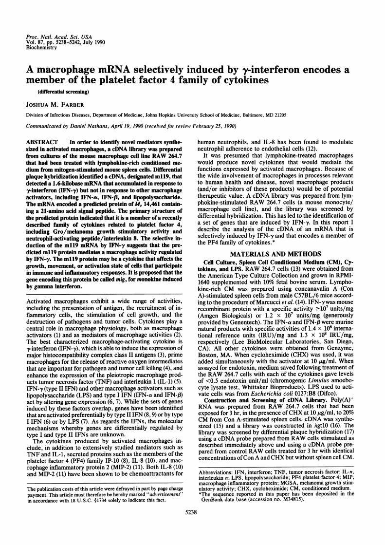
from Con A-stimulated mouse spleen cells. Screening bydifferential plaque hybridization led to the isolation of cDNAclones that identified 11 mRNA species that accumulated inthe RAW 264.7 cells following exposure to the spleen cellCM. As shown in Fig. 1A, one cDNA clone, 1.2 kilobases(kb) long and designated m119, hybridized to a major mRNAspecies of approximately 1.6 kb that was induced in RAWcells by the CM from Con A-stimulated spleen cells but notdetectable in control RAW cells even with long exposure ofthe autoradiograph. CHX did not inhibit expression of them119 mRNA, demonstrating that new protein synthesis was
C
Lru(N Ln
Hours o o~o~ - cl an v o co N"
xx
+
2z v4 z:.Y HN[
o H0 0x Z +
U) N N
* 4.4
* 2.4
* 1.m119 9
AHdou as l ase
3 Hours 6 Hours
HN 0
0 0
C)
CD
+ + +
H H H H H 1
h X [4 [4 IS [~~4 -4H IHH HH S
~~E00H~~~~~~ 0~0 CD0C0 0 00D C' 0D :0 0 0D00 0D 0D 0 0
H H- H H H H H
f -U)
mo0
.H-4 4J5) -H
E *0
0 0aNz
-44'if
5 H N H N H N f
0 )-
H-4 -
4
v IL W 4
HH H H H NZ*9a
Aldolase *lWoUU
CRG-2 t A
FIG. 1. RNA blot analysis of m119 mRNA in the RAW 264.7 cell line. All Northern analysis was done using total RNA (19) andformaldehyde/agarose denaturing gels as described (20). (A) m119 mRNA in RAW 264.7 cells treated with CM from Con A-stimulated spleencells. Twenty micrograms of total RNA was loaded per lane and hybridized sequentially to 32P-labeled m119 and aldolase A cDNA probes. TheRAW 264.7 cells had been treated for 3 hr as follows (from left to right): 20o CM from Con A-stimulated spleen cells plus CHX (10 kkg/ml);20% CM from unstimulated spleen cells plus Con A (10 ,ug/ml) and CHX (10 ,g/ml); 20%o CM from Con A-stimulated spleen cells; 20%o CMfrom unstimulated spleen cells plus Con A (10 ,ug/ml). In this and subsequent experiments, the 32P-labeled m119 cDNA probe was preparedby the nick-translation or random primer method from a 1.2-kb cDNA clone, and the aldolase A probe was prepared from a 1.4-kb cDNA clonekindly provided by A. Levy, L. Sanders, and D. Nathans. The RNA markers were obtained from Bethesda Research Laboratories and weredetected by hybridization to bacteriophage A DNA probe. In all experiments, autoradiography was done using an intensifying screen at -70'C,unless otherwise noted. The m119 signals shown here are from a 12-hr exposure. The aldolase signals are from a 24-hr exposure at room
temperature. The order of the lanes from the original blot has been changed for the figure. (B) m119 mRNA in RAW 264.7 cells treated withLPS and IFN-a, -a, and -y. Total RNA was prepared from RAW 264.7 cells treated for 3 or 6 hr with the stimuli as noted and was analyzedas in A. Duplicate 3- and 6-hr blots were hybridized to a Crg-2 cDNA probe. While polymyxin B (PB, 5 Ag/ml) significantly diminished theinduction of Crg-2 mRNA by LPS, it had no effect on the induction of m119 mRNA by IFN-y or on the induction of Crg-2 mRNA by the IFNs,suggesting that there were not trace amounts of contaminating LPS contributing to the IFN responses. The m119 signals are from a 4-dayexposure, and aldolase and Crg-2 signals from exposures at room temperature for 24 and 33 hr, respectively. U, units. (C) Time course of m119mRNA in RAW 264.7 cells in response to IFN-y. RNA was prepared from RAW 264.7 cells harvested after treatment with IFN-y (100 units/ml)for 0-24 hr, or after treatment with IFN-y (100 units/ml) and CHX (10 jug/ml) orCHX alone for 2 hr, or at 3 hr after mock manipulation withoutthe addition of IFN-y or CHX and was analyzed as in A. The m119 and aldolase signals are from 5-day and 12-hr exposures, respectively. Theorder of the lanes from the original blot has been changed for the figure.
ml19
Aldolase i oB
an+ H
H H
-i -4L CL
a 0a0' 0' 0
0 0 0) 0
CU, U) U) U)~"L. NlNl
m119
.A .014
Proc. Natl. Acad. Sci. USA 87 (1990)
not required for induction. In addition to the 1.6-kb band, theml19 probe identified prominent inducible species ofapprox-imately 3.2 and 1.8 kb. The 1.8-kb band was seen only in RNAfrom cells stimulated in the presence of CHX. Preliminaryevidence from analyses of multiple m119 cDNA clones andm119 genomic clones suggests that the 1.8-kb species is analternatively spliced m119 mRNA. The 3.2-kb species ispresumably a precursor of the 1.6-kb mRNA but has not beencharacterized. The level of aldolase A mRNA, shown as acontrol, was unaffected by exposure of RAW cells to CMfrom Con A-stimulated spleen cells.
Induction of m119 mRNA by Macrophage-Activating Fac-tors. To identify the lymphokines in the spleen cell CM thatmay have been responsible for inducing the m119 mRNA, aswell as to determine which other macrophage-activatingfactors were capable of enhancing expression of the m119gene, total RNA was prepared from RAW cells treated witha variety of agents for 3 and 6 hr and the RNA was analyzedby Northern blot. Significant induction of the m119 mRNAwas seen only with IFN-y. The results of the experimentswith IFN-a, IFN-,B, IFN-y and LPS are shown in Fig. 1B.The selective induction of the m119 mRNA in RAW 264.7cells by IFN-y was reproducible and was not due to aninability of RAW cells to respond to IFN-a, IFN-f3, or LPS.For example, shown in Fig. 1B is the induction by all the IFNsand LPS of crg-2, another of the genes that had beenidentified as responsive to the Con A-stimulated spleen cellCM. The sequence of the Crg-2 cDNA (34) suggests that theCrg-2 protein may be the murine homologue of IP-10, ahuman protein characterized as preferentially induced byIFN-y (8). In addition to the results with IFN-a, IFN-,l, andLPS, treatment of RAW cells with recombinant murineIL-la, recombinant murine IL-3, recombinant murine IL-4,recombinant murine granulocyte/macrophage colony-stimulating factor, recombinant human colony-stimulatingfactor 1, poly(I)-poly(C), the calcium ionophore A23187,phorbol 12-myristate 13-acetate, and the combination ofA23187 and phorbol myristate acetate all failed to induce them119 mRNA, nor was the m119 mRNA induced by serum(i.e., mitogen) stimulation of serum-starved BALB/c 3T3fibroblasts (data not shown).To determine the time course of induction of the m119
mRNA in response to IFN-y, RNA was prepared from RAWcells treated for 0-24 hr. The m119 mRNA was inducedrapidly and dramatically, as shown in Fig. 1C, reaching amaximum between 6 and 24 hr. As in the case of treatmentwith the spleen cell CM, induction by IFN-y did not requirenew protein synthesis. In fact, the addition of CHX led tosuperinduction of the m119 mRNA.While IFN-y led to an immediate and marked stimulation
of m119 gene expression, what is particularly striking is thedegree of specificity of induction of the m119 gene by IFN-y,exceptional even among those genes previously reported tobe induced by IFN-y preferentially (8, 9), which suggests aunique relationship between IFN-y and m119, both in termsof IFN--regulated gene expression and as regards the bio-logical actions of IFN-y.
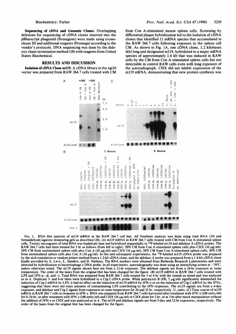
Induction of m119 mRNA in Peritoneal Macrophages. Todemonstrate the induction of m119 mRNA during the acti-vation of normal macrophages, m119 gene expression wasanalyzed in the adherent population from starch-elicitedperitoneal exudate cells obtained from both C3HeB/FeJ andBALB/cJ mice. As shown in Fig. 2, when these cells (>80%macrophages as determined by morphology) were exposed toCM from Con A-stimulated spleen cells, m119 mRNA wasinduced. Exposure of the C3HeB/FeJ cells to IFN-y likewiseled to the expression of the m119 gene. The electrophoreticmobilities of the m119 mRNA species from peritoneal cells ascompared to RAW cells were identical.
C3HeB/FeJ BALB/cJ
X< -Z-so.
mi~~~~~e~* 9*
< >:
O Eod aC±F.
d~ C, ZC'
FIG. 2. RNA blot analysis of m119 mRNA in peritoneal exudatecells. Peritoneal exudate cells were obtained as described (21).Samples were as follows (from left to right). From C3HeB/FeJ mice:1 mg of total RNA from cells exposed to 20% CM from ConA-stimulated spleen cells; 1 ug of total RNA from cells exposed to20% CM from unstimulated spleen cells plus Con A (10 /g/ml); 1 ugof total RNA from cells exposed to medium without additions; 0.8 jgof total RNA from cells treated with IFN-y [1000 units (u)/ml]; fromBALB/cJ mice: 0.4 yg of total RNA from cells exposed to 20% CMfrom Con A-stimulated spleen cells; 0.4 ug of total RNA from cellsexposed to 20%o CM from unstimulated spleen cells plus Con A (10pg/ml). The m119 and aldolase signals are from 2-week and 6-dayexposures, respectively.
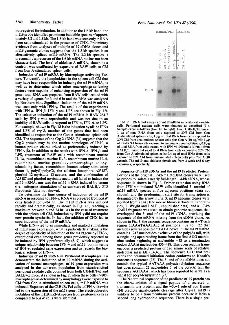
Sequence of m119 cDNAs and the mll9 Predicted Protein.Portions of the original 1.2-kb m119 cDNA clones were usedas probes to isolate a nearly full-length 1.4-kb cDNA, whosesequence is shown in Fig. 3. Primer extension using RNAfrom IFN-y-stimulated RAW cells identified 5' termini ofm119 mRNA species at five adjacent positions (data notshown), and the predominant start site for transcription isdesignated by the arrow in Fig. 3. ml19 genomic clones wereisolated from a BALB/c mouse library (Clontech Laborato-ries, T. Wright and J.M.F., unpublished data), and a 1.7-kbEcoRI fragment was used to obtain genomic sequence thatoverlapped the 5' end of the m119 cDNA, providing thesequence of the mRNA missing from the cDNA clone. Asshown in Fig. 3, the genomic sequence contains an A+T-richregion (TAAATAAATAT) at positions -32 to -22 thatincludes several possible "TATA boxes." The m119 mRNAcontains 1247 nucleotides exclusive of the poly(A) tail, witha single long open reading frame from the first AUG methio-nine codon beginning at nucleotide +58 to a terminationcodon UAA at nucleotides 436-438. This open reading frameencodes a predicted protein of 126 amino acids of relativemolecular mass (Mr) 14,461. The sequence GCC that pre-cedes the presumed initiation codon conforms to Kozak'sconsensus sequence (22). The 3' end of the cDNA does notcontain the typical AATAAA polyadenylylation sequencebut does contain, 22 nucleotides 5' of the poly(A) tail, thesequence AGTAAA, which has been reported to serve as asignal for polyadenylylation (23).The N-terminal sequence of the predicted ml19 protein has
the characteristics of a signal peptide of a secreted ortransmembrane protein, and the -3,-1 rule of von Heijne(24) predicts signal-peptide cleavage after Gly-21. m119 isunlikely to be a transmembrane protein because it lacks asecond long hydrophobic sequence. There is a single pre-
5240 Biochemistry: Farber
Proc. Natl. Acad. Sci. USA 87 (1990) 5241
14
GACTTCACTCCAACACAGTGACTCCACTCCAACACAGTGACTCAATAGAACTCAGCTCTGCC ATG AAG TCC GCT GTT
1 MET Lys Ser Ala Val
CTT TTC CTT TTG GGC ATC ATC TTC CTG GAG CAG TGT GGA GTT CGA GGAI6 Leu Phe Leu Leu Gly Ile Ile Phe Leu Glu Gln Cys Gly Val Arg Gly
ACC CTA GTG ATA AGG AAT GCA CGA TGC TCC TGC ATC AGC ACC AGC CGA22 Thr Leu Val Ile Arg Asn Ala Arg Cys Ser Cys Ile Ser Thr Ser Arg
GGC ACG ATC CAC TAC AAA TCC CTC AAA GAC CTC AAA CAG TTT GCC CCA38 Gly Thr Ile His Tyr Lys Ser Leu Lys Asp Leu Lys Gln Phe Ala Pro
AGC CCC AAT TGC AAC AAA ACT GAA ATC ATT GCT ACA CTG AAG AAC GGA54 Ser Pro Asn Cys Asn Lys Thr Glu Ile Ile Ala Thr Leu Lys Asn Gly
GAT CAA ACC TGC CTA GAT CCG GAC TCG GCA AAT GTG AAG AAG CTG ATG70 Asp Gln Thr Cys Leu Asp Pro Asp Ser Ala Asn Val Lys Lys Leu Met
AAA GAA TGG GAA AAG AAG ATC AAC CAA AAG AAA AAG CAA AAG AGG GGG86 Lys Glu Trp Glu Lys Lys Ile Asn Gln Lys Lys Lys Gln Lys Arg Gly
AAA AAA CAT CAA AAG AAC ATG AAA AAC AGA AAA CCC AAA ACA CCC CAA102 Lys Lys His Gln Lys Asn Met Lys Asn Arg Lys Pro Lys Thr Pro Gln
AGT CGT CGT CGT TCA AGG AAG ACT ACA TAA GAGACCATTACTTTACCAACAAG118 Ser Arg Arg Arg Ser Arg Lys Thr Thr *
CACCCTGAATCTTAATGGGTTTTAGATTGTACTGAAAAGCCTTCCCTGGCAGAGCAGCCTTTAATACATAGGCTTTTAATACATTAACTCAACTACAAAACATAAAGTGTTAATTTGAAATTATAACTAACTTTAGGAAGTTAATTGCAAAACTCCAATAGTAACAATTGCTAGAGGCAAAAACTCTGTGTTCTACACAGCCAACAAAATTTCATCACGCCCTTGAGCCTAGTCGTGATAACATCAGATCTGGGCAAGTGTCCCTTTCCTTCATAGCTATCCAATGCACAACAGCTGTCTGGCTTCCAGAGCCACACATTTGGCAGCCTCCGGAGACTTCTGAGGCTCACGTCACCAAGTCCCAGGCCTGTCTGTTTG
TAGAGCCCCTGCACACATTGTGTCTCAGAGATGGTGCTAATGGTTTTGGGGTTCTACAGTGGAGACCACCAGAGTT.GGCCTTCAGAACCTCCCACGTAGCTTTCGAGACCATGGGATTTCATTATTAACTTGATCCCATCTTCAGAGCTTATTCTAAGTTTGCCTCTTCAATAAAACTCTCCTAGAAGGTTGTGGCTGTAGCTTAGTGGCAGAACACTTGGTGTTGCAGGGACCAGGTCCTTCACTAACAGTGCAAAAACTTAACCAATTTAAAGAACATTTTCTGGCTACTCAAATTCTCTTAAATTTATTCCTGTTTCACAAGTAAACACTTCGCTGCTATCTA
72
120
FIG. 3. The cDNA sequence and inferred amino acid168 sequence of m119. Numbers at right margin refer to the
last nucleotide in each line (nucleotide 1 is marked by the216 arrow); numbers at left correspond to the first amino acid
in each line. The sequence from position -37 to +38 wasobtained from genomic DNA by using an oligonucleotide
264 primer complementary to m119 cDNA nucleotides 45-64, while sequence from position +20 to the poly(A) tailfollowing nucleotide 1247 was obtained from cDNA. A
312 TATA region at positions -32 to -22 is underlined. Avertical line marks the predicted cleavage site following
360 Gly-21. The underlined Asn-58 is a predicted N-linkedglycosylation site. The asterisk denotes the codon thatends the open reading frame. The possible poly(A)-
408 addition signal AGTAAA is underlined. AN representsthe poly(A) tail. Several m119 cDNA clones were miss-ing short stretches of nucleotides adjacent to the poly(A)
461 tail as compared with the cDNA sequence shown here.None of the cDNA clones, however, contained se-
524 quence in this region additional to that shown here, and587 the sequence shown here was found in two independent650 cDNA clones. The cDNA sequence displayed was ob-713 tained from a single 1.4-kb m119 cDNA clone. The776 nucleotide sequence of the open reading frame was839 confirmed by sequencing a second independent m1199025 cDNA clone. Both strands of the 1.4-kb cDNA clone9651028 were sequenced in their entirety with the exception of1091 the 31 nucleotides of the minus strand immediately1154 adjacent to the poly(A) [poly(T)] tail, and the sequence1217 of these 31 nucleotides from the minus strand was1248 obtained from a second m119 cDNA clone.
MKSAVLFLLGIIFLEOCGVVRGTLVI RNAR C S C ISTSRGTIHYKSLKDLKOFAPSPN C NKTE I I AT L-KNGDOT C LDPDSANVKK LMKEW EKK- m119
MNOSAAVIFCLiLLGLSGTOGIPLARTVR C N C I HI DDGPVRMRA I GK LEI I PAS LS C PR VE1lATMKKNDEOR C LNP ES KT I KNLMK A FS OK mCRG-2MNOTAI LICCLIFLTLSGIOGVPLSRTVR C T C ISISNQPVNPRSLEKLErI PASOF :C PRV1E IATMKKKGEKR C LNPESKAIKNLLK AVSKE- hiP-10
MIPATRSLLCAALLLLATSRLATGAPiANELR C C C LOTMAG-IHLKNIQSLKVLPSGPH C TOTEVIATL-KNGREA C L~PEAPLVOKIVOKMLKG- mKC/Gro-PSNPRLLRVALLLLLLVAAGRRAAGASVATELR C O C LOTLOG- HPKNI OSVNVKSPGPH C AOTEVJATL-KNGRKA C LNPASPIVKKIIEKMLNS- hGro/MGSRMTSK LAVALLAAFLISAALCEAVLPRSAKE L C C IKTYSKPFHPKFIKELRVIESGPH C ANTE1IVKL-SDGREL C LDPKIENWVORVVEKFLKR- hNfP/IL-8
N L A K G K E E S L D S D L Y A E L R C M C IKTTSG-HPKNQSLEVIGKGTH C NOVEVIATL-KDGR KI C LDPDAPRIKKIVOKKLAG- hCTRP I I IEAEEDGDLO C L C VKT-TSQVRPRHiTSL7VIKAGPH C PTAOL ATL-KNGRK I C DLODAPLYKK I IKKLLES hPf 4
MNGKLGAVLALLLVSAALSOGRTLVKMGNELR C O C IISTHSKFIHPKSIODVKLTPSGPH C K NV E I I ATL-KDGREV C LDPTAPWVO L I VK A LM AK- c9E3
HOMOLOGY TO ml 19
mCRG-2
hIP-10
mKC/Gro
hGro/MGSAhNAP/IL-8
hCTAP 111
hPF4
mMIP-2
c9E3
TOTAL
Values (T&o3 1/98(32)
32/98(33)
31/94(33)
33/95(35)27/95(28)24/86(28)18,71(25)4/32(13)
34/103(33)
CYS WINDOW
Values (%L
16/45(36)
18/45(40)
22/44(50)
21/44(48)
17/44(39)
18/44(41)
13/44(30)
3/22(14)
22/44(50)
FIG. 4. Amino acid sequences of m119 and closely related members of the PF4 family. KC is a platelet-derived growth factor-induced proteinand is the mouse homologue of the human Gro/MGSA (melanoma growth stimulatory activity) (26). Connective tissue-activating protein (CTAPIII) is the precursor of ,B-thromboglobulin (27), and is stored in the a granules of human platelets. 9E3 is a chicken protein induced followingtransformation with Rous sarcoma virus (28). The other proteins are described in the text. Sequences were aligned according to the fourconserved cysteine residues. Residues shared between m119 and another family member are shaded. Dashes terminate truncated sequences.Shown below the sequences are the numbers and percents of identical residues found in comparisons between m119 and each of the membersof the family, both over their total lengths as far as comparable (Total) and within the most highly related region bounded by the conservedcysteines (Cys window) according to Oquendo et al. (26). The lowercase prefixes refer to the species of origin: m, mouse; h, human; and c,chicken. References for the sequences are as follows: mCRG-2 (34); hIP-10 (8); mKC (26); hGro/MGSA (29); hNAP (neutrophil-activatingpeptide)/IL-8 (10); hCTAP III (27); hPF4 (30); c9E3 (28).
TTTCC AAATA'rGATCCCCAAGAACATGCTCTCIAAAGACATTCTCG
Biochemistry: Farber
Proc. Natl. Acad. Sci. USA 87 (1990)
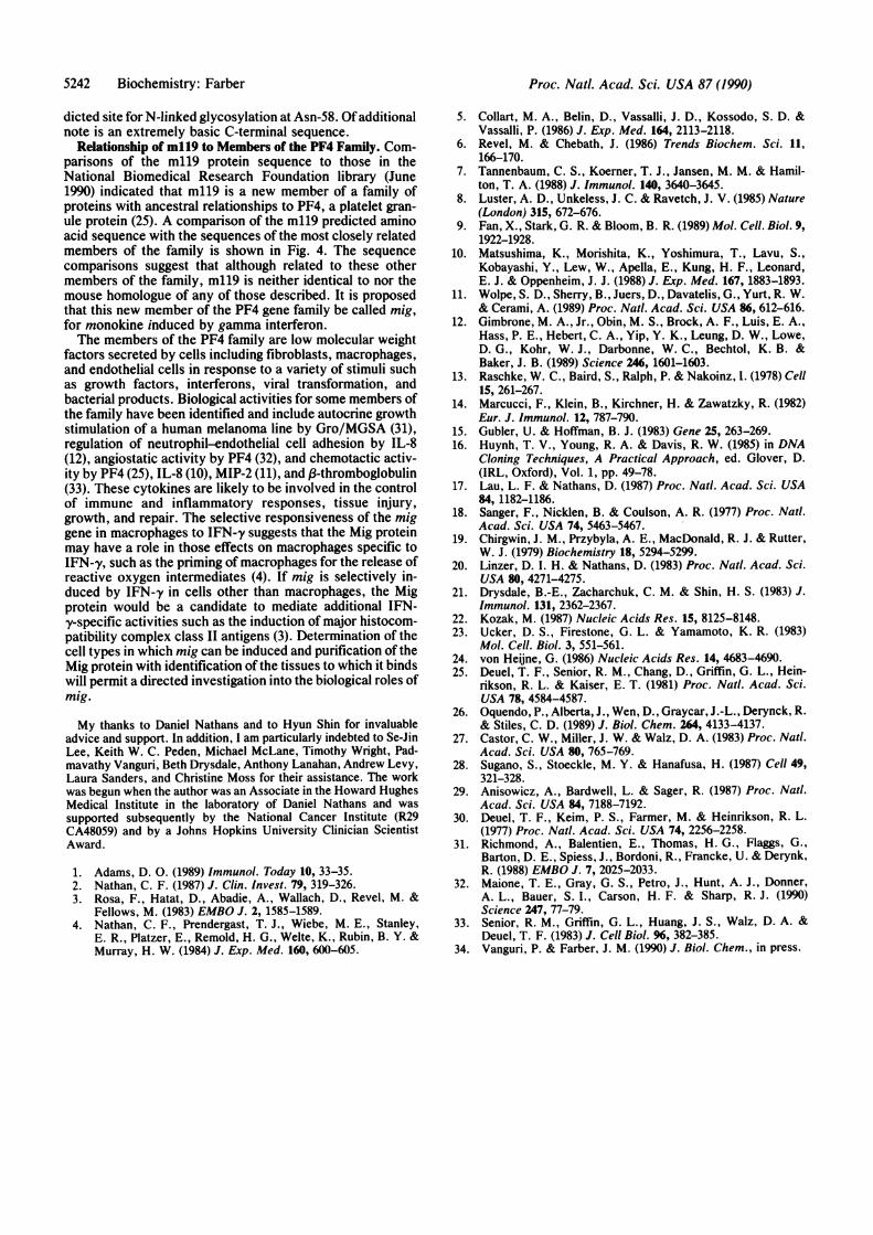
dicted site for N-linked glycosylation at Asn-58. Of additionalnote is an extremely basic C-terminal sequence.
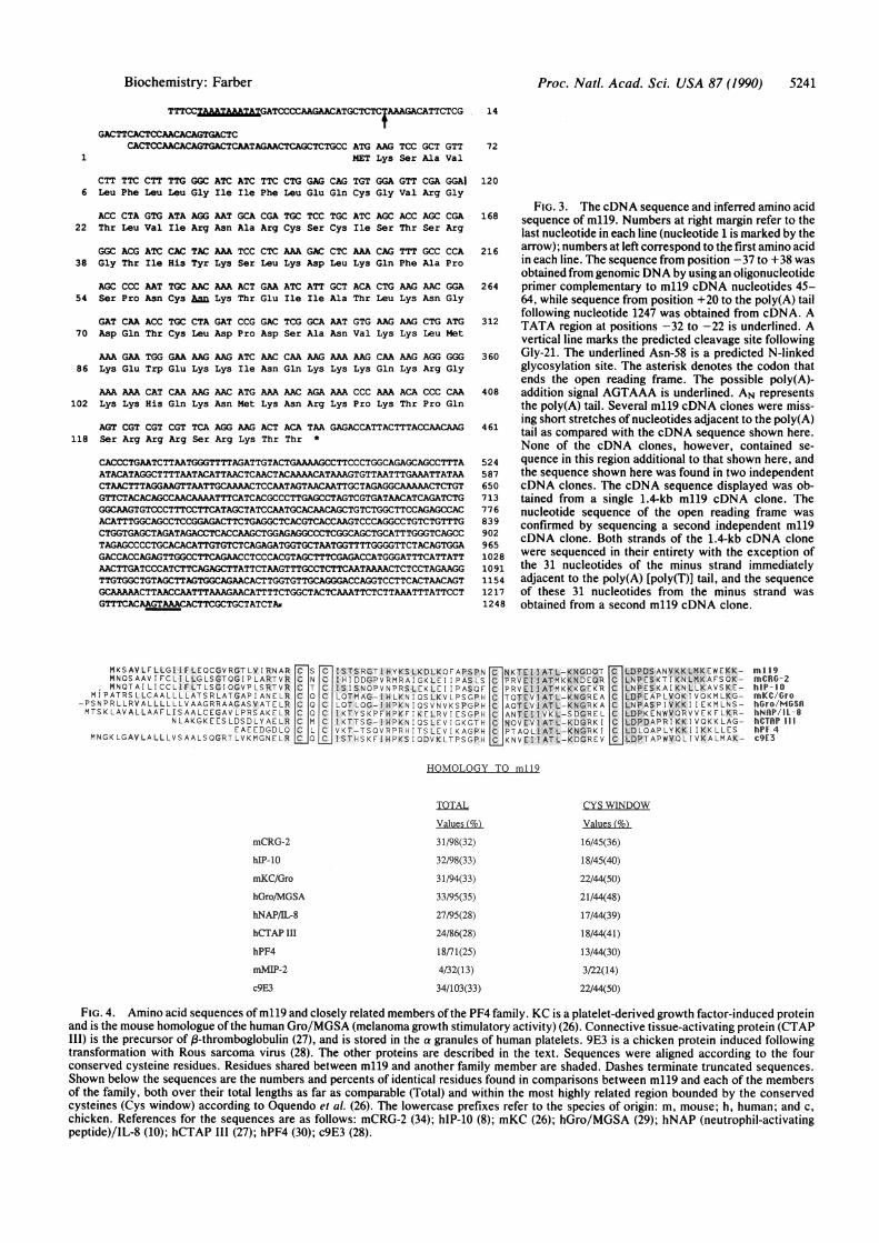
Relationship of m119 to Members of the PF4 Family. Com-parisons of the m119 protein sequence to those in theNational Biomedical Research Foundation library (June1990) indicated that m119 is a new member of a family ofproteins with ancestral relationships to PF4, a platelet gran-ule protein (25). A comparison of the m119 predicted aminoacid sequence with the sequences of the most closely relatedmembers of the family is shown in Fig. 4. The sequencecomparisons suggest that although related to these othermembers of the family, m119 is neither identical to nor themouse homologue of any of those described. It is proposedthat this new member of the PF4 gene family be called mig,for monokine induced by gamma interferon.The members of the PF4 family are low molecular weight
factors secreted by cells including fibroblasts, macrophages,and endothelial cells in response to a variety of stimuli suchas growth factors, interferons, viral transformation, andbacterial products. Biological activities for some members ofthe family have been identified and include autocrine growthstimulation of a human melanoma line by Gro/MGSA (31),regulation of neutrophil-endothelial cell adhesion by IL-8(12), angiostatic activity-by PF4 (32), and chemotactic activ-ity by PF4 (25), IL-8 (10), MIP-2 (11), and P-thromboglobulin(33). These cytokines are likely to be involved in the controlof immune and inflammatory responses, tissue injury,growth, and repair. The selective responsiveness of the miggene in macrophages to IFN-y suggests that the Mig proteinmay have a role in those effects on macrophages specific toIFN-y, such as the priming of macrophages for the release ofreactive oxygen intermediates (4). If mig is selectively in-duced by IFN-y in cells other than macrophages, the Migprotein would be a candidate to mediate additional IFN-y-specific activities such as the induction of major histocom-patibility complex class II antigens (3). Determination of thecell types in which mig can be induced and purification of theMig protein with identification of the tissues to which it bindswill permit a directed investigation into the biological roles ofmig.
My thanks to Daniel Nathans and to Hyun Shin for invaluableadvice and support. In addition, I am particularly indebted to Se-JinLee, Keith W. C. Peden, Michael McLane, Timothy Wright, Pad-mavathy Vanguri, Beth Drysdale, Anthony Lanahan, Andrew Levy,Laura Sanders, and Christine Moss for their assistance. The workwas begun when the author was an Associate in the Howard HughesMedical Institute in the laboratory of Daniel Nathans and wassupported subsequently by the National Cancer Institute (R29CA48059) and by a Johns Hopkins University Clinician ScientistAward.
1. Adams, D. 0. (1989) Immunol. Today 10, 33-35.2. Nathan, C. F. (1987) J. Clin. Invest. 79, 319-326.3. Rosa, F., Hatat, D., Abadie, A., Wallach, D., Revel, M. &
Fellows, M. (1983) EMBO J. 2, 1585-1589.4. Nathan, C. F., Prendergast, T. J., Wiebe, M. E., Stanley,
E. R., Platzer, E., Remold, H. G., Welte, K., Rubin, B. Y. &Murray, H. W. (1984) J. Exp. Med. 160, 600-605.
5. Collart, M. A., Belin, D., Vassalli, J. D., Kossodo, S. D. &Vassalli, P. (1986) J. Exp. Med. 164, 2113-2118.
6. Revel, M. & Chebath, J. (1986) Trends Biochem. Sci. 11,166-170.
7. Tannenbaum, C. S., Koerner, T. J., Jansen, M. M. & Hamil-ton, T. A. (1988) J. Immunol. 140, 3640-3645.
8. Luster, A. D., Unkeless, J. C. & Ravetch, J. V. (1985) Nature(London) 315, 672-676.
9. Fan, X., Stark, G. R. & Bloom, B. R. (1989) Mol. Cell. Biol. 9,1922-1928.
10. Matsushima, K., Morishita, K., Yoshimura, T., Lavu, S.,Kobayashi, Y., Lew, W., Apella, E., Kung, H. F., Leonard,E. J. & Oppenheim, J. J. (1988) J. Exp. Med. 167, 1883-1893.
11. Wolpe, S. D., Sherry, B., Juers, D., Davatelis, G., Yurt, R. W.& Cerami, A. (1989) Proc. NatI. Acad. Sci. USA 86, 612-616.
12. Gimbrone, M. A., Jr., Obin, M. S., Brock, A. F., Luis, E. A.,Hass, P. E., Hebert, C. A., Yip, Y. K., Leung, D. W., Lowe,D. G., Kohr, W. J., Darbonne, W. C., Bechtol, K. B. &Baker, J. B. (1989) Science 246, 1601-1603.
13. Raschke, W. C., Baird, S., Ralph, P. & Nakoinz, 1. (1978) Cell15, 261-267.
14. Marcucci, F., Klein, B., Kirchner, H. & Zawatzky, R. (1982)Eur. J. Immunol. 12, 787-790.
15. Gubler, U. & Hoffman, B. J. (1983) Gene 25, 263-269.16. Huynh, T. V., Young, R. A. & Davis, R. W. (1985) in DNA
Cloning Techniques, A Practical Approach, ed. Glover, D.(IRL, Oxford), Vol. 1, pp. 49-78.
17. Lau, L. F. & Nathans, D. (1987) Proc. Natl. Acad. Sci. USA84, 1182-1186.
18. Sanger, F., Nicklen, B. & Coulson, A. R. (1977) Proc. Natl.Acad. Sci. USA 74, 5463-5467.
19. Chirgwin, J. M., Przybyla, A. E., MacDonald, R. J. & Rutter,W. J. (1979) Biochemistry 18, 5294-5299.
20. Linzer, D. I. H. & Nathans, D. (1983) Proc. Natl. Acad. Sci.USA 80, 4271-4275.
21. Drysdale, B.-E., Zacharchuk, C. M. & Shin, H. S. (1983) J.Immunol. 131, 2362-2367.
22. Kozak, M. (1987) Nucleic Acids Res. 15, 8125-8148.23. Ucker, D. S., Firestone, G. L. & Yamamoto, K. R. (1983)
Mol. Cell. Biol. 3, 551-561.24. von Heijne, G. (1986) Nucleic Acids Res. 14, 4683-4690.25. Deuel, T. F., Senior, R. M., Chang, D., Griffin, G. L., Hein-
rikson, R. L. & Kaiser, E. T. (1981) Proc. Natl. Acad. Sci.USA 78, 4584-4587.
26. Oquendo, P., Alberta, J., Wen, D., Graycar, J.-L., Derynck, R.& Stiles, C. D. (1989) J. Biol. Chem. 264, 4133-4137.
27. Castor, C. W., Miller, J. W. & Walz, D. A. (1983) Proc. Natl.Acad. Sci. USA 80, 765-769.
28. Sugano, S., Stoeckle, M. Y. & Hanafusa, H. (1987) Cell 49,321-328.
29. Anisowicz, A., Bardwell, L. & Sager, R. (1987) Proc. Natl.Acad. Sci. USA 84, 7188-7192.
30. Deuel, T. F., Keim, P. S., Farmer, M. & Heinrikson, R. L.(1977) Proc. Natl. Acad. Sci. USA 74, 2256-2258.
31. Richmond, A., Balentien, E., Thomas, H. G., Flaggs, G.,Barton, D. E., Spiess, J., Bordoni, R., Francke, U. & Derynk,R. (1988) EMBO J. 7, 2025-2033.
32. Maione, T. E., Gray, G. S., Petro, J., Hunt, A. J., Donner,A. L., Bauer, S. I., Carson, H. F. & Sharp, R. J. (1990)Science 247, 77-79.
33. Senior, R. M., Griffin, G. L., Huang, J. S., Walz, D. A. &Deuel, T. F. (1983) J. Cell Biol. 96, 382-385.
34. Vanguri, P. & Farber, J. M. (1990) J. Biol. Chem., in press.
5242 Biochemistry: Farber