Embed Size (px)
Citation preview
A Mismatch-Based Model for Memory Reconsolidationand Extinction in Attractor NetworksRemus Osan1,2,3., Adriano B. L. Tort4,5., Olavo B. Amaral6*
1 Center for Neuroscience, Boston University, Boston, Massachusetts, United States of America, 2 Center for Biodynamics, Boston University, Boston, Massachusetts,
United States of America, 3 Department of Mathematics and Statistics, Boston University, Boston, Massachusetts, United States of America, 4 Brain Institute, Federal
University of Rio Grande do Norte, Natal, Rio Grande do Norte, Brazil, 5 Edmond and Lily Safra International Institute of Neuroscience of Natal, Natal, Rio Grande do Norte,
Brazil, 6 Institute of Medical Biochemistry, Federal University of Rio de Janeiro, Rio de Janeiro, Brazil
Abstract
The processes of memory reconsolidation and extinction have received increasing attention in recent experimentalresearch, as their potential clinical applications begin to be uncovered. A number of studies suggest that amnestic drugsinjected after reexposure to a learning context can disrupt either of the two processes, depending on the behavioralprotocol employed. Hypothesizing that reconsolidation represents updating of a memory trace in the hippocampus, whileextinction represents formation of a new trace, we have built a neural network model in which either simple retrieval,reconsolidation or extinction of a stored attractor can occur upon contextual reexposure, depending on the similaritybetween the representations of the original learning and reexposure sessions. This is achieved by assuming thatindependent mechanisms mediate Hebbian-like synaptic strengthening and mismatch-driven labilization of synapticchanges, with protein synthesis inhibition preferentially affecting the former. Our framework provides a unified mechanisticexplanation for experimental data showing (a) the effect of reexposure duration on the occurrence of reconsolidation orextinction and (b) the requirement of memory updating during reexposure to drive reconsolidation.
Citation: Osan R, Tort ABL, Amaral OB (2011) A Mismatch-Based Model for Memory Reconsolidation and Extinction in Attractor Networks. PLoS ONE 6(8): e23113.doi:10.1371/journal.pone.0023113
Editor: Gennady Cymbalyuk, Georgia State University, United States of America
Received June 10, 2011; Accepted July 6, 2011; Published August 3, 2011
Copyright: � 2011 Osan et al. This is an open-access article distributed under the terms of the Creative Commons Attribution License, which permitsunrestricted use, distribution, and reproduction in any medium, provided the original author and source are credited.
Funding: This work was supported by the Center for Neuroscience, Boston University, USA (RO), Conselho Nacional de Desenvolvimento Cientıfico eTecnologico, Brazil (ABLT and OBA), and Fundacao de Amparo a Pesquisa do Estado do Rio de Janeiro, Brazil (OBA). The funders had no role in study design, datacollection and analysis, decision to publish, or preparation of the manuscript.
Competing Interests: The authors have declared that no competing interests exist.
* E-mail: [email protected]
. These authors contributed equally to this work.
Introduction
The concept of memory reconsolidation was proposed more
than 40 years ago [1], but has recently regained considerable
attention in the literature [2]. Most of the data in favor of the
reconsolidation hypothesis has stemmed from the finding that
pharmacological agents can induce amnesia when administered
after reexposure to a context in which a memory was originally
learned [3,4]. This finding initially sparked controversy, as studies
of memory extinction had traditionally found a directly opposite
effect: namely, that the same drugs could block extinction,
therefore preserving the original memory [5,6].
A number of studies later tried to reconcile these apparently
paradoxical effects, showing that both phenomena are possible
outcomes of nonreinforced reexposure, and that the occurrence of
one or another depends on the experimental protocol: in conditions
in which extinction is observed in controls, amnestic drugs block
extinction and preserve the original memory; meanwhile, in
conditions causing no extinction, the same drugs cause amnesia,
putatively due to disruption of reconsolidation [7,8]. These results
led to the proposition that the ‘‘dominant trace’’ after reexposure is
the one made labile to amnestic agents [7].
The fact that not all studies could demonstrate reconsolidation
by post-reexposure interventions [9,10] also suggested that there
are ‘‘boundary conditions’’ which are necessary for trace
labilization [11,12]. One of these conditions has been proposed
to be the occurrence of memory updating during reexposure [13],
due to studies in which simple reexposure in the absence of new
information did not lead to reconsolidation, as shown by the lack
of effect of amnestic drugs [14,15]. Similarly, other studies have
shown that very short reexposure trials were also associated with
no effect of these drugs [8,16].
Understanding what determines the occurrence of these
phenomena is important, as modulations of both reconsolidation
and extinction have begun to be tested as therapeutic strategies in
anxiety disorders such as PTSD [17] and phobias [18]. To date,
no mechanism has been postulated to explain how changes in a
single variable such as reexposure duration can lead to these
different outcomes. Since the same drugs can block (or enhance)
both reconsolidation and extinction, however, it is feasible to
hypothesize that the differences between these processes depend
not only on their molecular features, but also – and perhaps
mainly – on their network properties.
Attractor network models have provided a general framework
through which information storage can be modeled in connected
networks, and the existence of attractors in brain structures such as
the hippocampus [19,20], neocortex [21] and olfactory bulb [22]
has received experimental support from electrophysiological
studies. By assuming that memory processing is based on attractor
dynamics, and that updating of a memory trace occurs based on
PLoS ONE | www.plosone.org 1 August 2011 | Volume 6 | Issue 8 | e23113
mismatch-induced synaptic changes, we propose a model which
can explain how contextual reexposure may lead to reconsolida-
tion or extinction. In this framework, the dominant process
occurring after reexposure depends on the degree of mismatch
between the animal’s current representation of a context and a
previously stored attractor. The model accounts for the different
effects of amnestic agents on reconsolidation and extinction, as
well as for the requirement of dissimilarities between the learning
and reexposure sessions for reconsolidation to occur.
Results
Model FrameworkTo study the processes described above computationally, we use
an adaptation of the classical attractor network model [23,24].
These highly connected neural networks, which can store memories
as neuronal activation patterns based on Hebbian modifications of
synaptic weights, have been proposed to be simple correlates of
autoassociative networks such as the one believed to exist in region
CA3 of the hippocampus [25,26]. Attractor-like functioning has
been shown to be compatible with both firing-rate and spike-time
dependent plasticity in spiking neuronal networks [27,28]. For the
sake of simplicity, however, and for better correlation with previous
models dealing with the effect of mismatch and memory
representations (e.g. [29]), we use the classical firing rate
implementation, which remains a useful tool for studying emergent
network properties related to learning and memory.
Neuronal activities in the attractor network (meant to represent
a hippocampal auto-associative storage network in our model) are
determined by equation (1):
tdui
dt~{uiz
1
21z tanh
XN
j~1wijujzIi
� �� �ð1Þ
where t is the neural time constant and ui represents the level of
activation of neuron i in a network comprised by N neuronal units,
varying continuously from 0 to 1 for each neuron, and not from
21 to 1 as in classical formulations (see Methods). This can reflect
the firing rate and connectivity of neurons in a more realistic way,
as it solves a series of biologically unfeasible features of the original
formulation, including (a) the requirement of symmetric connec-
tions between neurons, (b) the strengthening of connections
between neurons with low activity and (c) the occasional retrieval
of mirror patterns diametrically opposite to those originally
learned. The term {ui causes the activation level to decay
towards 0, while the termPN
j~1 wijuj represents the influence of
presynaptic neurons within the attractor network, weighed by the
strength of the synaptic connections wij . Finally, the term Ii
represents synaptic influences from cue inputs. These cue inputs
are thought to represent cortical afferents providing the hippo-
campus with the animal’s current representation of its environ-
ment, based both on external (i.e. sensory input) and internal
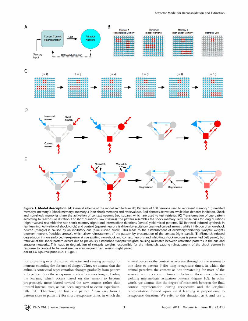
information (i.e. retrieved memories) (Figure 1A). The interplay
between sensory information and hippocampal feedback is not
modeled explicitly; instead, the presented cues will be modeled as
relying more on external or internal input depending on
behavioral parameters (see below).
Learning in the model occurs through presentation of an
activation pattern by the cue inputs, which leads to changes in the
synaptic weight matrix W~ wij
� �, as determined by equation (2):
DW~{cWzHLPzMID ð2Þwhere 0vcv1 is a time-dependent synaptic decay factor [30,31],
and HLP and MID stand for Hebbian Learning Plasticity and
Mismatch-Induced Degradation, respectively, expressed in array form.
Both of these matrices are dependent on the steady state pattern of
neuronal activation that is reached by the network upon cue
presentation (Eq. (1)). The precise meaning of the MID term and
its equation will be explained below; for now, we will mention that
all entries in theMID matrix are related to mismatch between the
cue and a retrieved attractor and, as such, equal zero during initial
learning. The HLP term represents a modified Hebbian learning
factor (see Methods), and it is given by
HLP~S(uT � u){S((1{u)T � u) ð3Þ
where the vector u~(u1,:::,uN ) is the steady state of the network
and S§0 corresponds to a factor representing a sum of the
biochemical requirements for Hebbian synaptic plasticity, such as
receptor activation, intracellular signaling and protein synthesis.
Thus, if two neurons are maximally active (ui = uj = 1), the uiRuj
connection gets reinforced by S; if the presynaptic neuron ui is
active and the postsynaptic neuron uj is silent (uj = 0), then the
connection uiRuj changes by 2S. If ui is silent, nothing happens to
the connection uiRuj. Intermediate values of ui and uj lead to
intermediate effects of these factors. The value of S is what is
modified in simulations studying the influence exerted by
pharmacological agents on initial memory consolidation, reconso-
lidation and extinction. The effect of protein synthesis inhibition
by anisomycin, for instance, is modeled by setting S to 0, thereby
blocking Hebbian plasticity.
Training, reexposure and testing in a simple one-trial learning
task, such as contextual fear conditioning, are modeled by setting
up appropriate cue patterns. Training sessions consist of
presentation of one of three complete patterns (Figure 1B): pattern
1, representing a memory which is unrelated to fear conditioning;
pattern 2, representing fear conditioning training, in which a set of
neurons representing the context is activated along with another
set of neurons representing the presence of danger or an aversive
stimulus (i.e. an electric shock); and pattern 3, representing fear
conditioning extinction, in which the same context neurons are
activated along with a different set of neurons representing
absence of danger. The use of a specific pattern to represent
extinction is motivated by experimental data suggesting that the
extinction process represents the active learning of a new memory
trace [5,6], as well as by studies suggesting that it may be encoded
by neuronal populations which are at least partially distinct from
those involved in the original learning [32,33].
Memory retrieval is tested by presenting the cue pattern that
represents the context (Figure 1B), and observing the attractor to
which the network evolves. We model the animal’s behavioral
response by assuming that retrieval of pattern 2 leads to a far
greater degree of conditioned behavior in response to danger than
when the network reaches another attractor (see Methods and
Figure S1). In analogy to the experimental literature, we refer to
the fear conditioned response as ‘‘freezing’’, and use the
percentage of time spent freezing during the test as a measure of
memory in the task.
Nonreinforced reexposure to the context is modeled similarly to
training, except that the cue pattern in this case is a mix of patterns
2 and 3. This is based on the assumption that, upon reexposure to
the context in which fear learning occurred, the memory network
will initially retrieve the aversive memory, with feedback from the
hippocampus signaling the activation of neurons representing
danger in the animal’s contextual representation. Later within the
trial, however, the absence of shock will lead the animal to start
perceiving the context as non-threatening, with sensory informa-
Attractor Model for Reconsolidation and Extinction
PLoS ONE | www.plosone.org 2 August 2011 | Volume 6 | Issue 8 | e23113
tion prevailing over the stored attractor and causing activation of
neurons encoding the absence of danger. Thus, we assume that the
animal’s contextual representation changes gradually from pattern
2 to pattern 3 as the reexposure session becomes longer, leading
the learning which occurs based on this session to become
progressively more biased toward the new context rather than
toward internal cues, as has been suggested to occur experimen-
tally [34]. Therefore, the final cue pattern I can vary from a
pattern close to pattern 2 (for short reexposure times, in which the
animal perceives the context as aversive throughout the session) to
one close to pattern 3 (for long reexposure times, in which the
animal perceives the context as non-threatening for most of the
session), with reexposure times in between these two extremes
yielding intermediate activation patterns (Figure 1C). In other
words, we assume that the degree of mismatch between the final
context representation during reexposure and the original
representation formed upon initial learning is proportional to
reexposure duration. We refer to this duration as t, and use a
Figure 1. Model description. (A) General scheme of the model architecture. (B) Patterns of 100 neurons used to represent memory 1 (unrelatedmemory), memory 2 (shock memory), memory 3 (non-shock memory) and retrieval cue. Red denotes activation, while blue denotes inhibition. Shockand non-shock memories share the activation of context neurons (red square), which are used to test retrieval. (C) Transformation of cue patternaccording to reexposure duration. For short durations (low t values), the pattern resembles the shock memory (left), while cues for long durations(high t values) resemble the non-shock memory (right) and intermediate durations (center) yield mixed patterns. (D) Retrieval-induced synthesis infear learning. Activation of shock (circle) and context (square) neurons is driven by excitatory cues (red curved arrows), while inhibition of a non-shockneuron (triangle) is caused by an inhibitory cue (blue curved arrow). This leads to the establishment of excitatory/inhibitory synaptic weightsbetween neurons (red/blue arrows), which allow reinstatement of the pattern by presentation of the context (right panel). (E) Mismatch-induceddegradation in nonreinforced reexposure. A cue exciting non-shock and context neurons and inhibiting shock neurons is presented (left panel), butretrieval of the shock pattern occurs due to previously established synaptic weights, causing mismatch between activation patterns in the cue andattractor networks. This leads to degradation of synaptic weights responsible for the mismatch, causing reinstatement of the shock pattern inresponse to context to be weakened in a subsequent test session (right panel).doi:10.1371/journal.pone.0023113.g001
Attractor Model for Reconsolidation and Extinction
PLoS ONE | www.plosone.org 3 August 2011 | Volume 6 | Issue 8 | e23113
transformation from pattern 2 to pattern 3 which is a function of t
to create the cue patterns representing different durations of
reexposure (see Methods).
As in the initial training session, synaptic weights are updated
after the reexposure session following equation (2), and the
Hebbian learning rule acts by means of the HLP term (Figure 1D).
However, the existence of a previously stored attractor for the
context in the reexposure session can lead the memory network to
retrieve an attractor which is different from the cue pattern
employed, leading to mismatch between the two patterns. We thus
introduce a memory updating system which degrades synaptic
weights between the different sets of neurons responsible for this
mismatch, reducing the strength of connections which cause
disagreement with the new cue pattern (Figure 1E). This effect is
modeled by the term MID in (2), which follows the equation:
MID~D(mT � u) ð4Þ
where the degradation factor D represents biochemical requirements
for mismatch-induced updating of synaptic connections – which
are thought to involve, among other things, protein degradation
[35,36] – and m~Inorm{u is the mismatch vector (where Inorm is a
normalized cue vector varying between 0 and 1). Note that when
the retrieved attractor is equal to the cue input (as during initial
learning) there is no mismatch, since u~Inorm in these cases,
leading all entries in vector m to equal zero.
Although the biochemical elements in the model are an obvious
simplification (i.e. synaptic plasticity is certainly more complex
than a synthesis/degradation balance, and involves many other
mechanisms), there is much evidence to suggest that protein
synthesis is a defining factor in long-term memory consolidation
[37], as well as some evidence [35,36] to suggest that protein
degradation through the ubiquitin-proteasome system is involved
in trace labilization during reconsolidation. Therefore, we focus on
these two parameters in our simulations of pharmacological
experiments. The synaptic weight changes induced by these
processes are modeled as occurring during the post-reexposure
period, based upon the activation state reached during the
reexposure session (which presumably sets in motion the
biochemical cascades and transcriptional information which will
drive the protein changes occurring later). Pharmacological
interventions after reexposure are thus modeled as changing
either S or D during the synaptic weight updating process caused
by the reexposure session (Eq. (2)), and the effects of these
interventions are measured by evaluating subsequent retrieval in
response to the cue representing the context.
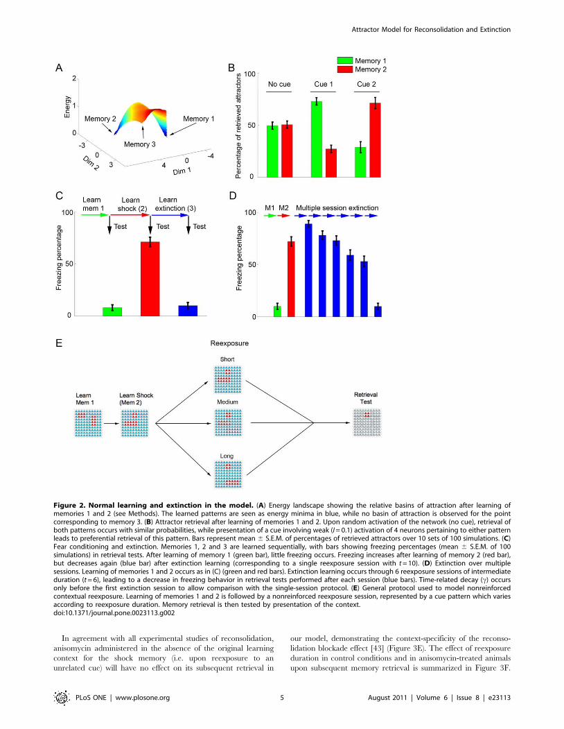
Learning and extinction in the modelFigure 2 shows normal learning in the model. We first present
the network with two orthogonal patterns with no overlapping
active neurons, one at a time: pattern 1 (an unrelated memory)
and pattern 2 (the shock memory). Presentation of these patterns
leads to the formation of local energy minima corresponding to the
two memories (Figure 2A). Retrieval of either one can occur upon
random network initialization, while presentation of a partial cue
for either of the two patterns biases retrieval towards the
corresponding attractor (Figure 2B). Although we perform our
simulations using only 3 patterns in a small network of 100
neurons, our network framework is capable of storing larger
numbers of memories, with the absolute capacity depending on
parameters such as network size and on the number of active
neurons in each memory pattern, as has been shown to be the case
for other attractor-based models [38,39]. Estimations of storage
capacities for different network sizes and sparseness values are
shown in Figure S2, demonstrating that the model can store a
reasonable number of memories, provided the number of neurons
is large enough and memory patterns are reasonably sparse.
Similarly to what occurs behaviorally, extinction in the model
(represented as learning of pattern 3) can occur either in a single
retrieval session with a cue similar to pattern 3 (i.e. a high t value,
representing a long retrieval session) (Figure 2C) or in multiple
retrieval sessions with intermediate cues (representing multiple
short sessions in which pattern 2 and 3 are both reflected in the
cue) (Figure 2D). Extinction over multiple sessions occurs due to
gradual weakening of the shock attractor, which is repeatedly
retrieved in the presence of mismatch and thus undergoes
degradation, allowing learning of a new attractor (the extinction
memory) to occur eventually. This is in contrast with single session
extinction, in which prompt learning of the extinction memory
prevents retrieval of the original attractor and weakening of the
shock representation (see Figure S3).
The sequence of patterns used to model learning followed by
nonreinforced reexposure to the context, which will be used
throughout the simulations concerning the effects of anisomycin, is
shown in Figure 2E. Learning of patterns 1 and 2 is followed by a
nonreinforced reexposure session of variable duration (modeled by
changing the value of t), and retrieval is later measured through
presentation of the context cue.
Effects of anisomycin on different reexposure protocolsFigure 3 shows the effects of anisomycin administration (i.e.
setting S to 0) in different learning and reexposure protocols.
During initial learning, blockade of protein synthesis inhibits
Hebbian modifications and prevents formation of the shock
memory (Figure 3A), a finding which is consistent with the effect of
anisomycin in various behavioral paradigms of learning, including
fear conditioning [40].
In Figures 3B to 3E, learning of the shock memory occurs
normally (S = 0.8), and anisomycin administration is modeled in
various nonreinforced reexposure protocols with different contex-
tual cues (see Figures 1C and 2E). In very short reexposure trials,
in which the shock memory is retrieved over the full course of the
retrieval session and dominates the contextual representation (i.e.
low t values), anisomycin will have little effect on subsequent
retrieval of that memory, as the degree of mismatch-induced
degradation will be small even in the absence of protein synthesis
(Figure 3B). This is compatible with the ‘‘simple retrieval’’
condition observed with short reexposure durations in experimen-
tal studies [8,16,41].
In reexposure trials with intermediate durations (i.e. ‘‘reconso-
lidation’’ conditions), inhibition of protein synthesis starts to exert
a significant amnestic effect on subsequent retrieval trials
(Figure 3C), as Hebbian learning is blocked and cannot
compensate for mismatch-induced degradation of the shock
memory. This effect is analogous to the reconsolidation blockade
effect described in various experimental studies [3,4]. Finally, in
long reexposure trials, in which the cue pattern will be distinct
enough from pattern 2 to prevent its retrieval, extinction (i.e.
formation of a new attractor representing pattern 3) will occur
after the reexposure session in control conditions. The burning of a
new attractor in the network will also prevent mismatch
degradation of the shock representation; in this case, therefore,
anisomycin will block formation of the extinction memory, but will
not affect the existing shock attractor, leading to preservation of
the shock memory in treated animals (Figure 3D). Such results
closely match the effects of reexposure time on reconsolidation and
extinction found in experimental studies [8,16,41,42].
Attractor Model for Reconsolidation and Extinction
PLoS ONE | www.plosone.org 4 August 2011 | Volume 6 | Issue 8 | e23113
In agreement with all experimental studies of reconsolidation,
anisomycin administered in the absence of the original learning
context for the shock memory (i.e. upon reexposure to an
unrelated cue) will have no effect on its subsequent retrieval in
our model, demonstrating the context-specificity of the reconso-
lidation blockade effect [43] (Figure 3E). The effect of reexposure
duration in control conditions and in anisomycin-treated animals
upon subsequent memory retrieval is summarized in Figure 3F.
Figure 2. Normal learning and extinction in the model. (A) Energy landscape showing the relative basins of attraction after learning ofmemories 1 and 2 (see Methods). The learned patterns are seen as energy minima in blue, while no basin of attraction is observed for the pointcorresponding to memory 3. (B) Attractor retrieval after learning of memories 1 and 2. Upon random activation of the network (no cue), retrieval ofboth patterns occurs with similar probabilities, while presentation of a cue involving weak (I = 0.1) activation of 4 neurons pertaining to either patternleads to preferential retrieval of this pattern. Bars represent mean 6 S.E.M. of percentages of retrieved attractors over 10 sets of 100 simulations. (C)Fear conditioning and extinction. Memories 1, 2 and 3 are learned sequentially, with bars showing freezing percentages (mean 6 S.E.M. of 100simulations) in retrieval tests. After learning of memory 1 (green bar), little freezing occurs. Freezing increases after learning of memory 2 (red bar),but decreases again (blue bar) after extinction learning (corresponding to a single reexposure session with t = 10). (D) Extinction over multiplesessions. Learning of memories 1 and 2 occurs as in (C) (green and red bars). Extinction learning occurs through 6 reexposure sessions of intermediateduration (t = 6), leading to a decrease in freezing behavior in retrieval tests performed after each session (blue bars). Time-related decay (c) occursonly before the first extinction session to allow comparison with the single-session protocol. (E) General protocol used to model nonreinforcedcontextual reexposure. Learning of memories 1 and 2 is followed by a nonreinforced reexposure session, represented by a cue pattern which variesaccording to reexposure duration. Memory retrieval is then tested by presentation of the context.doi:10.1371/journal.pone.0023113.g002
Attractor Model for Reconsolidation and Extinction
PLoS ONE | www.plosone.org 5 August 2011 | Volume 6 | Issue 8 | e23113
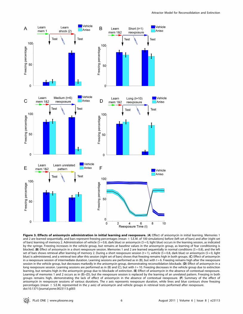
Figure 3. Effects of anisomycin administration in initial learning and reexposure. (A) Effect of anisomycin in initial learning. Memories 1and 2 are learned sequentially, and bars represent freezing percentages (mean 6 S.E.M. of 100 simulations) before (left set of bars) and after (right setof bars) learning of memory 2. Administration of vehicle (S = 0.8, dark blue) or anisomycin (S = 0, light blue) occurs in the learning session, as indicatedby the syringe. Freezing increases in the vehicle group, but remains at baseline values in the anisomycin group, as learning of fear conditioning isblocked. (B) Effect of anisomycin in a short reexposure session. Memories 1 and 2 are learned sequentially in normal conditions (S = 0.8), and the leftset of bars shows retrieval after learning of memory 2. During a short reexposure session (t = 1), vehicle (S = 0.8, dark blue) or anisomycin (S = 0, lightblue) is administered, and a retrieval test after this session (right set of bars) shows that freezing remains high in both groups. (C) Effect of anisomycinin a reexposure session of intermediate duration. Learning sessions are performed as in (B), but with t = 6. Freezing remains high after the reexposuresession in the vehicle group, but decreases markedly in the anisomycin group, demonstrating reconsolidation blockade. (D) Effect of anisomycin in along reexposure session. Learning sessions are performed as in (B) and (C), but with t = 10. Freezing decreases in the vehicle group due to extinctionlearning, but remains high in the anisomycin group due to blockade of extinction. (E) Effect of anisomycin in the absence of contextual reexposure.Learning of memories 1 and 2 occurs as in (B)–(D), but the reexposure session is replaced by the learning of an unrelated pattern. Freezing in bothgroups remains high, demonstrating the lack of effect of anisomycin in the absence of contextual reexposure. (F) Summary of the effect ofanisomycin in reexposure sessions of various durations. The x axis represents reexposure duration, while lines and blue contours show freezingpercentages (mean 6 S.E.M, represented in the y axis) of anisomycin and vehicle groups in retrieval tests performed after reexposure.doi:10.1371/journal.pone.0023113.g003
Attractor Model for Reconsolidation and Extinction
PLoS ONE | www.plosone.org 6 August 2011 | Volume 6 | Issue 8 | e23113
One can observe that the amnestic effect of anisomycin increases
along with reexposure duration until the minimum duration
required for extinction to occur in controls is reached (around
t = 8). In longer reexposure conditions, on the other hand, freezing
decreases in controls with increasing reexposure duration due to
extinction, while anisomycin preserves the original memory by
preventing extinction learning.
Reconsolidation and extinction after different strengthsof training
As observed experimentally [7,8,44], the protocols necessary to
induce reconsolidation and extinction in our model vary according
to the strength of the original learning. In some reexposure
conditions which normally induce reconsolidation in controls (and
amnesia in anisomycin treated animals) (Figure 4A), anisomycin
will have no effect if the initial learning of the shock memory is
made stronger by increasing S during the training session
(Figure 4B), as the stronger memory will not be as affected by
the degradation caused by reexposure. Such results are in
accordance with the behavioral data indicating that longer
reexposure trials are needed to induce reconsolidation of stronger
or more consolidated memories [8]. Another consequence of
strengthening the shock memory is that longer durations of
reexposure, which normally yield extinction (Figure 4C), will lead
to reconsolidation instead (Figure 4D). In this case, anisomycin will
not lead to memory preservation (as occurred after normal
training conditions) but to reconsolidation blockade and amnesia,
similarly to what has been described experimentally [7]. The effect
of reexposure duration on retrieval of the shock memory for
various strengths of initial learning is summarized in Figures 4E
(control conditions) and 4F (anisomycin treatment during
reexposure).
Effect of memory-enhancing drugs on differentreexposure protocols
Experimental data suggests that administration of memory-
enhancing drugs such as D-cycloserine (a partial agonist of the
coactivator site at the NR1 subunit of the NMDA receptor) during
contextual reexposure can improve either reconsolidation or
extinction, leading to an effect which is the opposite of that of
anisomycin [42]. We have simulated that by increasing the value
of S during the reexposure session, based on the enhancing effect
of such drugs upon synaptic plasticity [45]. As found experimen-
tally with D-cycloserine [42] and protein kinase A (PKA)
activation [46], stimulating Hebbian plasticity during reexposure
in reconsolidation conditions (t = 4) slightly improves subsequent
retrieval of the shock memory (Figure 5A, third set of bars). This
improvement was small in our simulations due to a ceiling effect,
as memory in controls already approached saturation values after
normal reconsolidation. On the other hand, increasing S during
extinction conditions (t = 8) improves extinction and lowers
subsequent fear memory retrieval (Figure 5A, fourth set of bars).
These trends hold true for a range of parameters, as shown in
Figure 5B, which summarizes the effects of increasing or
decreasing S during reexposure sessions of different durations.
Effects of blocking mismatch-induced degradationExperimental evidence for the effects of blocking protein
degradation on memory (usually achieved through the use of
inhibitors of the ubiquitin-proteasome cascade) is somewhat
controversial, with different effects described on initial learning
[36,47,48,49] and reconsolidation [35,36,47]. It has recently been
suggested, however, that protein degradation is necessary for the
amnestic effect of anisomycin on reconsolidation to occur [35,36].
This indeed occurs by blocking mismatch-induced degradation
(i.e. setting D to 0) in our model, which does not affect memory
reconsolidation by itself, but prevents the effect of anisomycin on
subsequent retrieval (Figure 6A). Blocking mismatch-induced
degradation will also prevent multiple session extinction
(Figure 6B), as shown experimentally in one of these studies
[36]. This result demonstrates that the mismatch-induced
degradation system has a physiologic role in our model, as it
allows nonreinforced trials of intermediate duration to lead to
extinction when performed repeatedly, as opposed to the
reinforcement of the original memory which occurs in the absence
of degradation. When compared to experimental findings, it also
suggests that protein degradation through the ubiquitin-protea-
some system could be one of the mechanisms involved in
mismatch-induced degradation of synaptic changes.
Discussion
The results presented show that our attractor network-based
model accounts for the main experimental results concerning the
effects of anisomycin on reconsolidation and extinction of fear
conditioning in different reexposure protocols. More specifically,
the model is in agreement with experimental data suggesting that
nonreinforced contextual reexposure has three possible outcomes,
namely: (a) simple retrieval (i.e. absence of reconsolidation or
extinction), in which anisomycin has no effect on memory; (b)
reconsolidation, in which anisomycin causes amnesia due to
blockade of this process; and (c) extinction, in which the behavioral
response to the original memory is reduced in controls, but
preserved in animals treated with anisomycin [8,16,41].
There are three main assumptions of the model that allow such
results to be obtained. The first one is the existence of attractor
dynamics in the brain, which produces nonlinear transitions
between the retrieval of an established attractor and the
instatement of a new attractor in the network as a function of
the perceived contextual representation during reexposure. The
second one is the existence of a protein-synthesis independent
system which acts to counteract Hebbian plastic changes in
synapses in response to mismatch between an established memory
and new information. The third assumption is that the degree of
dissimilarity between an animal’s contextual representation upon
nonreinforced reexposure and the representation of the original
learning will increase with longer durations of reexposure.
Concerning this last point, it should be noted that the data
presented can also be interpreted with t assumed to represent the
degree of dissimilarity between the reexposure session and the
original learning trial, rather than reexposure duration. In this
case, our results indicate that reconsolidation in our model can be
induced by an experience which is similar to that of the original
learning, but not by one which is identical to it (in which no
mismatch will occur) or radically different from it (which will lead
to instatement of a new attractor and also prevent mismatch). This
is in agreement with experimental data concerning the need of
both similarities (e.g. a similar environment) and differences (e.g.
absence of shock, differences in objects or platform location)
between the original learning trial and the reexposure trial for
reconsolidation to occur [14,15,50,51]. It is also in line with the
view that either updating of an existing memory trace or formation
of a new one can occur upon reexposure, depending on the degree
of contextual similarity [52].
It is noteworthy that the simple retrieval condition described in
our model provides an interesting framework to interpret studies
which failed to demonstrate reconsolidation, but used reexposure
Attractor Model for Reconsolidation and Extinction
PLoS ONE | www.plosone.org 7 August 2011 | Volume 6 | Issue 8 | e23113
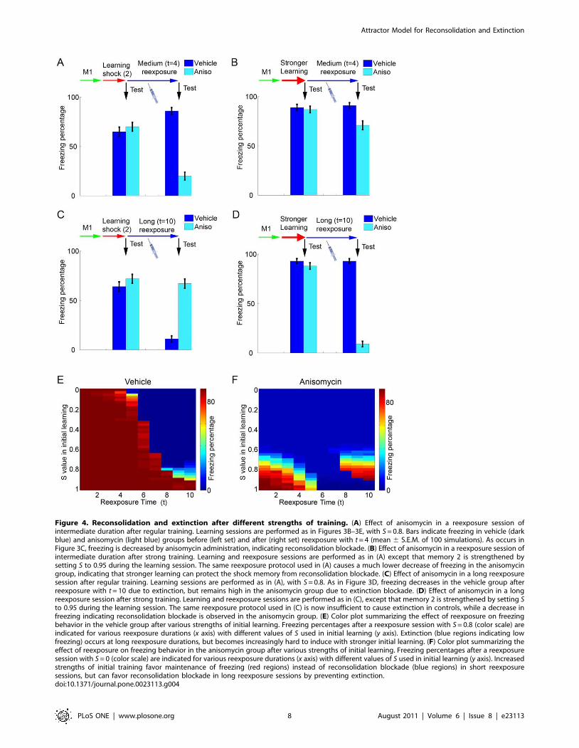
Figure 4. Reconsolidation and extinction after different strengths of training. (A) Effect of anisomycin in a reexposure session ofintermediate duration after regular training. Learning sessions are performed as in Figures 3B–3E, with S = 0.8. Bars indicate freezing in vehicle (darkblue) and anisomycin (light blue) groups before (left set) and after (right set) reexposure with t = 4 (mean 6 S.E.M. of 100 simulations). As occurs inFigure 3C, freezing is decreased by anisomycin administration, indicating reconsolidation blockade. (B) Effect of anisomycin in a reexposure session ofintermediate duration after strong training. Learning and reexposure sessions are performed as in (A) except that memory 2 is strengthened bysetting S to 0.95 during the learning session. The same reexposure protocol used in (A) causes a much lower decrease of freezing in the anisomycingroup, indicating that stronger learning can protect the shock memory from reconsolidation blockade. (C) Effect of anisomycin in a long reexposuresession after regular training. Learning sessions are performed as in (A), with S = 0.8. As in Figure 3D, freezing decreases in the vehicle group afterreexposure with t = 10 due to extinction, but remains high in the anisomycin group due to extinction blockade. (D) Effect of anisomycin in a longreexposure session after strong training. Learning and reexposure sessions are performed as in (C), except that memory 2 is strengthened by setting Sto 0.95 during the learning session. The same reexposure protocol used in (C) is now insufficient to cause extinction in controls, while a decrease infreezing indicating reconsolidation blockade is observed in the anisomycin group. (E) Color plot summarizing the effect of reexposure on freezingbehavior in the vehicle group after various strengths of initial learning. Freezing percentages after a reexposure session with S = 0.8 (color scale) areindicated for various reexposure durations (x axis) with different values of S used in initial learning (y axis). Extinction (blue regions indicating lowfreezing) occurs at long reexposure durations, but becomes increasingly hard to induce with stronger initial learning. (F) Color plot summarizing theeffect of reexposure on freezing behavior in the anisomycin group after various strengths of initial learning. Freezing percentages after a reexposuresession with S = 0 (color scale) are indicated for various reexposure durations (x axis) with different values of S used in initial learning (y axis). Increasedstrengths of initial training favor maintenance of freezing (red regions) instead of reconsolidation blockade (blue regions) in short reexposuresessions, but can favor reconsolidation blockade in long reexposure sessions by preventing extinction.doi:10.1371/journal.pone.0023113.g004
Attractor Model for Reconsolidation and Extinction
PLoS ONE | www.plosone.org 8 August 2011 | Volume 6 | Issue 8 | e23113
trials in which duration was short [9] or mismatch was not a
prominent feature [10], as these might have been insufficient to
induce significant updating of an existing attractor. The model
also accounts for the fact that different reexposure protocols are
required to induce reconsolidation and extinction of memories of
different strengths [7,8], which could furthermore explain the
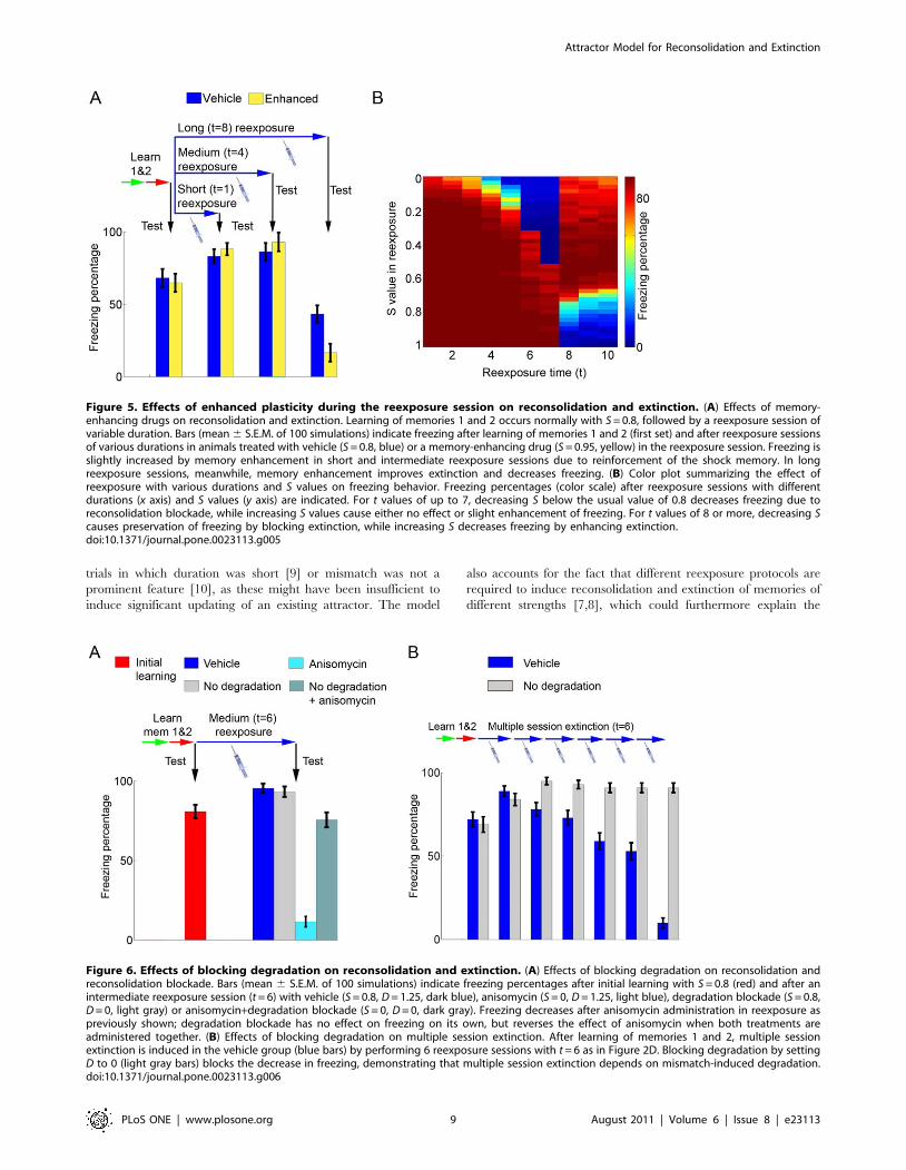
Figure 5. Effects of enhanced plasticity during the reexposure session on reconsolidation and extinction. (A) Effects of memory-enhancing drugs on reconsolidation and extinction. Learning of memories 1 and 2 occurs normally with S = 0.8, followed by a reexposure session ofvariable duration. Bars (mean 6 S.E.M. of 100 simulations) indicate freezing after learning of memories 1 and 2 (first set) and after reexposure sessionsof various durations in animals treated with vehicle (S = 0.8, blue) or a memory-enhancing drug (S = 0.95, yellow) in the reexposure session. Freezing isslightly increased by memory enhancement in short and intermediate reexposure sessions due to reinforcement of the shock memory. In longreexposure sessions, meanwhile, memory enhancement improves extinction and decreases freezing. (B) Color plot summarizing the effect ofreexposure with various durations and S values on freezing behavior. Freezing percentages (color scale) after reexposure sessions with differentdurations (x axis) and S values (y axis) are indicated. For t values of up to 7, decreasing S below the usual value of 0.8 decreases freezing due toreconsolidation blockade, while increasing S values cause either no effect or slight enhancement of freezing. For t values of 8 or more, decreasing Scauses preservation of freezing by blocking extinction, while increasing S decreases freezing by enhancing extinction.doi:10.1371/journal.pone.0023113.g005
Figure 6. Effects of blocking degradation on reconsolidation and extinction. (A) Effects of blocking degradation on reconsolidation andreconsolidation blockade. Bars (mean 6 S.E.M. of 100 simulations) indicate freezing percentages after initial learning with S = 0.8 (red) and after anintermediate reexposure session (t = 6) with vehicle (S = 0.8, D = 1.25, dark blue), anisomycin (S = 0, D = 1.25, light blue), degradation blockade (S = 0.8,D = 0, light gray) or anisomycin+degradation blockade (S = 0, D = 0, dark gray). Freezing decreases after anisomycin administration in reexposure aspreviously shown; degradation blockade has no effect on freezing on its own, but reverses the effect of anisomycin when both treatments areadministered together. (B) Effects of blocking degradation on multiple session extinction. After learning of memories 1 and 2, multiple sessionextinction is induced in the vehicle group (blue bars) by performing 6 reexposure sessions with t = 6 as in Figure 2D. Blocking degradation by settingD to 0 (light gray bars) blocks the decrease in freezing, demonstrating that multiple session extinction depends on mismatch-induced degradation.doi:10.1371/journal.pone.0023113.g006
Attractor Model for Reconsolidation and Extinction
PLoS ONE | www.plosone.org 9 August 2011 | Volume 6 | Issue 8 | e23113
different susceptibility of recent and remote memories to these
processes [3,8,53] if one assumes that reinforcement of memory
traces occurs over time [31]. This assumption can also reconcile
our model with results showing that amnesia induced by
reconsolidation blockade can be transient [54,55], as synaptic
reinforcement could lead to regeneration of the trace in the
hippocampus or neocortex [30]. Finally, if one assumes that
mismatch-induced synaptic changes involve protein degradation,
the model can explain the recently described dependence of
reconsolidation blockade on the activity of the ubiquitin-protea-
some system [35,36].
One should keep in mind that the dichotomy between retrieval-
induced, Hebbian plasticity based on protein synthesis and
mismatch-induced synaptic changes based on protein degradation
is an obvious biochemical simplification, and that both the HLP
and MID terms of the model certainly involve more than protein
synthesis and degradation in a biological setting. In fact, it is likely
that any plastic change in synapses involves both synthesis and
degradation of specific proteins, which would account for studies
showing the requirement of the ubiquitin-proteasome cascade for
initial learning [47,48,49] and normal reconsolidation [47] to
occur. It is also possible that both processes share other
mechanisms such as a dependence on NMDA receptors, as
blockade of these receptors has been shown either to induce
[42,56,57] or prevent [58] reconsolidation blockade depending on
the study. Nevertheless, our simplification is probably valid to
account for the experimental results we have tried to model, if one
assumes that protein synthesis blockade will preferentially affect
mechanisms underlying HLP, while degradation blockade will
preferentially have an effect on MID, at least under some
experimental conditions.
While HLP seems readily relatable to Hebbian-like plasticity
mechanisms such as long-term potentiation (LTP), the mecha-
nisms underlying mismatch detection and MID in the model
remain an open question; nevertheless, it seems natural to
speculate possible relationships of this process with long-term
depression (LTD) and synaptic depotentiation phenomena. LTD
has been shown to involve the ubiquitin-mediated degradation of
proteins such as PSD-95 [59] and, although it normally requires
protein synthesis [60], this requirement is not observed for all of its
forms [61]. Protein synthesis is also not required when low-
frequency stimulation is used to reverse preexisting LTP [62]; this
phenomenon, usually known as depotentiation, shares several
features with LTD [63], but is usually restricted to a short time
window (i.e. hours) following induction of LTP. However, since
MID in our model happens in many synapses which also exhibit
Hebbian plasticity during reexposure, it is possible that LTP-
mediated changes happening during reexposure could reinstate
the lability of synaptic weights to protein synthesis-independent
depotentiation.
If MID can indeed be related to LTD-like phenomena, then
changes in firing rates and spike synchronization among coactive
neurons during reexposure could mediate mismatch detection
through spike-time-dependent plasticity (STDP) [64,65], which
has recently been shown to be compatible with firing-rate based
models of LTP and LTD [66] and with autoassociative plasticity in
recurrent networks [28,67]. In this case, if cue currents to shock
and context neurons are unambiguous and synchronized (i.e.
simple retrieval), such neurons should spike at high firing rates and
in close temporal proximity, leading to the development of LTP in
their mutual connections. On the other hand, if cue inputs to
context and shock neurons become more distinct (i.e. reconsolida-
tion), firing rates and synchrony will decrease and LTD should
become more prominent. Thus, if LTP is disrupted by protein
synthesis inhibition, this could conceivably lead to reconsolidation
blockade. However, if reexposure is long enough to allow
extinction, silencing of the shock neurons would prevent the
occurrence of either LTP or LTD, as both depend on neuronal
spiking; thus, the disruptive effect of blocking protein synthesis on
the shock memory would not occur in extinction conditions, as
observed in our simulations.
The central core of our model’s results is accounted for by the
attractor properties of the Hopfield network, which allow it to
perform either pattern completion (leading to retrieval of the shock
memory upon presentation of the context alone) or pattern
separation (leading to the nonlinear transition between retrieval of
an established attractor and the formation of a new one). Attractor
networks have been classically thought to exist in the hippocam-
pus, particularly in CA3 [25,68], and both pattern completion and
separation have been suggested to occur in the hippocampal
formation by theoretical models [25,69] and behavioral findings
[70,71,72]. Moreover, electrophysiological evidence from place
field recordings suggests the occurrence of discontinuous attractor
transitions in the hippocampus [19]. If our attractor network is
thought to represent the CA3 region, an interesting finding is that
our results depend on a limited degree of overlap between the
shock and extinction patterns (7 to 28%, as observed in Figure S4),
which is in the range observed for place field representations of
similar environments in CA3 [73]; if overlap is higher, pattern
completion will prevent extinction learning. This is in line with the
view that some degree of pattern orthogonalization by the dentate
gyrus is necessary to allow CA3 to separate information between
similar contexts [74] – or, in the case of extinction, between
different representations of the same context.
Nevertheless, the existence of attractor dynamics in other
structures as well has also been suggested by experimental
evidence, such as the sustained activity of neocortical regions
after removal of sensory stimuli [21,75], the observation of abrupt
pattern transitions in response to gradually changing stimuli in the
olfactory bulb [22], and the recent electrophysiological demon-
strations of recurrent connectivity in the lateral amygdala [76].
Therefore, although we have developed our model to describe
phenomena thought to occur in the hippocampus, such as
contextual recognition, it is not dependent on specific features of
hippocampal anatomy (i.e. all it requires anatomically is the
existence of recurrent connections which can sustain attractor
properties). Thus, the mechanisms proposed by the model could
conceivably happen in other structures as well, as most of the
evidence showing the effects of reexposure duration on reconso-
lidation and extinction comes from studies using systemic
injections [8,14,16,41,42].
The amygdala in particular has been shown to be involved in
the reconsolidation of auditory fear conditioning [4], and
pharmacological transitions from reconsolidation to extinction
according to reexposure duration have been observed with intra-
amygdala injections [42,77]. Therefore, it is possible that recurrent
connectivity in this structure [76] could support local attractor
functioning, as proposed by other authors [78]. The observation of
reconsolidation and extinction in invertebrates such as crabs [79]
and fruitflies [41] also suggests that network effects mediating these
processes can exist in the absence of a hippocampus-like structure;
in this sense, our demonstration that a simple network with
recurrent connections can mediate attractor properties subserving
reconsolidation and extinction could be significant for interpreting
these data.
Still, although these systemic interactions mediating reconsoli-
dation and extinction are far from clear, and probably involve
complex modulations among structures [44], we propose that the
Attractor Model for Reconsolidation and Extinction
PLoS ONE | www.plosone.org 10 August 2011 | Volume 6 | Issue 8 | e23113
existence of attractor dynamics in the hippocampus could play a
central role in determining the dominant process induced by
contextual reexposure, at least in context-dependent and spatial
memory tasks. The bidirectional communication of this structure
with the entorhinal cortex could provide an anatomical substrate
for the feedback loop between online contextual representations
(mediated by the cortex) and the memory network (located in the
hippocampus) proposed in our framework. In particular, the CA1
area has been proposed to be responsible for comparing current
contextual representations (entorhinal cortex inputs) and stored
attractors (CA3 inputs) [80,81], and could be involved in detecting
mismatch between these representations. This hypothesis is further
strengthened by the recent demonstration that reexposure to a
learning context can induce transient weakening of LTP-related
changes in CA3-CA1 synapses [82]. In Figure S5 and in the
Supporting Text S1, we propose a simple model for how mismatch
detection by the CA1 region could mediate reconsolidation and
account for these results. In this model, differences in activity
between the CA3 and CA1 regions lead to mismatch-induced
degradation and reconsolidation phenomena, while transitions
between reconsolidation and extinction are accounted for by the
attractor dynamics in the CA3 region.
Still regarding the CA1 area, an interesting analogy can be
drawn between our computational results, based on fear
conditioning experiments, and electrophysiological data derived
from hippocampal place cell recordings in this region. Different
studies have suggested that ‘‘morphing’’ between two contexts with
different place fields can either lead to continuous updating of
these place fields [83] or to abrupt changes from one place field
representation to another [19], probably depending on the
protocol through which intermediate patterns are presented
[29]. These findings have been suggested to be related to attractor
dynamics [19,29] and show some similarities with the results of our
model, in which fear-related attractors can be either ‘‘updated’’
through reconsolidation or replaced with a new attractor encoding
extinction. The fact that fear conditioning can induce partial
remapping of place fields for the conditioning context [84] suggests
that such an analogy between shock/non-shock representations of
a context and different place field representations may be
warranted.
As expected from a theoretical model, our framework generates
a number of predictions concerning the effects of amnestic agents
on different learning and reexposure protocols. Some simple
experimental predictions on the behavioral, biochemical and
electrophysiological levels which would lend support to the general
framework of the model are the following:
(i) Only an attractor which is actively retrieved by the
network should be affected by reconsolidation blockade, as
mismatch-induced degradation will act upon the connec-
tions that maintain this attractor. Such specificity has
already been shown for memories learned in different
contexts [43] and our data predict that it can also happen
for different representations of the same environment. This
suggests that the extinction memory itself could be subject
to reconsolidation once it becomes dominant over the
original memory, if some form of mismatch (e.g. a
reminder of the shock memory) is introduced in a
reexposure session (for an example of this, see Figure S6).
The first experimental evidence for this prediction has
recently been reported using the inhibitory avoidance task
[85].
(ii) If the effects we describe are indeed happening in the
hippocampus, one would expect them to be observed not
only in contextual fear conditioning, but also in spatial
memory paradigms. Therefore, in spatial tasks in which
more than one behavioral strategy can be learned, such as
reversal learning protocols in the Morris water maze in
which two distinct platform locations are learned in
sequence, our model predicts that the one which is
retrieved by an animal at the time of nonreinforced
reexposure should be the one subject to reconsolidation
blockade. Besides providing support for the model’s
general framework, such a finding would strengthen the
case for involvement of the hippocampus in mediating
reconsolidation/extinction transitions.
(iii) The mismatch degradation system described in our model
should be activated more strongly after reconsolidation-
inducing reexposure protocols than after extinction-
inducing ones. This leads to the prediction that molecular
cascades involved in this system should be activated
differently after short and long reexposure durations, and
that signatures of this differential activation could be
detected through molecular/biochemical analysis of brain
tissue. Interesting candidates to be evaluated for this
purpose include the ubiquitin-proteasome system [36], and
possibly the endocannabinoid system, which has shown to
modulate reconsolidation and extinction in opposite ways
[86].
(iv) If one assumes the analogy between shock/non-shock
representations and place field representations as valid, this
means that extinction-inducing protocols should lead to a
partial remapping of place cells in the conditioning
context, similar to the one observed with initial condition-
ing [84]. This more indirect prediction is based on the
assumption that place field representations can also be
stored as attractors, as suggested by electrophysiological
data [19].
(v) If the abovementioned prediction is proved true, an
additional electrophysiological prediction is that the time
for place cell remapping during fear extinction should
match the time course of the transition between reconso-
lidation and extinction in the behavioral protocol used
(which has usually been shown to be in the range of 5–
30 minutes in studies of contextual fear conditioning tasks
in rodents [8,16]).
Finally, although our model argues for a network view of
reconsolidation and extinction, this does not mean that differences
between the two processes do not exist at the biochemical level.
On the contrary, it is likely that dissimilarities between them also
depend on the activation of different molecular cascades, as
suggested by some studies which have pointed out pharmacolog-
ical and biochemical differences between the two processes
[8,86,87,88]. In this sense, our model provides at least one
explanation why some drugs could have differential effects on
reconsolidation and extinction – namely, that they could be
targeting mechanisms which are not involved in classical Hebbian
plasticity, but rather in trace labilization (i.e. affecting MID but not
HLP in our model). If this is the case, the same drug could produce
differential effects in reconsolidation and extinction trials under
some conditions (see Figure S7), as has been recently shown with
drugs acting on the CB1 receptor [86]. Naturally, it is also possible
that there are other instances of memory modulation that were not
included in our model and could account for these effects.
In summary, by assuming the existence of attractor dynamics
and mismatch-induced updating of plastic changes in neural
networks, we provide a parsimonious explanation for the
Attractor Model for Reconsolidation and Extinction
PLoS ONE | www.plosone.org 11 August 2011 | Volume 6 | Issue 8 | e23113
occurrence of reconsolidation and extinction after nonreinforced
reexposure in fear conditioning tasks. Although in a biological
setting the modulation of these processes probably involves many
other factors as well, we believe our model is an interesting proof
of principle of the fact that both reconsolidation and extinction can
be explained by a unified set of plasticity mechanisms (i.e. the HLP
and MID terms in our equations), albeit operating in different
synapses. Therefore, the usual tenet that reconsolidation and
extinction represent distinct processes at the cellular and molecular
level might not be entirely true, as differences between the network
aspects of the two processes could be more important in their
distinction. This view is supported by the striking similarities
between the pharmacology of reconsolidation and that of
extinction, which certainly outnumber their dissimilarities in the
existing literature. Such aspects should be taken into account for
adequately translating knowledge from animal studies of memory
into useful clinical approaches for the treatment of psychiatric
disorders.
Methods
General model frameworkIn line with previous research, we model the attractor network
responsible for storing the memory patterns as a fully connected
neural network [24,29,30,31]. Neuronal activities in this network
are determined by Eq. (1), which fully defines its dynamics and
constrains neural activation (u) to values between 0 and 1 through
the term K(1+tanh(Swijuj + Ij). This represents a change from the
original Hopfield formulation, in which u is unbounded and can
achieve negative values as well. In that formulation, however, u is
typically regarded as the membrane potential, while
V(u) = (tanh(u)+1)/2 would represent the firing rate or activity
level of a neuron. In this sense, in our model u can be thought of as
a direct measure of the firing rate, without the intermediate step of
calculating the membrane potential.
As mentioned in the results session, the 0/1 implementation can
reflect the firing rate and connectivity of neurons in a more
realistic way, as it does not assume unrealistic features such as
symmetric connectivity and reinforcement between silent neurons;
this kind of change from the original Hopfield formulation has also
been implemented by other authors in different ways [89,90].
Furthermore, the use of a 0/1 activation scheme also prevents the
retrieval of ‘‘mirror attractors’’ (i.e. retrieval of a pattern
diametrically opposite to the one which was originally learned)
and diminishes the retrieval of spurious patterns when sequences
of correlated patterns are learned, as in the case of our simulations,
since it prevents the strengthening of connection between inactive
neurons, which can lead to the development of abnormal
connectivity between neuronal populations when the patterns
used are not completely arbitrary.
Modeling of synaptic weight strengthening anddegradation
The final steady state pattern achieved by Eq. (1) will in turn
induce changes in the synaptic weight matrix W~ wij
� �as
determined by Eq. (2), with the HLP and MID terms defined in
equations (3) and (4), respectively. In these equations, the notation
B*A denotes connections from A to B. Therefore, note that the first
term in Eq. (3) represents the strengthening of positive synaptic
connections among coactive neurons, while the second term models
a reduction in the strength of synaptic connectivity from active to
inactive neurons. Moreover, as the entries of W can achieve
negative values, the second term in (3) can bring about inhibition
from active to inactive neurons, as in classical formulations (see
Figure 1D). To prevent a memory or a set of memories from
completely dominating and suppressing the other memories, we
require that the magnitude of synaptic entries in the matrix W
saturates at a maximum value s0. We implement this by truncating
the entries that become too large back to s0, and by using a similar
procedure for synaptic values that decrease below 2s0.
After reaching the steady state upon a cue presentation, all units
belong to one of four categories: AA, SA, AS and SS, where A stands
for Active and S stands for Suppressed, with the first letter indicating
the nature of the cue currents and the second letter denoting the
final unit activity upon reaching the steady state. For the
computation of the mismatch vector, we normalize the cue
currents I , making them achieve values between 0 and 1 through a
transformation defined by Inorm~(I{Imin)=(Imax{Imin). There-
fore, it follows from Eq. (4) that when the retrieved pattern is in
complete agreement with the input cue, all entries in the matrix
MID have zero values, since all units belong either to category AA
or to category SS.
When mismatch occurs between the attractor network pattern
and the cue currents, this means that there are units pertaining to
either AS or SA categories – that is, there are neurons that were
suppressed in the retrieved pattern despite activation by the cue
current and, conversely, neurons that were active despite cue
suppression. The synaptic changes induced by mismatch occur
only at the connections linking: (a) active units (AA or SA) to AS,
and (b) active units to SA. As a result, in the first case, mismatch-
induced degradation acts to decrease the inhibition from active
units towards units that are rendered inactive despite the existence
of excitatory cue currents arriving at these neurons (AS units; see
Figure 1E). Thus, upon subsequent presentation of the same cue
pattern, the overall drive (i.e., cue currents plus within network
influences) to the AS units is increased, making these units more
likely to switch to the AA category. Similarly, in the second case,
the strength of connections from active units to SA units decays to
lower values as a result of the mismatch-induced degradation
(Figure 1E). Consequently, SA units become more likely to switch
to the SS category upon subsequent presentation of the same
pattern. Note that the effects mentioned here are a consequence of
the definition of the mismatch vector (m~Inorm{u), upon which
MID is dependent (Eq. (4)). For instance, an AS unit j has its
correspondent m entry value equal to mj = +120 = +1, whereas a
SA unit k has mk = 021 = 21. Therefore, by employing
MID~D(mT � u), the connection strength from an active unit i
(i.e, ui = +1) to an AS unit j will be changed by MIDji = +D, whereas
the connection to a SA unit k will change by MIDki = 2D. Taken
together, these two types of mismatch-induced degradation
facilitate the learning of new memory patterns that introduce
conflicting information.
Modeling of retrieval tests and nonreinforced reexposureMemory retrieval is tested by presenting the cue pattern which
represents the context, with Ij~0:1 of its strength at training for
context neurons j and 0 for other neurons (Figure 1B), and
observing the attractor to which the network evolves. In order to
have a closer correlation between attractor retrieval in our
computational model and the behavioral measures of memory
used in experimental studies of fear conditioning, we model the
retrieval of a particular memory pattern as leading to a certain
amount of freezing during the test session. Therefore, we assume
that upon retrieval of the shock pattern the animal exhibits a high
amount of freezing (90% of the test duration), while other memory
patterns induce a low, baseline freezing time (10% of the test
duration) (see Figure S1).
Attractor Model for Reconsolidation and Extinction
PLoS ONE | www.plosone.org 12 August 2011 | Volume 6 | Issue 8 | e23113
Cue inputs during the reexposure sessions are modeled by:
I~I2z(I3{I2) � f (t) ð5Þ
where vectors I2 and I3 represent the cue inputs for patterns 2 and
3 (Figure 1B), respectively, and t represents the amount of
reexposure time (which varies between minimum and maximum
values tmin = 0 and tmax = 10 in the simulations). The function f (t)monotonically varies between 0 and 1 and determines the ratio
between pattern 3 and pattern 2 present in the cue input I , which
is thus assumed to depend on the reexposure time t. We use a
sigmoid defined by f (t)~1=(1z exp (tmax=2{t)), but any
monotonically increasing function onto [0,1] provides qualitatively
similar results.
Visualization of attractor basinsIn agreement with previous research, the strength of the stored
memories could be estimated from statistics of full pattern retrieval
induced by either partial cue presentation or random initialization
of the neural units. In addition, we also developed a new method
(used in Figure 2 and Figures S3 and S6) to estimate the basins of
attraction for these patterns, defined as follows. Although each
pattern constitutes a point in a large N-dimensional space
(N = 100), the number of patterns P presented to the network is
low (P = 3). This allowed us to use Multiple Discriminant Analysis
(MDA) [91] to project these patterns into a low-dimensional
encoding subspace of dimension P21 (in the examples used in this
work the resulting dimension is D = 2, a plane). This projection can
be obtained by performing and eigenvalue/eigenvector decompo-
sition of the total covariance matrix Sb given by the formula:
SB~XP
k~1
(Ik{I0)T (Ik{I0), I0~1
P
XP
k~1
Ik ð6Þ
Here, Ik is the corresponding pattern for each class and I0 is the
global mean vector. This method allows the projection of
continuous N-dimensional neural states into this subspace, using
the matrix comprised by the first P21 eigenvectors. We then
compute their corresponding energy (Lyapunov) function [24] in
the original space, using the formula:
E~{1
2
Xi,j
wijuiujz1
2
Xi
ui ð7Þ
Finally, the average energy corresponding to a region in the
low-dimensional space is determined as the local mean energy
over a set of nearest neighbors and displayed as a 3D color map.
While we do not prove that network dynamics converge to a local
minimum for all possible initial states, numerical simulations
indicate that this is indeed true for all cases analyzed with the 0/1
network used in our work. As a result, this method is useful for
providing enhanced intuition about the relative strength of the
stored patterns, allowing direct visualization of their corresponding
basins of attraction.
Model parametersThe model parameter values employed in simulations were
t= 1 and N = 100 and c= 0.15; s0 = 1; S = 0.8; D = 1.25 (unless
where otherwise noted). Ii varied from 25 to +5 for each neuron
according to the learning pattern presented (see Figures 1C and
2E). Although parameters such as S and D cannot be directly
estimated from experimental evidence, results qualitatively similar
to ours can be obtained for a reasonably wide range of parameters,
albeit with reconsolidation and extinction occurring at different
values of t, as observed in Figure S8.
For each protocol studied, we ran 100 simulations with different
initial conditions of activation randomly chosen from a uniform
distribution on [0,0.1], and the reported result constitutes the
mean 6 S.E.M. of these simulations, unless where otherwise
specified.
Supporting Information
Figure S1 Comparison of attractor retrieval and freez-ing percentages. (A) After learning unrelated memory 1, testing
yields retrieval of this attractor (green bar, mean 6 S.E.M. of 10
sets of 100 simulations) even in the presence of contextual cues,
leading to a low percentage of freezing (grey bar). (B) After
learning of memory 2, retrieval of its attractor (red bar)
predominates over that of memory 1, leading to substantially
higher freezing. (C) After a long duration of reexposure leading to
extinction (t = 8), retrieval of attractor 3 becomes dominant (blue
bar), causing decrease of freezing. (D) Correlation between
retrieval of attractor 2 (x axis) and freezing percentages (y axis,
mean 6 S.E.M.) demonstrates a linear relation.
(PDF)
Figure S2 Effect of network size and pattern sparsenesson memory storage capacity. (A) Storage capacity of
networks with different number of neurons. Different numbers
of arbitrary patterns (x axis) with random overlap are stored in the
synaptic weight matrices W of networks of different sizes, varying
from 100 to 700 neurons (color lines), using the same learning rule
as in our model (Eq. (3)). Parameters are similar to those used in
other simulations, including the size of the memory patterns (14
neurons) and corresponding cues (4 neurons). Retrieval is
evaluated by providing the network with a cue pertaining to one
of the learned memory patterns and testing the correlation
between the activity of neurons in the retrieved pattern and in the
original memory pattern, as done by other authors [92,93]. For
each point, 200 retrieval trials using randomly chosen cues and
random initial conditions were performed, and the percentage of
trials in which successful retrieval occurred (defined as an r
value . 0.7 for the correlation) is shown. Memory capacity
increases steadily with network size, showing that our network
model is able to store a large amount of patterns if sufficient
neurons are added to the network. (B) Same as in (A), except that
the patterns and cues used are half the size as in (A) (i.e. 7
neurons/pattern, 2 neurons/cue). One can observe that memory
capacity greatly increases using sparser patterns, as reported to be
the case in previous attractor models.
(PDF)
Figure S3 Comparison of relative memory strengthsafter single session and multiple session extinction. (A)
Basins of attraction after single session extinction. Learning of
memories 1, 2 and 3 occurs as in Figure 2C, with extinction
learned in a single reexposure session with t = 10. Energy
landscape shows relative basins of attraction after learning of the
3 memories. The energy minimum for memory 2 persists, but is
higher than that of memory 3, leading to behavioral dominance of
the extinction memory. (B) Basins of attraction after multiple
session extinction. Learning of memories 1 and 2 occurs as in (A),
while extinction occurs through 6 reexposure sessions with t = 6 as
in Figure 2D. The energy minimum for memory 2 is significantly
reduced by multiple sessions of extinction when compared to a
Attractor Model for Reconsolidation and Extinction
PLoS ONE | www.plosone.org 13 August 2011 | Volume 6 | Issue 8 | e23113
single session, due to the effects of mismatch-induced degradation.
This could be related to the finding that spaced extinction trials
can lead to less spontaneous recovery and renewal of the original
memory than massed trials [94].
(PDF)
Figure S4 Effect of pattern overlap on the developmentof reconsolidation and extinction. (A) Freezing rates in a
retrieval test performed after a reexposure session of variable
duration in control conditions (S = 0.8) with different degrees of
overlap between memory 2 and memory 3 used in the simulations.
The percentage of coactive neurons in both patterns is indicated in
the y axis, while the duration of reexposure (t) is indicated in the x
axis, with the color scale representing freezing at a subsequent
retrieval test. One can observe that above a certain degree of
overlap (around 30% of active neurons in common), extinction
does not occur with the parameters used in the simulations,
suggesting that some degree of pattern separation is necessary for
extinction learning. (B) Freezing rates after reexposure of various
durations under anisomycin (S = 0). Blockade of HLP shows that
reconsolidation blockade occurs in a wider range of overlap (from
7 to 80% of active neurons, approximately) than extinction
blockade, which is only observed with up to 30% overlap, as in (A).
(PDF)
Figure S5 CA3-CA1 model for mismatch detection,reconsolidation and extinction. (A) Model scheme. Output
from the entorhinal cortex (EC) reaches both the CA3 and CA1
regions, providing information on the current context. CA3
neurons possess autoassociative connections, and send information
on their retrieved attractor to CA1. (B) Initial learning,
reconsolidation and extinction of aversive memories in the CA3-
CA1 model. Left column shows a more detailed view of the model
scheme, with sample neurons representing the context (square),
shock (circle) and absence of shock (triangle). Middle column
shows activation of the same neurons during initial learning,
reconsolidation and extinction, while the right column shows the
synaptic weight changes caused by this activation. (I) Initial
learning. Context and shock neurons are activated in all three
networks (middle column), leading to strengthening of synapses
(solid arrows) between coactive neurons (both in CA3 collaterals
and in CA3-CA1 synapses) and inhibition of non-shock neurons in
CA3 (circle-capped lines), as shown in the right column. (II)
Reconsolidation. Ambiguous information from the EC leads to
partial activation of shock and non-shock neurons in CA1, while
CA3 still retrieves the original pattern (middle column). The
mismatch generated between CA3 and CA1 shock neurons leads
to mismatch-induced degradation of their connections (discontin-
uous arrow), which is compensated by Hebbian learning both
within CA3 and in CA3-CA1 connections. (III) Extinction. Cue
patterns indicating absence of shock lead to instatement of this
pattern both in CA1 and CA3 (middle column). Synaptic weight
changes show formation of a new attractor representing extinction
in CA3 and strengthening of connections between non-shock
neurons in CA3 and CA1, while connections between shock
neurons remain unaltered. (C) Effect of anisomycin in reexposure
sessions of various durations in the CA3-CA1 model. The x axis
represents reexposure duration, while the lines show freezing
percentages (y axis) of vehicle (red) and anisomycin (blue) groups in
retrieval tests performed after reexposure. The dependence of
retrieval on reexposure duration in both groups is qualitatively
similar to what is observed with the general model in Figure 3F.
(PDF)
Figure S6 Selectivity of reconsolidation blockade to theretrieved attractor. (A) Reconsolidation of fear conditioning.
Bars show freezing percentages after learning memories 1 and 2
(left) and after a subsequent reexposure session with t = 5 (right) in
animals treated with vehicle and anisomycin in reexposure. As in
Figure 3C, freezing remains high after reexposure in the vehicle
group, but decreases due to reconsolidation blockade in the
anisomycin group. (B) Reconsolidation of fear extinction. Left bars
show freezing after learning of memories 1 and 2 followed by fear
extinction with a long reexposure session (t = 10), which leads to
learning of memory 3. As occurs in Figures 2C and 3C, memory 3
is dominant after this protocol, leading to low freezing.
Subsequently, a reexposure session with the same duration as in
(A) is performed; vehicle or anisomycin is administered in this
session, and right bars show freezing in a subsequent retrieval test.
Freezing remains low in the vehicle group, but increases in the
anisomycin treated group, indicating that the extinction memory,
which was preferentially retrieved during reexposure, becomes
susceptible to reconsolidation blockade. (C) Energy landscapes
showing relative basins of attraction for memories 1, 2 and 3 after
learning of memories 1 and 2 (left) and after a subsequent
reexposure session (t = 5) with vehicle (middle) or anisomycin
(right). Right panel shows a decrease in the basin of attraction of
memory 2 (shock memory). (D) Energy landscapes showing
relative basins of attraction for memories 1, 2 and 3 after learning
of memories 1, 2 and 3 (left) and after a subsequent reexposure
session (t = 5) with vehicle (middle) or anisomycin (right). Right
panel shows a decrease in the basin of attraction of memory 3
(extinction memory).
(PDF)
Figure S7 Differential effects of enhanced mismatch-induced degradation on reconsolidation and multiplesession extinction. (A) After learning of memory 2, increasing
D from 1.25 (blue bar) to 1.5 (orange bar) during a reexposure
session (t = 7.5) leads to a decrease in freezing as compared to the
vehicle group, as the reinforcement of memory 2 is impaired. (B)
In a multiple session extinction protocol (6 sessions with t = 6), the
same increase in D used in (A) leads to an acceleration of memory
extinction.
(PDF)
Figure S8 Effect of S and D variations on the develop-ment of reconsolidation and extinction. (A) Freezing rates
in a retrieval test performed after a reexposure session of variable
duration (x axis) with different values of S (y axis) used throughout
the simulations (i.e. learning of memories 1 and 2 and reexposure
sessions), while D is fixed at 1.25. One can observe that, with the
other parameters kept fixed, extinction happens for S values
varying from 0.6 to 0.9, although at different reexposure
durations. (B) Freezing rates retrieved as in (A), but with
anisomycin administration (S = 0) simulated during reexposure,
with different values of S used during learning of memories 1 and
2. Reconsolidation occurs for values of S varying from 0.7 to 1,
albeit at variable reexposure durations. (C) Freezing rates retrieved
as in (A) and (B), but with different values of D throughout the
simulations, while S is fixed at 0.8. D values have little effect on the
occurrence of extinction, as there is no mismatch between current
context and retrieved attractors in extinction conditions; however,
high values of D (i.e. above 1.6) can lead to an amnestic effect
under reconsolidation conditions, even in the absence of
anisomycin. (D) Freezing rates retrieved as in (C), but with
anisomycin administration during reexposure. Reconsolidation
blockade is observed at all values of D included in the simulations,
although higher values lead to amnesia at progressively shorter
reexposure durations.
(PDF)
Attractor Model for Reconsolidation and Extinction
PLoS ONE | www.plosone.org 14 August 2011 | Volume 6 | Issue 8 | e23113
Text S1
(PDF)
Acknowledgments
R.O. and A.B.L.T. thank Nancy Kopell for her support of their work.
Author Contributions
Conceived and designed the experiments: OBA. Performed the experi-
ments: RO ABLT. Analyzed the data: RO ABLT OBA. Wrote the paper:
RO ABLT OBA.
References
1. Misanin JR, Miller RR, Lewis DJ (1968) Retrograde amnesia produced by
electroconvulsive shock after reactivation of a consolidated memory trace.Science 160: 554–555.
2. Nader K, Hardt O (2009) A single standard for memory: the case forreconsolidation. Nat Rev Neurosci 10: 224–234.
3. Milekic MH, Alberini CM (2002) Temporally graded requirement for proteinsynthesis following memory reactivation. Neuron 36: 521–525.
4. Nader K, Schafe GE, LeDoux JE (2000) Fear memories require protein synthesisin the amygdala for reconsolidation after retrieval. Nature 406: 722–726.
5. Berman DE, Dudai Y (2001) Memory extinction, learning anew, and learningthe new: dissociations in the molecular machinery of learning in cortex. Science
291: 2417–2419.
6. Vianna MRM, Szapiro G, McGaugh JL, Medina JH, Izquierdo I (2001)
Retrieval of memory for fear-motivated training initiates extinction requiring
protein synthesis in the rat hippocampus. Proc Natl Acad Sci USA 98:12251–12254.
7. Eisenberg M, Kobilo T, Berman DE, Dudai Y (2003) Stability of retrievedmemory: inverse correlation with trace dominance. Science 301: 1102–1104.
8. Suzuki A, Josselyn SA, Frankland PW, Masushige S, Silva AJ, et al. (2004)Memory reconsolidation and extinction have distinct temporal and biochemical
signatures. J Neurosci 24: 4787–4795.
9. Biedenkapp JC, Rudy JW (2004) Context memories and reactivation: constraints
on the reconsolidation hypothesis. Behav Neurosci 118: 956–964.
10. Cammarota M, Bevilaqua LR, Medina JH, Izquierdo I (2004) Retrieval does not
induce reconsolidation of inhibitory avoidance memory. Learn Mem 11:
572–578.
11. Dudai Y (2006) Reconsolidation: the advantage of being refocused. Curr Opin
Neurobiol 16: 174–178.
12. Tronson NC, Taylor JR (2007) Molecular mechanisms of memory reconsolida-
tion. Nat Rev Neurosci 8: 262–275.
13. Lee JLC (2009) Reconsolidation: maintaining memory relevance. Trends
Neurosci 32: 413–420.
14. Pedreira ME, Perez-Cuesta LM, Maldonado H (2004) Mismatch between what
is expected and what actually occurs triggers memory reconsolidation orextinction. Learn Mem 11: 579–585.
15. Morris RG, Inglis J, Ainge JA, Olverman HJ, Tulloch J, et al. (2006) Memoryreconsolidation: sensitivity of spatial memory to inhibition of protein synthesis in
dorsal hippocampus during encoding and retrieval. Neuron 50: 479–489.
16. Bustos SG, Maldonado H, Molina VA (2009) Disruptive effect of midazolam on
fear memory reconsolidation: decisive influence of reactivation time span andmemory age. Neuropsychopharmacology 34: 446–457.
17. Brunet A, Orr SP, Tremblay J, Robertson K, Nader K, et al. (2008) Effect ofpost-retrieval propranolol on psychophysiologic responding during subsequent
script-driven traumatic imagery in post-traumatic stress disorder. J Psychiatr Res
42: 503–506.
18. Ressler KJ, Rothbaum BO, Tannenbaum L, Anderson P, Graap K, et al. (2004)
Cognitive enhancers as adjuncts to psychotherapy: use of D-cycloserine inphobic individuals to facilitate extinction of fear. Arch Gen Psychiatry 61:
1136–1144.
19. Wills TJ, Lever C, Cacucci F, Burgess N, O’Keefe J (2005) Attractor dynamics in
the hippocampal representation of the local environment. Science 308: 873–876.
20. Colgin LL, Leutgeb S, Jezek K, Leutgeb JK, Moser EI, et al. (2010) Attractor-
map versus autoassociation based attractor dynamics in the hippocampalnetwork. J Neurophysiol 104: 35–50.
21. Miyashita Y, Chang HS (1988) Neuronal correlate of pictorial short-term
memory in the primate temporal cortex. Nature 331: 68–70.
22. Niessing J, Friedrich RW (2010) Olfactory pattern classification by discrete
neuronal network states. Nature 465: 47–52.
23. Cohen M, Grossberg S (1983) Absolute stability of global pattern-formation and
parallel memory storage by competitive neural networks. IEEE Trans Syst ManCybern 13: 815–826.
24. Hopfield JJ (1982) Neural networks and physical systems with emergentcollective computational abilities. Proc Natl Acad Sci USA 79: 2554–2558.
25. Rolls ET (2007) An attractor network in the hippocampus: Theory andneurophysiology. Learn Mem 14: 714–731.
26. Tsodyks M (1999) Attractor neural network models of spatial maps inhippocampus. Hippocampus 9: 481–489.
27. Anishchenko A, Treves A (2006) Autoassociative memory retrieval andspontaneous activity bumps in small-world networks of integrate-and-fire
neurons. J Physiol Paris 100: 225–236.
28. Bush D, Philippides A, Husbands P, O’Shea M (2010) Dual coding with STDP
in a spiking recurrent neural network model of the hippocampus. PLoS ComputBiol 6: e1000839.
29. Blumenfeld B, Preminger S, Sagi D, Tsodyks M (2006) Dynamics of memory
representations in networks with novelty-facilitated synaptic plasticity. Neuron
52: 383–394.
30. Amaral OB, Osan R, Roesler R, Tort ABL (2008) A synaptic reinforcement-
based model for transient amnesia following disruptions of memory consolida-
tion and reconsolidation. Hippocampus 18: 584–601.
31. Wittenberg GM, Sullivan MR, Tsien JZ (2002) Synaptic reentry reinforcement
based network model for long-term memory consolidation. Hippocampus 12:
637–647.
32. Tronson NC, Schrick C, Guzman YF, Huh KH, Srivastava DP, et al. (2009)
Segregated populations of hippocampal principal CA1 neurons mediating
conditioning and extinction of contextual fear. J Neurosci 29: 3387–3394.
33. Herry C, Ciocchi S, Senn V, Demmou L, Muller C, et al. (2008) Switching on
and off fear by distinct neuronal circuits. Nature 454: 600–606.
34. Eisenhardt D, Menzel R (2007) Extinction learning, reconsolidation and the
internal reinforcement hypothesis. Neurobiol Learn Mem 87: 167–173.
35. Lee JLC (2008) Memory reconsolidation mediates the strengthening of
memories by additional learning. Nat Neurosci 11: 1264–1266.
36. Lee SH, Choi JH, Lee N, Lee HR, Kim JI, et al. (2008) Synaptic protein
degradation underlies destabilization of retrieved fear memory. Science 319:
1253–1256.
37. Hernandez PJ, Abel T (2008) The role of protein synthesis in memory
consolidation: progress amid decades of debate. Neurobiol Learn Mem 89:
293–311.
38. Treves A, Rolls ET (1991) What determines the capacity of autoassociative
memories in the brain? Network 2: 371–397.
39. Golomb D, Rubin N, Sompolinsky H (1990) Willshaw model: associate memory
with sparse coding and low fire rates. Phys Rev A 41: 1843–1854.
40. Schafe GE, Nadel NV, Sullivan GM, Harris A, Le Doux JE (1999) Memory
consolidation for contextual and auditory fear conditioning is dependent on
protein synthesis, PKA, and MAP kinase. Learn Mem 6: 97–110.
41. Lagasse F, Devaud J-M, Mery F (2009) A switch from cycloheximide-resistant
consolidated memory to cycloheximide-sensitive reconsolidation and extinction
in Drosophila. J Neurosci 29: 2225–2230.
42. Lee JLC, Milton AL, Everitt BJ (2006) Reconsolidation and extinction of
conditioned fear: inhibition and potentiation. J Neurosci 26: 10051–10056.
43. Doyere V, Debiec J, Monfils MH, Schafe G, LeDoux JE (2007) Synapse-specific
reconsolidation of distinct fear memories in the lateral amygdala. Nat Neurosci
10: 414–416.
44. Wang SH, de Oliveira Alvares L, Nader K (2009) Cellular and systems
mechanisms of memory strength as a constraint on auditory fear reconsolidation.
Nat Neurosci 12: 905–912.
45. Zhang XL, Sullivan JA, Moskal JR, Stanton PK (2008) A NMDA receptor
glycine site partial agonist, GLYX-13, simultaneously enhances LTP and
reduces LTD at Schaffer collateral-CA1 synapses in hippocampus. Neurophar-
macology 55: 1238–1250.
46. Tronson NC, Wiseman SL, Olausson P, Taylor JR (2006) Bidirectional
behavioral plasticity of memory reconsolidation depends on amygdalar protein
kinase A. Nat Neurosci 9: 167–169.
47. Artinian J, McGauran AT, De Jaeger X, Mouledous L, Frances B, et al. (2008)
Protein degradation, as with protein synthesis, is required during not only long-
term spatial memory consolidation but also reconsolidation. Eur J Neurosci 27:
3009–3019.
48. Lopez-Salon M, Alonso M, Vianna MRM, Viola H, Mello e Souza T, et al.
(2001) The ubiquitin-proteasome cascade is required for mammalian long-term
memory formation. Eur J Neurosci 14: 1820–1826.
49. Merlo E, Romano A (2007) Long-term memory consolidation depends on
proteasome activity in the crab Chasmagnathus. Neuroscience 147: 46–52.
50. Rossato JI, Bevilaqua LR, Medina JH, Izquierdo I, Cammarota M (2006)
Retrieval induces hippocampal-dependent reconsolidation of spatial memory.
Learn Mem 13: 431–440.
51. Rossato JI, Bevilaqua LR, Myskiw JC, Medina JH, Izquierdo I, et al. (2007) On
the role of hippocampal protein synthesis in the consolidation and reconsolida-
tion of object recognition memory. Learn Mem 14: 36–46.
52. Hupbach A, Hardt O, Gomez R, Nadel L (2008) The dynamics of memory:
Context-dependent updating. Learn Mem 15: 574–579.
53. Lopez J, Vasconcelos AP, Cassel JC (2008) Differential sensitivity of recent vs.
remote memory traces to extinction in a water-maze task in rats. Neurobiol
Learn Mem 90: 460–466.
54. Lattal KM, Abel T (2004) Behavioral impairments caused by injections of the
protein synthesis inhibitor anisomycin after contextual retrieval reverse with
time. Proc Natl Acad Sci USA 101: 4667–4672.
Attractor Model for Reconsolidation and Extinction
PLoS ONE | www.plosone.org 15 August 2011 | Volume 6 | Issue 8 | e23113
55. Amaral OB, Luft T, Cammarota M, Izquierdo I, Roesler R (2007) Temporary
inactivation of the dorsal hippocampus induces a transient impairment inretrieval of aversive memory. Behav Brain Res 180: 113–118.
56. Pedreira ME, Perez-Cuesta LM, Maldonado H (2002) Reactivation and
reconsolidation of long-term memory in the crab Chasmagnathus: proteinsynthesis requirement and mediation by NMDA-type glutamatergic receptors.
J Neurosci 22: 8305–8311.57. Przybyslawski J, Sara SJ (1997) Reconsolidation of memory after its reactivation.
Behav Brain Res 84: 241–246.
58. Mamou CB, Gamache K, Nader K (2006) NMDA receptors are critical forunleashing consolidated auditory fear memories. Nat Neurosci 9: 1237–1239.
59. Colledge M, Snyder EM, Crozier RA, Soderling JA, Jin Y, et al. (2003)Ubiquitination regulates PSD-95 degradation and AMPA receptor surface
expression. Neuron 40: 595–607.60. Kauderer BS, Kandel ER (2000) Capture of a protein synthesis-dependent
component of long-term depression. Proc Natl Acad Sci USA 97: 13342–13347.
61. Naie K, Manahan-Vaughan D (2005) Investigations of the protein synthesisdependency of mGluR-induced long-term depression in the dentate gyrus of
freely moving rats. Neuropharmacology 49: 35–44.62. Zhang L, Meng K, Li YH, Han TZ (2009) NR2A-containing NMDA receptors
are required for L-LTP induction and depotentiation in CA1 region of
hippocampal slices. Eur J Neurosci 29: 2137–2144.63. Wagner JJ, Alger BE (1996) Homosynaptic LTD and depotentiation: Do they
differ in name only? Hippocampus 6: 24–29.64. Bi GQ, Poo MM (1998) Synaptic modifications in cultured hippocampal
neurons: dependence on spike timing, synaptic strength, and postsynaptic celltype. J Neurosci 18: 10464–10472.
65. Markram H, Lubke J, Frotscher M, Sakmann B (1997) Regulation of synaptic
efficacy by coincidence of postsynaptic APs and EPSPs. Science 275: 213–215.66. Izhikevich EM, Desai NS (2003) Relating STDP to BCM. Neural Comput 15:
1511–1523.67. Gilson M, Burkitt A, van Hemmen LJ (2010) STDP in recurrent neuronal
networks. Front Comput Neurosci 4: 12.
68. McNaughton BL, Morris RGM (1987) Hippocampal synaptic enhancement andinformation storage within a distributed memory system. Trends Neurosci 10:
408–415.69. Marr D (1971) Simple memory: a theory for archicortex. Philos Trans R Soc
Lond B 262: 23–81.70. Gold AE, Kesner RP (2005) The role of the CA3 subregion of the dorsal
hippocampus in spatial pattern completion in the rat. Hippocampus 15:
808–814.71. McHugh TJ, Jones MW, Quinn JJ, Balthasar N, Coppari R, et al. (2007)
Dentate gyrus NMDA receptors mediate rapid pattern separation in thehippocampal network. Science 317: 94–99.
72. Nakazawa K, Quirk MC, Chitwood RA, Watanabe M, Yeckel MF, et al. (2002)
Requirement for hippocampal CA3 NMDA receptors in associative memoryrecall. Science 297: 211–218.
73. Leutgeb S, Leutgeb JK, Treves A, Moser MB, Moser EI (2004) Distinctensemble codes in hippocampal areas CA3 and CA1. Science 305: 1295–1298.
74. Kesner RP (2007) A behavioral analysis of dentate gyrus function. Prog BrainRes 163: 567–576.
75. Amit DJ, Brunel N (1997) Model of global spontaneous activity and local
structured activity during delay periods in the cerebral cortex. Cereb Cortex 7:237–252.
76. Johnson LR, Hou M, Ponce-Alvarez A, Gribelyuk L, Alphs H, et al. (2008) A
recurrent network in the lateral amygdala: a mechanism for coincidencedetection. Front Neural Circuits 2: 3.
77. Mamiya N, Fukushima H, Suzuki A, Matsuyama Z, Homma S, et al. (2009)Brain region-specific gene expression activation required for reconsolidation and
extinction of contextual fear memory. J Neurosci 29: 402–413.
78. Rolls ET, Stringer SM (2001) A model of the interaction between mood andmemory. Network 12: 89–109.
79. Pedreira ME, Maldonado H (2003) Protein synthesis subserves reconsolidationor extinction depending on reminder duration. Neuron 38: 863–869.
80. Hasselmo ME, Wyble BP (1997) Free recall and recognition in a network modelof the hippocampus: simulating effects of scopolamine on human memory
function. Behav Brain Res 89: 1–34.
81. Lisman JE, Grace AA (2005) The hippocampal-VTA loop: controlling the entryof information into long-term memory. Neuron 46: 703–713.
82. Clarke JR, Cammarota M, Gruart A, Izquierdo I, Delgado-Garcıa JM (2010)Plastic modifications induced by object recognition memory processing. Proc
Natl Acad Sci USA 107: 2652–2657.
83. Leutgeb JK, Leutgeb S, Treves A, Meyer R, Barnes CA, et al. (2005) Progressivetransformation of hippocampal neuronal representations in ‘‘morphed’’
environments. Neuron 48: 345–358.84. Moita MAP, Rosis S, Zhou Y, LeDoux JE, Blair HT (2004) Putting fear in its
place: remapping of hippocampal place cells during fear conditioning. J Neurosci24: 7015–7023.
85. Rossato JI, Bevilaqua LR, Izquierdo I, Medina JH, Cammarota M (2010)
Retrieval induces reconsolidation of fear extinction memory. Proc Natl Acad SciUSA 107: 1017–1020.
86. de Oliveira Alvares L, Pasqualini Genro B, Diehl F, Molina VA, Quillfeldt JA(2008) Opposite action of hippocampal CB1 receptors in memory reconsolida-
tion and extinction. Neuroscience 154: 1648–1655.
87. Merlo E, Romano A (2008) Memory extinction entails the inhibition of thetranscription factor NF-kappaB. PLoS ONE 3: e3687.
88. Yamada D, Zushida K, Wada K, Sekiguchi M (2009) Pharmacologicaldiscrimination of extinction and reconsolidation of contextual fear memory by
a potentiator of AMPA receptors. Neuropsychopharmacology 34: 2574–2584.89. Tsodyks MV, Feigel’man MV (1988) The enhanced storage capacity in neural
networks with low activity level. Europhys Lett 6: 101–105.
90. Wilson RC (2009) Parallel Hopfield networks. Neural Comput 21: 831–850.91. Osan R, Zhu L, Shoham S, Tsien JZ (2007) Subspace projection approaches to
classification and visualization of neural network-level encoding patterns. PLoSONE 2: e404.
92. Rolls ET, Treves A, Foster D, Perez-Vicente C (1997) Simulation studies of the
CA3 hippocampal subfield modelled as an attractor network. Neural Netw 9:1559–1569.
93. Simmen MW, Treves A, Rolls ET (1996) Pattern retrieval in threshold-linearassociative nets. Network 7: 109–122.
94. Urcelay GP, Wheeler DS, Miller RR (2009) Spacing extinction trials alleviatesrenewal and spontaneous recovery. Learn Behav 37: 60–73.
Attractor Model for Reconsolidation and Extinction
PLoS ONE | www.plosone.org 16 August 2011 | Volume 6 | Issue 8 | e23113