Embed Size (px)
Citation preview
Vol.:(0123456789)
SN Applied Sciences (2021) 3:783 | https://doi.org/10.1007/s42452-021-04767-2
Research Article
A microfluidic approach to studying the injection flow of concentrated albumin solutions
Alfredo Lanzaro1,2
Received: 1 October 2020 / Accepted: 13 August 2021
© The Author(s) 2021 OPEN
Abstract Subcutaneous injection by means of prefilled syringes allows patients to self-administrate high-concentration (100 g/L or more) protein-based drugs. Although the shear flow of concentrated globulins or monoclonal antibodies has been intensively studied and related to the injection force proper of SC processes, very small attention has been paid to the extensional behavior of this category of complex fluids. This work focuses on the flow of concentrated bovine serum albumin (BSA) solutions through a microfluidic “syringe-on-chip” contraction device which shares some similarities with the geometry of syringes used in SC self-injection. By comparing the velocity and pressure measurements in complex flow with rheometric shear measurements obtained by means of the “Rheo-chip” device, it is shown that the extensional viscosity plays an important role in the injection process of protinaceous drugs.
Article Highlights
• A microfluidic “syringe on chip” device mimicking the injection flow of protinaceous drugs has been devel-oped.
• The velocity field of concentrated BSA solutions through the “syringe on chip” is Newtonian-like.
• The extensional viscosity of concentrated protein solu-tions should also be considered when computing injec-tion forces through needles.
Keywords Biopharmaceuticals · Concentrated protein solutions · Microfluidics · Complex flows · Extensional rheometry
1 Introduction
Macromolecules such as monoclonal antibodies (mAbs) are increasingly being used for treating a large variety of diseases, including immunodeficiency, leukemia and arthritis [17], while globular proteins such as recombinant serum albumin are often employed as drug stabilizers [28, 59]. Subcutaneous (SC) injection by means of prefilled
syringes coupled with autoinjector devices constitutes a viable alternative to intravenous delivery, as it allows the patients to self-administrate the protinaceous drug at home. Perceived pain and back pressure typically limit the highest deliverable volume to 1.5 mL [37, 58, 62]; there-fore, biopharmaceuticals need to be concentrated above 100 g/L in order for the drug to be effective. Such large values of protein concentrations typically translate into
* Alfredo Lanzaro, [email protected] | 1Present Address: Institute for Systems Rheology, Guangzhou University, Guangzhou 510006, People’s Republic of China. 2School of Chemical Engineering and Analytical Science, Faculty of Engineering and Physical Sciences, University of Manchester, Manchester M13 9PL, UK.
Vol:.(1234567890)
Research Article SN Applied Sciences (2021) 3:783 | https://doi.org/10.1007/s42452-021-04767-2
large solution viscosity (up to 1 Pa s [2, 63]) as well as into shear thinning behavior [8, 11, 18, 68] at the shear rates typical of SC injection ( 104 ≤ �̇� ≤ 105 s−1 for 27-G needle syringes, see [1]).
Several previous works have produced a calculation of the injection force of concentrated mAbs through syringes by assuming either Newtonian or shear-thinning viscos-ity [1, 7, 48]. It must however be observed that the flow of protein solutions through needles can be considered as a complex flow, because it contains not only a shear component, but also a well-defined extensional contribu-tion which is localized in the contraction part between the syringe barrel and the needle. Thus, in order to develop a complete understanding of the origin of the forces arising when a biopharmaceutical drug is delivered by means of SC injection, one must characterize not only the shear, but also the extensional viscosity of concentrated protein solu-tions. While there is a large amount of works which focus on bulk shear viscosity [29, 36, 53, 68], high-frequency vis-coelasticity [42, 43, 65] or interfacial shear rheometry [26, 57] of dense protein solutions, very little attention has so far been paid to the elongational behavior of this category of complex fluids.
The extensional viscosity of complex fluids is directly related to the amount of deformation underwent by mac-romolecules under flow. Following de Gennes’ work [10], a polymer in extensional flow undergoes a transition from coiled to uncoiled state if it is deformed with a strain rate whose magnitude is comparable to the inverse of the longest polymer relaxation time, and for a long enough time such that a sufficient amount of strain can be accu-mulated. For example, �-bacteriophage DNA has a relaxa-tion time of approximately 0.1 s [45], and hence it reaches the uncoiled state if extensional rates of 10 s−1 or above are applied. While there is a general consensus in the lit-erature on the flow conditions that need to be achieved in order to stretch DNA and linear polymer chains, the data related to proteins are more controversial. Jaspe and Hagen [27] estimated that horse cytochrome unravels under purely extensional flow at strain rates of 107 s−1 or above. Churchich et al. [9] suggest that also the relaxa-tion time of a much larger globulin such as bovine serum albumin (BSA) is on the order of 10−7 s. This is in contrast to the experimental work of Bekard et al. [5], which suggest that bovine serum albumin (BSA) unfolds in Couette flows at shear rates as low as 102 s−1 . By means of a microfluidic device, Schneider [54] demonstrated that the von Wille-brand factor, a protein commonly found in human blood, can unravel at shear rates of about 103 s−1 . Computer simulations [60, 61] showed that the unraveling under shear flow of ubiquitin and integrin gives rise to many more intermediates than in the case of homopolymers. The dynamics of unraveling also depend on whether the
protein is anchored or not, and on which specific protein site is anchored. More recently, Dobson et al. [13] showed that BSA and monoclonal antibodies can also aggregate when they undergo multi-pass extensional flow at strain rates �̇� ≅ 104 s−1 . The authors suggest that the aggregation of protein molecules at large extensional rates is caused by flow-driven protein unfolding, which is in turn related to strong hydrodynamic forces.
The advent of microfluidics has made it possible to study flow regimes which could not be previously inves-tigated. Due to characteristic sizes of just a few microns, microfabricated devices allow for fluids to be tested under very large ( �̇� ≥ 104 s−1 ) rates of deformation, while Reyn-olds numbers (hence inertial effects) are almost negli-gible. When macromolecular solutions are subjected to large deformation rates in microscopic flows through abrupt [30, 35, 49, 50] and hyperbolic contractions [32] or “cross-slots” [3, 24, 33], nonlinear flow effects such as asym-metries in the flow field and vortex development are often witnessed. While a considerable amount of effort has been spent in studying microscopic flows of macromolecular fluids such as polymers, DNA and other biopolymers [19–21, 23], or wormlike micellar solutions [22, 44, 69], very little attention has so far been paid to the behavior of con-centrated protein solutions at high deformation rate, com-plex flow conditions which mimic practical processes. It is then natural to ask if the flow-induced protein unraveling and/or aggregation phenomena discussed above can lead to nonlinear flow effects similar to what has been previ-ously observed for other types of macromolecules.
In the present work, the commonly made assumption that the injection force of protinaceous drugs is made only by a purely shear contribution is critically assessed. This is done by means of a microfluidic platform which mimics the flow behavior of concentrated protein solu-tions when flowing through syringes. Three BSA solutions in a pH = 7.0 buffer condition and at c2 = 100, 200 and 300 g/L were used here. Such concentrations are above the physiological value ( 7 × 10⁻⁴ M, equivalent to c2 ≈ 50 g/L) typically found in human blood [14]. Nevertheless, BSA solutions at this pH and over this range of c2 are expected to have a similar shear and extensional viscos-ity with respect to that of mAbs-based biopharmaceuti-cals [53]. The microscopic geometries employed here are straight channel and a “syringe-on-chip” device consisting in several sudden planar contractions with same contrac-tion ratio as those found in 26-G, 27-G and 30-G syringe needles. From the first geometry, the steady shear viscos-ity 𝜂(�̇�) corresponding to a range of deformation rates ( 10 s−1 ≤ �̇� ≤ 104 s−1 ) comparable with that of the syringe injection process is extracted. From the “syringe-on-chip” geometry, the velocity and local strain rate fields within a similar range of nominal deformation rates are measured
Vol.:(0123456789)
SN Applied Sciences (2021) 3:783 | https://doi.org/10.1007/s42452-021-04767-2 Research Article
by means of particle-image velocimetry (PIV) [39, 41], so that it is verified whether the flow of the BSA solutions retains a Newtonian character. Finally, the extra pressure drops from the “syringe-on-chip” device are critically com-pared with the results obtained from the steady shear flow measurements, so that the relative importance of shear and extensional viscosity in the overall injection force can be assessed.
2 Materials
BSA (Cat. no. A7906) was obtained from Sigma-Aldrich. The protein was initially dissolved at a concentration c2 = 300 g/L in a pre-filtered buffer made by NaCl (145 mM), sodium octanoate (8 mM) and Tween 80 (0.05 g/L) at a pH = 7.0 and let to dissolve for 72 h at 5 ◦ C. The initial BSA sample was filtered with a 0.22-microns filter (Anotop 10 filters, GE Healthcare, Piscataway, CA) to remove irreversible aggre-gates, and then fresh buffer was used for all subsequent dilutions up to the desired c2 = 100 , 200 or 300 g/L. The concentration of the protein samples was confirmed by a UV-1600PC spectrophotometer (VWR) operating at a wavelength of 280 nm. The small-angle X-ray scattering (SAXS) measurements performed by Sønderby et al. [59] on recombinant serum albumin, a globulin very similar to BSA, demonstrated that protein–protein interactions are repulsive under the chosen buffer conditions.
3 Experimental methodology
3.1 Rheo‑chip platform
All microfluidic experiments presented in this work were run by means of the “Rheo-chip” platform [67]. It consists of a series of microchannels made in polymethilmetaac-rilate (PMMA) by Epigem Ltd (Redcar, TS, UK). Each micro-chip features a delivery module where inlets, outlets and pressure taps are obtained. Pressure measurements were taken by means of Honeywell strain gauge sensors 26PCGFM6G (range 0–250 psi). The transient pressure sig-nals were collected at 50 Hz by a cDAQ data acquisition system (National Instruments) and a LabVIEW software. The flow through microfluidic devices was controlled by means of a Nexus 6000 high force syringe pump (Chemyx Inc, Stafford, Tx) equipped with a 1-mL glass syringe (SGE Analytical, ID = 4.5 mm). Note that, differently from other microfluidics-based rheometric techniques available else-where [46], the Rheo-chip technology employs replace-able pressure sensors, so that only the microfluidic chips need to be replaced if microchannels are clogged. This
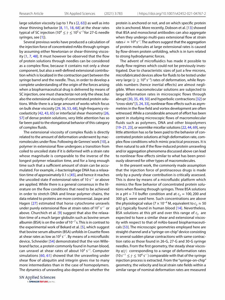
makes the rheometric characterisation of precious mate-rials such as concentrated protein solutions much more cost-effective. The schematic diagrams of the “Rheo-chip” coupled with the pressure sensors are given in Fig. 1.
3.2 Shear viscosity measurements
The steady shear viscosities of the model protein solu-tions were measured by means of Rheo-chips featur-ing a straight channel with width wc = 800 μ m, depth hnominal = 50 μ m and length L = 30 mm. Two pressure taps were located along the microchannel at a distance of L = 30 mm from each other, far away from the inlet and outlet, to measure the overall pressure drop �Ps . The pressure signals were collected by imposing an initial flow rate Q max=10 mL/h and then waiting for the sig-nal to reach a steady-state value. The flow rate was then reduced to 8 mL/h, and the procedure was repeated until a steady-state pressure drop corresponding to a flow rate Qmin = 0.25 mL/h was measured [46]. For all samples studied in this paper, the nominal shear rate at walls is defined as
where htrue is found by fitting the steady-state pressure drop measurements of DI water with the analytical predic-tion for Newtonian flows through rectangular ducts given by White [64]. The corresponding shear viscosity is
where the calibration coefficient 1K=
𝛥Pbuffer
�̇� is obtained by
averaging the ratios between steady-state pressure drops of buffer and the corresponding shear rates, and �buffer ≅ 1 mPa s is the shear viscosity of buffer as measured by a TA
(1)�̇�N =2Q
h2true
wc
,
(2)𝜂N = 𝜂.buffer
K𝛥Ps(�̇�)
�̇�,
Fig. 1 Schematic diagram of a “Rheo-chip” microdevice. 1 Delivery module. 2 Microfluidic flow geometry. 3 Inlet. 4 Outlet. 5 and 6 Hon-eywell pressure sensors
Vol:.(1234567890)
Research Article SN Applied Sciences (2021) 3:783 | https://doi.org/10.1007/s42452-021-04767-2
ARES-G2 rheometer equipped with cone and plate meas-urement tool. The overall measurement time for each sam-ple was 20 mins, approximately, and an amount of sample between 0.6 and 1 mL was used for each measurement. The impact of viscous heating on the rheometric measure-ments was estimated by the Nahme–Griffith number
where � ≈0.5918 W/(m K) is the thermal conductivity of buffer and � =
T
�buffer
|||d�
dT
|||T=T0
≈ 2.6 is the logarithmic deriva-
tive of shear viscosity of buffer with temperature T evalu-ated at T=T0 = 25 ◦ C. It expresses the ratio between the heat generated by viscous heating of samples and the heat released by thermal diffusion at the surface of the micro-channels [46]. Na resulted to be ≤ 10−5 for all the samples and shear rates studied here, which systematically excludes the influence of thermal effects.
3.3 “Syringe‑on‑chip” microdevices
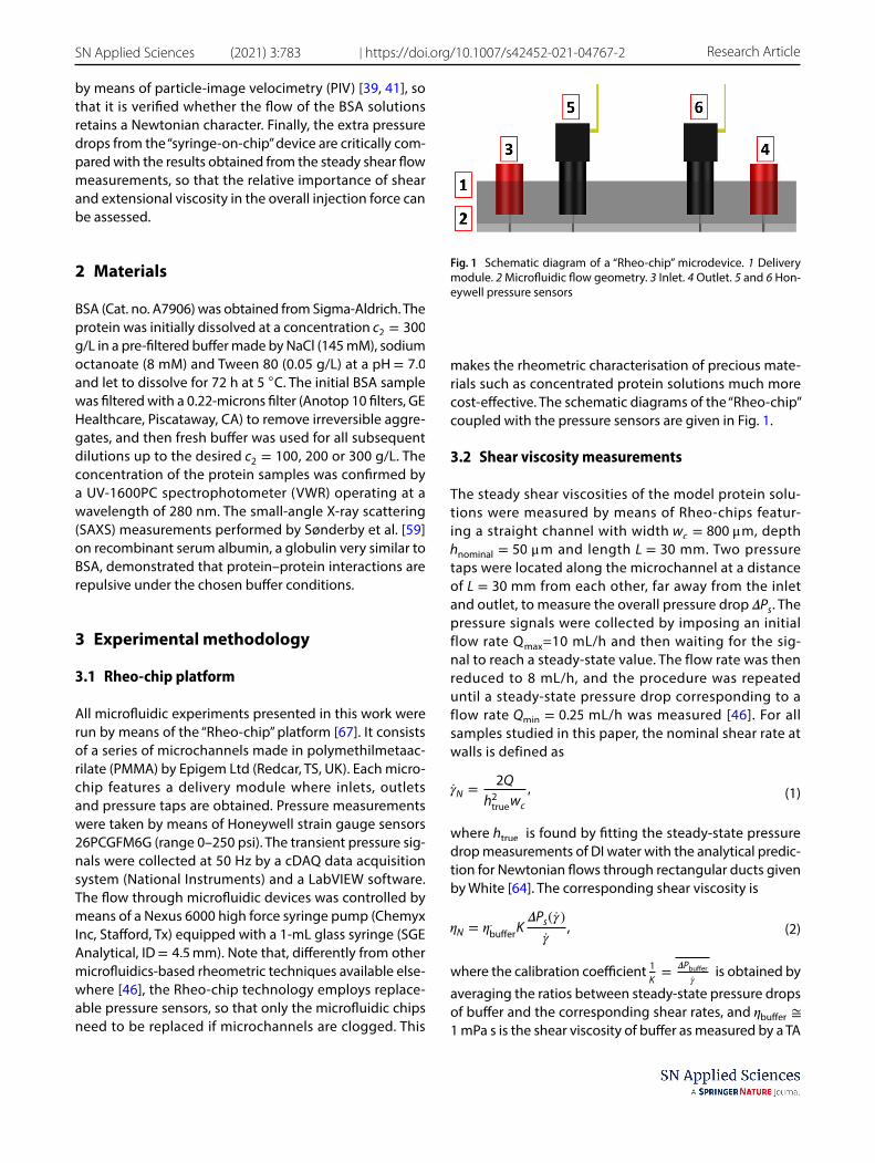
The two “syringe-on-chip” microdevices that are used in this work are presented in Fig. 2. They consist of pla-nar sudden contraction devices with an upstream width wu = 800 μ m, channel depth h = 50 μ m and downstream widths wd = 47 and 38 μ m. The corresponding contrac-tion ratios �=17.02 and 21.05 are the same as what is typi-cally found in syringes where the plunger diameter is 4.5
(3)Na =𝜂0𝛽d
2
h�̇�2
𝜅T,
mm and the needle is a 26G or 27G [66]. Hence, from here onwards such microcontractions will be labeled as “26G” and “27G” geometry, respectively. Note that, due to fact that real syringes have axisymmetric instead of planar abrupt geometries, the flow through the syringe-on-chip devices feature lower Hencky strains �H = ln � compared to real syringes case, where �H = 2 ln � . Each flow geom-etry contains a slot for a pressure sensor as in the figure. The outlet of each microchannel is connected to a fluid reservoir by means of a stainless steel tube with length Ltube = 100 mm and inner diameter ID = 1∕16 inches (Cat. No. 56720-U, Sigma-Aldrich). The total pressure drop �Ptotal measured by the sensor is related to the pressure drop �P across the device by �Ptotal = �P + �Ptube , where �Ptube =
128�LQ
�ID4 is estimated by means of the Hagen–Poi-
seuille law. The �Ptube contribution resulted to be 5 % or less of the total pressure drop. The details of the flow geometries are given in Table 1. The flow of the concen-trated BSA solutions in complex geometry is characterized in terms of the Reynolds number Re
where < vd > , � ≈ 1000 g/L and �0 are the average speed in the downstream channel, the fluid density and zero-shear viscosity, respectively, and Dh =
2hwd
h+wd
is the hydraulic
diameter of the downstream channel. In addition to Re, the Péclet number
(4)Re =𝜌 < vd > Dh
𝜂0,
Table 1 The details of “syringe-on-chip” flow geometries
� and � denote the upstream aspect ratio and contraction ratio, respectively. �H= ln � is the Hencky
strain
Flow cell wu [μm] wd [μm] h [μm] � � �H
26G 800 47 50 20 17.02 2.8327G 800 38 50 20 21.05 3.05
Fig. 2 Schematic diagram of the “syringe-on-chip” microflu-idic geometry. The dimensions wu and wd are given in Table 1, and Lp=14 mm, Lu = 32 mm and Lc = 12 mm
Vol.:(0123456789)
SN Applied Sciences (2021) 3:783 | https://doi.org/10.1007/s42452-021-04767-2 Research Article
expresses the ratio between the characteristic timescales of flow, 𝜏F =
1
�̇�=
wd
2<vd> and Brownian diffusion [34]. The
diffusion time is �D =d2
h
DSL
, where d h = 7 nm is the hydrody-
namic diameter of BSA [4], and DSL is the longtime self-
diffusion coefficient of the BSA molecule, which is esti-mated by the generalized Stokes–Einstein relationship [51] as
with kB and T ≈ 25 ◦ C being the Boltzmann constant and temperature, respectively.
3.4 Particle‑image velocimetry and data analysis method
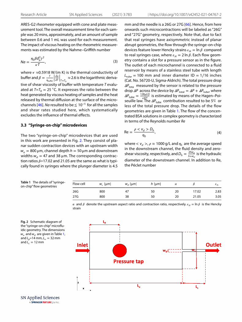
Velocity fields at the center plane of the “syringe-on-chip” flow channels were measured by a �-PIV system (TSI Instruments Ltd, USA). It consists of a pulsed Nd:YAG laser emitting at a wavelength of 532 nm, a Nikon TE2000-E flu-orescent microscope, and an 8 Hz CCD camera with a reso-lution of 1280 × 1024 × 12 bits. A Nikon objective lens with magnification M = 10X and numerical aperture NA = 0.3 was used. In Fig. 3, a schematic diagram of the “syringe-on-chip” device coupled with the PIV system is shown.
The BSA solutions were seeded with 0.01 wt% epi-f l u o r e s c e n t p a r t i c l e s ( d i a m e t e r dp = 1.0 μ m , Exmax∕Emmax = 542∕612 , Duke Scientific Co.). For the above setup, the estimated measurement depth is 26 μ m, equivalent to 52% of the channel depth [38]. All velocity measurements were taken after the pressure drop reached a steady value. Velocity fields were obtained by means of a Nyquist grid engine and a
(5)Pe =�D
�F
(6)DSL=
kBT
3��0�dh,
cross-correlation PIV algorithm supplied by TSI. All the �-PIV measurements presented in this work were per-formed at the middleplane of flow channel, determined by positioning the focus plane at half of the depth between the top and bottom surfaces of the microchan-nel, using a high-precision y-axis linear motor with step resolution 50 nm. The error in the velocity measurements is about 12–16% below the true value. This systematic error could be reduced where a higher magnification objective lens with a larger NA used. The details of the �-PIV techniques can be found in [39, 52]. All the velocity fields presented in this work were obtained by averaging 50 consecutive instantaneous velocity fields, corre-sponding to an overall measurement time of 12.5 s. From the measured two-dimensional planar velocity field v = v(x, z) , the largest eigenvalue �1 of the strain rate ten-sor D =
1
2
(∇v + ∇vT
) is then evaluated to construct the
local extensional rate field �̇�(x, z) . The uncertainty about the PIV velocity measurements is about 12–16%. More information on the computation of �̇�1(x, z) can be found in [33].
4 Results
4.1 BSA solutions under steady shear flow
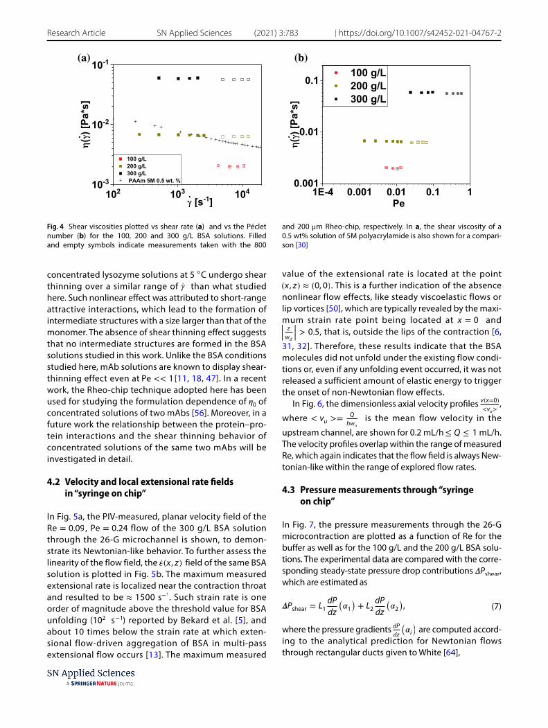
The shear viscosity of the BSA solutions is plotted as a function of the shear rate 200 s−1 ≤ �̇� ≤ 2 × 10⁴ s−1 in Fig. 4a. The viscosity curve of a weakly shear thin-ning, semi-dilute polyacrylamide (PAAm) solution [30] is also shown for a comparison. The rheograms of BSA appeared to be essentially flat, which is attributed to Pe being below unity throughout the imposed range of �̇� (see Fig. 4b). A recent work [12] demonstrated that
Fig. 3 Schematic diagram of a typical “syringe-on-chip” experimental setup. The LabVIEW-based data acquisi-tion platform allows to impose a flow rate Q to the syringe pump, and to measure the total pressure drop �Ptotal from the pressure sensor. 1 Sample-filled glass syringe. 2 Syringe pump. 3 “Syringe-on-chip” microdevice. 4 Magnification lens. 5 Outlet piping
Vol:.(1234567890)
Research Article SN Applied Sciences (2021) 3:783 | https://doi.org/10.1007/s42452-021-04767-2
concentrated lysozyme solutions at 5 ◦ C undergo shear thinning over a similar range of �̇� than what studied here. Such nonlinear effect was attributed to short-range attractive interactions, which lead to the formation of intermediate structures with a size larger than that of the monomer. The absence of shear thinning effect suggests that no intermediate structures are formed in the BSA solutions studied in this work. Unlike the BSA conditions studied here, mAb solutions are known to display shear-thinning effect even at Pe << 1 [11, 18, 47]. In a recent work, the Rheo-chip technique adopted here has been used for studying the formulation dependence of �0 of concentrated solutions of two mAbs [56]. Moreover, in a future work the relationship between the protein–pro-tein interactions and the shear thinning behavior of concentrated solutions of the same two mAbs will be investigated in detail.
4.2 Velocity and local extensional rate fields in “syringe on chip”
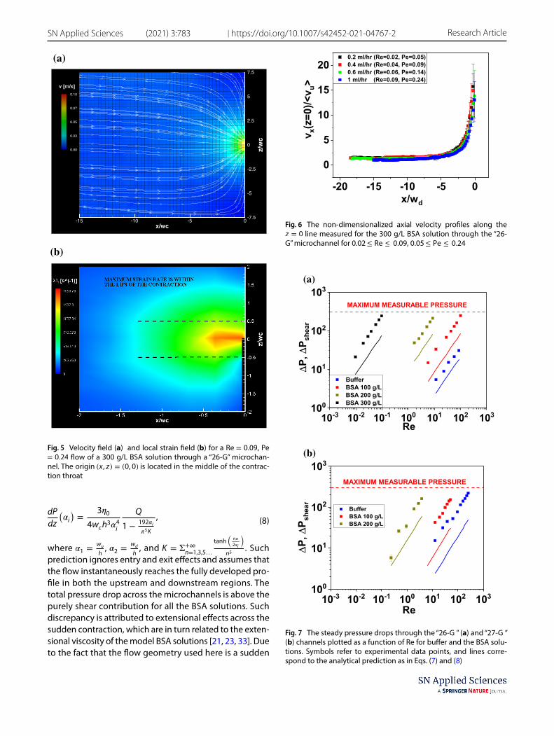
In Fig. 5a, the PIV-measured, planar velocity field of the Re = 0.09 , Pe = 0.24 flow of the 300 g/L BSA solution through the 26-G microchannel is shown, to demon-strate its Newtonian-like behavior. To further assess the linearity of the flow field, the �̇�(x, z) field of the same BSA solution is plotted in Fig. 5b. The maximum measured extensional rate is localized near the contraction throat and resulted to be ≈ 1500 s−1 . Such strain rate is one order of magnitude above the threshold value for BSA unfolding ( 102 s −1 ) reported by Bekard et al. [5], and about 10 times below the strain rate at which exten-sional flow-driven aggregation of BSA in multi-pass extensional flow occurs [13]. The maximum measured
value of the extensional rate is located at the point (x, z) ≈ (0, 0) . This is a further indication of the absence nonlinear flow effects, like steady viscoelastic flows or lip vortices [50], which are typically revealed by the maxi-mum strain rate point being located at x = 0 and |||z
wd
|||> 0.5 , that is, outside the lips of the contraction [6,
31, 32]. Therefore, these results indicate that the BSA molecules did not unfold under the existing flow condi-tions or, even if any unfolding event occurred, it was not released a sufficient amount of elastic energy to trigger the onset of non-Newtonian flow effects.
In Fig. 6, the dimensionless axial velocity profiles v(x=0)<vu>
,
where < vu >=Q
hwu
is the mean flow velocity in the
upstream channel, are shown for 0.2 mL/h ≤ Q ≤ 1 mL/h. The velocity profiles overlap within the range of measured Re, which again indicates that the flow field is always New-tonian-like within the range of explored flow rates.
4.3 Pressure measurements through “syringe on chip”
In Fig. 7, the pressure measurements through the 26-G microcontraction are plotted as a function of Re for the buffer as well as for the 100 g/L and the 200 g/L BSA solu-tions. The experimental data are compared with the corre-sponding steady-state pressure drop contributions �Pshear , which are estimated as
where the pressure gradients dPdz
(�i) are computed accord-
ing to the analytical prediction for Newtonian flows through rectangular ducts given to White [64],
(7)�Pshear = L1dP
dz
(�1)+ L2
dP
dz
(�2),
(a) (b)
Fig. 4 Shear viscosities plotted vs shear rate (a) and vs the Péclet number (b) for the 100, 200 and 300 g/L BSA solutions. Filled and empty symbols indicate measurements taken with the 800
and 200 μ m Rheo-chip, respectively. In a, the shear viscosity of a 0.5 wt% solution of 5M polyacrylamide is also shown for a compari-son [30]
Vol.:(0123456789)
SN Applied Sciences (2021) 3:783 | https://doi.org/10.1007/s42452-021-04767-2 Research Article
where �1 =wu
h , �2 =
wd
h , and K = Σ+∞
n=1,3,5…
tanh
(n�
2�i
)
n5 . Such
prediction ignores entry and exit effects and assumes that the flow instantaneously reaches the fully developed pro-file in both the upstream and downstream regions. The total pressure drop across the microchannels is above the purely shear contribution for all the BSA solutions. Such discrepancy is attributed to extensional effects across the sudden contraction, which are in turn related to the exten-sional viscosity of the model BSA solutions [21, 23, 33]. Due to the fact that the flow geometry used here is a sudden
(8)dP
dz
(�i)=
3�0
4wch3�4
i
Q
1 −192�i
�5K
,
x/wc
z/wc
-15 -10 -5 0 -7.5
-5
-2.5
0
2.5
5
7.5
0.10
0.07
0.05
0.03
0.00
(a)
(b)
Fig. 5 Velocity field (a) and local strain field (b) for a Re = 0.09 , Pe = 0.24 flow of a 300 g/L BSA solution through a “26-G” microchan-nel. The origin (x, z) = (0, 0) is located in the middle of the contrac-tion throat
Fig. 6 The non-dimensionalized axial velocity profiles along the z = 0 line measured for the 300 g/L BSA solution through the “26-G” microchannel for 0.02 ≤ Re ≤ 0.09, 0.05 ≤ Pe ≤ 0.24
(a)
(b)
Fig. 7 The steady pressure drops through the “26-G ” (a) and “27-G ” (b) channels plotted as a function of Re for buffer and the BSA solu-tions. Symbols refer to experimental data points, and lines corre-spond to the analytical prediction as in Eqs. (7) and (8)
Vol:.(1234567890)
Research Article SN Applied Sciences (2021) 3:783 | https://doi.org/10.1007/s42452-021-04767-2
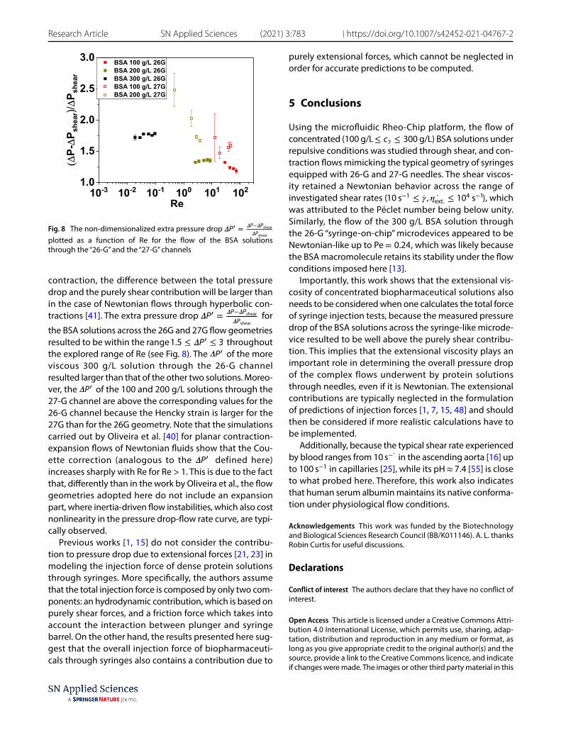
contraction, the difference between the total pressure drop and the purely shear contribution will be larger than in the case of Newtonian flows through hyperbolic con-tractions [41]. The extra pressure drop �P� = �P−�Pshear
�Pshear for
the BSA solutions across the 26G and 27G flow geometries resulted to be within the range 1.5 ≤ �P′ ≤ 3 throughout the explored range of Re (see Fig. 8). The �P′ of the more viscous 300 g/L solution through the 26-G channel resulted larger than that of the other two solutions. Moreo-ver, the �P′ of the 100 and 200 g/L solutions through the 27-G channel are above the corresponding values for the 26-G channel because the Hencky strain is larger for the 27G than for the 26G geometry. Note that the simulations carried out by Oliveira et al. [40] for planar contraction-expansion flows of Newtonian fluids show that the Cou-ette correction (analogous to the �P′ defined here) increases sharply with Re for Re > 1. This is due to the fact that, differently than in the work by Oliveira et al., the flow geometries adopted here do not include an expansion part, where inertia-driven flow instabilities, which also cost nonlinearity in the pressure drop-flow rate curve, are typi-cally observed.
Previous works [1, 15] do not consider the contribu-tion to pressure drop due to extensional forces [21, 23] in modeling the injection force of dense protein solutions through syringes. More specifically, the authors assume that the total injection force is composed by only two com-ponents: an hydrodynamic contribution, which is based on purely shear forces, and a friction force which takes into account the interaction between plunger and syringe barrel. On the other hand, the results presented here sug-gest that the overall injection force of biopharmaceuti-cals through syringes also contains a contribution due to
purely extensional forces, which cannot be neglected in order for accurate predictions to be computed.
5 Conclusions
Using the microfluidic Rheo-Chip platform, the flow of concentrated (100 g/L ≤ c2 ≤ 300 g/L) BSA solutions under repulsive conditions was studied through shear, and con-traction flows mimicking the typical geometry of syringes equipped with 26-G and 27-G needles. The shear viscos-ity retained a Newtonian behavior across the range of investigated shear rates ( 10 s−1 ≤ �̇� , ̇𝜂ext. ≤ 104 s−1 ), which was attributed to the Péclet number being below unity. Similarly, the flow of the 300 g/L BSA solution through the 26-G “syringe-on-chip” microdevices appeared to be Newtonian-like up to Pe = 0.24 , which was likely because the BSA macromolecule retains its stability under the flow conditions imposed here [13].
Importantly, this work shows that the extensional vis-cosity of concentrated biopharmaceutical solutions also needs to be considered when one calculates the total force of syringe injection tests, because the measured pressure drop of the BSA solutions across the syringe-like microde-vice resulted to be well above the purely shear contribu-tion. This implies that the extensional viscosity plays an important role in determining the overall pressure drop of the complex flows underwent by protein solutions through needles, even if it is Newtonian. The extensional contributions are typically neglected in the formulation of predictions of injection forces [1, 7, 15, 48] and should then be considered if more realistic calculations have to be implemented.
Additionally, because the typical shear rate experienced by blood ranges from 10 s−1 in the ascending aorta [16] up to 100 s−1 in capillaries [25], while its pH ≈ 7.4 [55] is close to what probed here. Therefore, this work also indicates that human serum albumin maintains its native conforma-tion under physiological flow conditions.
Acknowledgements This work was funded by the Biotechnology and Biological Sciences Research Council (BB/K011146). A. L. thanks Robin Curtis for useful discussions.
Declarations
Conflict of interest The authors declare that they have no conflict of interest.
Open Access This article is licensed under a Creative Commons Attri-bution 4.0 International License, which permits use, sharing, adap-tation, distribution and reproduction in any medium or format, as long as you give appropriate credit to the original author(s) and the source, provide a link to the Creative Commons licence, and indicate if changes were made. The images or other third party material in this
Fig. 8 The non-dimensionalized extra pressure drop �P� = �P−�Pshear
�Pshear
plotted as a function of Re for the flow of the BSA solutions through the “26-G” and the “27-G” channels
Vol.:(0123456789)
SN Applied Sciences (2021) 3:783 | https://doi.org/10.1007/s42452-021-04767-2 Research Article
article are included in the article’s Creative Commons licence, unless indicated otherwise in a credit line to the material. If material is not included in the article’s Creative Commons licence and your intended use is not permitted by statutory regulation or exceeds the permitted use, you will need to obtain permission directly from the copyright holder. To view a copy of this licence, visit http:// creat iveco mmons. org/ licen ses/ by/4. 0/.
References
1. Allmendinger A, Fischer S, Huwyler J, Mahler HC, Schwarb E, Zarraga IE, Mueller R (2014) Rheological characterization and injection forces of concentrated protein formulations: an alter-native predictive model for non-newtonian solutions. Eur J Pharm Biopharm 87(2):318–328
2. Amin S, Barnett GV, Pathak JA, Roberts CJ, Sarangapani PS (2014) Protein aggregation, particle formation, characterization & rhe-ology. Curr Opin Colloid Interface Sci 19(5):438–449
3. Arratia PE, Thomas C, Diorio J, Gollub JP (2006) Elastic insta-bilities of polymer solutions in cross-channel flow. Phys Rev Let 96(14):144502
4. Axelsson I (1978) Characterization of proteins and other mac-romolecules by agarose gel chromatography. J Chromatogr A 152(1):21–32
5. Bekard IB, Asimakis P, Teoh CL, Ryan T, Howlett GJ, Bertolini J, Dunstan DE (2012) Bovine serum albumin unfolds in Couette flow. Soft Matter 8(2):385–389
6. Binding D (1988) An approximate analysis for contraction and converging flows. J Non-Newton Fluid Mech 27(2):173–189
7. Burckbuchler V, Mekhloufi G, Giteau AP, Grossiord J, Huille S, Agnely F (2010) Rheological and syringeability properties of highly concentrated human polyclonal immunoglobulin solu-tions. Eur J Pharm Biopharm 76(3):351–356
8. Castellanos MM, Pathak JA, Colby RH (2014) Both protein adsorption and aggregation contribute to shear yielding and viscosity increase in protein solutions. Soft Matter 10(1):122–131
9. Churchich JE (1962) Polarization of fluorescence studies of reduced bovine serum albumin. Arch Biochem Biophys 97(3):574–577
10. De Gennes PG (1979) Scaling concepts in polymer physics. Cor-nell University Press
11. Dear BJ, Hung JJ, Truskett TM, Johnston KP (2017) Contrasting the influence of cationic amino acids on the viscosity and stabil-ity of a highly concentrated monoclonal antibody. Pharm Res 34(1):193–207
12. Dharmaraj VL, Godfrin PD, Liu Y, Hudson SD (2016) Rheology of clustering protein solutions. Biomicrofluidics 10(4):043509
13. Dobson J, Kumar A, Willis LF, Tuma R, Higazi DR, Turner R, Lowe DC, Ashcroft AE, Radford SE, Kapur N, Brockwell DJ (2017) Induc-ing protein aggregation by extensional flow. Proc Natl Acad Sci 114(18):4673–4678. https:// doi. org/ 10. 1073/ pnas. 17027 24114
14. Fasano M, Curry S, Terreno E, Galliano M, Fanali G, Narciso P, Notari S, Ascenzi P (2005) The extraordinary ligand binding properties of human serum albumin. IUBMB Life 57(12):787–796
15. Fischer I, Schmidt A, Bryant A, Besheer A (2015) Calculation of injection forces for highly concentrated protein solutions. Int J Pharm 493(1):70–74. https:// doi. org/ 10. 1016/j. ijpha rm. 2015. 07. 054
16. Gabe IT, Gault JH, Ross JRJ, Mason DT, Mills CJ, Schillingford JP, Braunwald E (1969) Measurement of instantaneous blood flow velocity and pressure in conscious man with a catheter-tip velocity probe. Circulation 40(5):603–614
17. Garidel P, Kuhn AB, Schäfer LV, Karow-Zwick AR, Blech M (2017) High-concentration protein formulations: how high is high? Eur J Pharm Biopharm 119:353–360
18. Godfrin PD, Zarraga IE, Zarzar J, Porcar L, Falus P, Wagner NJ, Liu Y (2016) Effect of hierarchical cluster formation on the viscosity of concentrated monoclonal antibody formulations studied by neutron scattering. J Phys Chem B 120(2):278–291
19. Gulati S, Liepmann D, Muller SJ (2008a) Elastic secondary flows of semidilute DNA solutions in abrupt 90 microbends. Phys Rev E 78(3):036314
20. Gulati S, Muller SJ, Liepmann D (2008b) Direct measurements of viscoelastic flows of DNA in a 2: 1 abrupt planar micro-con-traction. J Non-Newton Fluid Mech 155(1):51–66
21. Haward SJ (2014) Characterization of hyaluronic acid and syno-vial fluid in stagnation point elongational flow. Biopolymers 101(3):287–305
22. Haward SJ, McKinley GH (2012) Stagnation point flow of worm-like micellar solutions in a microfluidic cross-slot device: effects of surfactant concentration and ionic environment. Phys Rev E 85(3):031502
23. Haward SJ, Odell JA, Berry M, Hall T (2011) Extensional rheology of human saliva. Rheol Acta 50(11–12):869–879
24. Haward SJ, McKinley GH, Shen AQ (2016) Elastic instabilities in planar elongational flow of monodisperse polymer solutions. Sci Rep 6:1–18
25. Ivanov K, Kalinina M, Levkovich YI (1981) Blood flow velocity in capillaries of brain and muscles and its physiological signifi-cance. Microvascu Res 22(2):143–155
26. Jaishankar A, McKinley GH (2013) Power-law rheology in the bulk and at the interface: quasi-properties and fractional con-stitutive equations. Proc R Soc A 469:20120284
27. Jaspe J, Hagen SJ (2006) Do protein molecules unfold in a simple shear flow? Biophys J 91(9):3415–3424
28. Jorgensen L, Hostrup S, Moeller EH, Grohganz H (2009) Recent trends in stabilising peptides and proteins in pharmaceutical formulation-considerations in the choice of excipients. Expert Opin Drug Deliv 6(11):1219–1230
29. Kanai S, Liu J, Patapoff TW, Shire SJ (2008) Reversible self-associ-ation of a concentrated monoclonal antibody solution mediated by fab-fab interaction that impacts solution viscosity. J Pharm Sci 97(10):4219–4227
30. Lanzaro A, Yuan XF (2011) Effects of contraction ratio on non-lin-ear dynamics of semi-dilute, highly polydisperse paam solutions in microfluidics. J Non-Newton Fluid Mech 166(17):1064–1075
31. Lanzaro A, Yuan XF (2014) A quantitative analysis of spatial extensional rate distribution in nonlinear viscoelastic flows. J Non-Newton Fluid Mech 207:32–41
32. Lanzaro A, Li Z, Yuan XF (2015) Quantitative characterization of high molecular weight polymer solutions in microfluidic hyper-bolic contraction flow. Microfluid Nanofluid 18(5–6):819–828
33. Lanzaro A, Corbett D, Yuan XF (2017) Non-linear dynamics of semi-dilute paam solutions in a microfluidic 3D cross-slot flow geometry. J Non-Newton Fluid Mech 242:57–65
34. Larson RG (1999) The structure and rheology of complex fluids, vol 150. Oxford University Press, New York
35. Li Z, Yuan XF, Haward SJ, Odell JA, Yeates S (2011) Non-linear dynamics of semi-dilute polydisperse polymer solutions in microfluidics: a study of a benchmark flow problem. J Non-Newton Fluid Mech 166(16):951–963
36. Liu J, Nguyen MD, Andya JD, Shire SJ (2005) Reversible self-asso-ciation increases the viscosity of a concentrated monoclonal antibody in aqueous solution. J Pharm Sci 94(9):1928–1940
37. Mathaes R, Koulov A, Joerg S, Mahler HC (2016) Subcutaneous injection volume of biopharmaceuticals: pushing the bounda-ries. J Pharm Sci 105(8):2255–2259
Vol:.(1234567890)
Research Article SN Applied Sciences (2021) 3:783 | https://doi.org/10.1007/s42452-021-04767-2
38. Meinhart C, Wereley S, Gray M (2000) Volume illumination for two-dimensional particle image velocimetry. Measur Sci Tech-nol 11(6):809–14
39. Meinhart CD, Wereley ST, Santiago JG (1999) PIV measurements of a microchannel flow. Exp Fluids 27(5):414–419
40. Oliveira MS, Rodd LE, McKinley GH, Alves MA (2008) Simula-tions of extensional flow in microrheometric devices. Microfluid Nanofluid 5(6):809
41. Oliveira MSN, Alves MA, Pinho FT, McKinley GH (2007) Viscous flow through microfabricated hyperbolic contractions. Exp Flu-ids 43(2–3):437–451
42. Pan W, Filobelo L, Pham ND, Galkin O, Uzunova VV, Vekilov PG (2009) Viscoelasticity in homogeneous protein solutions. Phys Rev Lett 102(5):058101
43. Patel AR, Kerwin BA, Kanapuram SR (2009) Viscoelastic charac-terization of high concentration antibody formulations using quartz crystal microbalance with dissipation monitoring. J Pharm Sci 98(9):3108–3116
44. Pathak JA, Hudson SD (2006) Rheo-optics of equilibrium poly-mer solutions: wormlike micelles in elongational flow in a micro-fluidic cross-slot. Macromolecules 39(25):8782–8792
45. Perkins TT, Smith DE, Chu S (1997) Single polymer dynamics in an elongational flow. Science 276(5321):2016–2021
46. Pipe CJ, Majmudar TS, McKinley GH (2008) High shear rate vis-cometry. Rheol Acta 47(5–6):621–642
47. Ramallo N, Paudel S, Schmit J (2019) Cluster formation and entanglement in the rheology of antibody solutions. J Phys Chem B 123(18):3916–3923
48. Rathore N, Pranay P, Bernacki J, Eu B, Ji W, Walls E (2012) Charac-terization of protein rheology and delivery forces for combina-tion products. J Pharm Sci 101(12):4472–4480
49. Rodd L, Cooper-White J, Boger D, McKinley G (2007) Role of the elasticity number in the entry flow of dilute polymer solutions in micro-fabricated contraction geometries. J Non-Newton Fluid Mech 143(2):170–191
50. Rodd LE, Scott TP, Boger DV, Cooper-White JJ, McKinley GH (2005) The inertio-elastic planar entry flow of low-viscosity elas-tic fluids in micro-fabricated geometries. J Non-Newton Fluid Mech 129(1):1–22
51. Roos M, Ott M, Hofmann M, Link S, Rossler E, Balbach J, Krush-elnitsky A, Saalwachter K (2016) Coupling and decoupling of rotational and translational diffusion of proteins under crowd-ing conditions. J Am Chem Soc 138(32):10365–10372
52. Santiago JG, Wereley ST, Meinhart CD, Beebe D, Adrian RJ (1998) A particle image velocimetry system for microfluidics. Exp Fluids 25(4):316–319
53. Sarangapani PS, Hudson SD, Migler KB, Pathak JA (2013) The limitations of an exclusively colloidal view of protein solution hydrodynamics and rheology. Biophys J 105(10):2418–2426
54. Schneider S, Nuschele S, Wixforth A, Gorzelanny C, Alexander-Katz A, Netz R, Schneider M (2007) Shear-induced unfolding trig-gers adhesion of von willebrand factor fibers. Proc Natl Acad Sci 104(19):7899–7903
55. Severinghaus J (1958) Oxyhemoglobin dissociation curve cor-rection for temperature and pH variation in human blood. J Appl Physiol 12(3):485–486
56. Shah M, Corbett D, Lanzaro A, Roche A, Sibanda N, Davis P, Uddin S, van der Walle C, Curtis R, Pluen A (2020) Micro-and macro-viscosity relations in high concentration antibody solutions. Eur J Pharm Biopharm 153:211–221
57. Sharma V, Jaishankar A, Wang YC, McKinley GH (2011) Rheol-ogy of globular proteins: apparent yield stress, high shear rate viscosity and interfacial viscoelasticity of bovine serum albumin solutions. Soft Matter 7(11):5150–5160
58. Shire SJ, Shahrokh Z, Liu J (2004) Challenges in the develop-ment of high protein concentration formulations. J Pharma Sci 93(6):1390–1402
59. Sønderby P, Bukrinski JT, Hebditch M, Peters GH, Curtis RA, Harris P (2018) Self-interaction of human serum albumin: a formula-tion perspective. ACS Omega 3(11):16105–16117
60. Szymczak P, Cieplak M (2006) Stretching of proteins in a uniform flow. J Chem Phys 125(16):164903
61. Szymczak P, Cieplak M (2007) Proteins in a shear flow. J Chem Phys 127(15):10B618
62. Viola M, Sequeira J, Seiça R, Veiga F, Serra J, Santos AC, Ribeiro AJ (2018) Subcutaneous delivery of monoclonal antibodies: how do we get there? J Controll Release 286:301–314
63. Wang WH, Lilyestrom WG, Hu ZY, Scherer TM (2018) Cluster size and quinary structure determine the rheological effects of antibody self-association at high concentrations. J Phys Chem B 122(7):2138–2154
64. White FM, Corfield I (2006) Viscous fluid flow, vol 3. McGraw-Hill Higher Education, Boston
65. Yadav S, Sreedhara A, Kanai S, Liu J, Lien S, Lowman H, Kalo-nia DS, Shire SJ (2011) Establishing a link between amino acid sequences and self-associating and viscoelastic behav-ior of two closely related monoclonal antibodies. Pharm Res 28(7):1750–1764
66. Yentis SM, Hirsch NP, Ip J (2018) Anaesthesia and intensive care AZ E-book: an encyclopaedia of principles and practice. Elsevier Health Sciences
67. Yuan Xf (2016) Rheometry apparatus. US Patent 9,354,152 68. Zarraga IE, Taing R, Zarzar J, Luoma J, Hsiung J, Patel A, Lim FJ
(2013) High shear rheology and anisotropy in concentrated solu-tions of monoclonal antibodies. J Pharm Sci 102(8):2538–2549
69. Zhao Y, Cheung P, Shen AQ (2014) Microfluidic flows of wormlike micellar solutions. Adv Colloid Interface Sci 211:34–46
Publisher’s Note Springer Nature remains neutral with regard to jurisdictional claims in published maps and institutional affiliations.