Embed Size (px)
Citation preview
Faraday DiscussionsCite this: DOI: 10.1039/c4fd00062e
PAPER
Publ
ishe
d on
15
July
201
4. D
ownl
oade
d by
Uni
vers
ity C
olle
ge L
ondo
n on
15/
10/2
014
14:0
5:00
.
View Article OnlineView Journal
A low cost synthesis method forfunctionalised iron oxide nanoparticles formagnetic hyperthermia from readilyavailable materials†
Joseph C. Bear,*a Bin Yu,a Cristina Blanco-Andujar,b
Paul D. McNaughter,c Paul Southern,b Marc-Krystelle Mafina,d
Quentin A. Pankhurstb and Ivan P. Parkina
Received 9th April 2014, Accepted 15th July 2014
DOI: 10.1039/c4fd00062e
The synthesis of iron oxide nanocrystals from reagents taken from high street sources
using thermal decomposition of an iron–fatty acid precursor in a high boiling point
solvent in the presence of surfactants is presented. The nanocrystals were characterised
using a variety of techniques including: electron microscopy, X-ray dispersive
spectroscopy, infrared spectroscopy, X-ray diffraction (XRD), X-ray photoelectron
spectroscopy (XPS) and magnetometry. Thermogravimetric analysis (TGA) is also used
to compare the decomposition behaviour of iron oleate and iron palmitate, our
nanoparticle precursors. The nanoparticles also exhibit shape anisotropy when prepared
under optimum conditions. We show that these nanoparticles have potential in
magnetic hyperthermia after transfer to aqueous media via an amphiphilic polymer.
Introduction
The large scale and inexpensive synthesis of monodisperse nanocrystals fromnon-hazardous precursors is an area of intense interest in nanoparticulateresearch.1–5
Synthesis methods such as continuous hydrothermal ow synthesis and ballmilling generate kilogram-scale volumes of nanoparticles, but oen at the
aMaterials Chemistry Research Centre, Department of Chemistry, University College London, 20 Gordon Street,
London, WC1H 0AJ, UK. E-mail: [email protected] Healthcare Biomagnetics Laboratories, Royal Institution of Great Britain, 21 Albemarle Street, London,
W1S 4BS, UKcSchool of Chemistry, University of East Anglia, Norwich Research Park, Norwich, NR4 7TJ, UKdSchool of Engineering and Materials Science, Queen Mary, University of London, Mile End Road, London, E1
4NS, UK
† Electronic supplementary information (ESI) available: Contains additional TEM, SAED, EDX and FTIRcharacterisation, differential scanning calorimetry curves of iron oleate and iron palmitate. See DOI:10.1039/c4fd00062e
This journal is © The Royal Society of Chemistry 2014 Faraday Discuss.
Faraday Discussions PaperPu
blis
hed
on 1
5 Ju
ly 2
014.
Dow
nloa
ded
by U
nive
rsity
Col
lege
Lon
don
on 1
5/10
/201
4 14
:05:
00.
View Article Online
expense of product crystallinity and monodispersity.6–9 On the other hand, thenanoparticle quality control gained using hot-injection and “heat-up”methods isexcellent.3,10,11 However, these methods oen utilise expensive reagents and arenot applicable for large scale processes due to large temperature gradients placedacross synthesis solutions. The vast majority of “heat-up” methods involve thethermal decomposition of iron–fatty acid complexes such as iron oleate. Iron–fatty acid complexes are either isolated and decomposed in the presence ofsurfactants12 or formed in situ.13 In their landmark paper, Hyeon and co-workersdecomposed a pre-synthesised iron oleate complex in the presence of oleic acidand 1-octadecene to produce highly monodisperse and size-controllable Fe3O4
nanocrystals on a batch-level scale.3 In their paper, they allude to the possibility ofa scale-up process and draw attention to the inexpensive nature and low toxicity ofthe precursors (Fig. 4, ESI†).
In particular, iron oxide has received a great deal of interest for magnetic uidhyperthermia (MFH); a cancer treatment which uses radiofrequency to heat upsuperparamagnetic nanoparticles in vivo to thermally ablate/treat tumourcells.14–17 The best nanoparticles for MFH are oen a compromise between size,biocompatibility, cost, ease of synthesis, ability to respond to an external stimulusand heating ability.17–19 Iron oxide has been of particular interest due to itsfullment of the majority of these desired properties.20,21
In this paper, we have developed a synthesis using a similar thermal decom-position method to the one developed by Hyeon et al., but with precursors solelybought from high street shops and supermarkets. We report the batch-synthesisof high-quality iron oxide nanocrystals at a fraction of the cost compared to theHyeon paper. We also demonstrate their suitability and effectiveness for magnetichyperthermia, aer transfer to aqueous media via amphiphilic polymercoating.22,23
The nanoparticles demonstrate signicant temperature rises at low nano-particle concentrations, particularly the nanostructures obtained from thedecomposition of iron palmitate in shark liver oil. The methods and reagentsproposed in this paper therefore represent a viable, facile and cheap route toeffective hyperthermia agents.
Experimental detailsCharacterisation techniques
Transmission electron microscopy (TEM) images were recorded using a JEOL JEM1200EX with a 4 megapixel Gatan Orius SC200 charge-coupled device (CCD)camera at an acceleration voltage of 120 kV. High resolution transmission elec-tron microscopy (HRTEM) measurements were collected using a Philips CM200FEG TEM tted with a Gatan GIF 200 imaging lter and an Oxford InstrumentsUTW EDX detector running ISIS soware. XPS measurements were recorded on aThermo Scientic K-alpha spectrometer. XRD patterns were obtained on a Pan-Alytical diffractometer using Co Ka radiation, l ¼ 1.789010 A. ATR-FTIRmeasurements were taken over a range of 450 to 4000 cm�1 using a Perkin-ElmerSpectrum-100 equipped with a universal ATR attachment. Magnetisation data wastaken using a Quantum Design MPMS SQUID VSM Magnetometer (San Diego,USA) at 300 K using a eld range of�7 T. Mossbauer spectroscopy was carried outusing a SEE Co. Model W302 Resonant Gamma Ray Spectrometer, with a 57Co(Rh)
Faraday Discuss. This journal is © The Royal Society of Chemistry 2014
Paper Faraday DiscussionsPu
blis
hed
on 1
5 Ju
ly 2
014.
Dow
nloa
ded
by U
nive
rsity
Col
lege
Lon
don
on 1
5/10
/201
4 14
:05:
00.
View Article Online
gamma ray source operating at 300 K. Isomer shis were measured relative to a-Feat 300 K. Hyperthermia experiments were carried out using a MACH system(Magnetic Alternating Current Hyperthermia) designed and built by ResonantCircuits Limited.24 The temperature was monitored using a uoroptic tempera-ture probe (Luxtron FOT Lab Kit, Lumasense California USA).
Materials
Iron(III) chloride hexahydrate (98%) was purchased from VWR International Ltd.Iron tablets were purchased from Boots Ltd. Olive oil and “Sainsbury basics” soapwas purchased form J. Sainsbury Ltd. Shark liver oil (60 � 1000 mg capsules) waspurchased from Shark Liver Oil UK.
All other laboratory chemicals used in this investigation were purchased fromSigma-Aldrich Chemical Co; including: 1-octadecene (technical grade, 90%), oleicacid (technical grade, 90%), oleylamine (technical grade, #70%), poly(maleicanhydride-alt-1-octadecene) (average Mn ¼ 30 000–50 000), tetramethyl ammo-nium hydroxide pentahydrate ($97%) and sodium oleate (#82% fatty acidcontent). Laboratory solvents were purchased from Fisher Scientic Ltd. and ofthe highest possible grade.
Nanoparticle synthesis and functionalisation
Iron oxide nanoparticle synthesis. Iron oxide nanoparticles were synthesisedby thermal decomposition of iron oleate, according to Park et al.3 The iron oleatewas prepared by heating a suspension of iron(III) chloride hexahydrate (10.8 g, 40mmol) and sodium oleate (36.5 g, 120 mmol) in a solvent mixture of n-hexane (140ml), deionised water (60 ml) and ethanol (80 ml) to 70 �C for 4 hours. The organiclayer was separated and washed with 3 � 30 ml portions of distilled water toremove sodium chloride. The dark brown organic layer was separated and dried invacuo, to remove hexane, and isolated as a waxy solid (yield �70%).
Iron oleate (18.0 g, 20 mmol) and oleic acid (2.35 ml, 10 mmol) were dissolvedin 1-octadecene (100 g, 396.1 mmol) and stirred thoroughly at room temperature.The reactionmixture was heated to 320 �C at a rate of 3.3 �Cmin�1 under nitrogenand held at 320 �C for 1 hour. The resulting black solution was allowed to cool toroom temperature before addition of ethanol (250 ml) to precipitate the particles.The solution was centrifuged at 600g for 10 minutes, giving solid particleprecipitates. If the precipitate was not solid, excess oleate-type species wereremoved by washing with ethanol (2� 80ml). The supernatant was discarded andthe solid particle precipitates dispersed in chloroform (30 ml total).
Modied Fe3O4 nanoparticle synthesis. Iron palmitate was prepared by heat-ing a suspension of homogenised iron tablets (20 tablets each containing 14 mgof iron as iron gluconate) and soap (assumed to be 65% sodium palmitate, 0.321g, 1.15 mmol) in a solvent mixture of n-hexane (140 ml), deionised water (60 ml)and ethanol (80 ml) to 70 �C for 4 hours. The organic layer was separated andwashed with 3� 30 ml portions of distilled water to remove sodium chloride. Thedark organic layer was dried in vacuo to remove hexane, yielding the iron oleatecomplex as a waxy solid.
For �12 nm particles and rods. Iron palmitate (2 g, 2.4 mmol) and olive oil(65% oleic acid, 0.59 ml, 1.2 mmol) were dissolved in 1-octadecene (20 ml,62.5 mmol) or shark liver oil (20 ml) and stirred thoroughly at room temperature.
This journal is © The Royal Society of Chemistry 2014 Faraday Discuss.
Faraday Discussions PaperPu
blis
hed
on 1
5 Ju
ly 2
014.
Dow
nloa
ded
by U
nive
rsity
Col
lege
Lon
don
on 1
5/10
/201
4 14
:05:
00.
View Article Online
A molar ratio of 1 : 1 iron palmitate to the oleic acid content of olive oil was usedto grow the rods (Fig. 2(E) and (F)), whilst the solvent was changed to shark liveroil. The reaction mixture was heated to 320 �C at a rate of 3.3 �C min�1 undernitrogen and held at 320 �C for 1 hour. The resulting black solution was allowed tocool to room temperature before addition of ethanol (250 ml) to precipitate theparticles. The solution was centrifuged at 600g for 10 minutes, giving solidparticle precipitates. If the precipitate was not solid, excess fatty acid-type specieswere removed by washing with ethanol (2 � 80 ml). The supernatant was dis-carded and the solid particle precipitates dispersed in chloroform (30 ml total).
Phase transfer of iron oxide nanoparticles
Oleic acid-coated nanoparticles were transferred from organic solvents to wateraccording to a protocol developed by Lees et al.22 Iron oxide nanoparticles inhexane (5 ml) were precipitated with ethanol (�100 ml), centrifuged and the solidresidue re-dispersed in chloroform (10 ml). Poly(maleic anhydride-alt-1-octade-cene) (10 mg) was dissolved in chloroform (10 ml) and added under vigorousstirring to the nanoparticle solution and le to stir for 1 hour. The chloroform wasremoved slowly in vacuo (750 mbar, 25 �C water bath) and then subsequentlyplaced under a �10�2 mbar vacuum for 24 hours. 20 ml of a 0.1 M solution oftetramethyl ammonium hydroxide was added, and le overnight to react. Oncethe nanoparticles were fully dispersed, the solution was acidied to pH 8 withacetic acid before concentration with centrifuge lters (Centriprep YM-10, 10 kDaMW cutoff for 3 � 20 minute cycles at 3000g).
Magnetic hyperthermia testing
A sample of polymer-coated water-transferred iron oxide nanoparticles (1 ml) wasplaced into a 1.5 ml Eppendorf® tube and situated centrally in a 6 turn roomtemperature controlled solenoid coil within the MACH system. An alternatingeld strength of 6.6 kA m�1 at a frequency of 945 kHz was used throughout themagnetic hyperthermia experiments. The experiment was run until the samplereached temperature saturation, typically of the order of ve minutes. Tempera-ture data was collected using a non-metallic uoroptic probe in order to preventeddy current heating within the sample.
Results and discussion
Colloidal iron oxide nanoparticles were prepared by the high temperaturethermal decomposition of iron–fatty acid complexes in the presence of surfac-tants. Reaction solutions turned from brown to black during the reaction, indi-cating the formation of colloidal nanoparticles. Nanoparticles were isolated byprecipitation with ethanol and centrifugation and dispersed readily in hexane. Itis noteworthy that the obtained samples had signicant organic components,which were difficult to remove without the use of magnetic separation.
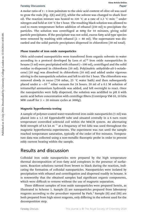
Three different samples of iron oxide nanoparticles were prepared herein, asillustrated in Scheme 1. Sample (I) are nanoparticles prepared from laboratoryreagents according to the procedure reported by Park.3 Sample (II) and (III) areboth prepared from high street reagents, only differing in the solvent used for thedecomposition step.
Faraday Discuss. This journal is © The Royal Society of Chemistry 2014
Scheme 1 Sample (I) is the route to iron oxide nanoparticles proposed by Park et al.,3
Sample (II) is the synthesis of iron palmitate from high street sources and its subsequentdecomposition in 1-octadecene and Sample (III) the decomposition of iron palmitate inshark liver oil.
Paper Faraday DiscussionsPu
blis
hed
on 1
5 Ju
ly 2
014.
Dow
nloa
ded
by U
nive
rsity
Col
lege
Lon
don
on 1
5/10
/201
4 14
:05:
00.
View Article Online
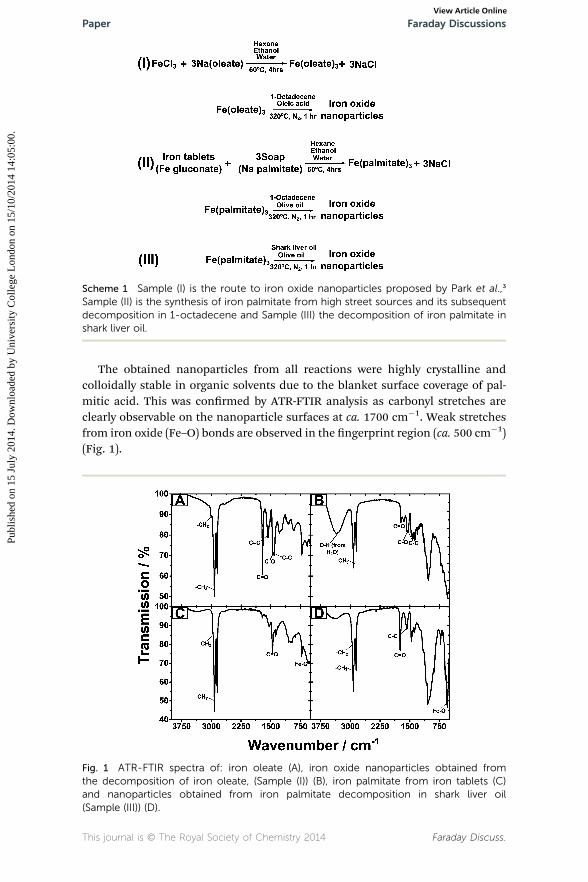
The obtained nanoparticles from all reactions were highly crystalline andcolloidally stable in organic solvents due to the blanket surface coverage of pal-mitic acid. This was conrmed by ATR-FTIR analysis as carbonyl stretches areclearly observable on the nanoparticle surfaces at ca. 1700 cm�1. Weak stretchesfrom iron oxide (Fe–O) bonds are observed in the ngerprint region (ca. 500 cm�1)(Fig. 1).
Fig. 1 ATR-FTIR spectra of: iron oleate (A), iron oxide nanoparticles obtained fromthe decomposition of iron oleate, (Sample (I)) (B), iron palmitate from iron tablets (C)and nanoparticles obtained from iron palmitate decomposition in shark liver oil(Sample (III)) (D).
This journal is © The Royal Society of Chemistry 2014 Faraday Discuss.
Faraday Discussions PaperPu
blis
hed
on 1
5 Ju
ly 2
014.
Dow
nloa
ded
by U
nive
rsity
Col
lege
Lon
don
on 1
5/10
/201
4 14
:05:
00.
View Article Online
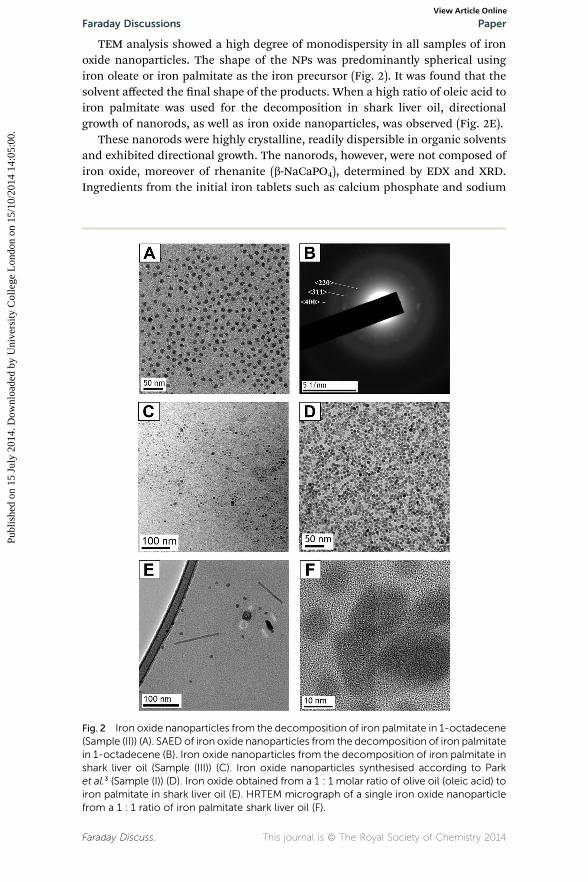
TEM analysis showed a high degree of monodispersity in all samples of ironoxide nanoparticles. The shape of the NPs was predominantly spherical usingiron oleate or iron palmitate as the iron precursor (Fig. 2). It was found that thesolvent affected the nal shape of the products. When a high ratio of oleic acid toiron palmitate was used for the decomposition in shark liver oil, directionalgrowth of nanorods, as well as iron oxide nanoparticles, was observed (Fig. 2E).
These nanorods were highly crystalline, readily dispersible in organic solventsand exhibited directional growth. The nanorods, however, were not composed ofiron oxide, moreover of rhenanite (b-NaCaPO4), determined by EDX and XRD.Ingredients from the initial iron tablets such as calcium phosphate and sodium
Fig. 2 Iron oxide nanoparticles from the decomposition of iron palmitate in 1-octadecene(Sample (II)) (A). SAED of iron oxide nanoparticles from the decomposition of iron palmitatein 1-octadecene (B). Iron oxide nanoparticles from the decomposition of iron palmitate inshark liver oil (Sample (III)) (C). Iron oxide nanoparticles synthesised according to Parket al.3 (Sample (I)) (D). Iron oxide obtained from a 1 : 1 molar ratio of olive oil (oleic acid) toiron palmitate in shark liver oil (E). HRTEM micrograph of a single iron oxide nanoparticlefrom a 1 : 1 ratio of iron palmitate shark liver oil (F).
Faraday Discuss. This journal is © The Royal Society of Chemistry 2014
Paper Faraday DiscussionsPu
blis
hed
on 1
5 Ju
ly 2
014.
Dow
nloa
ded
by U
nive
rsity
Col
lege
Lon
don
on 1
5/10
/201
4 14
:05:
00.
View Article Online
chloride contributed to the nanorod formation. This is possibly due to the organicspecies present in shark liver oil.
The constituents of shark liver oil itself vary according to the depth at whichthe shark normally resides.25–28 Deep-sea sharks can approach neutral buoyancythrough storage of low density lipids stored in the liver. A large contributor issqualene (density 0.858 g ml�1 at 25 �C, which increases at cold, deep seatemperatures), which has been shown to inuence iron oxide nanoparticleshape.29 Other constituents include: diacyl glyceryl ether, triacylgycerol and waxesters, all of which could contribute to directional growth of nanoparticles.30–33
By varying the amount of oleic acid used in the synthesis, the particle sizecould be tuned, with particle size increasing with smaller amounts of oleic acid(viz. olive oil). Average NP sizes were calculated from TEM micrographs as �12.7,5.8 and 3.6 nm for 1.2, 6, 24 mmol of oleic acid in olive oil, respectively (whendecomposing 2 g of iron palmitate).
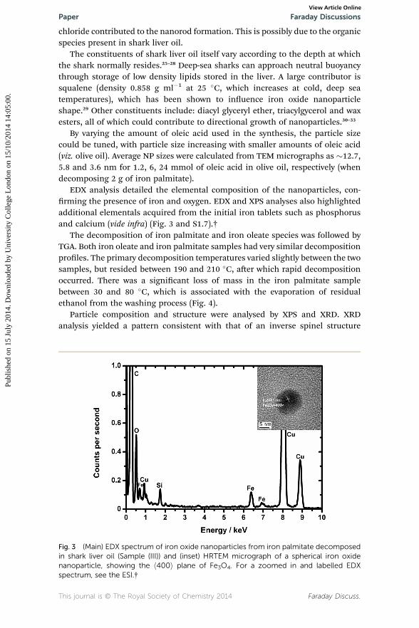
EDX analysis detailed the elemental composition of the nanoparticles, con-rming the presence of iron and oxygen. EDX and XPS analyses also highlightedadditional elementals acquired from the initial iron tablets such as phosphorusand calcium (vide infra) (Fig. 3 and S1.7).†
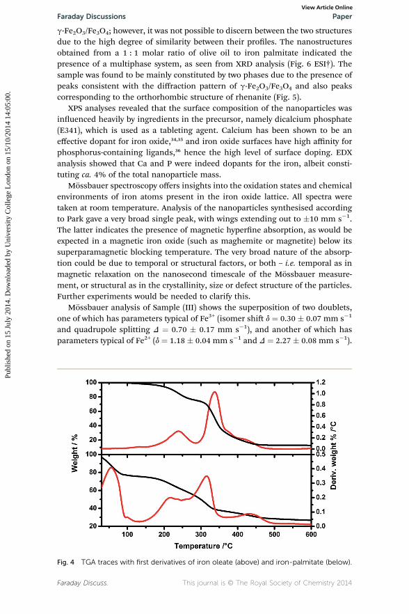
The decomposition of iron palmitate and iron oleate species was followed byTGA. Both iron oleate and iron palmitate samples had very similar decompositionproles. The primary decomposition temperatures varied slightly between the twosamples, but resided between 190 and 210 �C, aer which rapid decompositionoccurred. There was a signicant loss of mass in the iron palmitate samplebetween 30 and 80 �C, which is associated with the evaporation of residualethanol from the washing process (Fig. 4).
Particle composition and structure were analysed by XPS and XRD. XRDanalysis yielded a pattern consistent with that of an inverse spinel structure
Fig. 3 (Main) EDX spectrum of iron oxide nanoparticles from iron palmitate decomposedin shark liver oil (Sample (III)) and (inset) HRTEM micrograph of a spherical iron oxidenanoparticle, showing the h400i plane of Fe3O4. For a zoomed in and labelled EDXspectrum, see the ESI.†
This journal is © The Royal Society of Chemistry 2014 Faraday Discuss.
Faraday Discussions PaperPu
blis
hed
on 1
5 Ju
ly 2
014.
Dow
nloa
ded
by U
nive
rsity
Col
lege
Lon
don
on 1
5/10
/201
4 14
:05:
00.
View Article Online
g-Fe2O3/Fe3O4; however, it was not possible to discern between the two structuresdue to the high degree of similarity between their proles. The nanostructuresobtained from a 1 : 1 molar ratio of olive oil to iron palmitate indicated thepresence of a multiphase system, as seen from XRD analysis (Fig. 6 ESI†). Thesample was found to be mainly constituted by two phases due to the presence ofpeaks consistent with the diffraction pattern of g-Fe2O3/Fe3O4 and also peakscorresponding to the orthorhombic structure of rhenanite (Fig. 5).
XPS analyses revealed that the surface composition of the nanoparticles wasinuenced heavily by ingredients in the precursor, namely dicalcium phosphate(E341), which is used as a tableting agent. Calcium has been shown to be aneffective dopant for iron oxide,34,35 and iron oxide surfaces have high affinity forphosphorus-containing ligands,36 hence the high level of surface doping. EDXanalysis showed that Ca and P were indeed dopants for the iron, albeit consti-tuting ca. 4% of the total nanoparticle mass.
Mossbauer spectroscopy offers insights into the oxidation states and chemicalenvironments of iron atoms present in the iron oxide lattice. All spectra weretaken at room temperature. Analysis of the nanoparticles synthesised accordingto Park gave a very broad single peak, with wings extending out to �10 mm s�1.The latter indicates the presence of magnetic hyperne absorption, as would beexpected in a magnetic iron oxide (such as maghemite or magnetite) below itssuperparamagnetic blocking temperature. The very broad nature of the absorp-tion could be due to temporal or structural factors, or both – i.e. temporal as inmagnetic relaxation on the nanosecond timescale of the Mossbauer measure-ment, or structural as in the crystallinity, size or defect structure of the particles.Further experiments would be needed to clarify this.
Mossbauer analysis of Sample (III) shows the superposition of two doublets,one of which has parameters typical of Fe3+ (isomer shi d ¼ 0.30 � 0.07 mm s�1
and quadrupole splitting D ¼ 0.70 � 0.17 mm s�1), and another of which hasparameters typical of Fe2+ (d ¼ 1.18 � 0.04 mm s�1 and D ¼ 2.27 � 0.08 mm s�1).
Fig. 4 TGA traces with first derivatives of iron oleate (above) and iron-palmitate (below).
Faraday Discuss. This journal is © The Royal Society of Chemistry 2014
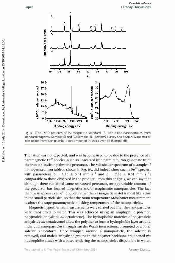
Fig. 5 (Top) XRD patterns of (A) magnetite standard, (B) iron oxide nanoparticles fromstandard reagents (Sample (I)) and (C) Sample (II). (Bottom) Survey and Fe2p XPS spectra ofiron oxide from iron palmitate decomposed in shark liver oil (Sample (III)).
Paper Faraday DiscussionsPu
blis
hed
on 1
5 Ju
ly 2
014.
Dow
nloa
ded
by U
nive
rsity
Col
lege
Lon
don
on 1
5/10
/201
4 14
:05:
00.
View Article Online
The latter was not expected, and was hypothesised to be due to the presence of aparamagnetic Fe2+ species, such as unreacted iron palmitate/iron gluconate fromthe iron tablets/iron palmitate precursor. The Mossbauer spectrum of a sample ofhomogenised iron tablets, shown in Fig. 6A, did indeed show such a Fe2+ species,with parameters (d ¼ 1.20 � 0.01 mm s�1 and D ¼ 2.23 � 0.01 mm s�1)comparable to those observed in the product. From this analysis, we can say thatalthough there remained some unreacted percursor, an appreciable amount ofthe precursor has formed magnetite and/or maghemite nanoparticles. The factthat these appear as a Fe3+ doublet rather than amagnetic sextet is most likely dueto the small particle size, so that the room temperature Mossbauer measurementis above the superparamagnetic blocking temperature of the nanoparticles.
Magnetic hyperthermiameasurements were carried out aer the nanoparticleswere transferred to water. This was achieved using an amphiphilic polymer,poly(maleic anhydride-alt-octadecene). The hydrophobic moieties of poly(maleicanhydride-alt-octadecene) allow the polymer to form a hydrophobic layer aroundindividual nanoparticles through van der Waals interactions, promoted by a polarsolvent, chloroform. Once wrapped around a nanoparticle, the solvent isremoved, and maleic anhydride groups in the polymer backbone are opened vianucleophilic attack with a base, rendering the nanoparticles dispersible in water.
This journal is © The Royal Society of Chemistry 2014 Faraday Discuss.
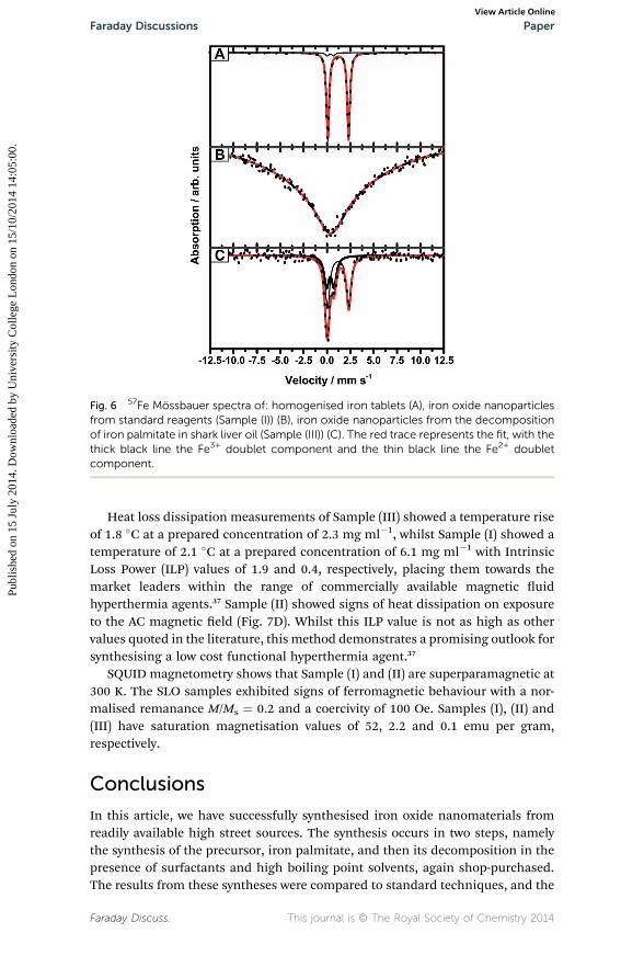
Fig. 6 57Fe Mossbauer spectra of: homogenised iron tablets (A), iron oxide nanoparticlesfrom standard reagents (Sample (I)) (B), iron oxide nanoparticles from the decompositionof iron palmitate in shark liver oil (Sample (III)) (C). The red trace represents the fit, with thethick black line the Fe3+ doublet component and the thin black line the Fe2+ doubletcomponent.
Faraday Discussions PaperPu
blis
hed
on 1
5 Ju
ly 2
014.
Dow
nloa
ded
by U
nive
rsity
Col
lege
Lon
don
on 1
5/10
/201
4 14
:05:
00.
View Article Online
Heat loss dissipation measurements of Sample (III) showed a temperature riseof 1.8 �C at a prepared concentration of 2.3 mg ml�1, whilst Sample (I) showed atemperature of 2.1 �C at a prepared concentration of 6.1 mg ml�1 with IntrinsicLoss Power (ILP) values of 1.9 and 0.4, respectively, placing them towards themarket leaders within the range of commercially available magnetic uidhyperthermia agents.37 Sample (II) showed signs of heat dissipation on exposureto the AC magnetic eld (Fig. 7D). Whilst this ILP value is not as high as othervalues quoted in the literature, this method demonstrates a promising outlook forsynthesising a low cost functional hyperthermia agent.37
SQUID magnetometry shows that Sample (I) and (II) are superparamagnetic at300 K. The SLO samples exhibited signs of ferromagnetic behaviour with a nor-malised remanance M/Ms ¼ 0.2 and a coercivity of 100 Oe. Samples (I), (II) and(III) have saturation magnetisation values of 52, 2.2 and 0.1 emu per gram,respectively.
Conclusions
In this article, we have successfully synthesised iron oxide nanomaterials fromreadily available high street sources. The synthesis occurs in two steps, namelythe synthesis of the precursor, iron palmitate, and then its decomposition in thepresence of surfactants and high boiling point solvents, again shop-purchased.The results from these syntheses were compared to standard techniques, and the
Faraday Discuss. This journal is © The Royal Society of Chemistry 2014
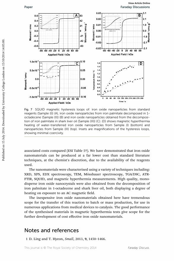
Fig. 7 SQUID magnetic hysteresis loops of: iron oxide nanoparticles from standardreagents (Sample (I)) (A), iron oxide nanoparticles from iron palmitate decomposed in 1-octadecene (Sample (II)) (B) and iron oxide nanoparticles obtained from the decomposi-tion of iron palmitate in shark liver oil (Sample (III)) (C). (D) shows magnetic hyperthermiaheating of water-transferred iron oxide nanoparticles from Sample (I) (bottom) andnanoparticles from Sample (III) (top). Insets are magnifications of the hysteresis loops,showing minimal coercivity.
Paper Faraday DiscussionsPu
blis
hed
on 1
5 Ju
ly 2
014.
Dow
nloa
ded
by U
nive
rsity
Col
lege
Lon
don
on 1
5/10
/201
4 14
:05:
00.
View Article Online
associated costs compared (ESI Table 5†). We have demonstrated that iron oxidenanomaterials can be produced at a far lower cost than standard literaturetechniques, at the chemist's discretion, due to the availability of the reagentsused.
The nanomaterials were characterised using a variety of techniques including:XRD, XPS, EDX spectroscopy, TEM, Mossbauer spectroscopy, TGA/DSC, ATR-FTIR, SQUID, and magnetic hyperthermia measurements. High quality, mono-disperse iron oxide nanocrystals were also obtained from the decomposition ofiron palmitate in 1-octadecene and shark liver oil, both displaying a degree ofheating on exposure to an AC magnetic eld.
The inexpensive iron oxide nanomaterials obtained here have tremendousscope for the transfer of this reaction to batch or mass production, for use innumerous applications from medical devices to catalysis. The good performanceof the synthesised materials in magnetic hyperthermia tests give scope for thefurther development of cost effective iron oxide nanomaterials.
Notes and references
1 D. Ling and T. Hyeon, Small, 2013, 9, 1450–1466.
This journal is © The Royal Society of Chemistry 2014 Faraday Discuss.
Faraday Discussions PaperPu
blis
hed
on 1
5 Ju
ly 2
014.
Dow
nloa
ded
by U
nive
rsity
Col
lege
Lon
don
on 1
5/10
/201
4 14
:05:
00.
View Article Online
2 N.-N. Song, H.-T. Yang, X. Ren, Z.-A. Li, Y. Luo, J. Shen, W. Dai, X.-Q. Zhang andZ.-H. Cheng, Nanoscale, 2013, 5, 2804–2810.
3 J. Park, K. An, Y. Hwang, J.-G. Park, H.-J. Noh, J.-Y. Kim, J.-H. Park,N.-M. Hwang and T. Hyeon, Nat. Mater., 2004, 3, 891–895.
4 M. Mahmoudi, S. Sant, B. Wang, S. Laurent and T. Sen, Adv. Drug Delivery Rev.,2011, 63, 24–46.
5 W. Cai and J. Wan, J. Colloid Interface Sci., 2007, 305, 366–370.6 A. Guittoum, A. Layadi, H. Tafat and N. Souami, J. Magn. Magn. Mater., 2010,322, 566–571.
7 A. Tavakoli, M. Sohrabi and A. Kargari, Chem. Pap., 2007, 61, 151–170.8 A. S. Teja and P.-Y. Koh, Prog. Cryst. Growth Charact. Mater., 2009, 55, 22–45.9 S. Laurent, D. Forge, M. Port, A. Roch, C. Robic, L. Vander Elst andR. N. Muller, Chem. Rev., 2008, 108, 2064–2110.
10 S. Xu, J. Ziegler and T. Nann, J. Mater. Chem., 2008, 18, 2653–2656.11 M. Lattuada and T. A. Hatton, Langmuir, 2007, 23, 2158–2168.12 L. M. Bronstein, X. Huang, J. Retrum, A. Schmucker, M. Pink, B. D. Stein and
B. Dragnea, Chem. Mater., 2007, 19, 3624–3632.13 S. Sun and H. Zeng, J. Am. Chem. Soc., 2002, 124, 8204–8205.14 J. Bear, G. Charron, M. T. Fernandez-Arguelles, S. Massadeh, P. McNaughter
and T. Nann, in BetaSys: Systems Biology of Regulated Exocytosis in Pancreaticb-Cells, Springer, New York, 2011, vol. 2, pp. 185–220.
15 G. Vallejo-Fernandez, O. Whear, A. G. Roca, S. Hussain, J. Timmis, V. Patel andK. O'Grady, J. Phys. D: Appl. Phys., 2013, 46, 312001.
16 R. Hergt, R. Hiergeist, I. Hilger, W. A. Kaiser, Y. Lapatnikov, S. Margel andU. Richter, J. Magn. Magn. Mater., 2004, 270, 345–357.
17 M. A. Gonzalez-Fernandez, T. E. Torres, M. Andres-Verges, R. Costo, P. de laPresa, C. J. Serna, M. P. Morales, C. Marquina, M. R. Ibarra and G. F. Goya,J. Solid State Chem., 2009, 182, 2779–2784.
18 S. E. Barry, Int. J. Hyperthermia, 2008, 24, 451–466.19 L. A. Thomas, L. Dekker, M. Kallumadil, P. Southern, M. Wilson, S. P. Nair,
Q. A. Pankhurst and I. P. Parkin, J. Mater. Chem., 2009, 19, 6529.20 I. Baker, Q. Zeng, W. Li and C. R. Sullivan, J. Appl. Phys., 2006, 99, 08H106.21 M. Gonzales-Weimuller, M. Zeisberger and K. M. Krishnan, J. Magn. Magn.
Mater., 2009, 321, 1947–1950.22 E. E. Lees, T.-L. Nguyen, A. H. A. Clayton, B. W. Muir and P. Mulvaney, ACS
Nano, 2009, 3, 2049.23 T. Pellegrino, L. Manna, S. Kudera, T. Liedl, D. Koktysh, A. L. Rogach, S. Keller,
J. Radler, G. Natile and W. J. Parak, Nano Lett., 2004, 4, 703–707.24 http://resonantcircuits.com/.25 B. M. Wetherbee and P. D. Nichols, Comp. Biochem. Physiol., Part B: Biochem.
Mol. Biol., 2000, 125, 511–521.26 M. J. Bakes and P. D. Nichols, Comp. Biochem. Physiol., Part B: Biochem. Mol.
Biol., 1995, 110, 267–275.27 J. C. Cain and R. A. Morton, Biochem. J., 1955, 60, 274–283.28 I. Batista and M. L. Nunes, Fish. Res., 1992, 14, 329–334.29 B. Rodriguez-Gonzalez, M. Spasova, M. Farle, L. Liz-Marzan and A. Shavel, Adv.
Funct. Mater., 2007, 17, 3870–3876.30 C. Yang, J. Wu and Y. Hou, Chem. Commun., 2011, 47, 5130–5141.
Faraday Discuss. This journal is © The Royal Society of Chemistry 2014
Paper Faraday DiscussionsPu
blis
hed
on 1
5 Ju
ly 2
014.
Dow
nloa
ded
by U
nive
rsity
Col
lege
Lon
don
on 1
5/10
/201
4 14
:05:
00.
View Article Online
31 M. V. Kovalenko, M. I. Bodnarchuk, R. T. Lechner, G. Hesser, F. Schaffler andW. Heiss, J. Am. Chem. Soc., 2007, 129, 6352–6353.
32 J. Cheon, N.-J. Kang, S.-M. Lee, J.-H. Lee, J.-H. Yoon and S. J. Oh, J. Am. Chem.Soc., 2004, 126, 1950–1951.
33 L. M. Bronstein, J. E. Atkinson, A. G. Malyutin, F. Kidwai, B. D. Stein,D. G. Morgan, J. M. Perry and J. A. Karty, Langmuir, 2011, 27, 3044–3050.
34 L. J. Berchmans, M. Myndyk, K. L. Da Silva, A. Feldhoff, J. Subrt, P. Heitjans,K. D. Becker and V. Sepelak, J. Alloys Compd., 2010, 500, 68–73.
35 R. Dom, R. Subasri, K. Radha and P. H. Borse, Solid State Commun., 2011, 151,470–473.
36 Y. Sahoo, H. Pizem, T. Fried, D. Golodnitsky, L. Burstein, C. N. Sukenik andG. Markovich, Langmuir, 2001, 17, 7907–7911.
37 M. Kallumadil, M. Tada, T. Nakagawa, M. Abe, P. Southern andQ. A. Pankhurst, J. Magn. Magn. Mater., 2009, 321, 1509–1513.
This journal is © The Royal Society of Chemistry 2014 Faraday Discuss.