Embed Size (px)
Citation preview

21st Computer Vision Winter WorkshopLuka Cehovin, Rok Mandeljc, Vitomir Struc (eds.)Rimske Toplice, Slovenia, February 3–5, 2016
A Longitudinal Diffeomorphic Atlas-Based Tissue Labeling Framework forFetal Brains using Geodesic Regression
Roxane Licandro1,2
[email protected] Institute of Computer Aided Automation, Computer Vision Lab, Vienna University of Technology,
http://www.caa.tuwien.ac.at/cvl
2 Department of Radiology and Image-guided Therapy, Computational Imaging Research Lab,Medical University of Vienna,
http://www.cir.meduniwien.ac.at
Georg Langs2 Gregor Kasprian2 Robert Sablatnig1 Daniela Prayer2
Ernst Schwartz2
Abstract. The human brain undergoes structuralchanges in size and in morphology between the sec-ond and the third trimester of pregnancy, corre-sponding to accelerated growth and the progress ofcortical folding. To make fetal brains comparable,spatio-temporal atlases are used as a standard spacefor studying brain development, fetal pathology loca-tions, fetal abnormalities or anatomy. The aim of thiswork is to provide a continuous model of brain devel-opment and to use it as base for an automatic tissuelabeling framework. This paper provides a novel lon-gitudinal fetal brain atlas construction concept forgeodesic image regression using three different age-ranges which are parametrized according to the de-velopmental stage of the fetus. The dataset used forevaluation contains 45 T2−weighted Magnetic Res-onance (MR) volumes between Gestation Week (GW)18.0 and GW 30 day 2. The automatic tissue label-ing framework estimates cortical segmentations witha Dice Coefficient (DC) of up to 0.85 and ventriclesegmentations with a DC of up to 0.60.
1. Introduction
The aim of brain mapping experiments is to cre-ate maps (models), based on studies, to understandstructural and functional brain organization. To thisend, neuroimaging methods as well as knowledge ofneuroanatomy and physiology are combined. Due tothe fundamental changes occurring in the human fe-
tal brain during pregnancy, a single map is not suf-ficient to model brain development [19]. Changesin size, according to accelerated growth, changes inmorphology, due to the progress of cortical foldingand deceleration of the proliferation of ventricularprogenitor cells [16] occur and are illustrated in Fig-ure 1a. Thus, a collection of brain maps is neededto describe these alterations as a function of time.For studying the brain organisation during its de-velopment, abnormalities, and locations of patholo-gies, brain maps are used as a reference model [18].Newly acquired brain images are labelled to iden-tify structures and possible abnormal changes or tofind indicators for diseases. This labeling can be per-formed manually by annotating the images, whichneeds an expert, time and consequently leads to in-creased costs compared to an automatic labeling pro-cedure [3]. In this case, labels for non annotatedimages are estimated automatically by software us-ing a brain model for the mapping. Such an auto-mated labeling procedure on the one hand and a ref-erence model on the other form an atlas. To cover thetime-dependent development of the fetal brain, time-varying reference models are considered for buildingspatio-temporal atlases.
1.1. State-of-the-Art
State-of-the-art approaches [8, 10, 13, 17, 21] forcomputing a spatio-temporal atlas combine registra-tion methods and interpolation techniques to obtain
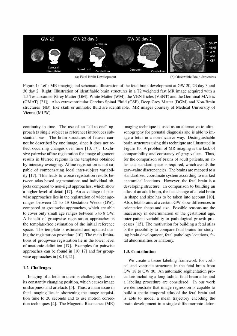
(a) Fetal Brain Development (b) Observable Brain Structures
Figure 1: Left: MR imaging and schematic illustration of the fetal brain development at GW 20, 23 day 3 and30 day 2. Right: Illustration of identifiable brain structures in a T2 weighted fast MR image acquired with a1.5 Tesla scanner (Grey Matter (GM), White Matter (WM), the VENTricles (VENT) and the Germinal MATrix(GMAT) [21]). Also extraventricular Cerebro Spinal Fluid (CSF), Deep Grey Matter (DGM) and Non-Brainstructures (NB), like skull or amniotic fluid are identifiable. MR images courtesy of Medical University ofVienna (MUW).
continuity in time. The use of an ”all-to-one” ap-proach (a single subject as reference) introduces sub-stantial bias. The brain structures of fetuses can-not be described by one image, since it does not re-flect occurring changes over time [10, 17]. Exclu-sive pairwise affine registration for image alignmentresults in blurred regions in the templates obtainedby intensity averaging. Affine registration is not ca-pable of compensating local inter-subject variabil-ity [17]. This leads to worse registration results be-tween atlas-based segmentations and individual ob-jects compared to non-rigid approaches, which showa higher level of detail [17]. An advantage of pair-wise approaches lies in the registration of wider age-ranges between 15 to 18 Gestation Weeks (GW),compared to groupwise approaches, which are ableto cover only small age ranges between 5 to 8 GW.A benefit of groupwise registration approaches isthe template-free estimation of the initial referencespace. The template is estimated and updated dur-ing the registration procedure [10]. The main limita-tions of groupwise registration lie in the lower levelof anatomic definition [17]. Examples for pairwiseapproaches can be found in [10, 17] and for group-wise approaches in [8, 13, 21].
1.2. Challenges
Imaging of a fetus in utero is challenging, due toits constantly changing position, which causes imageunsharpness and artefacts [5]. Thus, a main issue infetal imaging lies in shortening the image acquisi-tion time to 20 seconds and to use motion correc-tion techniques [4]. The Magnetic Resonance (MR)
imaging technique is used as an alternative to ultra-sonography for prenatal diagnosis and is able to im-age a fetus in a non-invasive way. Distinguishablebrain structures using this technique are illustrated inFigure 1b. A problem of MR imaging is the lack ofcomparability and constancy of gray-values. Thus,for the comparison of brains of adult patients, an at-las as a standard space is required, which avoids thegray-value discrepancies. The brains are mapped to astandardized coordinate system according to markedanatomical locations. However, the fetal brain is adeveloping structure. In comparison to building anatlas of an adult brain, the fast change of a fetal brainin shape and size has to be taken into account [10].Also, fetal brains at a certain GW show differences inorientation shape and size. Possible reasons are theinaccuracy in determination of the gestational age,inter-patient variability or pathological growth pro-cesses [15]. The motivation for building a fetal atlasis the possibility to compare fetal brains for study-ing brain development, fetal pathology locations, fe-tal abnormalities or anatomy.
1.3. Contribution
We create a tissue labeling framework for corti-cal and ventricle structures in the fetal brain fromGW 18 to GW 30. An automatic segmentation pro-cedure including a longitudinal fetal brain atlas anda labeling procedure are considered. In our workwe demonstrate that image regression is capable tobuild a spatio-temporal atlas of the fetal brain andis able to model a mean trajectory encoding thebrain development in a single diffeomorphic defor-

mation, instead of calculating discrete age-dependenttemplates combined with interpolation. As foundin literature [7, 9, 11], image regression for time-series data have been evaluated only using adult- andchild-brain datasets, which record changes of brainstructure over time. In the proposed work the lo-cal inter-subject variability is considered to be mod-elled continuously in time and non-rigidly in spaceby geodesic regression [1, 2]. The computed atlasis used as a prior of the Graph Cut (GC) approachfor multi label segmentation proposed by Yuan etal. [20].The paper is organized as follows. In Section 2 anoverview of the methodology used and the conceptof the tissue labeling framework proposed is pre-sented. The results and the corresponding discussionare given in Section 3. This work concludes with asummary of the contributions in Section 4.
2. Methodology
The framework proposed is illustrated in Figure 2.The input represents a gray value image Inew at timepoint tnew, which is preprocessed in a first step, byperforming motion correction, rigid alignment, im-age masking and image cropping. Subsequently, thelongitudinal diffeomorphic fetal brain atlas is used toestimate a time point tnew corresponding diffeomor-phic transformation for computing a time-dependentintensity image IA and a time-dependent segmenta-tion for ventricular and cortical tissue Stissue
A in atlasspace. In a pairwise registration procedure, a trans-formation T from the preprocessed input (AlignedInew) to the atlas-based intensity image IA is esti-mated. The inverse of the computed transformationT−1 is used to transform the atlas based segmenta-tions Stissue
A to the subject’s space (StissueA T−1 =
StissueGC ). As next step the transformed segmentationsStissueGC and Inew are used as input parameters for the
multi label GC segmentation refinement. The outputof the framework are segmentations for ventricularand cortical brain tissues Stissue
new of the input imageInew.
2.1. Image Acquisition and Preprocessing
The time series MR image dataset used consistsof 45 healthy fetal brains with an age range between18 and 30 GW. The MR image acquisition is per-formed using an 1.5 Philips Gyroscan superconduct-ing unit scanner performing a single-shot, fast spin-echo T2-weighted MR sequence: In-plane resolu-
tion = 0.78 - 0.9 pixels per mm, Slice thickness =3 - 4.4mm, Acquisition matrix = 256×256, Fieldof view = 200 - 230mm, Specific Absorption Rate(SAR) = < 100% /4.0W/kg, Image acquisition time= ≤ 20s, TE (Echo Time) = 100 - 140ms, TR (Rep-etition Time) = 9000 - 19000ms. The dataset of MRimages used for atlas learning are preprocessed usingthe pipeline illustrated in Figure 2. First the imagesare motion corrected using the toolkit for fetal brainMR images published by Rousseau et al. [14]. Sub-sequently, the manual annotation of the cortex, leftand right eye, ventricle and occipital foramen mag-num is performed by an expert. After this step, thedata is rigidly aligned, the surrounding mother tissueis excluded in a masking step and the volumes arecropped to reduce computational costs in the longi-tudinal registration procedure using a bounding boxof size 90× 140× 140 voxels.
2.2. Spatio Temporal Atlas Learning
The algorithm used for Diffeomorphic Anatom-ical RegistraTion using Exponential Lie algebra(DARTEL) of Ashburner et al. [1, 2] for geodesicregression is integrated in the Statistical ParaMetric(SPM) tool box - release SPM8 1. This approachis used to encode the brain development in a singlediffeomorphic deformation by optimising the energyterm E expressed in Equation 1 [2].
E =1
2‖Lv0‖2+
1
2
N∑n=1
( ∫x∈Ω
‖It0 − Itn(ϕtn)‖2 dx
)(1)
The term ϕtn denotes the forward deformation fromsource It0 to target Itn at time point tn, where n =1, . . . , N and L represents a model of the ”inertia”of the system, i.e. a linear operator which operateson a time-dependent velocity that mediates the defor-mation over unit time [2]. It is introduced to derivean initial momentum m0 through an initial velocityv0. The velocity field v(x) learned at position x isparametrised using a linear combination of i basisfunctions. Such basis functions consist of a vectorof coefficients ci and a ith first degree B-spline basisfunction ρi(x) (cf. Equation 2) [1].
v(x) =∑i
ciρi(x) (2)
The aim of the DARTEL implementation is to esti-mate an optimized parametrisation of c. The energy
1http://www.fil.ion.ucl.ac.uk/spm/;[accessed 07 December 2015]

Figure 2: Fetal brain tissue labeling framework. MR images courtesy of MUW.
cost term E in Equation 1 is reformulated in termsof finding the coefficients of c for a given dataset Dwith maximum probability (cf. Equation 3). A maxi-mization of the probability leads to the minimizationof its negative logarithm and thus, is used to interpretregistration of data D as a minimization procedureof the objective function (− log p(c,D)) expressedin Equation 3, consisting of a prior term (− log p(c))and a likelihood term (− log p(D|c)) [1].
− log p(c,D) = − log p(c)− log p(D|c) (3)
The prior term denotes the prior probability p(c).Ashburner et al. [1] use a concentration matrix (in-verse of a covariance matrix) K to encode spa-tial variability. The parameters [λ1, λ2, λ0, λ, µ],which have to be predefined to computeK, influencethe behaviour of the deformation (bending energy,stretching, shearing) as well as the divergence andamount of volumetric expansion or contraction [1].The term λ0 encodes the penalisation of absolute dis-placements, λ1 penalises the difference between twoneighboured vectors by observing the first derivatives(linear term) of the displacements, λ2 penalises thedifference between the first derivatives of two neigh-boured vectors by observing the second derivativesof the displacements and λ denotes the variability ofthe spatial locations (divergence of each point in theflow field) with a constant value. Increasing λ leadsto increasing smoothing of the flow vector field andpreserves volumes during the transformation. The
term µ encodes the variance according to symmetriccomponents, rotations and the penalisation of scalingand shearing. The likelihood term encodes the prob-ability of c given the data D [1] and corresponds tothe mean-squared difference between a warped tem-plate deformed by the calculated transformation andthe target image.
2.2.1 Optimisation Procedure
A Full Multi Grid (FMG) approach is used to solvethe equation (cf. Equation 4) which is needed to up-date the vector field during a Gauß-Newton opti-mising procedure, where H iter denotes the Hessian,giter the gradient and K the concentration matrix.Details regarding the computation of viter+1
0 are ex-plained in [1, 2].
viter+10 = viter0 − ε(K +Hiter)−1
(Kviter0 + giter) (4)
For this task images are observed in different scales.For every resolution level multigrid methods recur-sively estimate the field, starting at the coarsest scaleand computing the residual to solve the update equa-tions on the current grid. Subsequently, the solutionis prolongated to the next finer grid [1].
2.3. Automatic Tissue Labeling using Graph Cuts
For tissue labeling, we use a continuous max flowformulation of a multi label GC [20]. Three inputparameters are necessary for performing tissue seg-mentation. A data term (gray value volume Inew

at age tnew), a cost (unary) term, and a penalty(binary) term. For computing a unary term, atlasbased segmentations for cortex and ventricle tissueStissue = Scortex, Sventricle at age t are estimatedand smoothed with a Gaussian filter G. The parame-ter δ is defined to weight the smoothed result with aconstant factor. The unary term is illustrated in Equa-tion 5, where ? denotes the convolution operator.
C = δ ∗ (Stissue ? G) (5)
Three different binary terms are evaluated:Penalty term 1 (P1) is a weighted norm of the gra-dient of the data term D (cf. Equation 6), where δdenotes the same weighting term as used in Equation5 and a, b are constant weighting parameters.
P1 = δ ∗ b
1 + (a ∗ ‖∇D‖)(6)
Penalty term 2 (P2) denotes an intensity based termand is calculated separately for cortex and ventri-cle segmentation (cf. Equation 7). Tissue type spe-cific gray values are modelled as Gaussian distribu-tions N∼(µtissue, σtissue), which parameters µtissueand σtissue are estimated using the a-priori atlas seg-mentation. These parameters are used to calculatethe probability of every pixel belonging to cortex orventricle. Subsequently, the gradient of the resultingprobability map P and its norm are computed andweighted by the parameters δ, a, b as shown in Equa-tion 6.
P2 = δ ∗ b
1 + (a ∗ ‖∇P (µtissue, σtissue)‖)(7)
Penalty term 3 (P3) represents an exponential for-mulation and is expressed in Equation 8. The param-eter u is a constant and v a linear weighting parame-ter. The term w weights the norm of the image’s Dgradient non-linearly in the exponential term.
P3 = u+ v ∗ exp(−‖∇D‖
w
)(8)
3. Results
Evaluation of the proposed framework is per-formed using leave-one-out cross validation. In thispaper a novel longitudinal registration procedure isformulated by dividing the data set into three ageranges, based on the developmental stage of the fetus.Age range 1 reaches from 20 GW day 6 (146 GD) to23 GW day 3 (164 GD), age range 2 from 23 GW
day 3 (164 GD) to 26 GW day 2 (184 GD) and agerange 3 from 26 GW day 2 (184 GD) to 30 GW day 2(212 GD). The first part of the evaluation documentsthe atlas learning results for each age range. Subse-quently, the atlases computed are used to evaluate thetissue labeling procedure as a second part of the eval-uation. Estimated atlas templates at the testing time-point are pairwise registered to the test MR volume toobtain a transformation T . The inverse T−1 is usedto transform the atlas based segmentation to the test-subject’s space. As last step the segmentation of thetest volume using the transformed atlas is computed.To evaluate our approach, we report the overlap be-tween automatic- and manual segmentations of thefetal cortex and ventricles. In the leave-one-out crossvalidation, we compare the Dice Coefficient (DC) [6]between the groundtruth annotation and different au-tomatic segmentations based on (1) the atlas, (2) thetransformed atlas, and (3) the GC segmentation opti-mization.Furthermore, we report the volume of cortex andventricles, and the area of the cortical surface of theatlas based segmentations.
3.1. Results Spatio-Temporal Atlas Learning
The deformation behaviour of image regres-sion using 21 different regularisation kernelsK [λ1, λ2, λ0, λ, µ] (cf. Section 2.2) is evaluated forevery age range. Beside the DC also the behaviourof the regularisation of the volume expansion andchanges of the area of cortical surface have to betaken into account, when choosing a suitable ker-nel. Atlas-based cortical and ventricle segmentationsare studied. According to the evaluation results, ker-nel 1 (K1
[0.01, 0.01, 9e−6, 1e−5, 1e−5
]) is chosen
as suitable regularisation for age range 1, kernel 4(K4
[0.01, 9e−6, 9e−6, 0.01, 1e−5
]) for age range 2
and kernel 7 (K7
[0.01, 0.01, 9e−6, 0.01, 1e−5
]) for
age range 3. Figure 3a shows examples of the at-las templates learned and Figure 3b illustrates theanatomical details of these at age GW 21 day 4 (GD151), GW 24 day 3 (GD 171) and GW 29 (GD 203).In both figures the growth of the brain structures isobservable. The brain model at age range 1 is char-acterised by a smoother cortex surface in compari-son to a brain at a higher age range. It also visu-alises the increase of the cortical folding grade. Ac-cording to Pugash et al. [12], the ventricles achievetheir thickest size in early gestation and regress in thethird trimester, which is not visible. The regularisa-

GD 148 GD 150 GD 153 GD 156 GD 159
ATLAS BASED TEMPLATES AGE RANGE 1 KERNEL 1
GD 164 GD 168 GD 172 GD 177 GD 181
ATLAS BASED TEMPLATES AGE RANGE 2KERNEL 4
GD 184 GD 190 GD 194 GD 208
ATLAS BASED TEMPLATES AGE RANGE 3 KERNEL 7
GD 205GD 200 GD 212
GD 163
(a) Atlas based templates (b) Details Atlas based templates
Figure 3: Left: Atlas based templates of age range 1, 2 and 3 between GW 21 day 1 (GD 148) and GW 30day 2 (GD 212). Right: Anatomical details of atlas based templates at age GW 21 day 4 (GD 151), GW 24day 3 (GD 171) and GW 29 (GD 203). Coronal (first row), axial (second row) and sagital (third row) slicesare illustrated. Denoted structures: Sylvian Fissure (SF), InterHemispheric Fissure (IHF), Germinal MATrix(GMAT), Lateral-VENTricle (L-VENT), Cingulate Sulcus (CiS), ColLateral Sulcus (CLS), Cavum of SeptumPellucidum (CSP), Occipital Lobe (OL), Frontal Lobe (FL), Central Sulcus (CeS), PreCentral Gyrus (PreCG),PostCentral Gyrus (PostCG), ParietoOccipital Sulcus (POS) and Calcarine Sulcus (CaS).
tion term for geodesic regression is not able to modellocation specific volume expansion and shrinkage atthe same time. This leads to worse modelling resultsfor ventricles, compared to cortical structure, since akernel is chosen which models expansion. Addition-ally, the subject specific variability of age-dependentventricle size in the dataset and the complex form ofventricles complicate the determination of a suitablekernel and consequently the registration procedure.Observable structures at every age range are SylvianFissures (SF), Lateral VENTricle (L-VENT), Inter-Hemispheric Fissure (IHF), Cavum of Septum Pellu-cidum (CSP), Occipital Lobe (OL) and Frontal Lobe(FL). The SF show in the coronal and axial slices asmooth bending at age range 1 and develop to a deepfold at the lateral side of the brain at age range 3.Also the IHF shows a deeper folding at age range3 with Cingulate Sulcus (CiS) as additional form-ing compared to age range 1. The Germinal MATrix(GMAT) is existent until age range 2 and disappearslater in the third trimester of pregnancy. The CentralSulcus (CeS) formation starts at age range 2 and getsmore apparent at age range 3 as well as the develop-ing of the PreCentral Gyrus (PreCG) and PostCen-tral Gyrus (PostCG). The ColLateral Sulcus (CLS) isvisible at age range 3 as well as the Calcarine Sulcus
(CaS) and PreOccipital Sulcus (POS).
3.2. Results Automatic Tissue Labeling
For pairwise registration kernel A(KA
[5e−3, 5e−3, 3e−5, 1e−5, 9e−6
]) is used
for regularisation. The DC distributions of seg-mentations of the cortex for age range 1, 2 and3 are illustrated in Figure 4 on the top and forventricle segmentations on the bottom. The DCdistribution of atlas based and transformed atlas-based segmentations using pairwise registration areillustrated and the three dotted lines visualise theDCs of GC based segmentations computed usingpenalty terms 1, 2 and 3. For age range 1 thehighest DC improvement from 0.727 to 0.771 atGD 158 is achieved by pairwise registration and GCrefinement compared to atlas based segmentations.In contrast to this no improvement is reached at GD151, but shows the highest DC of about 0.851. AtGDs older than 154 the GC refining using penalty1 and penalty 2 achieve a higher DC increase ofabout 0.02 compared to using penalty 3. At agerange 2 no improvement of transformed atlas basedsegmentations is observed after pairwise registration,which leads to a decrease of the DC. It is observedthat the labeling result of the pairwise registration

0,55
0,65
0,75
0,85
150151154158164164165170171180184184186191196197199203206208210
DC(C
ORT
EX) ATLAS
PW
GC-P1
GC-P2
GC-P3
0
0,2
0,4
0,6
150151154158164164165170171180184184186191196197199203206208210
DC(V
ENTR
ICLE)
GESTATIONALDAYS
ATLASPWGC-P1GC-P2GC-P3
AgeRange1 AgeRange2 AgeRange3
Figure 4: DCs of automatically estimated labels of the cortex and ventricle at age range 1, 2 and 3.
GD171
DATA ATLAS PW GC M
GD203
Figure 5: Top: Coronal view - segmentations of the cortex at GD 171 (GW 24 day 3), bottom: sagital view- segmentations of the ventricle at GD 203 (GW 29). Segmentations are illustrated estimated by the atlas(ATLAS), after the pairwise registration procedure (PW), estimated by the GC approach (GC) and manualannotations (M).
has an influence on the GC labeling since it acts asinitialization of this procedure, best visible at GD184. The GC refinement is able to compensate theresults of the pairwise registration between GD 164and 184 and shows an increase of the DC betweenatlas and graph-cut based segmentations in averageof about 0.02. At age range 3 an increase of DC atevery age range is achievable using GC refinement.The highest improvement between atlas-based seg-mentations and GC based segmentations is reachedat GD 206 with a DC increase from 0.71 to 0.795.The highest DC at age range 3 of about 0.819 isachieved at GD 203 and the lowest of about 0.575 atGD 184. It is observable that pairwise registration
is not capable to compensate differences in volumesize or absolute displacements. If an estimatedsegmentation has a bigger volume than the structureto be segmented or is displaced, then the borders ofneighboured tissue prevents the GC approach fromcutting through regions of a high gradient, sincethis would lead to increasing costs in the energyminimisation procedure. Consequently, the GC isnot capable to refine the segmentation. In Figure 5an example for a misaligned segmentation and itsdeformation through the labeling procedure is illus-trated. The displacement is observable at the IHF inthe first column and the superior part of the anteriorhorn of the ventricle in the second column. Test

data and corresponding estimated segmentations,transformed segmentations to subject’s space andGC based segmentations of the cortex at GD 171(top) and of ventricular tissue at GD 203 (bottom)are shown. The GC segmentations are computedusing the penalty term 3, since it shows the bestimprovement between atlas-based and GC basedsegmentations.
4. Conclusion
In this paper an automatic fetal brain tissue label-ing framework using geodesic image regression waspresented and was identified to be suitable as regis-tration approach to longitudinally model the changesof the brain during the 18th and 30th GW. The advan-tage is the provision of a time-dependent transforma-tion from a source to a target brain, instead of com-bining a template building technique and interpola-tion technique to obtain continuity in time. A novellongitudinal registration scheme was proposed, usingseparate age ranges for flexible regularisation of thedeformation behaviour due to the age range depen-dent changes. The atlas learned was evaluated us-ing a leave-one-out cross validation approach for ev-ery age range and 21 different regularisation kernelswere analysed according to their behaviour regard-ing volume expansion, modelling of cortical surfaceand Dice similarity to manual annotations. The fe-tal brain atlas proposed is not capable of modellingthe thinning of ventricles from age range 1 to agerange 3. Since the proposed method uses one regu-larisation kernel per age range, geodesic regressionis not able to regularise location specific volume ex-pansion and shrinkage at the same time. To overcomethis issue, the usage of tissue specific regularisationand consequently the computation of separate ven-tricle atlases are a possible solution. In contrast tothis, the increase of the cortical folding grade and ofthe volume over time are integrated in the proposedspatio-temporal model. The quality of transformedatlas based segmentations to subject’s space usingpairwise registration leads to the conclusion that thekernel for pairwise registration has to be defined dif-ferently according to the age range and also tissuetype, for being able to improve the graph cut initiali-sation term. Additionally, it is shown that the qualityof graph cut labeling is dependent on the initialisa-tion cost term (atlas segmentation) and the penaltyterm. A false or displaced atlas segmentation hindersas cost term the refinement of the graph cut based
labeling. Finally the proposed framework is able toestimate cortex segmentations with a DC up to 0.85and ventricle segmentations up to 0.60. We showthat image regression is capable to model the vari-ability of fetal brains in time and is qualified to beused for building a spatio-temporal atlas as basis forfetal brain tissue segmentation. The evaluation of thecortical labeling results for age range 1, 2 and 3 showthat a single kernel for pairwise registration for everyage range is not suitable. Thus, a main focus of futurework will lie in the improvement of the labeling pro-cedure, by evaluating age range and tissue dependentregularisation, to improve the quality of graph cutbased segmentation. Additionally, a combination ofglobal rigid and local deformable pairwise registra-tion could be analysed for transforming atlas basedsegmentations to the subject’s space as extension tothis work.
Acknowledgements
This work was co-funded by ZIT - Life Sciences2014, grant number 1207843, Project Flowcluster,and by OeNB (15929).
References
[1] J. Ashburner. A fast diffeomorphic image regis-tration algorithm. NeuroImage, 38(1):95–113,Oct. 2007. 3, 4
[2] J. Ashburner and K. Friston. Diffeomorphicregistration using geodesic shooting and Gauss-Newton optimisation. NeuroImage, 55(3):954–967, Apr. 2011. 3, 4
[3] M. Becker and N. Magnenat-Thalmann. De-formable models in medical image segmenta-tion. In N. Magnenat-Thalmann, O. Ratib, andH. Choi, editors, 3D Multiscale PhysiologicalHuman, pages 81–106. Springer London, Jan.2014. 1
[4] L. Breysem, H. Bosmans, S. Dymarkowski,D. V. Schoubroeck, I. Witters, J. Deprest,P. Demaerel, D. Vanbeckevoort, C. Vanhole,P. Casaer, and M. Smet. The value of fast MRimaging as an adjunct to ultrasound in prenataldiagnosis. European Radiology, 13(7):1538–1548, July 2003. 2
[5] M. Clemence. How to shorten MRI sequences.In D. Prayer, editor, Fetal MRI, Medical Radiol-ogy, pages 19–32. Springer Berlin Heidelberg,2011. 2

[6] L. Dice. Measures of the amount of ecologic as-sociation between species. Ecology, 26(3):297–302, July 1945. 5
[7] S. Durrleman, X. Pennec, A. Trouve, J. Braga,G. Gerig, and N. Ayache. Toward a comprehen-sive framework for the spatiotemporal statisti-cal analysis of longitudinal shape data. Interna-tional Journal of Computer Vision, 103(1):22–59, May 2013. 3
[8] P. Habas, K. Kim, J. Corbett-Detig,F. Rousseau, O. Glenn, A. Barkovich, andC. Studholme. A spatiotemporal atlas of MRintensity, tissue probability and shape of thefetal brain with application to segmentation.NeuroImage, 53(2):460–470, Nov. 2010. 1, 2
[9] Y. Hong, Y. Shi, M. Styner, M. Sanchez, andM. Niethammer. Simple geodesic regressionfor image time-series. In B. Dawant, G. Chris-tensen, J. Fitzpatrick, and D. Rueckert, editors,Biomedical Image Registration, number 7359in Lecture Notes in Computer Science, pages11–20. Springer Berlin Heidelberg, Jan. 2012.3
[10] M. Kuklisova-Murgasova, P. Aljabar, L. Srini-vasan, S. Counsell, V. Doria, A. Serag, I. Gou-sias, J. Boardman, M. Rutherford, A. Edwards,J. Hajnal, and D. Rueckert. A dynamic 4Dprobabilistic atlas of the developing brain. Neu-roImage, 54(4):2750–2763, Feb. 2011. 1, 2
[11] M. Niethammer, Y. Huang, and F. Vialard.Geodesic regression for image time-series. In-ternational Conference MICCAI 2011, 14(Pt2):655–662, 2011. 3
[12] D. Pugash, U. Nemec, P. Brugger, andD. Prayer. Fetal MRI of Normal Brain Devel-opment. In D. Prayer, editor, Fetal MRI, Medi-cal Radiology, pages 147–175. Springer BerlinHeidelberg, Jan. 2011. 5
[13] L. Risser, F. Vialard, A. Serag, P. Ajabar, andD. Rueckert. Construction of diffeomorphicspatio-temporal atlases using Krcher means andLDDMM: Application to early cortical devel-opment. In Workshop on Image Analysis of Hu-man Brain Development (IAHBD), in Interna-tional Conference MICCAI 2011, Sept. 2011.1, 2
[14] F. Rousseau, E. Oubel, J. Pontabry,M. Schweitzer, C. Studholme, M. Koob,and J. Dietemann. BTK: An Open-Source
Toolkit for Fetal Brain MR Image Process-ing. Computer methods and programs inbiomedicine, 109(1):65–73, Jan. 2013. 3
[15] T. Saul, R. Lewiss, and M. Rivera. Accuracyof emergency physician performed bedside ul-trasound in determining gestational age in firsttrimester pregnancy. Critical Ultrasound Jour-nal, 4(1):1–5, Dec. 2012. 2
[16] J. Scott, P. Habas, K. Kim, V. Rajagopalan,K. Hamzelou, J. Corbett-Detig, A. Barkovich,O. Glenn, and C. Studholme. Growth trajecto-ries of the human fetal brain tissues estimatedfrom 3D reconstructed in utero MRI. Interna-tional Journal of Developmental Neuroscience,29(5):529–536, Aug. 2011. 1
[17] A. Serag, P. Aljabar, G. Ball, S. Counsell,J. Boardman, M. Rutherford, A. Edwards,J. Hajnal, and D. Rueckert. Construction ofa consistent high-definition spatio-temporal at-las of the developing brain using adaptive ker-nel regression. NeuroImage, 59(3):2255–2265,Feb. 2012. 1, 2
[18] C. Studholme. Mapping fetal brain develop-ment in utero using magnetic resonance imag-ing: the big bang of brain mapping. Annualreview of biomedical engineering, 13:345–368,Aug. 2011. 1
[19] A. Toga and P. Thompson. 1 - an introductionto maps and atlases of the brain. In A.W. Togaand J.C. Mazziotta, editors, Brain Mapping:The Systems, pages 3–32. Academic Press, SanDiego, 2000. 1
[20] J. Yuan, E. Bae, X. Tai, and Y. Boykov. Acontinuous max-flow approach to potts model.In K. Daniilidis, P. Maragos, and N. Paragios,editors, Computer Vision ECCV 2010, num-ber 6316 in Lecture Notes in Computer Sci-ence, pages 379–392. Springer Berlin Heidel-berg, Jan. 2010. 3, 4
[21] J. Zhan, I. Dinov, J. Li, Z. Zhang, S. Hobel,Y. Shi, X. Lin, A. Zamanyan, L. Feng, G. Teng,F. Fang, Y. Tang, F. Zang, A. Toga, and S. Liu.Spatialtemporal atlas of human fetal brain de-velopment during the early second trimester.NeuroImage, 82:115–126, Nov. 2013. 1, 2


![Deep Diffeomorphic Transformer Networkssohau/papers/cvpr2018/... · curacy [25,42] or maintained the same performance level Original. Accuracy: 0.78. Diffeomorphic. Accuracy: 0.87](https://img.pdfslide.us/doc/110x75/60b2406c2d608f30644cde45/deep-diffeomorphic-transformer-sohaupaperscvpr2018-curacy-2542-or-maintained.jpg)