Embed Size (px)
Citation preview
ISSN 1672-9145 Acta Biochimica et Biophysica Sinica 2005, 37(8): 515–524 CN 31-1940/Q
©Institute of Biochemistry and Cell Biology, SIBS, CAS
A Lipidomic Study of the Effects of N-methyl-N'-nitro-N-nitrosoguanidineon Sphingomyelin Metabolism
Yun HUANG&#, Jing SHEN#, Ting WANG, Yan-Ke YU, Fanqing F. CHEN1, and Jun YANG*Department of Pathology and Pathophysiology, Center for Environmental Genomics, Zhejiang University School of Medicine, Hangzhou 310031, China;
1 Molecular Biology Branch, Life Science Division, Lawrence Berkeley National Laboratory, University of California at Berkeley, Berkeley, CA 94720, USA
Abstract Systems biology is a new and rapidly developing research area in which, by quantitativelydescribing the interaction among all the individual components of a cell, a systems-level understanding of abiological response can be achieved. Therefore, it requires high-throughput measurement technologies forbiological molecules, such as genomic and proteomic approaches for DNA/RNA and protein, respectively.Recently, a new concept, lipidomics, which utilizes the mass spectrometry (MS) method for lipid analysis,has been proposed. Using this lipidomic approach, the effects of N-methyl-N'-nitro-N-nitrosoguanidine (MNNG)on sphingomyelin metabolism, a major class of sphingolipids, were evaluated. Sphingomyelin moleculeswere extracted from cells and analyzed by matrix-assisted laser desorption ionization-time of flight MS. Itwas found that MNNG induced profound changes in sphingomyelin metabolism, including the appearance ofsome new sphingomyelin species and the disappearance of some others, and the concentrations of severalsphingomyelin species also changed. This was accompanied by the redistribution of acid sphingomyelinase(ASM), a key player in sphingomyelin metabolism. On the other hand, imipramine, an inhibitor of ASM,caused the accumulation of sphingomyelin. It also prevented some of the effects of MNNG, as well as theredistribution of ASM. Taken together, these data suggested that the lipidomic approach is highly effectivefor the systematic analysis of cellular lipids metabolism.
Key words lipidomics; mass spectrometry; ceramide; sphingomyelin; acid sphingomyelinase
The completion of the human genome project has led toa revolution in the world of biological science: the generationof “genomics”. Following this event, “omics” in other dis-ciplines also emerged, such as proteomics, metabonomics,toxicogenomics and pharmacogenomics [1,2]. All of these“omics”, genomics and proteomics in particular, form the
foundation for a new research field, systems biology. Thegoal of systems biology is to formulate a computational/mathematical model that describes the structure of thesystem and its response to individual perturbations throughthe monitoring of systematic changes of all cellularcomponents (genes, proteins, or signaling pathways) inresponse to any type of perturbation (biological, genetic,or chemical) [3,4]. Therefore, it requires certain technicalapproaches which can define many cellular molecules atmultiple levels; microarray for DNA analysis in genomicsand 2-dimensional (2-D) gel electrophoresis combined withmass spectrometry (MS) for protein analysis in proteomicsare just such methods.
It has been gradually recognized that studying DNAand protein alone does not engender a full understandingof a complex biological response, as other major cellular
Received: March 20, 2005 Accepted: May 23, 2005This work was supported by the grants from the National Key Basic
Research and Development Program (2002CB512901), National Hi-Tech Research and Development Program (2004AA649120), NaturalScience Foundation of China (30300277 and 30471956), the InitiativeFund for Returned Oversea Chinese Scholars, Ministry of Education,China, and the Y.C. TANG Disciplinary Development Fund, ZhejiangUniversity, National Institute of Health (NIH CA95393-01 and NIHP50 grant CA112970) and National Aeronautics and Space Administra-tion (NASA NNA04CA75I), USA
& Present address: Department of Chemistry, Georgia StateUniversity, Atlanta, GA 30303, USA
# These authors contributed equally to this work*Corresponding author: Tel/Fax, 86-571-87217149; E-mail,
[email protected] DOI: 10.1111/j.1745-7270.2005.00073.x
516 Acta Biochim Biophys Sin Vol. 37, No. 8
©Institute of Biochemistry and Cell Biology, SIBS, CAS
constituents including lipids and carbohydrates are alsoinvolved in many physiological processes. Consequently, thelack of such information would hamper the constructionof a computational model for systems biology. Recently,a new concept, “lipidomics”, has been proposed [5,6].Lipidomics is a comprehensive analysis of lipid moleculeswhich, in combination with genomics and proteomics, isessential for the understanding of cellular physiology andpathology. Consequently, lipid biology has become a majorresearch target of the postgenomic revolution and systemsbiology [7].
Lipids are crucial structural/functional components ofcells. As structural material, they not only provide a physicalbarrier for cells, but also provide a platform (or lipid raft)for membrane protein-protein interaction. Even moreimportantly, many lipid species have distinct cellularfunctions. For example, diacylglycerol, ceramides,eicosanoids and lysolipids are all second messengers whichparticipate in various cellular events such as growth,proliferation, differentiation and cell death [8]. Sphingolipidsare a group of sphingoid-based lipids which are gainingincreasing attention from researchers. They are the majorcomponents for lipid raft. Furthermore, sphingolipids andtheir metabolites are involved in many important signaltransduction pathways which regulate such cellularprocesses as cell cycle arrest or apoptosis, proliferationand calcium homeostasis, as well as cancer development,multidrug resistance, and viral or bacterial infectionprocesses [9]. Clearly, the importance of this group oflipids should not be underestimated.
Unfortunately, the study of lipids is far behind those ofgenes and proteins. One major obstacle is the lack of high-throughput technologies in lipid analysis. The traditionalmethods, such as isotope labeling, thin-layer chromato-graphy and high performance liquid chromatography,could provide some useful information, but are far fromadequate. Nevertheless, until the application of MS insphingolipid study does a great amount of information isgenerated. Compared with traditional methods, MS analysisis more accurate, less labor-intensive and, most of all,can identify the molecular species of each class of lipids[10,11]. In our previous studies, using isotope labelingmethods as well as matrix-assisted laser desorptionionization-time of flight (MALDI-TOF) MS, it has beenshown that N-methyl-N'-nitro-N-nitrosoguanidine(MNNG), an alkylating agent which is a potent carcinogen,can affect ceramide metabolism [12,13]. Sphingomyelinis another important sphingolipid species closely relatedto ceramide metabolism, for example, sphingomyelin canbe hydrolyzed to generate ceramide [9]. Therefore, it is
quite reasonable to speculate that MNNG would also affectsphingomyelin metabolism.
In this research, we investigated the effects of MNNGon sphingomyelin metabolism. In addition, the cellulardistribution of acid sphingomyelinase (ASM), a key enzymein sphingomyelin metabolism, was also determined. Asreported here, MNNG induced the generation/loss of somesphingomyelin species, as well as the increase/decreaseof other sphingomyelin species.
Materials and Methods
Cell culture and reagents
Human amnion FL cells were cultured in Eagle’s mini-mum essential medium (EMEM; Invitrogen, Carlsbad,USA) containing 10% fetal bovine serum, supplementedwith 100 U/ml penicillin, 100 U/ml streptomycin and0.03% L-glutamine in a humidified incubator at 37 °Cwith 5% CO2. MNNG (Sigma, St. Louis, USA) wasdissolved in dimethylsulfoxide (DMSO) as a 10 mM stock.Imipramine (Sigma) was also dissolved in DMSO as a50 mM stock. For MNNG treatment, cells were treatedwith 10 µM of MNNG for 20 min. DMSO-treated oruntreated cells were used as solvent control or blankcontrol, respectively.
D-sphingosine, N-acetyl-D-sphingosine (C2-ceramide),N-hexanoyl-D-sphingosine (C6-ceramide), N-octanoyl-D-sphingosine (C8-ceramide), D-threo-ceramide C8,dihydrosphingosine, C2-dihydroceramide, C6-dihydroceramide, and C8-dihydroceramide were all pur-chased from Sigma; and each was dissolved following themanufacturer’s instructions.
Immunofluorescent microscopy
The translocation of ASM was observed by immuno-fluorescent microscopy as described before [14]. Briefly,1×105 FL cells were seeded into a 6-well culture platewith a glass cover slip in each well. After MNNG (10µM) treatment for 20 min, cells were fixed and permeatedwith 100% ice-cold methanol for 5 min, followed byblocking in a blocking solution (Zymed Laboratories Inc.,San Francisco, USA) for 2 h. The plate was washed withPBS three times and the polyclonal rabbit anti-ASMantibody (1:200; Santa Cruz Biotechnology, Santa Cruz,USA) was added and incubated for 90 min. Cy3-labeledgoat anti-rabbit secondary antibody (1:200; BosterBiological Technology Limited, Wuhan, China) was thenadded to the plate and incubated for 1 h. These cells were
Aug., 2005 Yun HUANG et al.: Effects of N-methyl-N'-nitro-N-nitrosoguanidine on Sphingomyelin Metabolism 517
http://www.abbs.info; www.blackwellpublishing.com/abbs
then washed and stained with 500 ng/ml FITC-choleratoxin B (Sigma) for 1 h. The cover slip was removed fromthe plate, mounted onto a glass slide, observed with anOlympus AX70 fluorescent microscope (Olympus, Tokyo,Japan), and analyzed using Image-Pro Plus software(MediaCybernetics, Silver Spring, USA). For imipraminetreatment, cells were pre-incubated with 50 µM imipraminefor 1 h before adding MNNG.
Sphingomyelin extraction and MALDI-TOF MS
Sphingomyelin was extracted as described before [12].In short, approximately 4×10⁷ cells were resolved in 500µl chloroform:methanol (2:1, V/V). 1 ml H2O was thenadded to each sample. The mixed samples were centri-fuged at 4770 g for 15 min and the lower phase was driedby vacuum centrifugation in a centrifugal evaporator(Speed-Vac, Thermo Savant, Holbrook, USA). Then, 500µl methanol containing 0.1 M NaOH was added into eachtube at 55 °C for 1 h to decompose glycerophospholipids.After neutralization with 100 µl methanol containing 1 MHCl, 500 µl hexane and one drop of water were added toeach sample. The mixture was then centrifuged again at4770 g for 15 min and the lower phase was dried in acentrifugal evaporator after the upper phase was removed.The residue was mixed with 0.8 ml theoretical lower phase(chloroform:methanol:water, 86:14:1, V/V) and 0.2 mltheoretical upper phase (chloroform:methanol:water,3:48:47, V/V) for the Folch partition, and centrifuged at4770 g for 15 min. The lower phase was evaporated in acentrifugal evaporator after removing the upper phase todiscard the salt. The residue crude sphingomyelin wasstored at –70 °C.
For MALDI-TOF MS analysis, each sample wasdissolved in 5 µl chloroform:methanol (2:1, V/V), followedby the addition of 5 µl matrix solution, ethylacetatecontaining 0.5 M 2,5-dihydroxyl-benzoic acid (2,5-DHB;Sigma) and 0.1% TFA, in a 0.5 ml Eppendorf tube. Thetube was agitated vigorously on a vortex mixer thencentrifuged in a microcentrifuge for 1 min. Then, 1 µlof mixture was directly added to the sample plate andrapidly dried under a warm stream of air in order to removethe organic solvent within seconds.
All samples were analyzed using a Voyager-DE STRMALDI-TOF mass spectrometer (ABI Applied Biosystem,Framingham, USA) with a 337 nm N2 UV laser. The massspectra of the samples were obtained in positive ion mode.Mass/charge (m/z) ratios were measured in the reflector/delayed extraction mode with an accelerating voltage of20 kV, grid voltage of 67% and delay time of 100 ns. C2-dihydroceramide (MW 343.6) was used to calibrate the
instrument. All sample lipid spectra were acquired using alow-mass gate at 400 Da. For each sample, 6 or 7 spectrawere obtained; only when a peak appeared in at least 5spectra with relatively stable intensity was it considered acandidate for analysis. All MS data were analyzed asdescribed before [15,16].
Results
Establishment of MS data analysis protocol
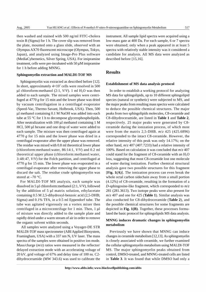
In order to establish a working protocol for analyzingMS data for sphingolipids, up to 10 different sphingolipidspecies (natural or synthetic) were subjected to MS, andthe major peaks from resulting mass spectra were calculatedto deduce the possible chemical structures. The majorpeaks from two sphingolipids molecules, C8-ceramide andC8-dihydroceramide, are listed in Table 1 and Table 2,respectively. 25 major peaks were generated by C8-ceramide during the ionization process, of which mostwere from the matrix 2,5-DHB. m/z 425 (425.6896)corresponded to the intact C8-ceramide. However, therelative intensity of this peak was only 14.17%; on theother hand, m/z 407 (407.7233) had a relative intensity of100%. Based on calculation it was concluded that m/z 407could stand for the fragment of C8-ceramide with an H2Oloss, suggesting that most C8-ceramide lost one moleculeof water during ionization. Further chemical structuralanalysis gave two possible structures for this fragment[Fig. 1(A)]. The ionization process can even break thewhole octal carbon sidechain away from a small portion(4.12%) of C8-ceramide, resulting in the formation of aD-sphingosine-like fragment, which corresponded to m/z281 (281.3613). Two isotope peaks were also present form/z 407 and one for 425 (Table 1). Similar analysis wasalso conducted for C8-dihydroceramide (Table 2), andthe possible chemical structures for some fragments aredepicted in Fig. 1(B). Together, these processes formu-lated the basic protocol for sphingolipids MS data analysis.
MNNG induces dramatic changes in sphingomyelinmetabolism
Previously we have shown that MNNG can inducechanges in ceramide metabolism [12,13]. As sphingomyelinis closely associated with ceramide, we further examinedthe cellular sphingomyelin metabolism using MALDI-TOFMS. The major sphingomyelin peaks obtained fromcontrol, DMSO-treated, and MNNG-treated cells are listedin Table 3. It was found that while DMSO had only a
518 Acta Biochim Biophys Sin Vol. 37, No. 8
©Institute of Biochemistry and Cell Biology, SIBS, CAS
Index Centroid mass (m/z) Charge (z) Relative intensity (%) Source
1 217.8414 0 6.23 Matrix2 231.6644 1 4.05 ?3 263.8577 0 6.38 ?4 264.1504 1 11.88 ?5 265.1066 1 5.16 ?6 272.5887 0 11.08 Matrix7 273.8165 0 15.67 Matrix8 281.3613 0 4.12 C18H35NO9 301.7986 0 4.32 Matrix
10 317.7744 0 3.60 Matrix11 329.7930 0 2.40 Matrix12 332.4761 1 34.47 ?13 333.4742 1 6.40 ?14 389.7198 1 4.76 Matrix15 390.6614 1 5.62 Matrix16 407.7233 1 100.00 C26H49NO2
17 408.6847 1 51.31 Isotope peak18 409.6962 1 15.02 Isotope peak19 414.5155 1 5.28 Matrix20 425.6896 1 14.17 C8-ceramide21 426.6879 1 3.64 Isotope peak22 436.4980 1 20.80 Matrix23 437.4921 1 4.99 Matrix24 447.6712 1 47.20 ?25 448.6611 1 16.08 ?
Table 1 MALDI-TOF MS analysis of C8-ceramide
Fig. 1 Proposed chemical structures for fragmented C8-ceramide and C8-dihydroceramide(A) m/z 407 from C8-ceramide. (B) m/z 238, 253 and 409 from C8-dihydroceramide.
Aug., 2005 Yun HUANG et al.: Effects of N-methyl-N'-nitro-N-nitrosoguanidine on Sphingomyelin Metabolism 519
http://www.abbs.info; www.blackwellpublishing.com/abbs
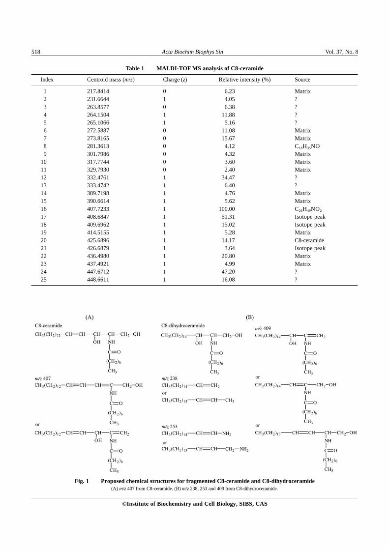
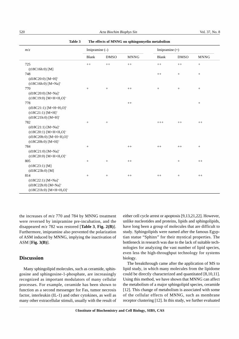
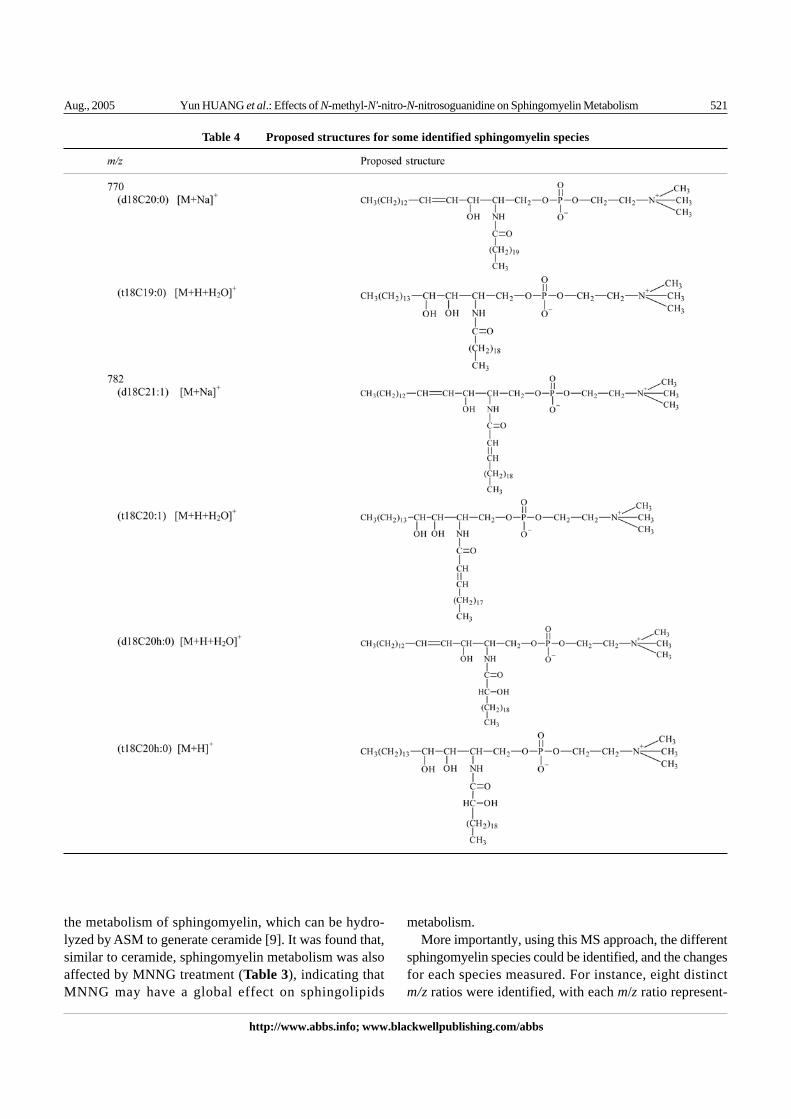
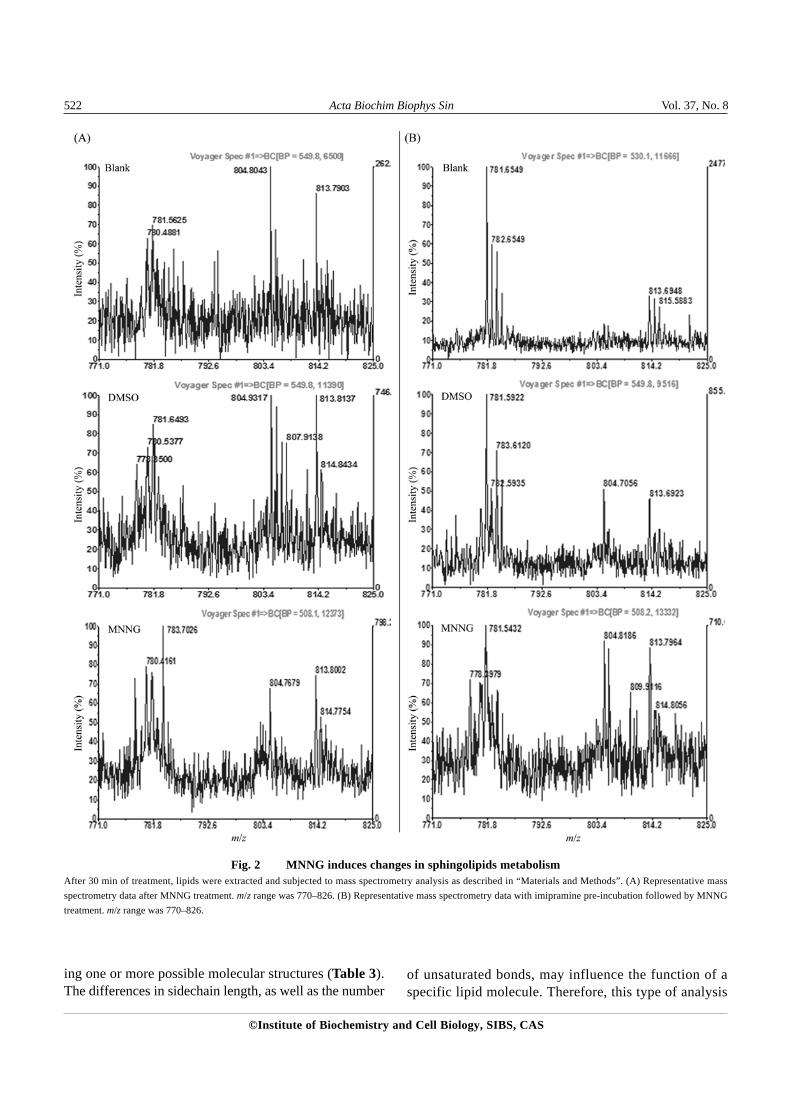
minor effect on sphingomyelin metabolism, there weresignificant differences between MNNG-treated and controlsamples for sphingomyelin. For example, m/z 778 wasnot present in control but appeared after MNNG treatment;whereas m/z 782 showed up in control but disappearedafter MNNG treatment (Table 3). In addition, the con-centrations of several sphingomyelin species, includingm/z 770, 784, 805 and 814, were increased. The massspectra data for some sphingomyelin species [Fig. 2(A),m/z 782, 784, 805 and 814] and possible structures forsome of the identified sphingomyelin species are alsopresented (Table 4).
MNNG induces the redistribution of ASM
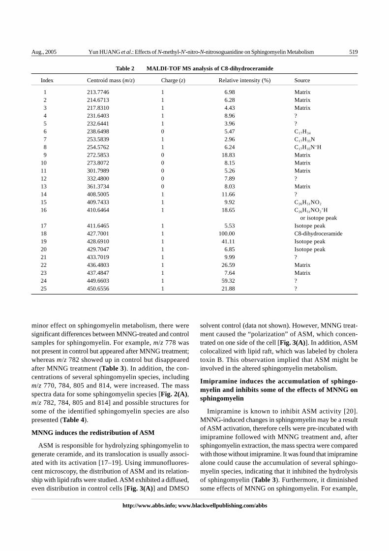
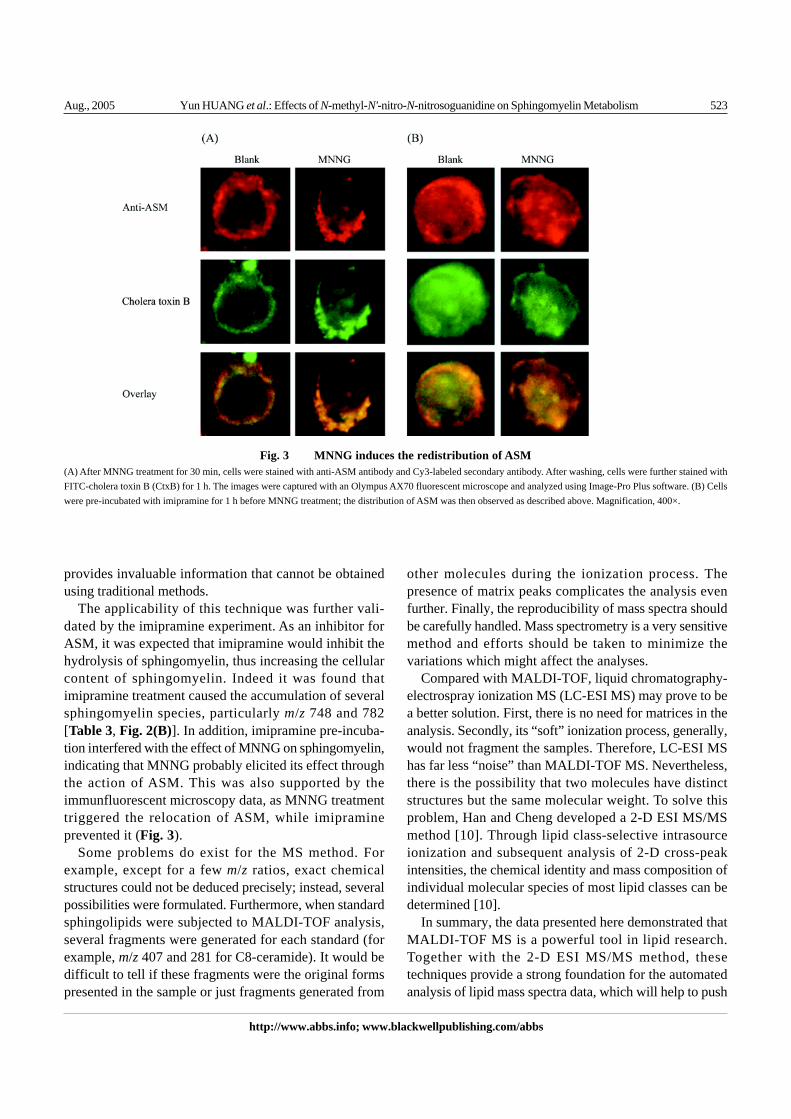
ASM is responsible for hydrolyzing sphingomyelin togenerate ceramide, and its translocation is usually associ-ated with its activation [17–19]. Using immunofluores-cent microscopy, the distribution of ASM and its relation-ship with lipid rafts were studied. ASM exhibited a diffused,even distribution in control cells [Fig. 3(A)] and DMSO
solvent control (data not shown). However, MNNG treat-ment caused the “polarization” of ASM, which concen-trated on one side of the cell [Fig. 3(A)]. In addition, ASMcolocalized with lipid raft, which was labeled by choleratoxin B. This observation implied that ASM might beinvolved in the altered sphingomyelin metabolism.
Imipramine induces the accumulation of sphingo-myelin and inhibits some of the effects of MNNG onsphingomyelin
Imipramine is known to inhibit ASM activity [20].MNNG-induced changes in sphingomyelin may be a resultof ASM activation, therefore cells were pre-incubated withimipramine followed with MNNG treatment and, aftersphingomyelin extraction, the mass spectra were comparedwith those without imipramine. It was found that imipraminealone could cause the accumulation of several sphingo-myelin species, indicating that it inhibited the hydrolysisof sphingomyelin (Table 3). Furthermore, it diminishedsome effects of MNNG on sphingomyelin. For example,
Index Centroid mass (m/z) Charge (z) Relative intensity (%) Source
1 213.7746 1 6.98 Matrix2 214.6713 1 6.28 Matrix3 217.8310 1 4.43 Matrix4 231.6403 1 8.96 ?5 232.6441 1 3.96 ?6 238.6498 0 5.47 C17H34
7 253.5839 1 2.96 C17H35N8 254.5762 1 6.24 C17H35N+H9 272.5853 0 18.83 Matrix
10 273.8072 0 8.15 Matrix11 301.7989 0 5.26 Matrix12 332.4800 0 7.89 ?13 361.3734 0 8.03 Matrix14 408.5005 1 11.66 ?15 409.7433 1 9.92 C26H51NO2
16 410.6464 1 18.65 C26H51NO2+H
or isotope peak17 411.6465 1 5.53 Isotope peak18 427.7001 1 100.00 C8-dihydroceramide19 428.6910 1 41.11 Isotope peak20 429.7047 1 6.85 Isotope peak21 433.7019 1 9.99 ?22 436.4803 1 26.59 Matrix23 437.4847 1 7.64 Matrix24 449.6603 1 59.32 ?25 450.6556 1 21.88 ?
Table 2 MALDI-TOF MS analysis of C8-dihydroceramide
520 Acta Biochim Biophys Sin Vol. 37, No. 8
©Institute of Biochemistry and Cell Biology, SIBS, CAS
the increases of m/z 770 and 784 by MNNG treatmentwere reversed by imipramine pre-incubation, and thedisappeared m/z 782 was restored [Table 3, Fig. 2(B)].Furthermore, imipramine also prevented the polarizationof ASM induced by MNNG, implying the inactivation ofASM [Fig. 3(B)].
Discussion
Many sphingolipid molecules, such as ceramide, sphin-gosine and sphingosine-1-phosphate, are increasinglyrecognized as important modulators of many cellularprocesses. For example, ceramide has been shown tofunction as a second messenger for Fas, tumor necrosisfactor, interleukin (IL-1) and other cytokines, as well asmany other extracellular stimuli, usually with the result of
either cell cycle arrest or apoptosis [9,13,21,22]. However,unlike nucleotides and proteins, lipids and sphingolipids,have long been a group of molecules that are difficult tostudy. Sphingolipids were named after the famous Egyp-tian statue “Sphinx” for their mystical properties. Thebottleneck in research was due to the lack of suitable tech-nologies for analyzing the vast number of lipid species,even less the high-throughput technology for systemsbiology.
The breakthrough came after the application of MS tolipid study, in which many molecules from the lipidomecould be directly characterized and quantitated [8,10,11].Using this method, we have shown that MNNG can affectthe metabolism of a major sphingolipid species, ceramide[12]. This change of metabolism is associated with someof the cellular effects of MNNG, such as membranereceptor clustering [12]. In this study, we further evaluated
Table 3 The effects of MNNG on sphingomyelin metabolism
m/z Imipramine (–) Imipramine (+)
Blank DMSO MNNG Blank DMSO MNNG
725 ++ ++ ++ ++ ++ +(t18C16h:0) [M]
748 ++ + +(d18C20:0) [M+H]+
(t18C16h:0) [M+Na]+
770 + + ++ + + +(d18C20:0) [M+Na]+
(t18C19:0) [M+H+H2O]+
778 ++ +(d18C21:1) [M+H+H2O]+
(t18C21:1) [M+H]+
(d18C21h:0) [M+H]+
782 + + +++ ++ ++(d18C21:1) [M+Na]+
(t18C20:1) [M+H+H2O]+
(d18C20h:0) [M+H+H2O]+
(t18C20h:0) [M+H]+
784 + ++ ++ ++ +(d18C21:0) [M+Na]+
(t18C20:0) [M+H+H2O]+
805 + + ++ + ++(t18C23:1) [M](d18C23h:0) [M]
814 + + ++ ++ + ++(t18C22:1) [M+Na]+
(d18C22h:0) [M+Na]+
(t18C21h:0) [M+H+H2O]+
Aug., 2005 Yun HUANG et al.: Effects of N-methyl-N'-nitro-N-nitrosoguanidine on Sphingomyelin Metabolism 521
http://www.abbs.info; www.blackwellpublishing.com/abbs
the metabolism of sphingomyelin, which can be hydro-lyzed by ASM to generate ceramide [9]. It was found that,similar to ceramide, sphingomyelin metabolism was alsoaffected by MNNG treatment (Table 3), indicating thatMNNG may have a global effect on sphingolipids
metabolism.More importantly, using this MS approach, the different
sphingomyelin species could be identified, and the changesfor each species measured. For instance, eight distinctm/z ratios were identified, with each m/z ratio represent-
Table 4 Proposed structures for some identified sphingomyelin species
522 Acta Biochim Biophys Sin Vol. 37, No. 8
©Institute of Biochemistry and Cell Biology, SIBS, CAS
ing one or more possible molecular structures (Table 3).The differences in sidechain length, as well as the number
of unsaturated bonds, may influence the function of aspecific lipid molecule. Therefore, this type of analysis
Fig. 2 MNNG induces changes in sphingolipids metabolismAfter 30 min of treatment, lipids were extracted and subjected to mass spectrometry analysis as described in “Materials and Methods”. (A) Representative massspectrometry data after MNNG treatment. m/z range was 770–826. (B) Representative mass spectrometry data with imipramine pre-incubation followed by MNNGtreatment. m/z range was 770–826.
Aug., 2005 Yun HUANG et al.: Effects of N-methyl-N'-nitro-N-nitrosoguanidine on Sphingomyelin Metabolism 523
http://www.abbs.info; www.blackwellpublishing.com/abbs
provides invaluable information that cannot be obtainedusing traditional methods.
The applicability of this technique was further vali-dated by the imipramine experiment. As an inhibitor forASM, it was expected that imipramine would inhibit thehydrolysis of sphingomyelin, thus increasing the cellularcontent of sphingomyelin. Indeed it was found thatimipramine treatment caused the accumulation of severalsphingomyelin species, particularly m/z 748 and 782[Table 3, Fig. 2(B)]. In addition, imipramine pre-incuba-tion interfered with the effect of MNNG on sphingomyelin,indicating that MNNG probably elicited its effect throughthe action of ASM. This was also supported by theimmunfluorescent microscopy data, as MNNG treatmenttriggered the relocation of ASM, while imipramineprevented it (Fig. 3).
Some problems do exist for the MS method. Forexample, except for a few m/z ratios, exact chemicalstructures could not be deduced precisely; instead, severalpossibilities were formulated. Furthermore, when standardsphingolipids were subjected to MALDI-TOF analysis,several fragments were generated for each standard (forexample, m/z 407 and 281 for C8-ceramide). It would bedifficult to tell if these fragments were the original formspresented in the sample or just fragments generated from
other molecules during the ionization process. Thepresence of matrix peaks complicates the analysis evenfurther. Finally, the reproducibility of mass spectra shouldbe carefully handled. Mass spectrometry is a very sensitivemethod and efforts should be taken to minimize thevariations which might affect the analyses.
Compared with MALDI-TOF, liquid chromatography-electrospray ionization MS (LC-ESI MS) may prove to bea better solution. First, there is no need for matrices in theanalysis. Secondly, its “soft” ionization process, generally,would not fragment the samples. Therefore, LC-ESI MShas far less “noise” than MALDI-TOF MS. Nevertheless,there is the possibility that two molecules have distinctstructures but the same molecular weight. To solve thisproblem, Han and Cheng developed a 2-D ESI MS/MSmethod [10]. Through lipid class-selective intrasourceionization and subsequent analysis of 2-D cross-peakintensities, the chemical identity and mass composition ofindividual molecular species of most lipid classes can bedetermined [10].
In summary, the data presented here demonstrated thatMALDI-TOF MS is a powerful tool in lipid research.Together with the 2-D ESI MS/MS method, thesetechniques provide a strong foundation for the automatedanalysis of lipid mass spectra data, which will help to push
Fig. 3 MNNG induces the redistribution of ASM(A) After MNNG treatment for 30 min, cells were stained with anti-ASM antibody and Cy3-labeled secondary antibody. After washing, cells were further stained withFITC-cholera toxin B (CtxB) for 1 h. The images were captured with an Olympus AX70 fluorescent microscope and analyzed using Image-Pro Plus software. (B) Cellswere pre-incubated with imipramine for 1 h before MNNG treatment; the distribution of ASM was then observed as described above. Magnification, 400×.
524 Acta Biochim Biophys Sin Vol. 37, No. 8
©Institute of Biochemistry and Cell Biology, SIBS, CAS
the study of systems biology to a new level.
Acknowledgements
The authors gratefully thank Dr. T. TAKETOMI forproviding detailed instructions for analyzing the MALDI-TOF mass spectrometry data, and Dr. X. HAN for thehelpful discussion regarding lipidomics.
References
1 Pognan F. Genomics, proteomics and metabonomics in toxicology: Hopefullynot ‘fashionomics’. Pharmacogenomics 2004, 5: 879–893
2 Kramer R, Cohen D. Functional genomics to new drug targets. Nat Rev DrugDiscov 2004, 3: 965–972
3 Aggarwal K, Lee KH. Functional genomics and proteomics as a foundationfor systems biology. Brief Funct Genomic Proteomic 2003, 2: 175–184
4 Kell DB. Metabolomics and systems biology: Making sense of the soup.Curr Opin Microbiol 2004, 7: 296–307
5 Han X, Gross RW. Global analyses of cellular lipidomes directly from crudeextracts of biological samples by ESI mass spectrometry: A bridge to lipidomics.J Lipid Res 2003, 44: 1071–1079
6 Lagarde M, Geloen A, Record M, Vance D, Spener F. Lipidomics is emerging.Biochim Biophys Acta 2003, 1634: 61
7 Fahy E, Subramaniam S, Brown HA, Glass CK, Merrill AH Jr, Murphy RC,Raetz CR et al. A comprehensive classification system for lipids. J Lipid Res2005, 46: 839–862
8 Han X, Yang J, Cheng H, Ye H, Gross RW. Toward fingerprinting cellularlipidomes directly from biological samples by two-dimensional electrosprayionization mass spectrometry. Anal Biochem 2004, 330: 317–331
9 Yang J, Yu Y, Sun S, Duerksen-Hughes PJ. Ceramide and other sphingolipidsin cellular responses. Cell Biochem Biophys 2004, 40: 323–350
10 Han X, Cheng H. Characterization and direct quantitation of cerebrosidemolecular species from lipid extracts by shotgun lipidomics. J Lipid Res
2005, 46: 163–17511 Han X, Gross RW. Shotgun lipidomics: Electrospray ionization mass
spectrometric analysis and quantitation of cellular lipidomes directly fromcrude extracts of biological samples. Mass Spectrom Rev 2005, 24: 367–412
12 Huang Y, Yang J, Shen J, Chen F, Yu Y. Sphingolipids are involved inN-methyl-N'-nitro-N-nitrosoguanidine-induced epidermal growth factor receptorclustering. Biochem Biophys Res Commun 2005, 330: 430–438
13 Yang J, Duerksen-Hughes PJ. Activation of a p53-independent, sphingolipid-mediated cytolytic pathway in p53-negative mouse fibroblast cells treatedwith N-methyl-N-nitro-N-nitrosoguanidine. J Biol Chem 2001, 276: 27129–27135
14 Gao Z, Yang J, Huang Y, Yu Y. N-methyl-N'-nitro-N-nitrosoguanidineinterferes with the epidermal growth factor receptor-mediated signaling pathway.Mutat Res 2005, 570: 175–184
15 Fujiwaki T, Yamaguchi S, Tasaka M, Sakura N, Taketomi T. Application ofdelayed extraction-matrix-assisted laser desorption ionization time-of-flightmass spectrometry for analysis of sphingolipids in pericardial fluid, peritonealfluid and serum from Gaucher disease patients. J Chromatogr B AnalytTechnol Biomed Life Sci 2002, 776: 115–123
16 Fujiwaki T, Yamaguchi S, Sukegawa K, Taketomi T. Application of delayedextraction matrix-assisted laser desorption ionization time-of-flight massspectrometry for analysis of sphingolipids in cultured skin fibroblasts fromsphingolipidosis patients. Brain Dev 2002, 24: 170–173
17 Grassme H, Cremesti A, Kolesnick R, Gulbins E. Ceramide-mediated clusteringis required for CD95-DISC formation. Oncogene 2003, 22: 5457–5470
18 Gulbins E, Grassme H. Ceramide and cell death receptor clustering. BiochimBiophys Acta 2002, 1585: 139–145
19 Gulbins E. Regulation of death receptor signaling and apoptosis by ceramide.Pharmacol Res 2003, 47: 393–399
20 Lacour S, Hammann A, Grazide S, Lagadic-Gossmann D, Athias A, Sergent O,Laurent G et al. Cisplatin-induced CD95 redistribution into membrane lipidrafts of HT29 human colon cancer cells. Cancer Res 2004, 64: 3593–3598
21 Pru JK, Hendry IR, Davis JS, Rueda BR. Soluble Fas ligand activates thesphingomyelin pathway and induces apoptosis in luteal steroidogenic cellsindependently of stress-activated p38(MAPK). Endocrinology 2002, 143:4350–4357
22 Luberto C, Hassler DF, Signorelli P, Okamoto Y, Sawai H, Boros E, Hazen-Martin DJ et al. Inhibition of tumor necrosis factor-induced cell death inMCF7 by a novel inhibitor of neutral sphingomyelinase. J Biol Chem 2002,277: 41128–41139
Edited byShu-Sen LIU


![Sphingomyelin Liposomes Containing Porphyrin phospholipid ...phosphoethanolamine-N-[methoxy(polyethylene glycol)-2000] (DSPE-PEG-2K, Avanti #880120P), and Sphingomyelin (SPM, # Coatsome](https://img.pdfslide.us/doc/110x75/5f3f9b782f336f6958157d47/sphingomyelin-liposomes-containing-porphyrin-phospholipid-phosphoethanolamine-n-methoxypolyethylene.jpg)