3
Dear Sir, Allergic granulomatous angiitis (Churg-Strauss syn- drome, CSS) is characterized by the triad of asthma, eosinophilia, and systemic vasculitis [1]. In addition to these classical findings, pulmonary infiltrates, skin le- sions, eye, gastrointestinal, and lymph node involvement has been reported [5]. There are rare cases of limited form of CSS with no clinical evidence of asthma [6]. The patient presented here had no history of asthma or pulmonary symptoms. The patient’s disease was limited to gastrointestinal system and the lymph nodes draining the involved bowel segment. The patient had an elevated white blood cell count (16,800/ml) with normal red cell indices. On peripheral smear eosinophils were significantly elevated (38%). Se- rological tests for hepatitis B virus and human immuno- deficiency virus were negative, as were those for anti- nuclear antibodies, anti-neutrophil cytoplasmic antibod- ies (ANCA), and rheumatic factor. Urinalysis showed no proteinuria or hematuria. Renal and liver function tests were within normal limits. IgE levels were normal. The patient’s neurological examination and electromyographic findings were normal. Chest computed tomography re- vealed multiple pulmonary nodules on both lungs. Aller- gy tests were performed and no allergic response was observed on screening tests. The patient was a 19-year-old man who had experi- ence nausea, diarrhea, and fatigue for 1–2 weeks. The patient presented to the emergency room with abdomi- nal colic. Following initial work-up the clinical suspi- cion of acute appendicitis was established. Emergency laparotomy was performed. On gross inspection the ap- pendix was found to be normal. However, multiple hemorrhagic serosal and mesenteric nodules and necrot- ic appearing ileum, cecum, and transverse colon were observed. A right hemicolectomy including the ileoce- cal region was performed followed by an ileotransver- sostomy. On gross inspection the bowel serosa appeared hem- orrhagic and focally necrotic. On sectioning the bowel mucosa appeared pale but not ulcerated. The serosal nodules were hemorrhagic. No tumor was identified. The appendix appeared normal. On microscopic evaluation a necrotizing eosinophilic vasculitis involving small and medium-sized arteries was seen (Fig. 1C, D). The bowel mucosa showed ischemic changes but no crypt abscesses were observed (Fig. 1A). In addition, there was a dense eosinophilic infiltrate throughout the bowel wall and the lymph node capsules, sparing the mucosa (Fig. 1B). The regional lymph nodes showed partial involvement with the vasculitic process. The lymph node sinuses were filled with eosinophils. Eosinophilic infiltrates extended to pericapsular areas. The lymph node cortexes retained the follicular architecture but focal areas contained eosi- nophilic and necrotizing granulomas (Fig. 1E, F). Eosi- nophilic vasculitis of the lymph node arterioles was also evident. Based on these pathological and laboratory find- ings, the diagnosis of limited form of CSS was estab- lished. The patient responded well to steroids and cyclophos- phamide and is on remission with no evidence of any or- gan involvement. Following therapy the patient’s periph- eral eosinophilia was reversed and the pulmonary nod- ules detected by chest computed tomography decreased in diameter. The case presented here lacks some of the classical features of CSS such as asthma and p-ANCA positivity. However, presence of eosinophilia in the peripheral blood, eosinophilic infiltrates in the tissues, and eosino- philic vasculitis and necrotizing granulomas are specific F. Kacar ( ) · E. Levi · E. Dikicioglu · I. Meteoglu · M. Uyar Department of Pathology, Adnan Menderes University Medical School, Aydin 09100 Turkey e-mail: [email protected] Tel.: +90-256-2124078 Fax: +90-256-2120146 H. Erpek Department of Pathology, Adnan Menderes University Medical School, Aydin 09100 Turkey Virchows Arch (2002) 441:632–634 DOI 10.1007/s00428-002-0688-8 LETTER TO THE EDITOR Füruzan Kacar · Edi Levi · Emel Dikicioglu Ibrahim Meteoglu · Hakan Erpek · Meral Uyar A limited form of Churg-Strauss syndrome presenting as acute abdominal catastrophe Received: 13 December 2001 / Accepted: 18 June 2002 / Published online: 12 July 2002 © Springer-Verlag 2002

A limited form of Churg-Strauss syndrome presenting as acute abdominal catastrophe

  • Upload
    meral

  • View
    214

  • Download
    2

Embed Size (px)

Citation preview

Dear Sir,

Allergic granulomatous angiitis (Churg-Strauss syn-drome, CSS) is characterized by the triad of asthma,eosinophilia, and systemic vasculitis [1]. In addition tothese classical findings, pulmonary infiltrates, skin le-sions, eye, gastrointestinal, and lymph node involvementhas been reported [5]. There are rare cases of limitedform of CSS with no clinical evidence of asthma [6].The patient presented here had no history of asthma orpulmonary symptoms. The patient’s disease was limitedto gastrointestinal system and the lymph nodes drainingthe involved bowel segment.

The patient had an elevated white blood cell count(16,800/ml) with normal red cell indices. On peripheralsmear eosinophils were significantly elevated (38%). Se-rological tests for hepatitis B virus and human immuno-deficiency virus were negative, as were those for anti-nuclear antibodies, anti-neutrophil cytoplasmic antibod-ies (ANCA), and rheumatic factor. Urinalysis showed noproteinuria or hematuria. Renal and liver function testswere within normal limits. IgE levels were normal. Thepatient’s neurological examination and electromyographicfindings were normal. Chest computed tomography re-vealed multiple pulmonary nodules on both lungs. Aller-gy tests were performed and no allergic response wasobserved on screening tests.

The patient was a 19-year-old man who had experi-ence nausea, diarrhea, and fatigue for 1–2 weeks. Thepatient presented to the emergency room with abdomi-

nal colic. Following initial work-up the clinical suspi-cion of acute appendicitis was established. Emergencylaparotomy was performed. On gross inspection the ap-pendix was found to be normal. However, multiplehemorrhagic serosal and mesenteric nodules and necrot-ic appearing ileum, cecum, and transverse colon wereobserved. A right hemicolectomy including the ileoce-cal region was performed followed by an ileotransver-sostomy.

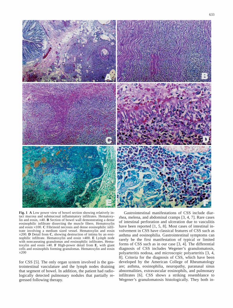
On gross inspection the bowel serosa appeared hem-orrhagic and focally necrotic. On sectioning the bowelmucosa appeared pale but not ulcerated. The serosalnodules were hemorrhagic. No tumor was identified. Theappendix appeared normal. On microscopic evaluation anecrotizing eosinophilic vasculitis involving small andmedium-sized arteries was seen (Fig. 1C, D). The bowelmucosa showed ischemic changes but no crypt abscesseswere observed (Fig. 1A). In addition, there was a denseeosinophilic infiltrate throughout the bowel wall and thelymph node capsules, sparing the mucosa (Fig. 1B). Theregional lymph nodes showed partial involvement withthe vasculitic process. The lymph node sinuses werefilled with eosinophils. Eosinophilic infiltrates extendedto pericapsular areas. The lymph node cortexes retainedthe follicular architecture but focal areas contained eosi-nophilic and necrotizing granulomas (Fig. 1E, F). Eosi-nophilic vasculitis of the lymph node arterioles was alsoevident. Based on these pathological and laboratory find-ings, the diagnosis of limited form of CSS was estab-lished.

The patient responded well to steroids and cyclophos-phamide and is on remission with no evidence of any or-gan involvement. Following therapy the patient’s periph-eral eosinophilia was reversed and the pulmonary nod-ules detected by chest computed tomography decreasedin diameter.

The case presented here lacks some of the classicalfeatures of CSS such as asthma and p-ANCA positivity.However, presence of eosinophilia in the peripheralblood, eosinophilic infiltrates in the tissues, and eosino-philic vasculitis and necrotizing granulomas are specific

F. Kacar (✉) · E. Levi · E. Dikicioglu · I. Meteoglu · M. UyarDepartment of Pathology, Adnan Menderes University Medical School, Aydin 09100 Turkeye-mail: [email protected].: +90-256-2124078Fax: +90-256-2120146

H. ErpekDepartment of Pathology, Adnan Menderes University Medical School, Aydin 09100 Turkey

Virchows Arch (2002) 441:632–634DOI 10.1007/s00428-002-0688-8

L E T T E R T O T H E E D I T O R

Füruzan Kacar · Edi Levi · Emel DikiciogluIbrahim Meteoglu · Hakan Erpek · Meral Uyar

A limited form of Churg-Strauss syndrome presenting as acute abdominal catastrophe

Received: 13 December 2001 / Accepted: 18 June 2002 / Published online: 12 July 2002© Springer-Verlag 2002

Verwendete Distiller 5.0.x Joboptions
Dieser Report wurde automatisch mit Hilfe der Adobe Acrobat Distiller Erweiterung "Distiller Secrets v1.0.5" der IMPRESSED GmbH erstellt. Sie koennen diese Startup-Datei für die Distiller Versionen 4.0.5 und 5.0.x kostenlos unter http://www.impressed.de herunterladen. ALLGEMEIN ---------------------------------------- Dateioptionen: Kompatibilität: PDF 1.2 Für schnelle Web-Anzeige optimieren: Ja Piktogramme einbetten: Ja Seiten automatisch drehen: Nein Seiten von: 1 Seiten bis: Alle Seiten Bund: Links Auflösung: [ 600 600 ] dpi Papierformat: [ 595 785 ] Punkt KOMPRIMIERUNG ---------------------------------------- Farbbilder: Downsampling: Ja Berechnungsmethode: Bikubische Neuberechnung Downsample-Auflösung: 150 dpi Downsampling für Bilder über: 225 dpi Komprimieren: Ja Automatische Bestimmung der Komprimierungsart: Ja JPEG-Qualität: Mittel Bitanzahl pro Pixel: Wie Original Bit Graustufenbilder: Downsampling: Ja Berechnungsmethode: Bikubische Neuberechnung Downsample-Auflösung: 150 dpi Downsampling für Bilder über: 225 dpi Komprimieren: Ja Automatische Bestimmung der Komprimierungsart: Ja JPEG-Qualität: Mittel Bitanzahl pro Pixel: Wie Original Bit Schwarzweiß-Bilder: Downsampling: Ja Berechnungsmethode: Bikubische Neuberechnung Downsample-Auflösung: 600 dpi Downsampling für Bilder über: 900 dpi Komprimieren: Ja Komprimierungsart: CCITT CCITT-Gruppe: 4 Graustufen glätten: Nein Text und Vektorgrafiken komprimieren: Ja SCHRIFTEN ---------------------------------------- Alle Schriften einbetten: Ja Untergruppen aller eingebetteten Schriften: Nein Wenn Einbetten fehlschlägt: Warnen und weiter Einbetten: Immer einbetten: [ ] Nie einbetten: [ ] FARBE(N) ---------------------------------------- Farbmanagement: Farbumrechnungsmethode: Alle Farben zu sRGB konvertieren Methode: Standard Arbeitsbereiche: Graustufen ICC-Profil: RGB ICC-Profil: sRGB IEC61966-2.1 CMYK ICC-Profil: U.S. Web Coated (SWOP) v2 Geräteabhängige Daten: Einstellungen für Überdrucken beibehalten: Ja Unterfarbreduktion und Schwarzaufbau beibehalten: Ja Transferfunktionen: Anwenden Rastereinstellungen beibehalten: Ja ERWEITERT ---------------------------------------- Optionen: Prolog/Epilog verwenden: Nein PostScript-Datei darf Einstellungen überschreiben: Ja Level 2 copypage-Semantik beibehalten: Ja Portable Job Ticket in PDF-Datei speichern: Nein Illustrator-Überdruckmodus: Ja Farbverläufe zu weichen Nuancen konvertieren: Nein ASCII-Format: Nein Document Structuring Conventions (DSC): DSC-Kommentare verarbeiten: Nein ANDERE ---------------------------------------- Distiller-Kern Version: 5000 ZIP-Komprimierung verwenden: Ja Optimierungen deaktivieren: Nein Bildspeicher: 524288 Byte Farbbilder glätten: Nein Graustufenbilder glätten: Nein Bilder (< 257 Farben) in indizierten Farbraum konvertieren: Ja sRGB ICC-Profil: sRGB IEC61966-2.1 ENDE DES REPORTS ---------------------------------------- IMPRESSED GmbH Bahrenfelder Chaussee 49 22761 Hamburg, Germany Tel. +49 40 897189-0 Fax +49 40 897189-71 Email: [email protected] Web: www.impressed.de
Adobe Acrobat Distiller 5.0.x Joboption Datei
<< /ColorSettingsFile () /AntiAliasMonoImages false /CannotEmbedFontPolicy /Warning /ParseDSCComments false /DoThumbnails true /CompressPages true /CalRGBProfile (sRGB IEC61966-2.1) /MaxSubsetPct 100 /EncodeColorImages true /GrayImageFilter /DCTEncode /Optimize true /ParseDSCCommentsForDocInfo false /EmitDSCWarnings false /CalGrayProfile () /NeverEmbed [ ] /GrayImageDownsampleThreshold 1.5 /UsePrologue false /GrayImageDict << /QFactor 0.9 /Blend 1 /HSamples [ 2 1 1 2 ] /VSamples [ 2 1 1 2 ] >> /AutoFilterColorImages true /sRGBProfile (sRGB IEC61966-2.1) /ColorImageDepth -1 /PreserveOverprintSettings true /AutoRotatePages /None /UCRandBGInfo /Preserve /EmbedAllFonts true /CompatibilityLevel 1.2 /StartPage 1 /AntiAliasColorImages false /CreateJobTicket false /ConvertImagesToIndexed true /ColorImageDownsampleType /Bicubic /ColorImageDownsampleThreshold 1.5 /MonoImageDownsampleType /Bicubic /DetectBlends false /GrayImageDownsampleType /Bicubic /PreserveEPSInfo false /GrayACSImageDict << /VSamples [ 2 1 1 2 ] /QFactor 0.76 /Blend 1 /HSamples [ 2 1 1 2 ] /ColorTransform 1 >> /ColorACSImageDict << /VSamples [ 2 1 1 2 ] /QFactor 0.76 /Blend 1 /HSamples [ 2 1 1 2 ] /ColorTransform 1 >> /PreserveCopyPage true /EncodeMonoImages true /ColorConversionStrategy /sRGB /PreserveOPIComments false /AntiAliasGrayImages false /GrayImageDepth -1 /ColorImageResolution 150 /EndPage -1 /AutoPositionEPSFiles false /MonoImageDepth -1 /TransferFunctionInfo /Apply /EncodeGrayImages true /DownsampleGrayImages true /DownsampleMonoImages true /DownsampleColorImages true /MonoImageDownsampleThreshold 1.5 /MonoImageDict << /K -1 >> /Binding /Left /CalCMYKProfile (U.S. Web Coated (SWOP) v2) /MonoImageResolution 600 /AutoFilterGrayImages true /AlwaysEmbed [ ] /ImageMemory 524288 /SubsetFonts false /DefaultRenderingIntent /Default /OPM 1 /MonoImageFilter /CCITTFaxEncode /GrayImageResolution 150 /ColorImageFilter /DCTEncode /PreserveHalftoneInfo true /ColorImageDict << /QFactor 0.9 /Blend 1 /HSamples [ 2 1 1 2 ] /VSamples [ 2 1 1 2 ] >> /ASCII85EncodePages false /LockDistillerParams false >> setdistillerparams << /PageSize [ 595.276 841.890 ] /HWResolution [ 600 600 ] >> setpagedevice

633

Fig. 1 A Low power view of bowel section showing relatively in-tact mucosa and submucosal inflammatory infiltrates. Hematoxy-lin and eosin, ×40. B Section of bowel wall demonstrating a denseeosinophilic infiltrate dissecting the muscle fibers. Hematoxylinand eosin ×100. C Fibrinoid necrosis and dense eosinophilic infil-trate involving a medium sized vessel. Hematoxylin and eosin×200. D Detail from C, showing destruction of intima by an eosi-nophilic infiltrate. Hematoxylin and eosin ×400. E Lymph nodewith noncaseating granulomas and eosinophilic infiltrates. Hema-toxylin and eosin ×40. F High-power detail from E, with giantcells and eosinophils forming granulomas. Hematoxylin and eosin×200

for CSS [5]. The only organ system involved is the gas-trointestinal vasculature and the lymph nodes drainingthat segment of bowel. In addition, the patient had radio-logically detected pulmonary nodules that partially re-gressed following therapy.

Gastrointestinal manifestations of CSS include diar-rhea, melena, and abdominal cramps [3, 4, 7]. Rare casesof intestinal perforation and ulceration due to vasculitishave been reported [1, 5, 8]. Most cases of intestinal in-volvement in CSS have classical features of CSS such asasthma and eosinophilia. Gastrointestinal symptoms canrarely be the first manifestation of typical or limitedforms of CSS such as in our case [3, 4]. The differentialdiagnosis of CSS includes Wegener’s granulomatosis,polyarteritis nodosa, and microscopic polyarteritis [3, 4,8]. Criteria for the diagnosis of CSS, which have beendeveloped by the American College of Rheumatologyare; asthma, eosinophilia, neuropathy, paranasal sinusabnormalities, extravascular eosinophils, and pulmonaryinfiltrates [6]. CSS shows a striking resemblance toWegener’s granulomatosis histologically. They both in-

diagnose. However, when the classical manifestations ofCSS such as asthma are lacking, and the patient presentswith gastrointestinal symptoms, the vasculitic syndromesincluding limited forms of CSS should also be includedin the differential diagnosis.

References

1. Churg J, Strauss L (1951) Allergic granulomatosis, allergic an-giitis and periarteritis nodosa. Am J Pathol 27:277–294

2. Cualing H, Schroder L, Perme C (2001) Allergic granulomato-sis secondary to a limited form of Churg-Strauss syndrome.Arch Pathol Lab Med 125:954–957

3. Kaneki T, Kawashima A, Hayano T, Honda T, Kubo K,Koizumi T, Sekiguchi M, Ichikawa H, Matsuzawa K, Katsuyama T (1998) Churg-Strauss syndrome (allergic granulo-matous angiitis) presenting with ileus caused by ischemic ilealulcer. J Gastroenterol 33:112–116

4. Lin TL, Wang CR, Liu MF, et al (2001) Multiple colonic ulcerscaused by Churg-Strauss syndrome in a 15-year-old girl. ClinRheumatol 20:362–364

5. Masi AT, Hunder GG, Lie JT (1990) The American College ofRheumatology 1990 criteria for the classification of Churg-Strauss syndrome. Arthritis Rheum 33:1094–1099

6. Ohwada S, Yanagisawa A, Joshita T, et al (1997) Necrotizinggranulomatous vasculitis of transverse colon and gallbladder.Hepatogastroenterology 44:1090–1094

7. Shimamato C, Hirata I, Ohshiba S, Fujiwara S, Nishio M(1990) Churg-Strauss syndrome [allergic granulomatous angii-tis] with peculiar multiple colonic ulcers. Am J Gastroenterol85:316–319

8. Solans R, Bosch JA, Perez-Bocanegra C, et al (2001) Churg-Strauss syndrome: outcome and long-term follow-up of 32 pa-tients. Rheumatology (Oxf) 40:763–771

9. Suen KC, Burton JD (1979) The spectrum of eosinophilic infil-tration of the gastrointestinal tract and its relationship to otherdisorders of angiitis and granulomatosis. Hum Pathol 10:31–43

634

volve the lungs presenting with necrotizing granulomas[2]. CSS is characterized by prominent eosinophilic in-filtrates and peripheral eosinophilia. Wegener’s granulo-matosis is characterized by c-ANCA (anti-proteinase III)as opposed to p-ANCA (anti-myeloperoxidase) com-monly found in CSS [5]. The absence of ANCA isagainst a diagnosis of Wegener’s granulomatosis. Theabsence of ANCA and the presence of eosinophilia isagainst a diagnosis of microscopic or macroscopic PAN[5]. The examination of the bowel did not reveal any in-flammatory lesions of the mucosa, and therefore a diag-nosis of inflammatory bowel disease was unlikely. Ex-amination of the feces and the tissues by special stainsdid not reveal any micro-organisms or parasites, there-fore an infectious cause was also eliminated. The pres-ence of vasculitis spreading to the lymph nodes excludesa diagnosis of eosinophilic gastroenteritis [9].

The cause of CSS is not known, however, ANCA(anti-myeloperoxidase) have been detected in approxi-mately 75% of the cases, and they are thought to play arole in the pathogenesis of this syndrome [8]. Associa-tion with asthma suggests the presence of an allergenicstimulus. The histopathological features of involvementof lymph nodes in CSS have been rarely reported [2]. Inour case the presence of dense eosinophilic infiltrates inthe lymph node sinuses and capsule and the presence ofnecrotizing epithelioid granulomas cuffed by dense eosi-nophilic infiltrates was striking.

We believe that this case is unique because most casesof CSS vasculitis with gastrointestinal involvement re-ported in the literature have a history of asthma and pe-ripheral eosinophilia [3, 4, 7]. These cases which presentwith the classical manifestations of disease are easier to