Embed Size (px)
Citation preview
A High-Content, Phenotypic ScreenIdentifies Fluorouridine as an Inhibitorof Pyoverdine Biosynthesis andPseudomonas aeruginosa Virulence
Daniel R. Kirienko, Alexey V. Revtovich, Natalia V. KirienkoDepartment of BioSciences, Rice University, Houston, Texas, USA
ABSTRACT Pseudomonas aeruginosa is an opportunistic pathogen that causes se-vere health problems. Despite intensive investigation, many aspects of microbial vir-ulence remain poorly understood. We used a high-throughput, high-content, whole-organism, phenotypic screen to identify small molecules that inhibit P. aeruginosavirulence in Caenorhabditis elegans. Approximately half of the hits were known anti-microbials. A large number of hits were nonantimicrobial bioactive compounds, in-cluding the cancer chemotherapeutic 5-fluorouracil. We determined that5-fluorouracil both transiently inhibits bacterial growth and reduces pyoverdine bio-synthesis. Pyoverdine is a siderophore that regulates the expression of several viru-lence determinants and is critical for pathogenesis in mammals. We show that5-fluorouridine, a downstream metabolite of 5-fluorouracil, is responsible for inhibit-ing pyoverdine biosynthesis. We also show that 5-fluorouridine, in contrast to5-fluorouracil, is a genuine antivirulence compound, with no bacteriostatic or bacte-ricidal activity. To our knowledge, this is the first report utilizing a whole-organismscreen to identify novel compounds with antivirulent properties effective againstP. aeruginosa.
IMPORTANCE Despite intense research effort from scientists and the advent ofthe molecular age of biomedical research, many of the mechanisms that underliepathogenesis are still understood poorly, if at all. The opportunistic human patho-gen Pseudomonas aeruginosa causes a variety of soft tissue infections and is respon-sible for over 50,000 hospital-acquired infections per year. In addition, P. aeruginosaexhibits a striking degree of innate and acquired antimicrobial resistance, complicat-ing treatment. It is increasingly important to understand P. aeruginosa virulence. Inan effort to gain this information in an unbiased fashion, we used a high-throughput phenotypic screen to identify small molecules that disrupted bacterialpathogenesis and increased host survival using the model nematode Caenorhabditiselegans. This method led to the unexpected discovery that addition of a modifiednucleotide, 5-fluorouridine, disrupted bacterial RNA metabolism and inhibited syn-thesis of pyoverdine, a critical toxin. Our results demonstrate that this compoundspecifically functions as an antivirulent.
KEYWORDS: 5-fluorouracil, 5-fluorouridine, Caenorhabditis elegans, Pseudomonasaeruginosa, pyoverdine, high-throughput screening, siderophores, virulencedeterminants
Pseudomonas aeruginosa is a Gram-negative opportunistic pathogen responsible fora variety of soft tissue infections. Several conditions, including cystic fibrosis,
immune deficiency, and hospitalization, predispose patients to infection. This pathogenis a key example of the reemergence of clinically relevant bacterial species due towidespread acquisition of drug resistance. For example, a pandrug-resistant strain of
Received 2 August 2016 Accepted 4 August2016 Published 24 August 2016
Citation Kirienko DR, Revtovich AV, KirienkoNV. 2016. A high-content, phenotypic screenidentifies fluorouridine as an inhibitor ofpyoverdine biosynthesis and Pseudomonasaeruginosa virulence. mSphere 1(4):e00217-16.doi:10.1128/mSphere.00217-16.
Editor Melanie Blokesch, Swiss FederalInstitute of Technology Lausanne
Copyright © 2016 Kirienko et al. This is anopen-access article distributed under the termsof the Creative Commons Attribution 4.0International license.
Address correspondence to Natalia V. Kirienko,[email protected].
RESEARCH ARTICLEHost-Microbe Biology
crossmark
Volume 1 Issue 4 e00217-16 msphere.asm.org 1
on March 25, 2020 by guest
http://msphere.asm
.org/D
ownloaded from
P. aeruginosa in Taiwan exhibited mortality rates of ~70% (1). Rates of antimicrobialresistance and the frequency of isolation of multidrug-resistant and extensively drug-resistant strains from clinical samples are increasing across all pathogenic bacterialgenera (2, 3). Meanwhile, a variety of challenges (including regulatory, commercial,intellectual property, and scientific constraints) have led to the reduction, or outrightelimination, of antimicrobial discovery programs by pharmaceutical companies (4–6).
One method to address this problem is to identify new types of treatments such asantivirulents. These drugs would prevent bacterial pathogenesis (preferably withoutlimiting bacterial growth or generating adaptive pressure). Identification of antivirulentcompounds is generally more difficult than identification of classical antimicrobials andrequires a better understanding of the molecular events underlying pathogenesis. Ittypically requires more complex screening regimes whose output metrics are com-prised of host health measures rather than bacterial growth rates. In return, compoundsare much less likely to engender and promote the acquisition and spread of resistancephenotypes. In addition, they are likely to be valuable tools for probing the complex-ities of host-pathogen interactions.
Recently, researchers have begun to test the viability of high-throughput, high-content, phenotypic, whole-animal screens. The model nematode Caenorhabditis el-egans is often used as a host due to its deep characterization, experimental simplicity,and powerful genetics (7–9). In addition, its small size allows assay miniaturization,facilitating screening in microtiter (i.e., 96- and 384-well) plates. This also facilitatesassay automation and screening of small-molecule-compound libraries to identifyvirulence inhibitors. For example, a pilot screen using the liquid killing assay describedhere identified a key role for pyoverdine in P. aeruginosa virulence in C. elegans (10).Pyoverdine, a siderophore (literally, iron carrier) secreted by P. aeruginosa to acquirethis crucial nutrient, also regulates the expression of several other pathogenic deter-minants, including exotoxin A, the endoprotease PrpL, and some types of biofilm(11–13), and can be considered a master virulence determinant. Indeed, several studieshave demonstrated that disrupting pyoverdine alone is sufficient to strongly attenuatevirulence in animal models of P. aeruginosa infection (10, 14–16).
Here we report that fluorinated pyrimidines strongly attenuate P. aeruginosapathogenesis, likely by compromising RNA metabolism. 5-Fluorouracil (FU) and5-fluorouridine (FUR) were shown to limit pyoverdine production. We determined thatFU also temporarily restricts bacterial growth, further limiting disease. This activity waslimited to FU, as the downstream metabolites FUR and 5-fluorodeoxyuridine (FUDR) didnot limit growth. Our data demonstrate that the relevant intermediate metabolite forlimiting virulence factor production is FUR.
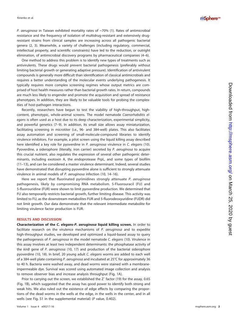
RESULTS AND DISCUSSIONCharacterization of the C. elegans-P. aeruginosa liquid killing screen. In order tofacilitate research on the virulence mechanisms of P. aeruginosa and to expeditehigh-throughput studies, we developed and optimized a liquid-based assay to querythe pathogenesis of P. aeruginosa in the model nematode C. elegans (10). Virulence inthis assay involves at least two independent determinants: the phosphatase activity ofthe kinB gene of P. aeruginosa (10, 17) and production of the bacterial siderophorepyoverdine (10, 18). In brief, 20 young adult C. elegans worms are added to each wellof a 384-well plate containing P. aeruginosa and incubated at 25°C for approximately 36to 40 h. Bacteria were washed away, and dead worms were stained with a membrane-impermeable dye. Survival was scored using automated image collection and analysisto remove observer bias and increase analysis throughput (Fig. 1A).
Prior to carrying out the screen, we established the Z= factor (19) for the assay, 0.65(Fig. 1B), which suggested that the assay has good power to identify both strong andweak hits. We also ruled out the existence of edge effects by comparing the propor-tions of the dead worms in the wells at the edge, in the wells in the center, and in allwells (see Fig. S1 in the supplemental material) (P value, 0.402).
Kirienko et al.
Volume 1 Issue 4 e00217-16 msphere.asm.org 2
on March 25, 2020 by guest
http://msphere.asm
.org/D
ownloaded from
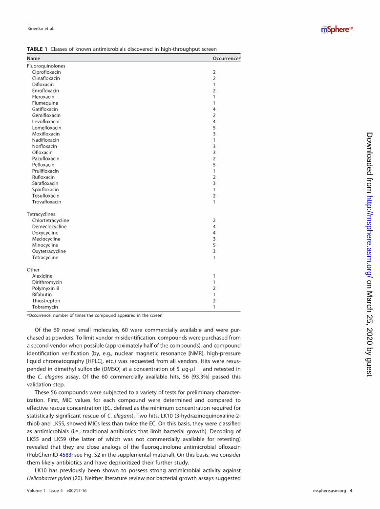
Compounds identified in the liquid killing screen include novel antimicro-bials. The liquid killing assay was used to screen 251 plates from various commerciallysupplied chemical diversity collections. Each plate contained between 320 and 352compounds (the remaining wells were dedicated to positive and negative controls).Combined, these plates comprised 86,441 wells containing test compounds, of which195 were considered hits (0.226% total rate, similar to the rates determined in previousC. elegans phenotypic screens) (8) (Fig. 1C). Initial analysis showed that 81 of these hitsrepresent known antimicrobials. We identified a total of 35 different compounds in thisgroup; each was independently found 1 to 5 times in the screen, and some compoundswere present in two or more plates. Most of the antimicrobials identified belong to thefluoroquinolone and tetracycline classes of antibiotics (Table 1). A total of 45 wells withhits contained known bioactive molecules, with 29 different structures identified. The69 remaining molecules fall into at least three nonexclusive classes: novel antimicro-bials (compounds that limit bacterial growth or kill bacteria), antivirulents (compoundsthat inhibit bacterial pathogenesis without affecting growth), and host immune stim-ulators (compounds that predominantly act by promoting host survival and have littleto no impact on the pathogen). All three categories may lead to the discovery of usefulmolecules with roles ranging from probe compounds to potential leads for therapeuticdevelopment.
Negative Control Hit Compound
Brig
htfie
ldFl
uore
scen
ce
A
C0
20
40
60
80
100
Per
cent
Dea
d
DMSOGentamicin
B
Z’=0.65
86, 441 wellsscreened
195 candidatewells
Antimicrobials81 (35)
Known Bioactives
45 (29)
Novel Small Molecules
69FIG 1 High-throughput screening of C. elegans-mediated liquid killing. (A) Images of worms exposed to P. aeruginosa andtreated with either solvent control DMSO (left) or a small-molecule compound that alleviates killing (right). (B) Quantification ofC. elegans death after treatment with P. aeruginosa, in the presence of either an antipseudomonal antimicrobial (gentamicin) ora vehicle control (DMSO). (C) Schematic showing the total number of wells screened, the number of hits, and the number of hitwells per category (numbers of unique structures are shown in parentheses).
Fluorouridine Inhibits Pyoverdine Biosynthesis
Volume 1 Issue 4 e00217-16 msphere.asm.org 3
on March 25, 2020 by guest
http://msphere.asm
.org/D
ownloaded from
Of the 69 novel small molecules, 60 were commercially available and were pur-chased as powders. To limit vendor misidentification, compounds were purchased froma second vendor when possible (approximately half of the compounds), and compoundidentification verification (by, e.g., nuclear magnetic resonance [NMR], high-pressureliquid chromatography [HPLC], etc.) was requested from all vendors. Hits were resus-pended in dimethyl sulfoxide (DMSO) at a concentration of 5 �g·�l�1 and retested inthe C. elegans assay. Of the 60 commercially available hits, 56 (93.3%) passed thisvalidation step.
These 56 compounds were subjected to a variety of tests for preliminary character-ization. First, MIC values for each compound were determined and compared toeffective rescue concentration (EC, defined as the minimum concentration required forstatistically significant rescue of C. elegans). Two hits, LK10 (3-hydrazinoquinoxaline-2-thiol) and LK55, showed MICs less than twice the EC. On this basis, they were classifiedas antimicrobials (i.e., traditional antibiotics that limit bacterial growth). Decoding ofLK55 and LK59 (the latter of which was not commercially available for retesting)revealed that they are close analogs of the fluoroquinolone antimicrobial ofloxacin(PubChemID 4583; see Fig. S2 in the supplemental material). On this basis, we considerthem likely antibiotics and have deprioritized their further study.
LK10 has previously been shown to possess strong antimicrobial activity againstHelicobacter pylori (20). Neither literature review nor bacterial growth assays suggested
TABLE 1 Classes of known antimicrobials discovered in high-throughput screen
Name Occurrencea
FluoroquinolonesCiprofloxacin 2Clinafloxacin 2Difloxacin 1Enrofloxacin 2Fleroxacin 1Flumequine 1Gatifloxacin 4Gemifloxacin 2Levofloxacin 4Lomefloxacin 5Moxifloxacin 3Nadifloxacin 1Norfloxacin 3Ofloxacin 3Pazufloxacin 2Pefloxacin 5Prulifloxacin 1Rufloxacin 2Sarafloxacin 3Sparfloxacin 1Tosufloxacin 2Trovafloxacin 1
TetracyclinesChlortetracycline 2Demeclocycline 4Doxycycline 4Meclocycline 3Minocycline 5Oxytetracycline 3Tetracycline 1
OtherAlexidine 1Dirithromycin 1Polymyxin B 2Rifabutin 1Thiostrepton 2Tobramycin 1
aOccurrence, number of times the compound appeared in the screen.
Kirienko et al.
Volume 1 Issue 4 e00217-16 msphere.asm.org 4
on March 25, 2020 by guest
http://msphere.asm
.org/D
ownloaded from
than any of the other 54 novel small molecules ordered are likely to function asantimicrobials. Our current hypothesis is that they are novel antivirulents (i.e., virulence-blocking compounds that do not interfere with bacterial growth) or else they promotehost survival by targeting the host (e.g., by stimulating tolerance or immunity).
5-Fluorouracil has potent activity against P. aeruginosa-mediated killing ofC. elegans. To prioritize compounds for in-depth analysis, we decided to initially focusour attention on the 29 bioactive compounds. These belonged to three generalcategories, including surface decontaminants (such as hexachlorophene, phenylmer-curic acetate, and thiomerosal), metal-chelating compounds (such as ciclopirox, piroc-tone, and nitroxamine), and fluoropyrimidines and their derivatives. Compounds in thefirst category most likely limit virulence by killing the pathogen and were thereforedeprioritized for further study. We have previously reported the identification ofciclopirox, which led to identification of pyoverdine as the most relevant virulencefactor in liquid-based killing (10). The other metal-chelating compounds may functionanalogously.
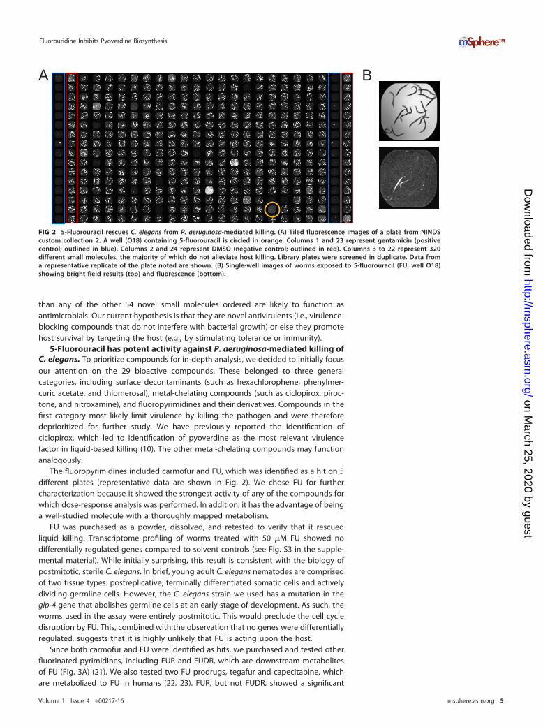
The fluoropyrimidines included carmofur and FU, which was identified as a hit on 5different plates (representative data are shown in Fig. 2). We chose FU for furthercharacterization because it showed the strongest activity of any of the compounds forwhich dose-response analysis was performed. In addition, it has the advantage of beinga well-studied molecule with a thoroughly mapped metabolism.
FU was purchased as a powder, dissolved, and retested to verify that it rescuedliquid killing. Transcriptome profiling of worms treated with 50 �M FU showed nodifferentially regulated genes compared to solvent controls (see Fig. S3 in the supple-mental material). While initially surprising, this result is consistent with the biology ofpostmitotic, sterile C. elegans. In brief, young adult C. elegans nematodes are comprisedof two tissue types: postreplicative, terminally differentiated somatic cells and activelydividing germline cells. However, the C. elegans strain we used has a mutation in theglp-4 gene that abolishes germline cells at an early stage of development. As such, theworms used in the assay were entirely postmitotic. This would preclude the cell cycledisruption by FU. This, combined with the observation that no genes were differentiallyregulated, suggests that it is highly unlikely that FU is acting upon the host.
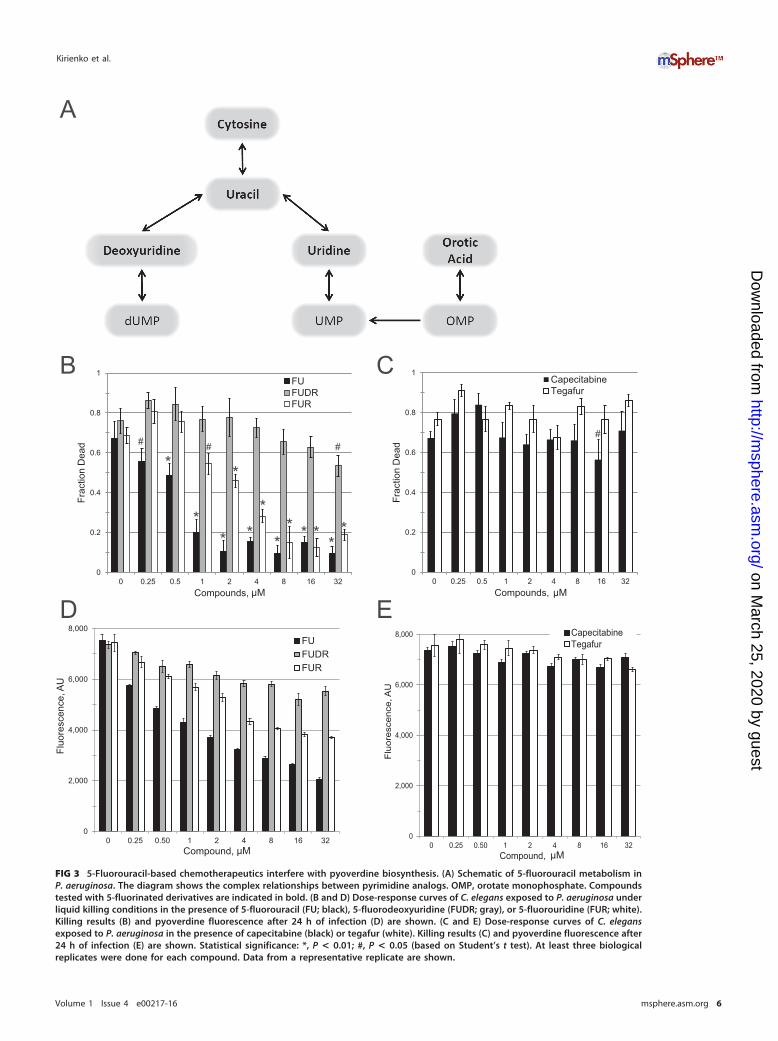
Since both carmofur and FU were identified as hits, we purchased and tested otherfluorinated pyrimidines, including FUR and FUDR, which are downstream metabolitesof FU (Fig. 3A) (21). We also tested two FU prodrugs, tegafur and capecitabine, whichare metabolized to FU in humans (22, 23). FUR, but not FUDR, showed a significant
A B
FIG 2 5-Fluorouracil rescues C. elegans from P. aeruginosa-mediated killing. (A) Tiled fluorescence images of a plate from NINDScustom collection 2. A well (O18) containing 5-fluorouracil is circled in orange. Columns 1 and 23 represent gentamicin (positivecontrol; outlined in blue). Columns 2 and 24 represent DMSO (negative control; outlined in red). Columns 3 to 22 represent 320different small molecules, the majority of which do not alleviate host killing. Library plates were screened in duplicate. Data froma representative replicate of the plate noted are shown. (B) Single-well images of worms exposed to 5-fluorouracil (FU; well O18)showing bright-field results (top) and fluorescence (bottom).
Fluorouridine Inhibits Pyoverdine Biosynthesis
Volume 1 Issue 4 e00217-16 msphere.asm.org 5
on March 25, 2020 by guest
http://msphere.asm
.org/D
ownloaded from
0
2,000
4,000
6,000
8,000
0 0.25 0.50 1 2 4 8 16 32
Fluo
resc
ence
, AU
Compound, uM
FUFUDRFUR
0
2,000
4,000
6,000
8,000
0 0.25 0.50 1 2 4 8 16 32
Fluo
resc
ence
, AU
Compound, uM
CapecitabineTegafur
A
0
0.2
0.4
0.6
0.8
1
0 0.25 0.5 1 2 4 8 16 32
Frac
tion
Dea
d
Compounds, uM
FUFUDRFUR
*** **
**
**
*
*
*# # #
0
0.2
0.4
0.6
0.8
1
0 0.25 0.5 1 2 4 8 16 32
Frac
tion
Dea
d
Compounds, uM
CapecitabineTegafur
#
CB
D EµM µM
µM µM
FIG 3 5-Fluorouracil-based chemotherapeutics interfere with pyoverdine biosynthesis. (A) Schematic of 5-fluorouracil metabolism inP. aeruginosa. The diagram shows the complex relationships between pyrimidine analogs. OMP, orotate monophosphate. Compoundstested with 5-fluorinated derivatives are indicated in bold. (B and D) Dose-response curves of C. elegans exposed to P. aeruginosa underliquid killing conditions in the presence of 5-fluorouracil (FU; black), 5-fluorodeoxyuridine (FUDR; gray), or 5-fluorouridine (FUR; white).Killing results (B) and pyoverdine fluorescence after 24 h of infection (D) are shown. (C and E) Dose-response curves of C. elegansexposed to P. aeruginosa in the presence of capecitabine (black) or tegafur (white). Killing results (C) and pyoverdine fluorescence after24 h of infection (E) are shown. Statistical significance: *, P < 0.01; #, P < 0.05 (based on Student’s t test). At least three biologicalreplicates were done for each compound. Data from a representative replicate are shown.
Kirienko et al.
Volume 1 Issue 4 e00217-16 msphere.asm.org 6
on March 25, 2020 by guest
http://msphere.asm
.org/D
ownloaded from
ability to inhibit C. elegans killing at low micromolar concentrations (Fig. 3B). Neithercapecitabine nor tegafur showed any significant ability to rescue worm killing (Fig. 3C),suggesting that the absorption and/or metabolism of these drugs to FU is inefficient inP. aeruginosa and/or C. elegans. Supplementation with nonfluorinated uracil also hadno discernible effect on pathogenesis at the concentrations tested for fluorinatedderivatives (see Fig. S4 in the supplemental material), indicating that these compoundsare not merely affecting cellular uracil flux.
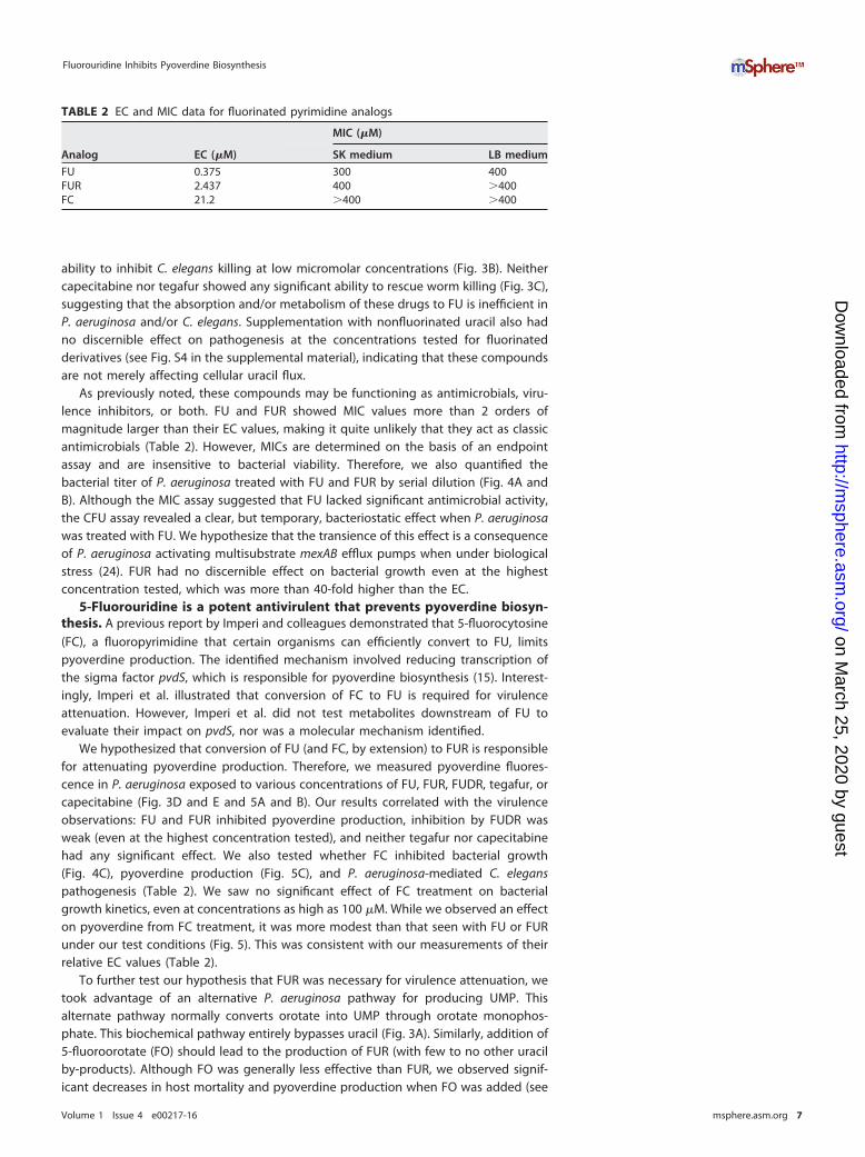
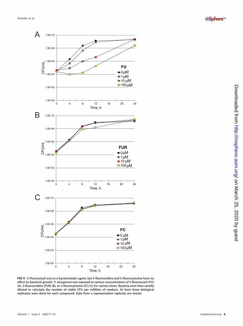
As previously noted, these compounds may be functioning as antimicrobials, viru-lence inhibitors, or both. FU and FUR showed MIC values more than 2 orders ofmagnitude larger than their EC values, making it quite unlikely that they act as classicantimicrobials (Table 2). However, MICs are determined on the basis of an endpointassay and are insensitive to bacterial viability. Therefore, we also quantified thebacterial titer of P. aeruginosa treated with FU and FUR by serial dilution (Fig. 4A andB). Although the MIC assay suggested that FU lacked significant antimicrobial activity,the CFU assay revealed a clear, but temporary, bacteriostatic effect when P. aeruginosawas treated with FU. We hypothesize that the transience of this effect is a consequenceof P. aeruginosa activating multisubstrate mexAB efflux pumps when under biologicalstress (24). FUR had no discernible effect on bacterial growth even at the highestconcentration tested, which was more than 40-fold higher than the EC.
5-Fluorouridine is a potent antivirulent that prevents pyoverdine biosyn-thesis. A previous report by Imperi and colleagues demonstrated that 5-fluorocytosine(FC), a fluoropyrimidine that certain organisms can efficiently convert to FU, limitspyoverdine production. The identified mechanism involved reducing transcription ofthe sigma factor pvdS, which is responsible for pyoverdine biosynthesis (15). Interest-ingly, Imperi et al. illustrated that conversion of FC to FU is required for virulenceattenuation. However, Imperi et al. did not test metabolites downstream of FU toevaluate their impact on pvdS, nor was a molecular mechanism identified.
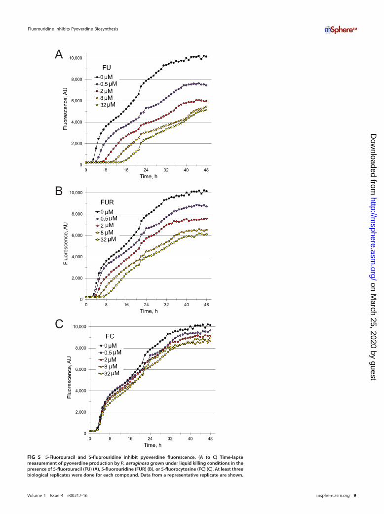
We hypothesized that conversion of FU (and FC, by extension) to FUR is responsiblefor attenuating pyoverdine production. Therefore, we measured pyoverdine fluores-cence in P. aeruginosa exposed to various concentrations of FU, FUR, FUDR, tegafur, orcapecitabine (Fig. 3D and E and 5A and B). Our results correlated with the virulenceobservations: FU and FUR inhibited pyoverdine production, inhibition by FUDR wasweak (even at the highest concentration tested), and neither tegafur nor capecitabinehad any significant effect. We also tested whether FC inhibited bacterial growth(Fig. 4C), pyoverdine production (Fig. 5C), and P. aeruginosa-mediated C. eleganspathogenesis (Table 2). We saw no significant effect of FC treatment on bacterialgrowth kinetics, even at concentrations as high as 100 �M. While we observed an effecton pyoverdine from FC treatment, it was more modest than that seen with FU or FURunder our test conditions (Fig. 5). This was consistent with our measurements of theirrelative EC values (Table 2).
To further test our hypothesis that FUR was necessary for virulence attenuation, wetook advantage of an alternative P. aeruginosa pathway for producing UMP. Thisalternate pathway normally converts orotate into UMP through orotate monophos-phate. This biochemical pathway entirely bypasses uracil (Fig. 3A). Similarly, addition of5-fluoroorotate (FO) should lead to the production of FUR (with few to no other uracilby-products). Although FO was generally less effective than FUR, we observed signif-icant decreases in host mortality and pyoverdine production when FO was added (see
TABLE 2 EC and MIC data for fluorinated pyrimidine analogs
Analog EC (�M)
MIC (�M)
SK medium LB medium
FU 0.375 300 400FUR 2.437 400 �400FC 21.2 �400 �400
Fluorouridine Inhibits Pyoverdine Biosynthesis
Volume 1 Issue 4 e00217-16 msphere.asm.org 7
on March 25, 2020 by guest
http://msphere.asm
.org/D
ownloaded from
1.0E+00
1.0E+02
1.0E+04
1.0E+06
1.0E+08
1.0E+10
0 4 8 12 16 20 24
CFU
/mL
Time, h
FU0 uM1 uM10 uM100 uM
A
µMµM
µMµM
µMµM
1.0E+00
1.0E+02
1.0E+04
1.0E+06
1.0E+08
1.0E+10
0 4 8 12 16 20 24
CFU
/mL
Time, h
FUR0 uM1 uM10 uM100 uMµM
µMµMµM
B
1.0E+00
1.0E+02
1.0E+04
1.0E+06
1.0E+08
1.0E+10
0 4 8 12 16 20 24
CFU
/mL
Time, h
FC0 uM1 uM10 uM100 uMµM
µMµMµM
C
FIG 4 5-Fluorouracil acts as a bacteriostatic agent, but 5-fluorouridine and 5-fluorocytosine have noeffect on bacterial growth. P. aeruginosa was exposed to various concentrations of 5-fluorouracil (FU)(A), 5-fluorouridine (FUR) (B), or 5-fluorocytosine (FC) (C) for various times. Bacteria were then seriallydiluted to calculate the number of viable CFU per milliliter of medium. At least three biologicalreplicates were done for each compound. Data from a representative replicate are shown.
Kirienko et al.
Volume 1 Issue 4 e00217-16 msphere.asm.org 8
on March 25, 2020 by guest
http://msphere.asm
.org/D
ownloaded from
0
2,000
4,000
6,000
8,000
10,000
0 8 16 24 32 40 48
Fluo
resc
ence
, AU
Time, h
FU0 uM0.5 uM2 uM8 uM32 uM
0
2,000
4,000
6,000
8,000
10,000
0 8 16 24 32 40 48
Fluo
resc
ence
, AU
Time, h
FUR0 uM0.5 uM2 uM8 uM32 uM
0
2,000
4,000
6,000
8,000
10,000
0 8 16 24 32 40 48
Fluo
resc
ence
, AU
Time, h
FC0 uM0.5 uM2 uM8 uM32 uM
A
B
C
µMµM
µMµMµM
µMµMµMµM
µM
µMµMµMµM
µM
FIG 5 5-Fluorouracil and 5-fluorouridine inhibit pyoverdine fluorescence. (A to C) Time-lapsemeasurement of pyoverdine production by P. aeruginosa grown under liquid killing conditions in thepresence of 5-fluorouracil (FU) (A), 5-fluorouridine (FUR) (B), or 5-fluorocytosine (FC) (C). At least threebiological replicates were done for each compound. Data from a representative replicate are shown.
Fluorouridine Inhibits Pyoverdine Biosynthesis
Volume 1 Issue 4 e00217-16 msphere.asm.org 9
on March 25, 2020 by guest
http://msphere.asm
.org/D
ownloaded from
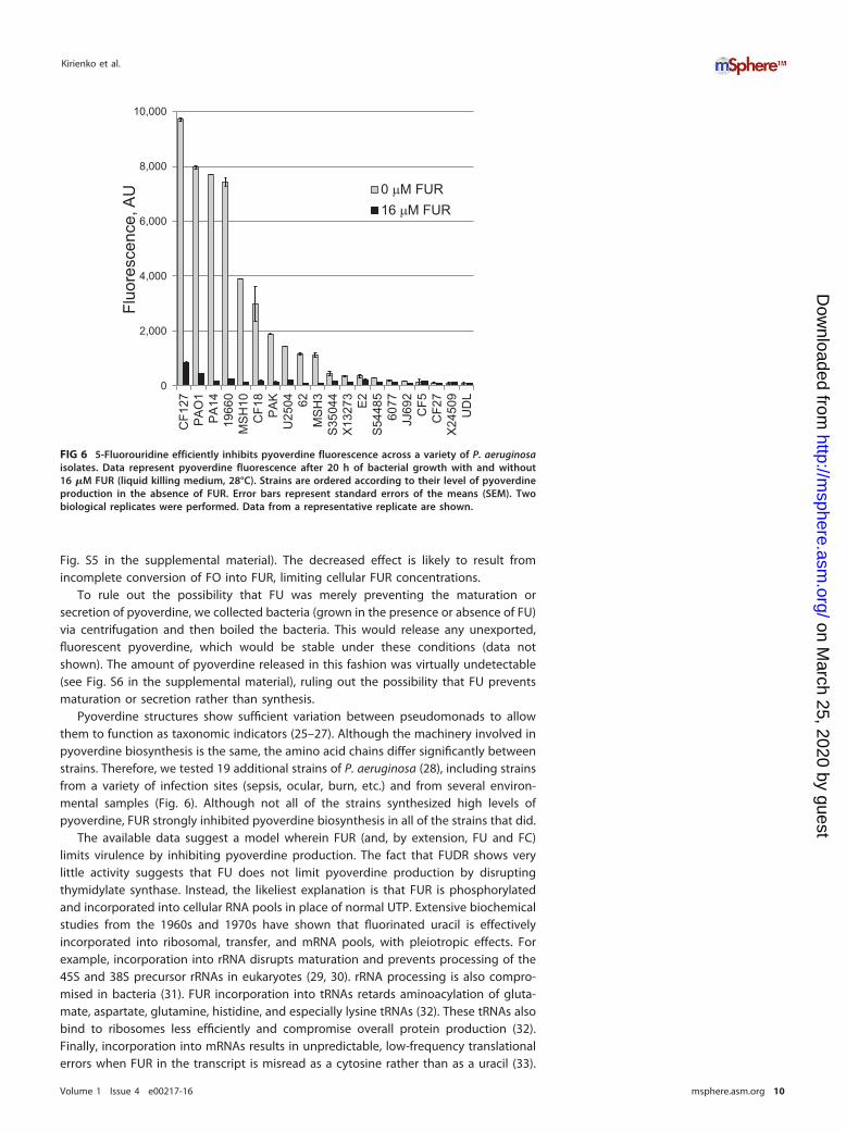
Fig. S5 in the supplemental material). The decreased effect is likely to result fromincomplete conversion of FO into FUR, limiting cellular FUR concentrations.
To rule out the possibility that FU was merely preventing the maturation orsecretion of pyoverdine, we collected bacteria (grown in the presence or absence of FU)via centrifugation and then boiled the bacteria. This would release any unexported,fluorescent pyoverdine, which would be stable under these conditions (data notshown). The amount of pyoverdine released in this fashion was virtually undetectable(see Fig. S6 in the supplemental material), ruling out the possibility that FU preventsmaturation or secretion rather than synthesis.
Pyoverdine structures show sufficient variation between pseudomonads to allowthem to function as taxonomic indicators (25–27). Although the machinery involved inpyoverdine biosynthesis is the same, the amino acid chains differ significantly betweenstrains. Therefore, we tested 19 additional strains of P. aeruginosa (28), including strainsfrom a variety of infection sites (sepsis, ocular, burn, etc.) and from several environ-mental samples (Fig. 6). Although not all of the strains synthesized high levels ofpyoverdine, FUR strongly inhibited pyoverdine biosynthesis in all of the strains that did.
The available data suggest a model wherein FUR (and, by extension, FU and FC)limits virulence by inhibiting pyoverdine production. The fact that FUDR shows verylittle activity suggests that FU does not limit pyoverdine production by disruptingthymidylate synthase. Instead, the likeliest explanation is that FUR is phosphorylatedand incorporated into cellular RNA pools in place of normal UTP. Extensive biochemicalstudies from the 1960s and 1970s have shown that fluorinated uracil is effectivelyincorporated into ribosomal, transfer, and mRNA pools, with pleiotropic effects. Forexample, incorporation into rRNA disrupts maturation and prevents processing of the45S and 38S precursor rRNAs in eukaryotes (29, 30). rRNA processing is also compro-mised in bacteria (31). FUR incorporation into tRNAs retards aminoacylation of gluta-mate, aspartate, glutamine, histidine, and especially lysine tRNAs (32). These tRNAs alsobind to ribosomes less efficiently and compromise overall protein production (32).Finally, incorporation into mRNAs results in unpredictable, low-frequency translationalerrors when FUR in the transcript is misread as a cytosine rather than as a uracil (33).
0
2,000
4,000
6,000
8,000
10,000
CF1
27P
AO
1P
A14
1966
0M
SH
10C
F18
PA
KU
2504 62
MS
H3
S35
044
X13
273
E2
S54
485
6077
JJ69
2C
F5C
F27
X24
509
UD
L
Fluo
resc
ence
, AU 0 µM FUR
16 µM FUR
FIG 6 5-Fluorouridine efficiently inhibits pyoverdine fluorescence across a variety of P. aeruginosaisolates. Data represent pyoverdine fluorescence after 20 h of bacterial growth with and without16 �M FUR (liquid killing medium, 28°C). Strains are ordered according to their level of pyoverdineproduction in the absence of FUR. Error bars represent standard errors of the means (SEM). Twobiological replicates were performed. Data from a representative replicate are shown.
Kirienko et al.
Volume 1 Issue 4 e00217-16 msphere.asm.org 10
on March 25, 2020 by guest
http://msphere.asm
.org/D
ownloaded from
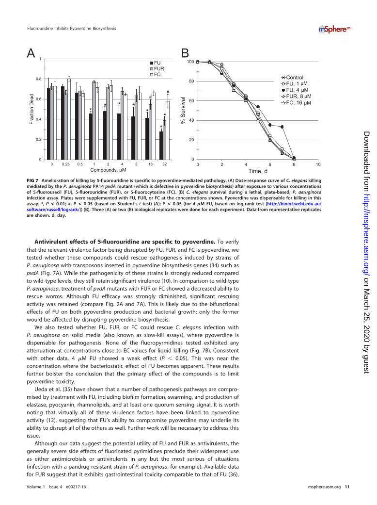
Antivirulent effects of 5-fluorouridine are specific to pyoverdine. To verifythat the relevant virulence factor being disrupted by FU, FUR, and FC is pyoverdine, wetested whether these compounds could rescue pathogenesis induced by strains ofP. aeruginosa with transposons inserted in pyoverdine biosynthesis genes (34) such aspvdA (Fig. 7A). While the pathogenicity of these strains is strongly reduced comparedto wild-type levels, they still retain significant virulence (10). In comparison to wild-typeP. aeruginosa, treatment of pvdA mutants with FUR or FC showed a decreased ability torescue worms. Although FU efficacy was strongly diminished, significant rescuingactivity was retained (compare Fig. 2A and 7A). This is likely due to the bifunctionaleffects of FU on both pyoverdine production and bacterial growth; only the formerwould be affected by disrupting pyoverdine biosynthesis.
We also tested whether FU, FUR, or FC could rescue C. elegans infection withP. aeruginosa on solid media (also known as slow-kill assays), where pyoverdine isdispensable for pathogenesis. None of the fluoropyrmidines tested exhibited anyattenuation at concentrations close to EC values for liquid killing (Fig. 7B). Consistentwith other data, 4 �M FU showed a weak effect (P � 0.05). This was near theconcentration where the bacteriostatic effect of FU becomes apparent. These resultsfurther bolster the conclusion that the primary effect of the compounds is to limitpyoverdine toxicity.
Ueda et al. (35) have shown that a number of pathogenesis pathways are compro-mised by treatment with FU, including biofilm formation, swarming, and production ofelastase, pyocyanin, rhamnolipids, and at least one quorum sensing signal. It is worthnoting that virtually all of these virulence factors have been linked to pyoverdineactivity (12), suggesting that FU’s ability to compromise pyoverdine may underlie itsability to disrupt all of the others as well. Further work will be necessary to address thisissue.
Although our data suggest the potential utility of FU and FUR as antivirulents, thegenerally severe side effects of fluorinated pyrimidines preclude their widespread useas either antimicrobials or antivirulents in any but the most serious of situations(infection with a pandrug-resistant strain of P. aeruginosa, for example). Available datafor FUR suggest that it exhibits gastrointestinal toxicity comparable to that of FU (36),
0
0.2
0.4
0.6
0.8
1
0 0.25 0.5 1 2 4 8 16 32
Frac
tion
Dea
d
Compounds, uM
FUFURFC
#
* * ** *
*
#
*
0
20
40
60
80
100
0 2 4 6 8 10
% S
urvi
val
Time, d
ControlFU, 1FU, 4FUR, 8FC, 16 uM
A B
µM
µMµM
µMµM
FIG 7 Amelioration of killing by 5-fluorouridine is specific to pyoverdine-mediated pathology. (A) Dose-response curve of C. elegans killingmediated by the P. aeruginosa PA14 pvdA mutant (which is defective in pyoverdine biosynthesis) after exposure to various concentrationsof 5-fluorouracil (FU), 5-fluorouridine (FUR), or 5-fluorocytosine (FC). (B) C. elegans survival during a lethal, plate-based, P. aeruginosainfection assay. Plates were supplemented with FU, FUR, or FC at the concentrations shown. Pyoverdine was dispensable for killing in thisassay. *, P < 0.01; #, P < 0.05 (based on Student’s t test) (A); P < 0.05 (for 4 �M FU, based on log-rank test [http://bioinf.wehi.edu.au/software/russell/logrank/]) (B). Three (A) or two (B) biological replicates were done for each experiment. Data from representative replicatesare shown. d, day.
Fluorouridine Inhibits Pyoverdine Biosynthesis
Volume 1 Issue 4 e00217-16 msphere.asm.org 11
on March 25, 2020 by guest
http://msphere.asm
.org/D
ownloaded from
but our data in C. elegans suggest that antivirulent doses of FUR exhibit relatively mildtoxicity (see Fig. S7 in the supplemental material). Further work remains to be done toclarify this matter. Regardless of their value as antivirulents in humans, fluorinatedpyrimidines will remain useful as tool compounds for studying the regulation ofpyoverdine production. This also highlights the utility of using unbiased approaches togain insights into host-pathogen interactions.
MATERIALS AND METHODSStrains. The SS104 [glp-4(bn2)] C. elegans strain was maintained on nematode growth medium (NGM)seeded with Escherichia coli strain OP50 at 15°C (37). For all experiments using wild-type P. aeruginosa,we used P. aeruginosa strain PA14, a clinical isolate described elsewhere (38). The PA14 pvdA mutant hasa Mariner transposon inserted into the pvdA locus (34) and was sequenced prior to use. The other 19strains of P. aeruginosa tested have been described previously (28).
Media. NGM (standard nematode growth medium), S basal minimal medium, LK medium (modifiedliquid NGM used for liquid killing assays with P. aeruginosa), and SK modified NGM used for plate-basedinfection with P. aeruginosa are all described elsewhere (39).
Liquid killing assay. The liquid killing assay was performed as previously described (39, 40). Z= factordeterminations were performed in 384-well plates, where half of the wells contained DMSO as a negativecontrol and half contained 100 �g·ml�1 gentamicin as a positive control. Unbiased discriminationbetween living and dead worms was performed on the basis of staining by the cell-impermeantfluorescent dye Sytox Orange (Invitrogen, Carlsbad, CA) (8). Cell Profiler software (http://cellprofiler.org/)was used for quantitative image processing and determination of worm mortality (41, 42).
High-throughput chemical screening was performed at the National Screening Laboratory for theRegional Centers of Excellence in Biodefense and Emerging Infectious Diseases (NSRB) at Harvard MedicalSchool. Compounds were screened in duplicate at a final concentration of approximately 20 �g·ml�1.Each 384-well plate contained at least 30 wells dedicated to solvent (DMSO) controls. Compounds wereconsidered primary hits if survival in both replicates was �3 standard deviations (SD) above the meanvalue for negative controls on the same plate.
EC values were determined in the liquid killing assay as the lowest compound concentration resultingin statistically significant rescue of worms (P � 0.05 [based on Student’s t test]). EC values for eachcompound represent an average of at least 4 biological replicates, with at least 4 wells per replicate.
Pyoverdine production under liquid killing conditions was measured spectrophotometrically (exci-tation wavelength [Ex], 405 nm; emission wavelength [Em], 460 nm) using a Cytation5 multimode platereader/imager (Molecular Devices, Sunnyvale, CA).
MIC assay. To determine the MIC of compounds for preventing bacterial growth, P. aeruginosa strainPA14 was grown in standard LB overnight and diluted 100,000-fold in either LB or in SK media.Compounds were 2-fold serially diluted and mixed with bacteria 1:1. Growth inhibition was visuallyscored on the basis of turbidity. Two wells were used per condition, and at least three biologicalreplicates were performed.
CFU assay. To determine the number of CFU, aliquots were taken at appropriate time points andserially diluted 5-fold in water prior to plating onto solid LB plates (2% agar). Colonies were countedunder a dissecting microscope.
RNA extraction and microarray analysis. Young adult worms (10,000) were plated onto 10-cmNGM plates supplemented with either DMSO or 50 �M FU. After 8 h, worms were washed into a 15-mlconical tube with S basal medium and rinsed twice. Afterward, worms were resuspended in TRI reagent(MRC, Inc., Cincinnati, OH) and frozen at �80°C. After thawing, RNA was extracted according tomanufacturer’s protocols and purified further using RNeasy columns (Qiagen, Gaithersburg, MD). cRNAsamples were prepared and hybridized to full-genome GeneChips for C. elegans (GPL200; Affymetrix,Santa Clara, CA) according to manufacturer’s protocols. Three biological replicates were tested for eachcondition. Gene expression was analyzed using GCRMA (http://www.bioconductor.org). Differentiallyregulated genes were determined as previously described (43). For nonparametric determination ofsignificantly affected genes, the following values were chosen: fold change, �2; modified Wilcoxon rankcoefficient, �1.5-fold; absolute expression level, �80 arbitrary units (AU). For the determination ofsignificantly affected genes using the assumption of normal distribution, MAS Suite and following criteriawere used: fold change, �2; Student’s t test value, �0.01 (after multiple sample correction); andAbsent/Present calls of MAS suite.
Slow-killing assay. The slow-killing assay was performed as previously described (39). At least twobiological replicates were performed for each experiment. Each biological replicate consisted of threeplates with 50 worms per plate. Worms that left the surface of the agar were eliminated from the scoring.
Accession number(s). Microarray data have been deposited in the NIH Gene Expression Omnibusdatabase under accession number GSE85342.
SUPPLEMENTAL MATERIALSupplemental material for this article may be found at http://dx.doi.org/10.1128/mSphere.00217-16.
Figure S1, PDF file, 0.3 MB.Figure S2, PDF file, 0.5 MB.
Kirienko et al.
Volume 1 Issue 4 e00217-16 msphere.asm.org 12
on March 25, 2020 by guest
http://msphere.asm
.org/D
ownloaded from
Figure S3, PDF file, 0.3 MB.Figure S4, PDF file, 0.3 MB.Figure S5, PDF file, 0.4 MB.Figure S6, PDF file, 0.3 MB.Figure S7, PDF file, 0.3 MB.
ACKNOWLEDGMENTSWe thank F. Ausubel for helpful comments on the manuscript, for the panel ofP. aeruginosa isolates, and for his outstanding mentorship. We express our gratitude toS. Chiang and the staff of the NSRB screening facility. We thank J. Larkins-Ford fortechnical assistance.
Additional support for the screening efforts was provided from grants U54 AI057159to the NSRB and R01 AI085581 to F. Ausubel. Equipment used in this study waspurchased with funds provided by the Cancer Prevention Research Institute of Texas toN.V.K. (RR150044). None of the funding agencies played a role in study design, datacollection, interpretation, or analysis, writing of the manuscript, or the decision topublish.
FUNDING INFORMATIONThis work, including the efforts of Natalia V. Kirienko, was funded by HHS | NIH |National Institute of Allergy and Infectious Diseases (NIAID) (F32 AI100501 and K22AI110552). This work, including the efforts of Natalia V. Kirienko, was funded by CancerPrevention and Research Institute of Texas (CPRIT) (RR150044).
REFERENCES1. Wang CY, Jerng JS, Chen KY, Cheng KY, Lee LN, Yu CJ, Hsueh PR,
Yang PC. 2006. Pandrug-resistant Pseudomonas aeruginosa amonghospitalised patients: clinical features, risk-factors and outcomes. ClinMicrobiol Infect 12:63– 68. http://dx.doi.org/10.1111/j .1469-0691.2005.01305.x.
2. Page MG, Bush K. 2014. Discovery and development of new antibac-terial agents targeting gram-negative bacteria in the era of pandrugresistance: is the future promising? Curr Opin Pharmacol 18:91–97.http://dx.doi.org/10.1016/j.coph.2014.09.008.
3. Karaiskos I, Giamarellou H. 2014. Multidrug-resistant and extensivelydrug-resistant gram-negative pathogens: current and emerging thera-peutic approaches. Expert Opin Pharmacother 15:1351–1370. http://dx.doi.org/10.1517/14656566.2014.914172.
4. Bumann D. 2008. Has nature already identified all useful antibacterialtargets? Curr Opin Microbiol 11:387–392. http://dx.doi.org/10.1016/j.mib.2008.08.002.
5. Projan SJ, Shlaes DM. 2004. Antibacterial drug discovery: is it alldownhill from here? Clin Microbiol Infect 10(Suppl 4):18 –22. http://dx.doi.org/10.1111/j.1465-0691.2004.1006.x.
6. Silver LL. 2005. A retrospective on the failures and successes of anti-bacterial drug discovery. IDrugs 8:651– 655.
7. Lakshmanan U, Yap A, Fulwood J, Yichun L, Hoon SS, Lim J, Ting A,Sem XH, Kreisberg JF, Tan P, Tan G, Flotow H. 2014. Establishment ofa novel whole animal HTS technology platform for melioidosis drugdiscovery Comb Chem High Throughput Screen 17:790 – 803. http://dx.doi.org/10.2174/1386207317666141019195031.
8. Moy TI, Conery AL, Larkins-Ford J, Wu G, Mazitschek R, Casadei G,Lewis K, Carpenter AE, Ausubel FM. 2009. High-throughput screen fornovel antimicrobials using a whole animal infection model. ACS ChemBiol 4:527–533. http://dx.doi.org/10.1021/cb900084v.
9. O’Reilly LP, Benson JA, Cummings EE, Perlmutter DH, Silverman GA,Pak SC. 2014. Worming our way to novel drug discovery with theCaenorhabditis elegans proteostasis network, stress response andinsulin-signaling pathways. Expert Opin Drug Discov 9:1021–1032.http://dx.doi.org/10.1517/17460441.2014.930125.
10. Kirienko NV, Kirienko DR, Larkins-Ford J, Wählby C, Ruvkun G,Ausubel FM. 2013. Pseudomonas aeruginosa disrupts Caenorhabdi-tis elegans iron homeostasis, causing a hypoxic response and death.Cel l Host Microbe 13:406 – 416. http://dx.doi .org/10.1016/j.chom.2013.03.003.
11. Visca P, Imperi F, Lamont IL. 2007. Pyoverdine siderophores: from
biogenesis to biosignificance. Trends Microbiol 15:22–30. http://dx.doi.org/10.1016/j.tim.2006.11.004.
12. Vasil ML. 2007. How we learnt about iron acquisition in Pseudomonasaeruginosa: a series of very fortunate events. Biometals 20:587– 601.http://dx.doi.org/10.1007/s10534-006-9067-2.
13. Wilderman PJ, Vasil AI, Johnson Z, Wilson MJ, Cunliffe HE, LamontIL, Vasil ML. 2001. Characterization of an endoprotease (PrpL) encodedby a PvdS-regulated gene in Pseudomonas aeruginosa. Infect Immun69:5385–5394. http://dx.doi.org/10.1128/IAI.69.9.5385-5394.2001.
14. Meyer JM, Neely A, Stintzi A, Georges C, Holder IA. 1996. Pyoverdinis essential for virulence of Pseudomonas aeruginosa. Infect Immun64:518 –523.
15. Imperi F, Massai F, Facchini M, Frangipani E, Visaggio D, Leoni L,Bragonzi A, Visca P. 2013. Repurposing the antimycotic drug flucyto-sine for suppression of Pseudomonas aeruginosa pathogenicity. ProcNatl Acad Sci U S A 110:7458 –7463. http://dx.doi.org/10.1073/pnas.1222706110.
16. Xiong YQ, Vasil ML, Johnson Z, Ochsner UA, Bayer AS. 2000. Theoxygen- and iron-dependent sigma factor pvdS of Pseudomonas aerugi-nosa is an important virulence factor in experimental infective endocar-ditis. J Infect Dis 181:1020 –1026. http://dx.doi.org/10.1086/315338.
17. Chand NS, Clatworthy AE, Hung DT. 2012. The two-component sensorKinB acts as a phosphatase to regulate Pseudomonas aeruginosa viru-lence. J Bacteriol 194:6537– 6547 http://dx.doi.org/10.1128/JB.01168-12.
18. Kirienko NV, Ausubel FM, Ruvkun G. 2015. Mitophagy confers resis-tance to siderophore-mediated killing by Pseudomonas aeruginosa.Proc Natl Acad Sci U S A 112:1821–1826. http://dx.doi.org/10.1073/pnas.1424954112.
19. Zhang JH, Chung TD, Oldenburg KR. 1999. A simple statisticalparameter for use in evaluation and validation of high throughputscreening assays. J Biomol Screen 4:67–73. http://dx.doi.org/10.1177/108705719900400206.
20. Gavrish E, Shrestha B, Chen C, Lister I, North EJ, Yang L, Lee RE, HanA, Williams B, Charnuska D, Coleman K, Lewis K, LaFleur MD. 2014.In vitro and in vivo activities of HPi1, a selective antimicrobial againstHelicobacter pylori. Antimicrob Agents Chemother 58:3255–3260.http://dx.doi.org/10.1128/AAC.02573-13.
21. Kanehisa M, Goto S. 2000. KEGG: Kyoto encyclopedia of genes andgenomes. Nucleic Acids Res 28:27–30. http://dx.doi.org/10.1093/nar/28.1.27.
22. El Sayed YM, Sadée W. 1983. Metabolic activation of R,S-1-(tetrahydro-
Fluorouridine Inhibits Pyoverdine Biosynthesis
Volume 1 Issue 4 e00217-16 msphere.asm.org 13
on March 25, 2020 by guest
http://msphere.asm
.org/D
ownloaded from
2-furanyl)-5-fluorouracil (ftorafur) to 5-fluorouracil by soluble enzymes.Cancer Res 43:4039 – 4044.
23. Ignoffo RJ. 1998. Capecitabine: a new oral fluoropyrimidine. CancerPract 6:302–304. http://dx.doi.org/10.1046/j.1523-5394.1998.00028.x.
24. Webber MA, Piddock LJ. 2003. The importance of efflux pumps inbacterial antibiotic resistance. J Antimicrob Chemother 51:9 –11. http://dx.doi.org/10.1093/jac/dkg050.
25. Fuchs R, Schäfer M, Geoffroy V, Meyer JM. 2001. Siderotyping—apowerful tool for the characterization of pyoverdines. Curr Top MedChem 1:31–57. http://dx.doi.org/10.2174/1568026013395542.
26. Meyer JM, Geoffroy VA, Baida N, Gardan L, Izard D, Lemanceau P,Achouak W, Palleroni NJ. 2002. Siderophore typing, a powerful tool forthe identification of fluorescent and nonfluorescent pseudomonads.Appl Environ Microbiol 68:2745–2753. http://dx.doi.org/10.1128/AEM.68.6.2745-2753.2002.
27. Meyer JM, Stintzi A, Coulanges V, Shivaji S, Voss JA, Taraz K,Budzikiewicz H. 1998. Siderotyping of fluorescent pseudomonads: char-acterization of pyoverdines of Pseudomonas fluorescens and Pseu-domonas putida strains from Antarctica. Microbiology 144:3119 –3126.http://dx.doi.org/10.1099/00221287-144-11-3119.
28. Lee DG, Urbach JM, Wu G, Liberati NT, Feinbaum RL, Miyata S,Diggins LT, He J, Saucier M, Déziel E, Friedman L, Li L, Grills G,Montgomery K, Kucherlapati R, Rahme LG, Ausubel FM. 2006.Genomic analysis reveals that Pseudomonas aeruginosa virulence iscombinatorial. Genome Biol 7:R90. http://dx.doi.org/10.1186/gb-2006-7-10-r90.
29. Wilkinson DS, Crumley J. 1977. Metabolism of 5-fluorouracil in sensi-tive and resistant Novikoff hepatoma cells. J Biol Chem 252:1051–1056.
30. Wilkinson DS, Pitot HC. 1973. Inhibition of ribosomal ribonucleic acidmaturation in Novikoff hepatoma cells by 5-fluorouracil and5-fluorouridine. J Biol Chem 248:63– 68.
31. Hahn GA, Mandel HG. 1971. Effects of fluorouracil on RNA synthesis inBacillus cereus. Biochem Pharmacol 20:1973–1990. http://dx.doi.org/10.1016/0006-2952(71)90397-2.
32. Ramberg ES, Ishaq M, Rulf S, Moeller B, Horowitz J. 1978. Inhibitionof transfer RNA function by replacement of uridine and uridine-derivednucleosides with 5-fluorouridine. Biochemistry 17:3978 –3985. http://dx.doi.org/10.1021/bi00612a016.
33. Gleason MK, Fraenkel-Conrat H. 1976. Biological consequences ofincorporation of 5-fluorocytidine in the RNA of 5-fluorouracil-treated
eukaryotic cells. Proc Natl Acad Sci U S A 73:1528 –1531. http://dx.doi.org/10.1073/pnas.73.5.1528.
34. Liberati NT, Urbach JM, Miyata S, Lee DG, Drenkard E, Wu G,Villanueva J, Wei T, Ausubel FM. 2006. An ordered, nonredundantlibrary of Pseudomonas aeruginosa strain PA14 transposon insertionmutants. Proc Natl Acad Sci U S A 103:2833–2838. http://dx.doi.org/10.1073/pnas.0511100103.
35. Ueda A, Attila C, Whiteley M, Wood TK. 2009. Uracil influences quo-rum sensing and biofilm formation in Pseudomonas aeruginosa andfluorouracil is an antagonist. Microb Biotechnol 2:62–74. http://dx.doi.org/10.1111/j.1751-7915.2008.00060.x.
36. Houghton JA, Houghton PJ, Wooten RS. 1979. Mechanism of induc-tion of gastrointestinal toxicity in the mouse by 5-fluorouracil,5-fluorouridine, and 5-fluoro-2=-deoxyuridine. Cancer Res 39:2406 –2413.
37. Stiernagle T. 11 February 2006. Maintenance of C. elegans. In The C.elegans Research Community, WormBook (ed), Wormbook. http://dx.doi.org/10.1895/wormbook.1.101.1.
38. Rahme LG, Stevens EJ, Wolfort SF, Shao J, Tompkins RG, AusubelFM. 1995. Common virulence factors for bacterial pathogenicity inplants and animals. Science 268:1899 –1902. http://dx.doi.org/10.1126/science.7604262.
39. Kirienko NV, Cezairliyan BO, Ausubel FM, Powell JR. 2014. Pseu-domonas aeruginosa PA14 pathogenesis in Caenorhabditis elegans.Methods Mol Biol 1149:653– 669. http://dx.doi.org/10.1007/978-1-4939-0473-0_50.
40. Conery AL, Larkins-Ford J, Ausubel FM, Kirienko NV. 2014. High-throughput screening for novel anti-infectives using a C. elegans patho-genesis model. Curr Protoc Chem Biol 6:25–37.
41. Moy TI, Ball AR, Anklesaria Z, Casadei G, Lewis K, Ausubel FM. 2006.Identification of novel antimicrobials using a live-animal infectionmodel. Proc Natl Acad Sci U S A 103:10414 –10419. http://dx.doi.org/10.1073/pnas.0604055103.
42. Kamentsky L, Jones TR, Fraser A, Bray MA, Logan DJ, Madden KL,Ljosa V, Rueden C, Eliceiri KW, Carpenter AE. 2011. Improved struc-ture, function and compatibility for CellProfiler: modular high-throughput image analysis software. BioInformatics 27:1179 –1180.http://dx.doi.org/10.1093/bioinformatics/btr095.
43. Kirienko NV, Fay DS. 2007. Transcriptome profiling of the C. elegans Rbortholog reveals diverse developmental roles. Dev Biol 305:674 – 684.http://dx.doi.org/10.1016/j.ydbio.2007.02.021.
Kirienko et al.
Volume 1 Issue 4 e00217-16 msphere.asm.org 14
on March 25, 2020 by guest
http://msphere.asm
.org/D
ownloaded from