Embed Size (px)
Citation preview

Eurographics Workshop on Visual Computing for Biology and Medicine (2017) Short PaperI. Hotz, D. Merhof, and C. Rieder (Editors)
A Guided Spatial Transformer Network for Histology CellDifferentiation
Marc Aubreville1, Maximilian Krappmann1, Christof Bertram2, Robert Klopfleisch2 and Andreas Maier1
1Pattern Recognition Lab, Friedrich-Alexander-Universität Erlangen-Nürnberg, Germany2Institute of Veterinary Pathology, Free University Berlin, Germany
Abstract
Identification and counting of cells and mitotic figures is a standard task in diagnostic histopathology. Due to the large overallcell count on histological slides and the potential sparse prevalence of some relevant cell types or mitotic figures, retrievingannotation data for sufficient statistics is a tedious task and prone to a significant error in assessment. Automatic classificationand segmentation is a classic task in digital pathology, yet it is not solved to a sufficient degree.We present a novel approach for cell and mitotic figure classification, based on a deep convolutional network with an incorpo-rated Spatial Transformer Network. The network was trained on a novel data set with ten thousand mitotic figures, about tentimes more than previous data sets. The algorithm is able to derive the cell class (mitotic tumor cells, non-mitotic tumor cellsand granulocytes) and their position within an image. The mean accuracy of the algorithm in a five-fold cross-validation is91.45 %.In our view, the approach is a promising step into the direction of a more objective and accurate, semi-automatized mitosiscounting supporting the pathologist.
CCS Concepts•Computing methodologies → Object detection; Neural networks; •Applied computing → Bioinformatics;
1. Introduction
The assessment of cell types in histology slides is a standard taskin pathology. Especially in tumor diagnostics, determining the rela-tive amount of mitotic figures, a marker for tumor proliferation andaggressiveness, is another important task for the diagnostic pathol-ogist [RRL∗13].
However, evaluation of the complete slide for mitotic figures(see figure 1) is usually too time consuming in routine diagnostics.Therefore it is suggested that only 10 high power fields (an area ofassumed equal size used for statistic comparison), presumed to con-tain the highest density of mitoses, are subjectively chosen by thepathologist. The area of these fields is, however, not well-defined,as it depends on the optical properties of the microscope and whichmay vary significantly in their content of mitotic figures [MMG16].The final count thus strongly depends on the randomly but not nec-essarily representatively selected high power fields thus the result-ing mitotic count is usually observer-dependent [VvDW∗14]. In ad-dition, mitotic figures may be very variable in their histologic phe-notype, which may also lead to inter-observer variability betweenpathologists.
The aim of this work is to develop a more objective and accu-rate, automatized approach to counting of mitotic figures by assist-
ing pathologists in the selection of fields with the highest mitoticcounts and with more constant parameters of mitotic figure identifi-cation. Detection and annotation of mitotic cells in histology slidesis a well-known task in images processing, and subject of severalchallenges in recent years [VvDW∗14, RRL∗13].
Mitosis comprises a number of different phases in the cell cycle(prophase, metaphase, anaphase, and telophase). In each phase, thenucleus is shaped differently. This means that the variance in im-ages showing a mitotic cell is high (see figure 1). On top of that,there is also atypical mitosis, adding yet another factor of varianceto the picture. However, publicly available databases for mitosisdetection feature a rather low number of mitotic figures (e.g. the2014 ICPR MITOS-ATYPIA-14 dataset with 873 images, the 2012ICPR dataset with 326 images [RRL∗13], or the AMIDA13 datasetwith 1083 images [VvDW∗14]), especially for robust detection.
Automatic detection of mitotic figures has been widely per-formed using the classical machine learning workflow on textural,morphological and shape features (e.g. [SFHG12,Irs13]).Ciresan etal. were the first to employ deep learning-based approaches for mi-tosis detection [CGGS13], yielding significant improvements overtraditional approaches [VvDW∗14]. Yet, deep learning technolo-gies suffer considerably from insufficient data amounts, as they
c© 2017 The Author(s)Eurographics Proceedings c© 2017 The Eurographics Association.

M. Aubreville, M. Krappmann, C. Bertram, R. Klopfleisch & A. Maier / A Guided Spatial Transformer Network for Histology Cell Differentiation
have a large number of trainable parameters and, because of this,are likely to overfit the data. Particularly in the field of mitosis de-tection, we assume that detection performance could be improvedif the whole variance of mitotic processes can be captured in thenetworks, requesting for a substantial increase in training data forsuch networks.
normal tumor cell mitotic tumor cell granulocyte
Figure 1: Examples of cropped cells, slides stained with hema-toxylin and eosin.
2. Related Work
Typically, the process of object detection is parted into two sub-processes: Segmentation and classification. This setup is especiallysensible for histology since the images represent a large amount ofdata and classification is usually the more complex process com-pared to segmentation. Sommer et al. used pixel-wise classifica-tion for candidate retrieval and then object shape and texture fea-tures for mitotic cell classification [SFHG12]. Irshad used activecontour models for candidate selection and statistical and morpho-logical features for classification [Irs13]. Those hand-crafted fea-tures have significant drawbacks, however: Given the often smalldata sets, automatic selection of features is prone to random corre-lation, while using higher-dimensional classification approaches onthe complete set increases overfitting [Lea96]. Further, it is ques-tionable, if those approaches can represent the variability in shapeand texture of mitotic figures [CDW∗16].
Triggered by the ground-breaking initial works of Lecun[LBBH98], Convolutional Neural Networks (CNN) have spreadwidely in the use for various image classification tasks. CNN-basedrecognition algorithms have won all major image recognition chal-lenges in recent years because of their ability to capture complexshapes and still remain sensitive to minor variations in the image.In the field of mitosis detection, CNN-based approaches have beenused for classification [CGGS13], feature extraction [WCRB∗14]as well as candidate generation [CDW∗16]. Yet, CNNs, throughtheir inherent ability to capture complex structures, are also proneto overfitting, a problem which is usually targeted by data augmen-tation and regularization strategies like dropout and other mech-anisms or by means of transfer learning. Another regularizationstrategy is to constrain the capacity of the approach [Goo16] byreducing effectively the free parameters of the model. We aim toattempt this by splitting the problem into an attention task and aclassification task. The general issue however, that the training datamight be a non-representative sample of the classification task andthus parts of real-world data are not recognized because the data set
does not generalize well, can be best targeted with a bigger trainingdata set, as it was the base for this work.
3. Material
For this study, digital histopathological images were acquired usingAperio ScanScope (Leica Biosystems Imaging, Inc., USA) slidescanner hardware at a magnification of 400x. Candidate patches forthree different cell types (mitotic cells, eosinophilic granulocytesand normal tumor cells) were annotated by an expert with profoundknowledge on cell differentiation and classified by a trained pathol-ogist. The cells were selected from histologic images of 12 differentparaffin-embedded canine mast cell tumors, stained with standardhematoxylin and eosin (H&E). In order to train a deep learningclassifier with a sufficient amount of data, our emphasis was not oncomplete annotation of the slides but on finding enough candidatesfor the above-mentioned cell types within the image.
More specifically, the emphasis was on finding mitotic cells.Commonly, in all major related works, the number of mitotic cellsin the data set was proportional to the actual occurrence in the re-spective slides, as whole slides where annotated, resulting in a rel-atively low number of mitotic cells. On the contrary, we purposelyselected a similar number of cells from each category to not assumeany priors in distribution.
We acknowledge that this procedure might add a certain biasin cell selection, and that our dataset might not be representative.However, this argument can also be made for the case where onlya small number of mitotic figures is available. Further, because ofthe high inter-rater variability in mitosis expert classifications, weassume that an unbiased ground truth is hard to retrieve and a mi-nor bias by image pre-selection can be tolerated. Finally, we do nottarget at finding all mitotic cells, but rather to guide the patholo-gist in finding a representative part of the slide and to thus reducevariability in expert grading.
In the data set, we have approx. 37,800 single annotations of cellsof the three different types (about 10,400 granulocytes, 10,800 mi-totic figures and 16,600 normal tumor cells). The majority of cellswas rated by the pathologist to be normal tumor cells, however alsoa significant amount of mitotic cells and eosinophil granulocyteswas annotated.
4. Methods
Spatial Transformer Networks (STN), first described by Jaderberget. al, provide a learnable method to focus the attention of a classifi-cation network on a specific subpart of the original image [JSZ∗15].To achieve this, parameters of an affine transformation matrix θ areregressed by the network, alongside with the optimization of theactual classifier.
Spatial Transformer Networks were originally successfully em-ployed on a distorted MNIST [LBBH98] data set, where transla-tion, scale, rotation and clutter were used to increase the difficultyfor the detection task. The approach has shown to be able to – with-out any prior knowledge about the actual transform that was appliedbeforehand – increase accuracy of the classification network by fo-cusing its attention to the area where the number was present and by
c© 2017 The Author(s)Eurographics Proceedings c© 2017 The Eurographics Association.

M. Aubreville, M. Krappmann, C. Bertram, R. Klopfleisch & A. Maier / A Guided Spatial Transformer Network for Histology Cell Differentiation
Figure 2: Image preprocessing. The offset ∆x, ∆y is set randomlywhile keeping the cell within the image.
compensating for the deformation [JSZ∗15]. The approach can beused in a joined learning approach, where both the transform andthe classification are learned end-to-end, something that could bedescribed as a weakly supervised learning approach for the trans-formation. The optimization on the MNIST data set is, however,a much easier task than on real-world data. In a typical patch ex-tracted from a histology slide, a lot of similar and valid objects maybe contained in the image, and joined optimization suffers from lo-cal extrema in the gradient descend approach.
In this work, we aim to use STN as a method of not only directingthe attention of a classification network to a sub-area of a largerimage, and thus hopefully improving classification performance,but also as a segmentation approach to derive the information aboutwhere the respective cells are located.
We believe that Spatial Transformer Networks are an ideal can-didate for this kind of task because they can be used to model twosources of natural variance into the machine learning process witha comparatively small overhead in complexity: Scaling and transla-tion. Scaling is relevant in microscopy for two reasons: Firstly, theactual magnification of the microscope is dependent on the opti-cal properties of the ocular, notably on the field number [MMG16].Secondly, cells differ in size, dependent on their function and thespecies they originate from.
4.1. Image Preprocessing
All images were cropped around the cell center in a first processingstep. In a second processing step, we introduce a random translation∆x, ∆y to the origin area of the input image before cropping, so itis no longer centered around the cell, i.e. the cell can be anywhereon the image, with the restriction that the whole cell will be withinthe image (see figure 2). From the introduced translation, we canderive a new ground truth transformation vector
θ =
[ϑs 0 ϑx0 ϑs ϑy
](1)
Localizer
dense dense
SpatialTransformerNetwork
20 6
256 3
dense dense
Classifier
32x32x3128x128x3
ϑ
Figure 3: Overview of the network. The cell’s position is estimatedby the localizer, which regresses an affine transform applied on theoriginal image, to feed the classifier with cell images.
where ϑs is the (in our case) fixed scaling vector. The scalingvector is dependent on the (manually chosen) expected cell size dc.For our data, prior investigation has shown that all typical cells inour case are fully contained within an area of 64 px around the cellcenter, so with di = 128 px being the length of the input image, wecan derive:
ϑs =di
dc= 0.5 (2)
The (relative) coordinate grid for the STN is spaced from -1.0 to1.0, with 0.0 being the center pixel. The translation elements of theground truth transformation vector in eqn. 1 thus become:
ϑ{x,y} =−2∆{x,y}
di(3)
4.2. Network layout
Our network consists of three main blocks, as depicted in Figure 3:The localizer, the classifier and the Spatial Transformer Network.The localizer is a deep convolutional network with two stackedconvolutional and max-pooling layers, one inception layer and twofully connected layers. It regresses an estimate θ of the transformmatrix θ.
The classifier is a rather small convolutional neural network with7 layers, using also convolutional, max-pooling and inception lay-ers. It outputs a vector of dimension 3, which represents the classprobabilities for the three cell types depicted in Figure 1.
Inception [SLJ∗14] blocks were introduced by Szegedy et al. in2014, and have been since then widely used in classification tasks.They are based on the idea that visual information should be pro-cessed at different scales, and described to be particularly useful forlocalization [SLJ∗14]. We incorporated an inception layer, muchlike Szegedy, between the initial convolutional and max-poolinglayers and the fully connected layers. In our case, the inceptionlayer increased convergence and performance in both localizer andclassifier.
c© 2017 The Author(s)Eurographics Proceedings c© 2017 The Eurographics Association.

M. Aubreville, M. Krappmann, C. Bertram, R. Klopfleisch & A. Maier / A Guided Spatial Transformer Network for Histology Cell Differentiation
4.3. Training
The network was trained with the TensorFlow framework using theAdam optimizer [KB14]. Each image was augmented with an arbi-trarily rotated copy of itself to increase robustness of the system. Tonot assume priors for the cell types, the distributions for the train-ing were made uniform by random deletion of non-minority classeswithin the training set. A five-fold cross-validation was used.
4.3.1. Classification network
In order to achieve good localization and classification perfor-mance, we propose a three stage process: In a first step, centeredcell images are presented to the network, and the classification-partof the network is trained for 50 epochs using an initial learningrate of 10−3. This serves as a good initialization of the network forlater use. As loss function, denoting the (one hot coded) ground-truth cell class c and the estimated class probabilities c, standardcross-entropy is used:
lcla =−3
∑i=1
ln(ci) · ci (4)
4.3.2. Training of the localization network
In the next step, the localization network is trained. For this, the im-ages were cropped with a random offset from the original image, asdescribed in section 4.1. Knowledge of this random offset enablesto define a ground truth transformation matrix θ for optimizing thenetwork. This is used to regress the estimated transformation vectorθ with its elements
θ =
[ϑ1 ϑ2 ϑx
ϑ3 ϑ4 ϑy
](5)
We want θ to be an affine transform with no skew and knownscale ϑs. To achieve this, we first derive the scaling of the estimatedtransform as:
ϑsx =
√ϑ1
2+ ϑ3
2(6)
ϑsy =
√ϑ2
2+ ϑ4
2(7)
Further, we want the diagonal elements ϑ1 and ϑ4 to be equaland the off-diagonal elements ϑ2 and ϑ3 equal with opposite sign,resulting in a rotation matrix with scale. These constraints compileinto the loss for the localization network:
lloc =∣∣∣ϑx−ϑx
∣∣∣2 + ∣∣∣ϑy−ϑy
∣∣∣2 + ∣∣∣ϑsx −ϑs
∣∣∣2+∣∣∣ϑsy −ϑs
∣∣∣2 + ∣∣∣ϑ1− ϑ4
∣∣∣2 + ∣∣∣ϑ2 + ϑ3
∣∣∣2 (8)
The rotation angle of the transform is a degree of freedom and
thus not covered by the loss. The localization part of the network istrained for 200 epochs using an initial learning rate of 10−4.
4.3.3. Final refinement of the classification network
Finally, the whole network is trained for 100 epochs, using an ini-tial learning rate of 10−4. This final step is calculated on the trans-lated images that were estimated by the localization network andthe STN, and it is using a combined loss:
l = lloc +κ · lcla (9)
This loss thus incorporates knowledge about the proper class ofthe image, about the position of the cell within the image and aboutthe scaling of the patch representing the cell, yet the rotation angleis not known.
4.4. Baseline comparison
It is hard to compare our results to other authors’ works, becauseunlike them, we consider different cell types within the image andour data set is sparse and not fully annotated. For a baseline com-parison, we took a 12-layer CNN like the one described by Ciresanet al. [CGGS13] for Mitosis detection, but aimed at a three classproblem and with an input size of 128x128 px. This classificationnetwork was trained for 200 epochs using an initial learning rate of10−3.
5. Results and Discussion
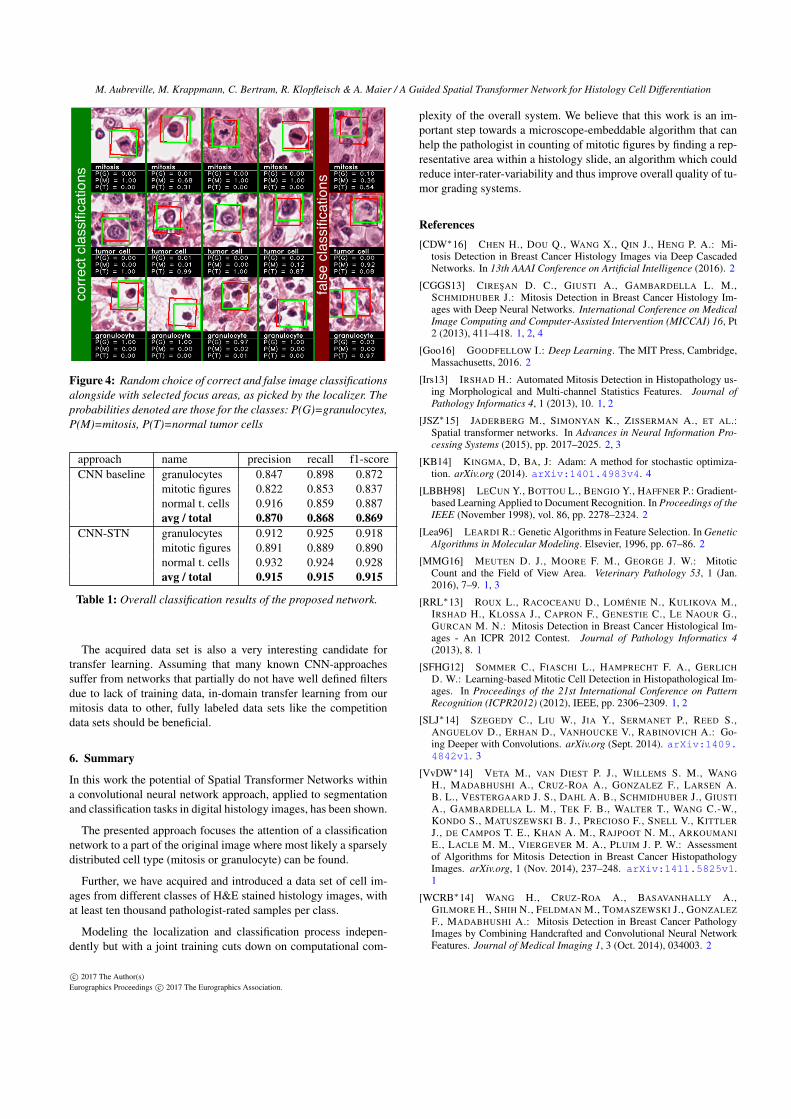
There were only minor differences in the results of the individ-ual test sets in cross-validation, which is why we concatenatedthe respective test vectors and calculated the following metrics onthe ensemble. We achieved an accuracy of 91.8%, with precisionsreaching from 90.4% to 93.4% and recall reaching from 90.1% to92.8%, as described in table 1. Compared to the baseline CNN de-scribed in section 4.4, this is a significant increase, with the addedbenefit of retrieving also segmentation information.
Regarding misclassifications, it is noteworthy that for many falsedecisions the root cause of error seems to be within the scope of thelocalizer (see right column of figure 4). In the top and bottom ex-amples depicted there, the localizer selected a different cell thanthe one originally annotated. Particularly for tumor cells, this is notalways a definite fault, since we do not consider annotation infor-mation of the direct environment of the annotated cell. If, in a directsurrounding of a tumor cell, a granulocyte or mitotic cell is present,the localizer in fact behaves completely correct in presenting thiscell to the classifier. Since we do not aim at finding or classifyingall cells, this is no major drawback. In fact, we inherently prioritizeclassification this way: Since we crop around a known sparse event(mitotic cells or granulocyte), and give this label to our classifier,we incorporate the knowledge that sparse events are more impor-tant than others into the loss function.
We think that the acquired data set provides a good fundamentfor further approaches in mitosis detection, where in our opinionthe lack of a sufficient amount of samples may limit the methodicprogress.
c© 2017 The Author(s)Eurographics Proceedings c© 2017 The Eurographics Association.
M. Aubreville, M. Krappmann, C. Bertram, R. Klopfleisch & A. Maier / A Guided Spatial Transformer Network for Histology Cell Differentiationco
rre
ct
cla
ssifi
ca
tion
s
fals
e c
lassifi
ca
tion
s
Figure 4: Random choice of correct and false image classificationsalongside with selected focus areas, as picked by the localizer. Theprobabilities denoted are those for the classes: P(G)=granulocytes,P(M)=mitosis, P(T)=normal tumor cells
approach name precision recall f1-scoreCNN baseline granulocytes 0.847 0.898 0.872
mitotic figures 0.822 0.853 0.837normal t. cells 0.916 0.859 0.887avg / total 0.870 0.868 0.869
CNN-STN granulocytes 0.912 0.925 0.918mitotic figures 0.891 0.889 0.890normal t. cells 0.932 0.924 0.928avg / total 0.915 0.915 0.915
Table 1: Overall classification results of the proposed network.
The acquired data set is also a very interesting candidate fortransfer learning. Assuming that many known CNN-approachessuffer from networks that partially do not have well defined filtersdue to lack of training data, in-domain transfer learning from ourmitosis data to other, fully labeled data sets like the competitiondata sets should be beneficial.
6. Summary
In this work the potential of Spatial Transformer Networks withina convolutional neural network approach, applied to segmentationand classification tasks in digital histology images, has been shown.
The presented approach focuses the attention of a classificationnetwork to a part of the original image where most likely a sparselydistributed cell type (mitosis or granulocyte) can be found.
Further, we have acquired and introduced a data set of cell im-ages from different classes of H&E stained histology images, withat least ten thousand pathologist-rated samples per class.
Modeling the localization and classification process indepen-dently but with a joint training cuts down on computational com-
plexity of the overall system. We believe that this work is an im-portant step towards a microscope-embeddable algorithm that canhelp the pathologist in counting of mitotic figures by finding a rep-resentative area within a histology slide, an algorithm which couldreduce inter-rater-variability and thus improve overall quality of tu-mor grading systems.
References[CDW∗16] CHEN H., DOU Q., WANG X., QIN J., HENG P. A.: Mi-
tosis Detection in Breast Cancer Histology Images via Deep CascadedNetworks. In 13th AAAI Conference on Artificial Intelligence (2016). 2
[CGGS13] CIRESAN D. C., GIUSTI A., GAMBARDELLA L. M.,SCHMIDHUBER J.: Mitosis Detection in Breast Cancer Histology Im-ages with Deep Neural Networks. International Conference on MedicalImage Computing and Computer-Assisted Intervention (MICCAI) 16, Pt2 (2013), 411–418. 1, 2, 4
[Goo16] GOODFELLOW I.: Deep Learning. The MIT Press, Cambridge,Massachusetts, 2016. 2
[Irs13] IRSHAD H.: Automated Mitosis Detection in Histopathology us-ing Morphological and Multi-channel Statistics Features. Journal ofPathology Informatics 4, 1 (2013), 10. 1, 2
[JSZ∗15] JADERBERG M., SIMONYAN K., ZISSERMAN A., ET AL.:Spatial transformer networks. In Advances in Neural Information Pro-cessing Systems (2015), pp. 2017–2025. 2, 3
[KB14] KINGMA, D, BA, J: Adam: A method for stochastic optimiza-tion. arXiv.org (2014). arXiv:1401.4983v4. 4
[LBBH98] LECUN Y., BOTTOU L., BENGIO Y., HAFFNER P.: Gradient-based Learning Applied to Document Recognition. In Proceedings of theIEEE (November 1998), vol. 86, pp. 2278–2324. 2
[Lea96] LEARDI R.: Genetic Algorithms in Feature Selection. In GeneticAlgorithms in Molecular Modeling. Elsevier, 1996, pp. 67–86. 2
[MMG16] MEUTEN D. J., MOORE F. M., GEORGE J. W.: MitoticCount and the Field of View Area. Veterinary Pathology 53, 1 (Jan.2016), 7–9. 1, 3
[RRL∗13] ROUX L., RACOCEANU D., LOMÉNIE N., KULIKOVA M.,IRSHAD H., KLOSSA J., CAPRON F., GENESTIE C., LE NAOUR G.,GURCAN M. N.: Mitosis Detection in Breast Cancer Histological Im-ages - An ICPR 2012 Contest. Journal of Pathology Informatics 4(2013), 8. 1
[SFHG12] SOMMER C., FIASCHI L., HAMPRECHT F. A., GERLICHD. W.: Learning-based Mitotic Cell Detection in Histopathological Im-ages. In Proceedings of the 21st International Conference on PatternRecognition (ICPR2012) (2012), IEEE, pp. 2306–2309. 1, 2
[SLJ∗14] SZEGEDY C., LIU W., JIA Y., SERMANET P., REED S.,ANGUELOV D., ERHAN D., VANHOUCKE V., RABINOVICH A.: Go-ing Deeper with Convolutions. arXiv.org (Sept. 2014). arXiv:1409.4842v1. 3
[VvDW∗14] VETA M., VAN DIEST P. J., WILLEMS S. M., WANGH., MADABHUSHI A., CRUZ-ROA A., GONZALEZ F., LARSEN A.B. L., VESTERGAARD J. S., DAHL A. B., SCHMIDHUBER J., GIUSTIA., GAMBARDELLA L. M., TEK F. B., WALTER T., WANG C.-W.,KONDO S., MATUSZEWSKI B. J., PRECIOSO F., SNELL V., KITTLERJ., DE CAMPOS T. E., KHAN A. M., RAJPOOT N. M., ARKOUMANIE., LACLE M. M., VIERGEVER M. A., PLUIM J. P. W.: Assessmentof Algorithms for Mitosis Detection in Breast Cancer HistopathologyImages. arXiv.org, 1 (Nov. 2014), 237–248. arXiv:1411.5825v1.1
[WCRB∗14] WANG H., CRUZ-ROA A., BASAVANHALLY A.,GILMORE H., SHIH N., FELDMAN M., TOMASZEWSKI J., GONZALEZF., MADABHUSHI A.: Mitosis Detection in Breast Cancer PathologyImages by Combining Handcrafted and Convolutional Neural NetworkFeatures. Journal of Medical Imaging 1, 3 (Oct. 2014), 034003. 2
c© 2017 The Author(s)Eurographics Proceedings c© 2017 The Eurographics Association.