Embed Size (px)
Citation preview
INDIAN PEDIATRICS 745 VOLUME 49__SEPTEMBER 16, 2012
CCCCC AAAAA SSSSS EEEEE R R R R R EEEEE PPPPP OOOOO RRRRR TTTTT SSSSS
A Fatal Outbreak of Trichosporon asahii Sepsis in a Neonatal IntensiveCare UnitVIPIN M VASHISHTHA, AMOL MITTAL AND *AMIT GARGFrom the Department of Pediatrics, Mangla Hospital and Research Center, Shakti Chowk, Bijnor, and *Department ofMicrobiology, LLRM Medical College, Meerut; Uttar Pradesh, India.
We describe an outbreak of Trichosporon asahii in 8 newborn infants with sepsis. Six out ofthese 8 infants died. The organism was identified on specific culture and morphologiccharacteristics. The organism was sensitive to amphotericin-B but resistant to fluconazole.Laminar flow unit was suspected to be the source of the outbreak.
Key words: India, Neonate, Outbreak, Sepsis, Trichosporon asahii.
Correspondence to:Dr Vipin M Vashishtha,Director and Consultant Pediatrician,Mangla Hospital and Research Center,Shakti Chowk, Bijnor 246 701, [email protected]: October 31, 2011;Initial review: March 15, 2012;Accepted: April 09, 2012.
Trichosporon asahii is an uncommon cause offungal sepsis among newborn infants, but, it isnow emerging as an important life-threateningopportunistic systemic pathogen, especially in
immuno-compromised hosts [1]. Trichosporonosis isusually an insidious disease and its diagnosis is likely tobe missed, particularly in developing countries, becauseof lack of awareness and lack of acquaintance with thesalient diagnostic feature of the etiologic agent. Barring afew isolated case-reports, there is no information on theprevalence of disseminated trichosporonosis in India. Wereport a fatal outbreak of T. asahii sepsis in eightnewborns in our neonatal intensive care unit (NICU).
CASE REPORT
Eight newborn infants admitted between 17th to 28th
August, 2011 in our NICU were found to be infected withT. asahii (Table I).
The first case was already on broad-spectrumantibiotics for last 12 days prior to admission. The bloodculture grew non-albicans candida species (unidentified)after 24 hours of aerobic incubation at 30º. Institution ofconventional IV amphotericin-B along with supportivetherapy led to clinical improvement. The baby was laterdischarged at day 10 of treatment with follow-up adviceof continuing amphotericin-B therapy for full 21 days.The Candida species on culture were later identified ascolonies of T. asahii.
Initial cultures of the second case were sterile. Babyimproved following partial exchange transfusion withnormal saline along with empiric antimicrobials, but later
developed feeding intolerance, abdominal distension,hematemesis and refractory shock. Amphotericin Badded empirically failed to improve general conditionand ultimately the baby died. The repeat blood culturegrew colonies of T. asahii.
The third case had early onset sepsis. Initial bloodculture grew E. coli which was sensitive to common beta-lactams. A dose of surfactant, assisted ventilation andappropriate antibiotics resulted in significantimprovement. Later, the infant developed features ofsepsis along with massive pulmonary hemorrhage andsuccumbed to his illness despite starting IV amphotericinB. The repeat blood culture again grew T. asahii.
The fourth case developed fulminant sepsis after 48hours of admission caused by extended-spectrum beta-lactamase producing Klebsiella pneumoniae. A course ofmeropenem and supportive therapy including ventilatorysupport resulted in improvement. Enteral feeds werestarted and the baby was weaned off from the ventilator.However, at 11th day of life, the baby again showedworsening of clinical and laboratory parameters. Repeatculture revealed growth of Trichosporon spp. LiposomalAmphotericin B was added in the regimen but the babydid not respond, and ultimately died at the age of 21 days.
The next infant had features of early onset sepsis butcultures were negative. The baby improved after a 7-daycourse of empiric antibiotics. Four days later, the babywas readmitted in the NICU for the treatment of jaundiceand later developed necrotizing enterocolitis (NEC),cholestasis and signs of sepsis. The repeat blood culture
INDIAN PEDIATRICS 746 VOLUME 49__SEPTEMBER 16, 2012
CASE REPORTS
revealed Trichosporon spp. With the addition ofamphotericin-B in the regimen, the infant graduallyresponded, blood culture became sterile at 10th day oftherapy, and the baby was discharged at 20th day of lifewith follow up advice of completing IV amphotericin Bcourse for total 21 days.
The sixth case was second born twin delivered to athird-gravida mother. After packed cell transfusion andsupportive treatment, the infant stabilized. However, thebaby later developed respiratory distress that necessitatedassisted ventilation. After weaning off from the ventilator,the baby developed repeated apneic spells and repeatculture grew fungal colonies identified as T. ashii.Addition of IV amphotericin-B to the antimicrobialregimen failed to salvage the baby who developedmassive gastrointestinal hemorrhage, perforation andshock, and died after 6 days of admission.
The next case was a premature infant who developedHyaline membrane disease soon after birth and wastreated successfully with surfactant and assistedventilation. Later, the infant developed signs of sepsis inform of apnea, pallor and hypotension. Repeat culturegrew yeast colonies, identified as as Trichosporon spp.The baby developed massive pulmonary hemorrhage anddied at the age of 7 days despite adding IV amphotericin-B to the regimen.
The last case was a term neonate who developedmeconium aspiration syndrome and was treated withhigh-flow oxygen, antibiotics and IV fluids. Five dayslater, the infant started exhibiting dullness, apnea, and
later gastrointestinal bleeding. The repeat blood culturegrew colonies of Trichosporon spp. Amphotericin B wasadded to the regimen. However, the baby developedfeatures of disseminated intravascular coagulation anddied at the post-natal age of 7 days.
Identification of T asahii: The blood culture was done byautomated BacT/ALERT 3D 120 blood culture System.Gram stain showed elongated blastoconidia and septatepseudohyphae. Broth from the positive blood culturesbottles were sub-cultured on blood agar and Sabouraud’sdextrose agar (SDA) with chromphenicol. The coloniesof yeast like fungi were isolated after 24 hrs ofincubation. Identification and sensitivity was done byVitek 2 Compact. The macroscopic and microscopicmorphology of T. asahii was compatible with thestandard description of the species.
DISCUSSION
Trichosporon asahii is opportunistic yeast described asan emerging pathogen in disseminated nosocomialinfections in NICUs [2-7]. Clinical manifestations ofinfection with this microorganism are non-specific andinfections often results in poor prognosis [2,6,7]. This isprobably the first report of an invasive outbreak in aneonatal unit in India. Case 1 probably represented theindex case; responsible for spreading the infection inother neonates who had nosocomial sepsis during thecourse of their stay.
Literature search revealed reports of T.asahiineonatal infection in 15 preterm newborns. Of these, 11weighed less than 1,000 g at birth and only one weighed
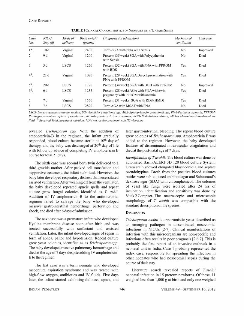
TABLE I CLINICAL CHARACTERISTICS OF NEONATES WITH T. ASAHII SEPSIS
Case NICU Mode of Birth weight Diagnosis (at admission) Mechanical OutcomeNo. Stay (d) delivery (grams) ventilation
1*. 10 d Vaginal 2400 Term-SGA with PNA with Sepsis No Improved2. 9 d Vaginal 1200 Preterm (35 week) SGA with Polycythemia No Died
with Sepsis3. 5 d LSCS 1250 Preterm (32 week) SGA with PNA with PPROM Yes Died
with RDS4$. 21 d Vaginal 1080 Preterm (29 week) SGA Breech presentation with Yes Died
PNA with PPROM5$. 20 d LSCS 1720 Preterm (34 week) SGA with BOH with PPROM No Improved6$. 6 d LSCS 1235 Preterm (28 week) AGA with PNA with twin Yes Died
pregnancy with PPROM with anemia7. 7 d Vaginal 1550 Preterm (31 weeks) SGA with RDS (HMD) Yes Died8. 7 d LSCS 2890 Term AGA with MSAF with PNA No Died
LSCS- Lower segment caesarean section; SGA-Small for gestational age; AGA-Appropriate for gestational age; PNA-Perinatal asphyxia; PPROM-Prolonged premature rupture of membranes, RDS-Respiratory distress syndrome; BOH- Bad obstetric history; MSAF- Meconium-stained amnioticfluid; $ Received Total parenteral nutrition; *Did not receive treatment with H2 –blockers.
INDIAN PEDIATRICS 747 VOLUME 49__SEPTEMBER 16, 2012
CASE REPORTS
more than 1,500 g at birth. All deaths (seven) occurred inthe extremely low birth weight group. However, in ourseries, the preterm infants were not extremely prematureand had comparatively higher weights, and even full termneonates were affected. Trichosporon infections inneonates have been almost uniformly fatal. In our seriesalso, six out of eight neonates died. Many of our patientshad one or more of the risk factors often blamed fornosocomial sepsis and fungal diseases [8].
August is the month of the year which has very highhumidity and high rates of neonatal admissions. Due tohigh work load, a breach in asepsis protocol might haveoccurred. Surprisingly on performing microbialsurveillance, we found that one surface culture fromlaminar flow unit yielded positive growth of Candida sppwith morphological features similar to T. asahii. Westopped using laminar flow for preparing intravenousfluids and the entire unit was thoroughly fumigated withformaldehyde. Hence, it can be presumed that laminarflow unit was probably the source of this outbreak.
Most strains of T. asahii may be confused withCandida spp. on initial culture examinations. Therefore,delays in appropriate treatment may occur. Severalstudies have demonstrated low in vitro sensitivity of T.asahii to commonly used antifungal agents [1,3,9]. Thefungus is known for varied susceptibility to amphotericinB and laboratory studies have shown that it is relativelyresistant to this agent [1,9]. On the other hand, manyauthors have described good results of earlyadministration of amphotericin B [2,4]. In our series also,all the isolates were sensitive to amphotericin-B, butresistant to fluconazole and flucytosine. However, afavourable clinical response was observed in only twocases despite using amphotericin B quite early on the firstsuspicion of nosocomial sepsis in most neonates. The invivo resistance of the drug can be explained due toformation of a biofilm by Trichosporon spp [10], whichmay explain persistence of the infection in spite of in vitrosensitivity of the drug.
Since there are no pathognomonic clinical features,the diagnosis of disseminated trichosporonosis dependsprimarily upon clinical suspicion, to be followed by
intensive mycological investigations. Infection with thisagent should be taken into consideration when dealingwith low birth weight preterm infants, particularly thosewith nosocomial sepsis having cocktail of broadspectrum antibiotics for a prolonged period but still withunfavourable clinical progress.Funding: None; Competing interests: None stated.
REFERENCES
1. Guého E, Improvisi L, de Hoog GS, Dupont B.Trichosporon on humans: a practical account. Mycoses.1994;37:3-10.
2. Gökahmetoglu S, Nedret Koç A, Günes T, Cetin N.Trichosporon mucoides infection in three prematurenewborns. Mycoses. 2002;45:123-5.
3. Panagopoulou P, Evdoridou J, Bibashi E, Filioti J,Sofianou D, Kremenopoulos G, et al. Trichosporonasahii: an unusual cause of invasive infection in neonates.Pediatr Infect Dis J. 2002;21:169-70.
4. Yildiran A, Kücüködük S, Saniç A, Belet N, Güvenli A.Disseminated Trichosporon asahii infection in a preterm.Am J Perinatol. 2003;20:269-71.
5. Maheshwari A, Stromquist CI, Pereda L, Emmanuel PJ.Mixed infection with unusual fungi and staphylococcalspecies in two extremely premature neonates. J Perinatol.2004;24:324-6.
6. Téllez-Castillo CJ, Gil-Fortuño M, Centelles-Sales I,Sabater-Vidal S, Pardo Serrano F. Trichosporon asahiifatal infection in a preterm newborn. Rev Chilena Infectol.2008;25: 213-5.
7. Pereira DN, Nader SS, Nader P, Martins PG, Furlan SP,Hentges CR. Disseminated Trichosporon spp infection inpreterm newborns: a case report. J Pediatr (Rio J).2009;85:459-61.
8. Saiman L, Ludington E, Pfaller M, Rangel-Frausto S,Wiblin RT, Dawson J, et al. Risk factors for candidemia inneonatal intensive care unit patients. The NationalEpidemiology of Mycosis Survey Study Group. PediatrInfect Dis J. 2000;19:319-24.
9. Walsh TJ, Melcher GP, Rinaldi MG, Lecciones J,McGough DA, Kelly P, et al. Trichosporon beigelii: anemerging pathogen resistant to amphotericin B. J ClinMicrobiol. 1990;28:1616-22.
10. Di Bonaventura G, Pompilio A, Picciani C, Iezzi M,D’Antonio D, Piccolomini R. Biofilm formation by theemerging fungal pathogen Trichosporon asahii:development, architecture, and antifungal resistance.Antimicrob Agents Chemother. 2006;50:3269-76.