Embed Size (px)
Citation preview
FULL PAPER
A Fast and Flexible MRI System for the Studyof Dynamic Vocal Tract Shaping
Sajan Goud Lingala,1* Yinghua Zhu,1 Yoon-Chul Kim,2 Asterios Toutios,1
Shrikanth Narayanan,1 and Krishna S. Nayak1
Purpose: The aim of this work was to develop and evaluate
an MRI-based system for study of dynamic vocal tract shap-ing during speech production, which provides high spatial and
temporal resolution.Methods: The proposed system utilizes (a) custom eight-channel upper airway coils that have high sensitivity to upper
airway regions of interest, (b) two-dimensional golden anglespiral gradient echo acquisition, (c) on-the-fly view-sharingreconstruction, and (d) off-line temporal finite difference con-
strained reconstruction. The system also provides simultane-ous noise-cancelled and temporally aligned audio. The system
is evaluated in 3 healthy volunteers, and 1 tongue cancerpatient, with a broad range of speech tasks.Results: We report spatiotemporal resolutions of 2.4�2.4 mm2
every 12 ms for single-slice imaging, and 2.4�2.4 mm2 every36 ms for three-slice imaging, which reflects roughly 7-fold
acceleration over Nyquist sampling. This system demonstratesimproved temporal fidelity in capturing rapid vocal tract shapingfor tasks, such as producing consonant clusters in speech, and
beat-boxing sounds. Novel acoustic-articulatory analysis wasalso demonstrated.
Conclusion: A synergistic combination of custom coils, spiralacquisitions, and constrained reconstruction enables visualiza-tion of rapid speech with high spatiotemporal resolution
in multiple planes. Magn Reson Med 000:000–000, 2016.VC 2016 Wiley Periodicals, Inc.
Key words: flexible MRI system; rapid vocal tract shaping;
custom upper-airway coil; spiral readouts; multi-slice; con-strained reconstruction
INTRODUCTION
Speech production involves complex spatiotemporalcoordination of several vocal organs in the upper andlower airways. Modalities to study speech productioninclude real-time MRI (RT-MRI), electromagnetic articu-lography (EMA), electropalatography, ultrasound, andX-ray, or videofluoroscopy (1). In comparison to alternatemodalities, RT-MRI provides distinct advantages in
terms of (a) noninvasiveness, as opposed to X-rays,videofluoroscopy, and (b) ability to image in arbitrary
planes and visualize deep structures (e.g., epiglottis, glot-tis), which are not possible with ultrasound and EMA.Applications of RT-MRI in speech science and vocal pro-
duction research are numerous; these include addressingopen questions pertaining to understanding the goals oflanguage production, language timing, speech errors, andother topics in phonetics and phonology (1–8) as well as
vocal production of song (9,10). It also has the potentialto manage and inform treatment plans in several clinicalapplications, such as clinical assessment of velopharyng-eal insufficiency (11–13), cleft palate repair and manage-
ment (14,15), and surgical planning and post-treatmentfunctional evaluation of speech and swallowing (16,17),in head and neck cancer.
The rates of movements of articulators are highly
dependent on the speech task and the subject’s speakingstyle (3,18,19). For example, in the articulation of sus-tained sounds, such as during singing, the spatial posi-tion of the articulators change on the order of seconds,
whereas in tasks involving flaps/trills and production ofconsonant clusters, the motion of articulators occur at amuch faster rate on the order of a few milliseconds (alsosee Figure 1) (3). Whereas modalities such as EMA can
operate up to a time resolution of 1 ms, the imagingspeed of RT-MRI is restricted by challenges posed as aresult of device physics.
Several schemes have been proposed to improve the
imaging speed of RT-MRI for speech studies. These canbe classified as on-the-fly or off-line schemes (3). On-the-fly schemes are referred to those that allow for immedi-ate visualization of the reconstructed images (with
latency less than 500 ms), whereas off-line schemes arereferred to those where the reconstructions are imple-mented off-line. Scott et al (20) utilized on-the-fly imag-ing with Cartesian trajectories and demonstrated
temporal resolutions between 111 and 50 ms at spatialresolution of 1.6–2.7 mm2 for velopharyngeal closure.Other investigators (1,21,22) utilized short spiral read-
outs to acquire images at a native time resolution of54–78 ms, and a spatial resolution of 3.0–2.4 mm2, andvisualize at 24 frames/sec using view sharing. View shar-ing was also used with radial imaging in (23,24). Freitas
et al (25) compares Cartesian, radial, and spiral trajecto-ries with view sharing and demonstrated spirals to pro-vide the best compromise in terms acquisition efficiency,motion robustness, and signal to noise (SNR).
Iterative constrained reconstruction schemes have
shown to enable greater acceleration factors. Utilizingradial trajectories, Niebergall et al (23) proposed
1Electrical Engineering, University of Southern California, Los Angeles, CA.2Samsung Medical Center, Seoul, South Korea.
Grant sponsor: National Institutes of Health; Grant number: NIH/NIDCDR01 DC007124.
*Correspondence to: Sajan Goud Lingala, Ph.D., Department of ElectricalEngineering, Viterbi School of Engineering, University of Southern California,3740 McClintock Avenue, Los Angeles, CA 90089. E-mail: [email protected]
Received 24 August 2015; revised 6 November 2015; accepted 24November 2015
DOI 10.1002/mrm.26090Published online 00 Month 2016 in Wiley Online Library (wileyonlinelibrary.com).
Magnetic Resonance in Medicine 00:00–00 (2016)
VC 2016 Wiley Periodicals, Inc. 1
nonlinear temporal regularized reconstruction to enablea temporal resolution of 33 ms, and a spatial resolutionof 1.5 mm2, and studied a variety of tasks (vowel, conso-nant sounds, and coarticulations events). More recently,Iltis et al (26) demonstrated an on-the-fly implementationof the iterative reconstruction by exploiting paralleliza-tion within the reconstruction along with efficient use ofgraphical processing units. This work used a 64-channelbrain coil and demonstrated 100 frames/sec at 1.5 mm2.Burdumy et al (24) applied off-line spatial total-variationregularization with radial trajectories to provide spatialresolution of 1.8 mm2, and a native time resolution of 55ms, and analyzed morphometric measurements of thevocal tract. Fu et al (27) utilized off-line reconstructionwith the partial separable (PS) model (28,29), along withspatial-spectral sparsity constraint (30), and demon-strated a frame rate of 102 frames/sec at a spatial resolu-tion of 2.2 mm2. The low-rank (by the PS model)constraint is essentially a data-driven retrospective bin-ning technique (31) and was fully exploited in Fu et al(27) by utilizing repeated speech utterances. Low-rankconstraints are unlikely to apply to short speech utteran-ces with no repetitions, or stimuli involving infrequentdistinct movements such as swallowing.
In this work, we developed a MRI-based system for
dynamic study of vocal tract shaping during speech pro-
duction to provide high spatiotemporal resolutions. We
propose a system that utilizes (a) a novel eight-channel
custom upper airway coil, which has improved
sensitivity in upper airway regions of interest (ROIs), (b)
a flexible slice selective spiral spoiled gradient echo
acquisition with golden angle time interleaving, (c) on-
the-fly view-sharing reconstruction, (d) off-line temporal
finite difference constrained reconstruction, (e) simulta-
neous audio acquisition, and off-line temporal alignment
of noise-cancelled audio with the reconstructions.
The innovation of our system lies in the synergistic
combination of the above components. We chose custom
upper airway coil design for its ability to provide supe-rior SNR across all articulators of interest. This is advan-
tageous because it can provide an important boost in
SNR while operating at 1.5 Tesla (T). Its combination
with spirals is complementary, because it enables
improved SNR at low fields, low off-resonance–relatedspiral artifacts, and high sampling efficiency. Multishot
short spirals (readout length of 2.4 ms) are used to
reduce off-resonance artifacts. Our rationale of choosing
spirals is that they have shown to provide improved
motion robustness and efficiency over alternate trajecto-ries. Temporal finite difference constraint was chosen
because it exploits redundancy based on the prior that
the desired information is contained in the moving
edges, which directly fits to the end goal assessment ofdynamics of air-tissue interfaces. Also, because it
exploits similarities among neighboring time frames, it is
applicable to a wide variety of speech tasks and does not
impose restrictions on the imaging task.We present several examples with the proposed sys-
tem for rapid RT-MRI of speech, including visualizationof interleaved consonant and vowel sounds, fluent
speech sentences that contain consonant clusters, as
well as beat-boxing sounds that involve rapid coordina-
tion between various vocal articulators, on three healthy
volunteers, and one tongue cancer patient referred toglossectomy.
METHODS
Simulation of Spatial Versus Time Resolution Trade-offsWith Spiral Sampling
A multishot short spiral readout spoiled gradient echo
pulse sequence (flip angle //FA: 15� degrees; slice
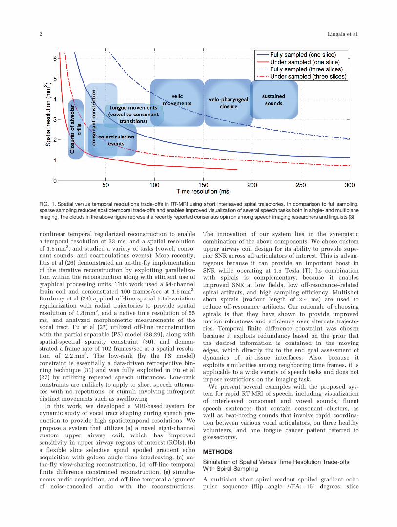
FIG. 1. Spatial versus temporal resolutions trade-offs in RT-MRI using short interleaved spiral trajectories. In comparison to full sampling,sparse sampling reduces spatiotemporal trade-offs and enables improved visualization of several speech tasks both in single- and multiplane
imaging. The clouds in the above figure represent a recently reported consensus opinion among speech imaging researchers and linguists (3).
2 Lingala et al.
thickness: 6 mm; readout time: 2.5 ms, repetition time[TR]¼ 6.004 ms), which was used in our previous stud-ies, was adapted in this study (32,33). We chose spiraltrajectories over alternate trajectories because they haveshown to provide a superior trade-off among spatial reso-lution, time resolution, and robustness to motion arti-facts. The spiral trajectories were designed to makemaximum use of gradients (40 mT/m maximum gradientamplitude and 150 mT/m/ms slew rate). Simulationswere performed to investigate the spatial versus timeresolution trade-offs for a field of view (FOV) of 20 cm2
at Nyquist (full) sampling and rate 6.5-fold undersam-pling for single-slice and concurrent three-slice imaging(Figure 1). Nyquist sampling was determined by varyingthe number of spiral interleaves such that the maximumspacing between interleaves equaled the reciprocal of theunaliased FOV. The Nyquist definition was definedbased on the assumption of spiral interleaving with auniform angle distribution. Figure 1 also duplicates theschematic placement of various speech tasks accordingto their spatial and temporal resolution requirements, asreported in Lingala et al (3).
Single-Slice and Multislice Golden Angle Spiral TimeInterleaving
A previously proposed golden angle time interleavedsampling pattern scheme in which successive spiral inter-leaves are separated by the golden angle 2p� 2/(�5þ1)was adapted in this work (32). The sampling schedulewas repeated after 144 interleaves. Two single-slicesequences with spatial resolutions of 2.4 and 1.76 mm2
were realized, which respectively corresponded to 13 and21 spiral interleaves/frame for Nyquist sampling. A flexi-ble multislice time interleaved sequence (33) was alsoadapted to realize an arbitrary three-slice select sequenceat 2.4 mm2. The golden angle increment for the three-slicesequence occurred every 3 TRs. It should be noted thatthe unaliased FOV with golden angle sampling slightlydiffers with that of uniform density sampling used for thesimulation in Figure 1 (32). Specifically, the point spreadfunction of golden angle spiral sampling provides morereduced side-lobe energies and provides improved trade-off of the achievable unaliased FOV in comparison to uni-form density sampling (32).
Custom Upper Airway Coil
All of our experiments were performed on a GE SignaExcite 1.5T scanner (GE Healthcare, Little Chalfont, UK)with a custom eight-channel upper airway receiver coilthat has four elements on either side of the jaw. The ele-ments were designed to be spatially localized and to bein close proximity to the upper airway to provide highSNR over all the important upper airway structures. Wechose custom coil design because of its ability to providesuperior SNR across all articulators of interest. This isadvantageous because it provides an important boost inSNR while operating at 1.5T, and enables efficient com-bination with spirals is highly complementary, becauseit together enables improved SNR at low fields, low off-resonance–related spiral artifacts, and high sampling effi-ciency. The custom coil was developed for two sizes for
adult and child arrays, although all the experiments inthe current study were performed on adults using theadult-sized array.
Reconstruction
On-the-Fly
Data acquisition was implemented within custom RT-hawk software (34). A view-sharing scheme was used,where data for each frame were combined respectivelyfrom 13 and 21 subsequent interleaves, respectively forthe 2.4 and 1.76 mm2 sequences. Images were recon-structed on-the-fly by using a fast implementation of thegridding algorithm within RT-hawk. The minimallatency allowed for instant feedback to the operator andenabled efficient scan plane localizations. In addition, itenabled on-the-fly adjustment of the center frequency tominimize off-resonance blurs. Specifically, the subjectbeing scanned was asked to open their mouth, and inthe midsagittal plane, the operator qualitatively adjustedthe center frequency such that the air-tissue (majorly:air-tongue, air-velum, air-lip) boundaries were sharp.
Offline
A sparse SENSE temporal finite difference constrainedreconstruction scheme was implemented offline. Thisconstraint exploits redundancy based on the fact that thedesired information is contained in the moving edges.This directly fits to the application of speech imaging,where the end goal is the assessment of interactionand timing of various articulators, or assessment of thedynamics of the air-tissue interfaces (moving edges). Spa-tial and temporal finite difference constraints have alsobeen previously used in several studies (e.g., (35–39).
The reconstruction is formulated as as shown by Equa-tion [1]:
minf ðx;tÞjjAðf Þ � bjj22 þ ljjDtðf Þjj1 [1]
where b is a concatenated vector containing the spiralnoisy k-t measurements from each coil and f(x,t) is thedynamic data to be reconstructed at a retrospectivelyspecified time resolution. A models coil sensitivityencoding as well as Fourier encoding on the specifiedspiral trajectory in each time frame; the coil sensitivitieswere assumed to be time-invariant, and were estimatedby an Eigen decomposition method using time-averagedimage data from each coil (40), and the nonuniform Fou-rier transform (nuFFT) implementation by Fessler andSutton (41) was used. The l1-sparsity-based temporalfinite difference (Dt) penalty is used to penalize pixelswith rapidly varying pixel time profiles. l is the regulari-zation parameter that controls the balance between thesparsity penalty and the data fidelity. Equation [1] wassolved by a nonlinear conjugate gradient (CG) algorithm(42). The algorithm was initialized with the f5AH(b) esti-mate and was terminated at 40 iterations, where qualita-tively there was no noticeable change in image quality.The reconstructions were implemented within MATLAB(The MathWorks, Inc., Natick, MA) on an Intel Core i73.5 GHz machine with 32-GB memory.
A Fast and Flexible MRI system to study vocal tract shaping 3
In Vivo Experiments and Speech Tasks
Three healthy volunteers (2 male, 1 female; median age:
29) and 1 male tongue cancer patient (62 years) were
scanned. The patient was scanned before clinical treat-
ment. All the stimuli were presented in the scanner
using a mirror projector setup. A variety of speech tasks
were considered. The midsagittal orientation was used
for single-slice sequences, whereas orientations for the
multislice sequence differed according to the speech
task. With the 2.4-mm2 single-slice sequence, volunteer
1 (male Indian English speaker) was scanned without
any speech stimuli on two separate instances: (a) using
an eight-channel head coil and (b) using the custom
eight-channel upper airway coil. With the custom upper
airway coil, the same volunteer was scanned with both
the single-slice sequences, using the stimuli: “one-two-
three-four-five” at a normal speech rate followed by a
rapid speech rate (approximately 4 times faster). A task
to produce interleaved consonant and vowel sounds by
the repetition of the phrase: “loo-lee-laa-za-na-za” at the
normal speech rate was considered on volunteer 1 and
imaged using the three-slice sequence (one mid-sagittal,
one axial plane at the level of mid-pharyngeal airway,
and one coronal plane at the middle of the tongue). Vol-
unteer 2 (male Chinese English speaker) was scanned
with the 1.76-mm2 single-slice sequence, while produc-
ing the sentence: “She had your dark suit in greasy
wash water,” which involves producing sounds that
involve rapid articulatory movements (e.g., coarticula-
tion events as a part of running speech). Volunteer 3
(female American English speaker) was a beat boxer and
was scanned while producing a variety of beat-boxing
sounds. The 2.4-mm2 single- and concurrent three-slice
(one midsagittal slice, one axial slice at the level of
velum, and one axial slice at the level of glottis) sequen-
ces were considered. The particular axial cuts were cho-
sen to capture the rapid velar and glottis movements
during beat boxing. Volunteer 4 (male American English
speaker) was a tongue cancer patient and was scanned
with the single-slice 2.4-mm2 sequence. Speech stimuli
comprising words, sentences, and a passage were pre-
sented, and the ability to produce speech was analyzed.
A small subset of these stimuli is presented in this
work, which pertain to short words that contain vowels
interleaved by consonants: “beat, bit, bait, bet, bat, pot,
bought, boat.”
Simultaneous Audio Collection
For 3 of 4 volunteers scanned, audio recordings were
obtained simultaneously at a sampling frequency of 20
KHz inside the scanner, while the subjects were being
imaged, using a commercial fiber optic microphone
(Optoacoustics Ltd., Or Yehuda, Israel) (43) and a cus-
tom recording setup. Noise cancellation was performed
using a data-driven adaptive signal processing algorithm,
which is blind to the acoustic properties of noise (44).
The final noise-cancelled audio was synchronized with
the reconstructed RT-MRI data to facilitate acoustic-
articulatory analysis.
Analysis
Comparison of SNR Between Coils
The SNR properties of the custom eight-channel upper air-
way coil were qualitatively compared with a commercial
eight-channel head (brain) coil. The single-slice (2.4 mm2)
sequence was used, where volunteer 1 was scanned with no
speech, and 55 interleaves were used to reconstruct
f5AH(b). The ROI SNR in different upper airway regions
were quantified as SNRROI¼m (S) / s (n), where S is a vector
with image intensities from the ROI containing a specific
upper airway structure, and n is a vector with intensities
from an ROI in the background capturing only noise. A total
of 10 ROIs were defined: 1) upper lip; 2) lower lip; 3) front
tongue; 4) middle tongue; 5) back tongue; 6) hard palate; 7)
velum; 8) pharyngeal wall; 9) epiglottis; and 10) glottis. A
relative measure of SNR between the two coils was eval-
uated by the factor SNRUA/SNRhead.
Choosing Regularization Parameter in ConstrainedReconstruction
The regularization parameter in the constrained recon-
struction was chosen empirically, with the best trade-off
between artifact removal, and temporal stair-casing. With
the single-slice 2.4-mm2 sequence, and a 12-ms time
resolution reconstruction, the effect of regularization
parameter on the reconstructions was studied. L-curves
were obtained for two data sets with different speech
stimuli from different volunteers: volunteer 1 with fast
speech stimuli of counting numbers and volunteer 3
with beat boxing. For generating the L-curve, the CG iter-
ations were set to a very high number of 120 to ensure
no bias in the norm calculations resulting from small
numerical errors; however, in practice, a lower number
of iterations of around 40 were sufficient for conver-
gence, where, qualitatively, there was no noticeable
change in image quality.
Comparison of Constrained Reconstructions at VariousReduction Factors
Golden-angle time interleaving allows for retrospective
reconstruction with arbitrary time resolution. Using the
speech stimuli of counting numbers at a rapid pace, recon-
structions were performed at 5, 3, 2, and 1 TR for the 2.4-
and 1.76-mm2 single-slice sequences. This corresponded
to reduction factors (R) between 2.6- and 13-fold for the
2.4-mm2 sequence and 4.2- to 21-fold for the 1.76-mm2
sequence. The regularization parameters were chosen
empirically for all cases. A l2.4mm2¼ 0.1 and l1.76mm2¼ 0.3
respectively for the 2.4- and 1.76-mm2 sequence was used
for the 5, 3, and 2 TR cases, whereas a higher l2.4mm2¼ 0.2
and l1.76mm2¼0.4 was used for the 1 TR case. The trade-off
between residual aliasing artifacts, and temporal blurring
(resulting from large temporal footprint), and reconstruc-
tion time was analyzed in all the cases.
Visualization of Various Stimuli With DifferentReconstructions
To qualitatively depict the gains in improved time resolu-
tion, and its utility in capturing fast articulatory
4 Lingala et al.
movements, constrained reconstructions (from sub-
sampled data) were qualitatively compared to gridding
reconstructions obtained at Nyquist (full) sampling using
the stimuli of counting numbers (normal pace, followed
by rapid pace). A reduction factor of 6.5- to 7.0-fold was
used in constrained reconstruction, which corresponded
to a temporal footprint of 2 and 3 TR, respectively, for the
single-slice 2.4- and 1.76-mm2 sequences. Constrained
reconstructions were also qualitatively compared to the
view-sharing reconstruction, which was used in our pre-
vious work (21,22). To ensure consistent comparisons, the
step size in view sharing was matched to the native time
resolution in the constrained reconstruction. Acoustic-
articulatory analysis along with articulatory image seg-
mentation was performed on the patient data set. An
upper airway image segmentation algorithm (45) was
modified to handle image space data and was applied to
segment all the important articulators in all the frames;
this enabled tracking of the articulators across time.
RESULTS
Simulation of Spatial Versus Time Resolution Trade-offsWith Spiral Sampling
Figure 1 shows the improved spatial and temporal resolu-
tion trade-off with rate 6.5 undersampled spiral imaging
over fully sampled spiral imaging. Fast articulatory move-
ments, such as consonant constrictions, coarticulation
events, and flaps, trills are expected to be well depicted
at a rate of 6.5-fold single-place spiral imaging over fully
sampled spiral imaging. Multiplane imaging compro-
mises the spatial and time resolutions for additional slice
coverage. At a rate of 6.5 fold undersampling, three-plane
imaging offers spatiotemporal resolutions of up to
2.4 mm2, and 36 ms/frame, which enables capture of
speech tasks, such as consonant constrictions, coarticula-
tion events, and all tongue movements. In contrast, with
full sampling, multiplane imaging is notoriously slow for
its efficient use in capturing fast articulatory shaping.
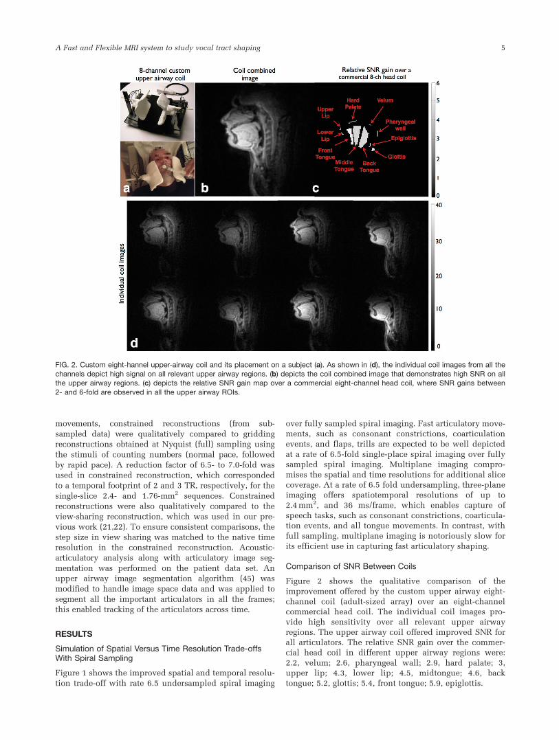
Comparison of SNR Between Coils
Figure 2 shows the qualitative comparison of the
improvement offered by the custom upper airway eight-
channel coil (adult-sized array) over an eight-channel
commercial head coil. The individual coil images pro-
vide high sensitivity over all relevant upper airway
regions. The upper airway coil offered improved SNR for
all articulators. The relative SNR gain over the commer-
cial head coil in different upper airway regions were:
2.2, velum; 2.6, pharyngeal wall; 2.9, hard palate; 3,
upper lip; 4.3, lower lip; 4.5, midtongue; 4.6, back
tongue; 5.2, glottis; 5.4, front tongue; 5.9, epiglottis.
FIG. 2. Custom eight-hannel upper-airway coil and its placement on a subject (a). As shown in (d), the individual coil images from all thechannels depict high signal on all relevant upper airway regions. (b) depicts the coil combined image that demonstrates high SNR on allthe upper airway regions. (c) depicts the relative SNR gain map over a commercial eight-channel head coil, where SNR gains between
2- and 6-fold are observed in all the upper airway ROIs.
A Fast and Flexible MRI system to study vocal tract shaping 5
Choosing Regularization Parameter in ConstrainedReconstruction
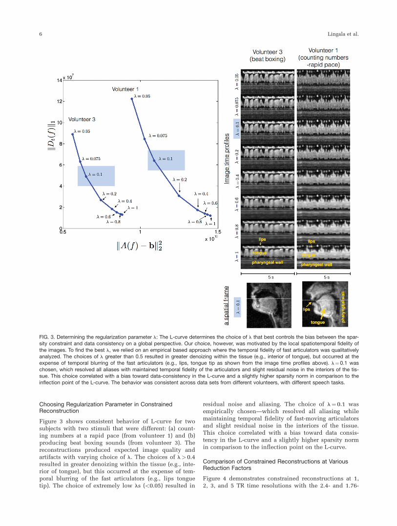
Figure 3 shows consistent behavior of L-curve for twosubjects with two stimuli that were different: (a) count-ing numbers at a rapid pace (from volunteer 1) and (b)producing beat boxing sounds (from volunteer 3). Thereconstructions produced expected image quality andartifacts with varying choice of l. The choices of l> 0.4resulted in greater denoizing within the tissue (e.g., inte-rior of tongue), but this occurred at the expense of tem-poral blurring of the fast articulators (e.g., lips tonguetip). The choice of extremely low ls (<0.05) resulted in
residual noise and aliasing. The choice of l¼ 0.1 wasempirically chosen—which resolved all aliasing whilemaintaining temporal fidelity of fast-moving articulatorsand slight residual noise in the interiors of the tissue.This choice correlated with a bias toward data consis-tency in the L-curve and a slightly higher sparsity normin comparison to the inflection point on the L-curve.
Comparison of Constrained Reconstructions at VariousReduction Factors
Figure 4 demonstrates constrained reconstructions at 1,2, 3, and 5 TR time resolutions with the 2.4- and 1.76-
FIG. 3. Determining the regularization parameter l: The L-curve determines the choice of l that best controls the bias between the spar-sity constraint and data consistency on a global perspective. Our choice, however, was motivated by the local spatiotemporal fidelity of
the images. To find the best l, we relied on an empirical based approach where the temporal fidelity of fast articulators was qualitativelyanalyzed. The choices of l greater than 0.5 resulted in greater denoizing within the tissue (e.g., interior of tongue), but occurred at theexpense of temporal blurring of the fast articulators (e.g., lips, tongue tip as shown from the image time profiles above). l¼0.1 was
chosen, which resolved all aliases with maintained temporal fidelity of the articulators and slight residual noise in the interiors of the tis-sue. This choice correlated with a bias toward data-consistency in the L-curve and a slightly higher sparsity norm in comparison to the
inflection point of the L-curve. The behavior was consistent across data sets from different volunteers, with different speech tasks.
6 Lingala et al.
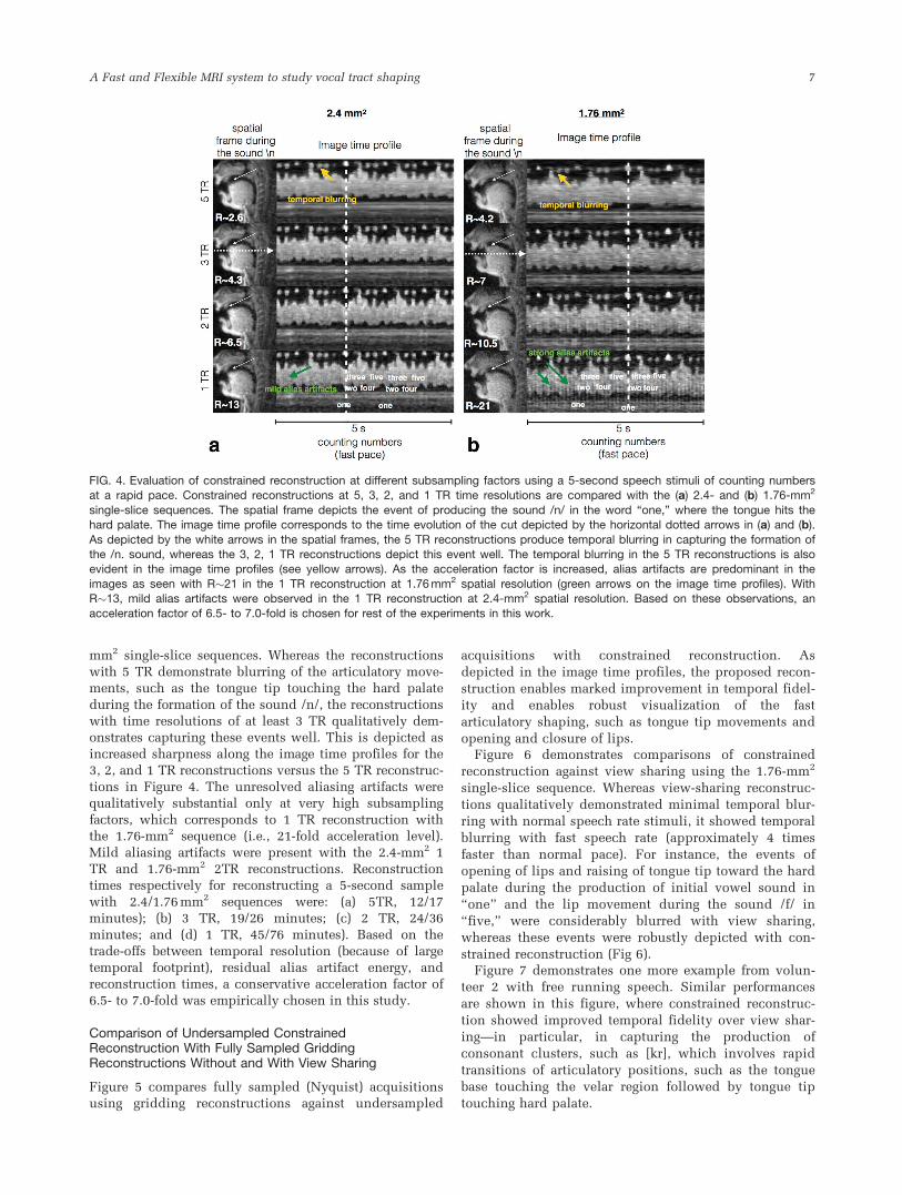
mm2 single-slice sequences. Whereas the reconstructions
with 5 TR demonstrate blurring of the articulatory move-ments, such as the tongue tip touching the hard palate
during the formation of the sound /n/, the reconstructionswith time resolutions of at least 3 TR qualitatively dem-onstrates capturing these events well. This is depicted as
increased sharpness along the image time profiles for the3, 2, and 1 TR reconstructions versus the 5 TR reconstruc-
tions in Figure 4. The unresolved aliasing artifacts werequalitatively substantial only at very high subsampling
factors, which corresponds to 1 TR reconstruction withthe 1.76-mm2 sequence (i.e., 21-fold acceleration level).
Mild aliasing artifacts were present with the 2.4-mm2 1TR and 1.76-mm2 2TR reconstructions. Reconstructiontimes respectively for reconstructing a 5-second sample
with 2.4/1.76 mm2 sequences were: (a) 5TR, 12/17minutes); (b) 3 TR, 19/26 minutes; (c) 2 TR, 24/36
minutes; and (d) 1 TR, 45/76 minutes). Based on thetrade-offs between temporal resolution (because of large
temporal footprint), residual alias artifact energy, andreconstruction times, a conservative acceleration factor of
6.5- to 7.0-fold was empirically chosen in this study.
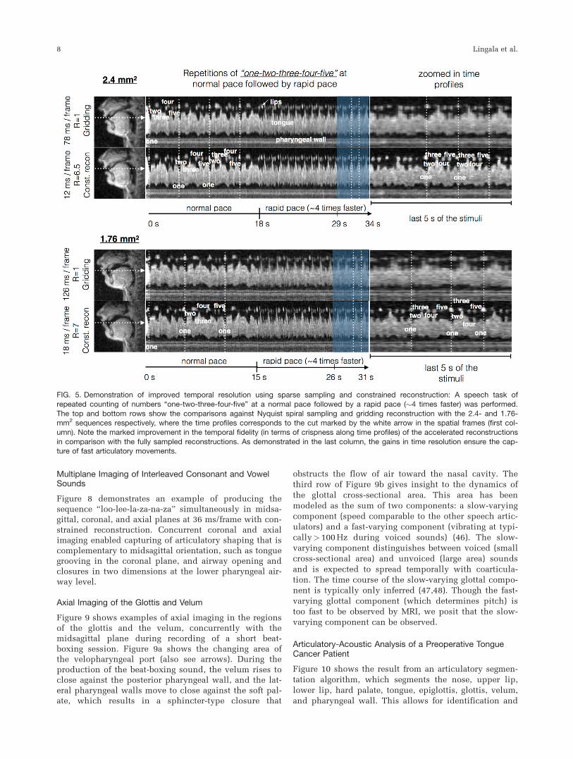
Comparison of Undersampled ConstrainedReconstruction With Fully Sampled GriddingReconstructions Without and With View Sharing
Figure 5 compares fully sampled (Nyquist) acquisitionsusing gridding reconstructions against undersampled
acquisitions with constrained reconstruction. As
depicted in the image time profiles, the proposed recon-
struction enables marked improvement in temporal fidel-
ity and enables robust visualization of the fast
articulatory shaping, such as tongue tip movements and
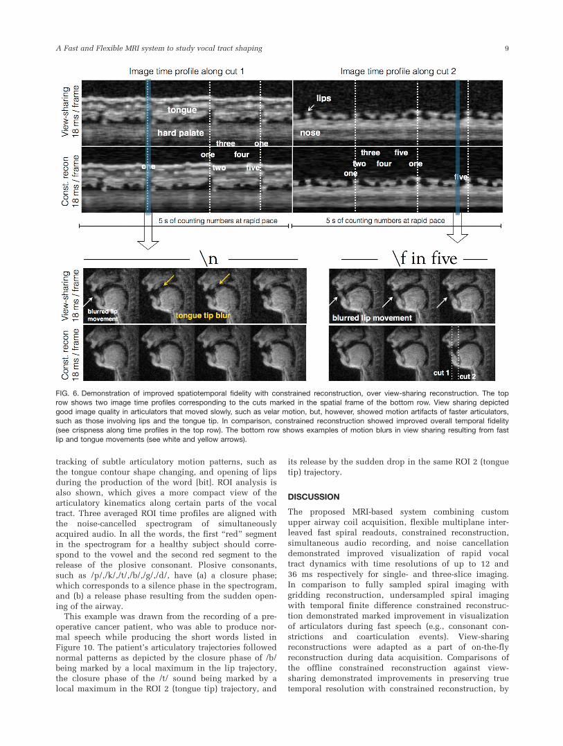
opening and closure of lips.Figure 6 demonstrates comparisons of constrained
reconstruction against view sharing using the 1.76-mm2
single-slice sequence. Whereas view-sharing reconstruc-
tions qualitatively demonstrated minimal temporal blur-
ring with normal speech rate stimuli, it showed temporal
blurring with fast speech rate (approximately 4 times
faster than normal pace). For instance, the events of
opening of lips and raising of tongue tip toward the hard
palate during the production of initial vowel sound in
“one” and the lip movement during the sound /f/ in
“five,” were considerably blurred with view sharing,
whereas these events were robustly depicted with con-
strained reconstruction (Fig 6).Figure 7 demonstrates one more example from volun-
teer 2 with free running speech. Similar performances
are shown in this figure, where constrained reconstruc-
tion showed improved temporal fidelity over view shar-
ing—in particular, in capturing the production of
consonant clusters, such as [kr], which involves rapid
transitions of articulatory positions, such as the tongue
base touching the velar region followed by tongue tip
touching hard palate.
FIG. 4. Evaluation of constrained reconstruction at different subsampling factors using a 5-second speech stimuli of counting numbersat a rapid pace. Constrained reconstructions at 5, 3, 2, and 1 TR time resolutions are compared with the (a) 2.4- and (b) 1.76-mm2
single-slice sequences. The spatial frame depicts the event of producing the sound /n/ in the word “one,” where the tongue hits thehard palate. The image time profile corresponds to the time evolution of the cut depicted by the horizontal dotted arrows in (a) and (b).
As depicted by the white arrows in the spatial frames, the 5 TR reconstructions produce temporal blurring in capturing the formation ofthe /n. sound, whereas the 3, 2, 1 TR reconstructions depict this event well. The temporal blurring in the 5 TR reconstructions is alsoevident in the image time profiles (see yellow arrows). As the acceleration factor is increased, alias artifacts are predominant in the
images as seen with R�21 in the 1 TR reconstruction at 1.76 mm2 spatial resolution (green arrows on the image time profiles). WithR�13, mild alias artifacts were observed in the 1 TR reconstruction at 2.4-mm2 spatial resolution. Based on these observations, an
acceleration factor of 6.5- to 7.0-fold is chosen for rest of the experiments in this work.
A Fast and Flexible MRI system to study vocal tract shaping 7
Multiplane Imaging of Interleaved Consonant and VowelSounds
Figure 8 demonstrates an example of producing thesequence “loo-lee-la-za-na-za” simultaneously in midsa-gittal, coronal, and axial planes at 36 ms/frame with con-strained reconstruction. Concurrent coronal and axialimaging enabled capturing of articulatory shaping that iscomplementary to midsagittal orientation, such as tonguegrooving in the coronal plane, and airway opening andclosures in two dimensions at the lower pharyngeal air-way level.
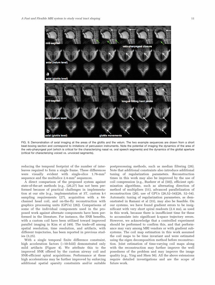
Axial Imaging of the Glottis and Velum
Figure 9 shows examples of axial imaging in the regionsof the glottis and the velum, concurrently with themidsagittal plane during recording of a short beat-boxing session. Figure 9a shows the changing area ofthe velopharyngeal port (also see arrows). During theproduction of the beat-boxing sound, the velum rises toclose against the posterior pharyngeal wall, and the lat-eral pharyngeal walls move to close against the soft pal-ate, which results in a sphincter-type closure that
obstructs the flow of air toward the nasal cavity. The
third row of Figure 9b gives insight to the dynamics of
the glottal cross-sectional area. This area has been
modeled as the sum of two components: a slow-varying
component (speed comparable to the other speech artic-
ulators) and a fast-varying component (vibrating at typi-
cally> 100 Hz during voiced sounds) (46). The slow-
varying component distinguishes between voiced (small
cross-sectional area) and unvoiced (large area) sounds
and is expected to spread temporally with coarticula-
tion. The time course of the slow-varying glottal compo-
nent is typically only inferred (47,48). Though the fast-
varying glottal component (which determines pitch) is
too fast to be observed by MRI, we posit that the slow-
varying component can be observed.
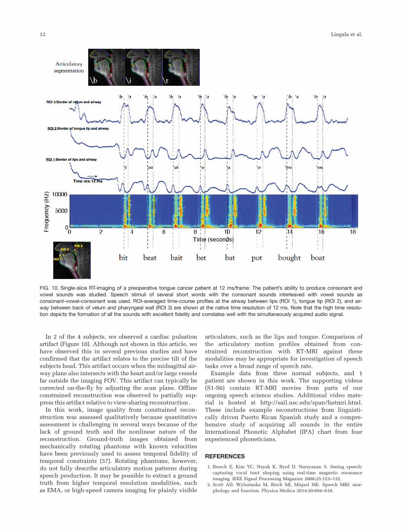
Articulatory-Acoustic Analysis of a Preoperative TongueCancer Patient
Figure 10 shows the result from an articulatory segmen-
tation algorithm, which segments the nose, upper lip,
lower lip, hard palate, tongue, epiglottis, glottis, velum,
and pharyngeal wall. This allows for identification and
FIG. 5. Demonstration of improved temporal resolution using sparse sampling and constrained reconstruction: A speech task ofrepeated counting of numbers “one-two-three-four-five” at a normal pace followed by a rapid pace (�4 times faster) was performed.
The top and bottom rows show the comparisons against Nyquist spiral sampling and gridding reconstruction with the 2.4- and 1.76-mm2 sequences respectively, where the time profiles corresponds to the cut marked by the white arrow in the spatial frames (first col-umn). Note the marked improvement in the temporal fidelity (in terms of crispness along time profiles) of the accelerated reconstructions
in comparison with the fully sampled reconstructions. As demonstrated in the last column, the gains in time resolution ensure the cap-ture of fast articulatory movements.
8 Lingala et al.
tracking of subtle articulatory motion patterns, such as
the tongue contour shape changing, and opening of lips
during the production of the word [bit]. ROI analysis is
also shown, which gives a more compact view of the
articulatory kinematics along certain parts of the vocal
tract. Three averaged ROI time profiles are aligned with
the noise-cancelled spectrogram of simultaneously
acquired audio. In all the words, the first “red” segment
in the spectrogram for a healthy subject should corre-
spond to the vowel and the second red segment to the
release of the plosive consonant. Plosive consonants,
such as /p/,/k/,/t/,/b/,/g/,/d/, have (a) a closure phase;
which corresponds to a silence phase in the spectrogram,
and (b) a release phase resulting from the sudden open-
ing of the airway.This example was drawn from the recording of a pre-
operative cancer patient, who was able to produce nor-
mal speech while producing the short words listed in
Figure 10. The patient’s articulatory trajectories followed
normal patterns as depicted by the closure phase of /b/
being marked by a local maximum in the lip trajectory,
the closure phase of the /t/ sound being marked by a
local maximum in the ROI 2 (tongue tip) trajectory, and
its release by the sudden drop in the same ROI 2 (tongue
tip) trajectory.
DISCUSSION
The proposed MRI-based system combining custom
upper airway coil acquisition, flexible multiplane inter-
leaved fast spiral readouts, constrained reconstruction,
simultaneous audio recording, and noise cancellation
demonstrated improved visualization of rapid vocal
tract dynamics with time resolutions of up to 12 and
36 ms respectively for single- and three-slice imaging.
In comparison to fully sampled spiral imaging with
gridding reconstruction, undersampled spiral imaging
with temporal finite difference constrained reconstruc-
tion demonstrated marked improvement in visualization
of articulators during fast speech (e.g., consonant con-
strictions and coarticulation events). View-sharing
reconstructions were adapted as a part of on-the-fly
reconstruction during data acquisition. Comparisons of
the offline constrained reconstruction against view-
sharing demonstrated improvements in preserving true
temporal resolution with constrained reconstruction, by
FIG. 6. Demonstration of improved spatiotemporal fidelity with constrained reconstruction, over view-sharing reconstruction. The toprow shows two image time profiles corresponding to the cuts marked in the spatial frame of the bottom row. View sharing depicted
good image quality in articulators that moved slowly, such as velar motion, but, however, showed motion artifacts of faster articulators,such as those involving lips and the tongue tip. In comparison, constrained reconstruction showed improved overall temporal fidelity(see crispness along time profiles in the top row). The bottom row shows examples of motion blurs in view sharing resulting from fast
lip and tongue movements (see white and yellow arrows).
A Fast and Flexible MRI system to study vocal tract shaping 9
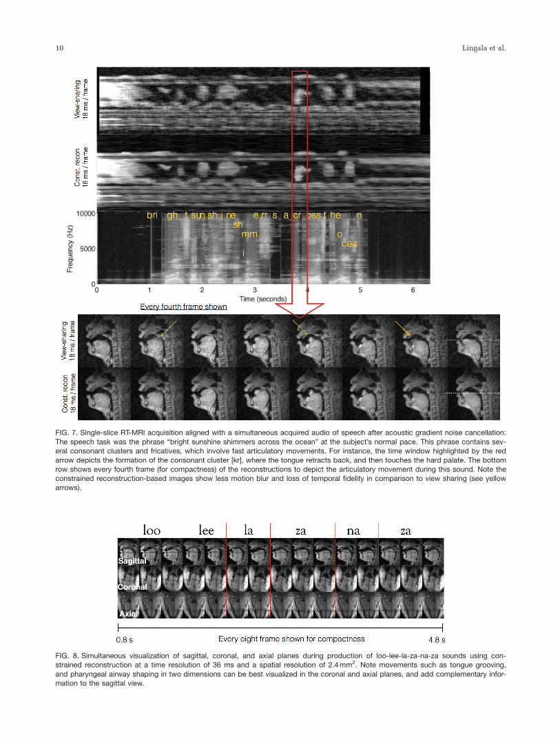
FIG. 7. Single-slice RT-MRI acquisition aligned with a simultaneous acquired audio of speech after acoustic gradient noise cancellation:
The speech task was the phrase “bright sunshine shimmers across the ocean” at the subject’s normal pace. This phrase contains sev-eral consonant clusters and fricatives, which involve fast articulatory movements. For instance, the time window highlighted by the red
arrow depicts the formation of the consonant cluster [kr], where the tongue retracts back, and then touches the hard palate. The bottomrow shows every fourth frame (for compactness) of the reconstructions to depict the articulatory movement during this sound. Note theconstrained reconstruction-based images show less motion blur and loss of temporal fidelity in comparison to view sharing (see yellow
arrows).
FIG. 8. Simultaneous visualization of sagittal, coronal, and axial planes during production of loo-lee-la-za-na-za sounds using con-strained reconstruction at a time resolution of 36 ms and a spatial resolution of 2.4 mm2. Note movements such as tongue grooving,
and pharyngeal airway shaping in two dimensions can be best visualized in the coronal and axial planes, and add complementary infor-mation to the sagittal view.
10 Lingala et al.
reducing the temporal footprint of the number of inter-leaves required to form a single frame. These differenceswere visually evident with single-slice 1.76-mm2
sequence and the multislice 2.4-mm2 sequences.A direct comparison of the proposed system against
state-of-the-art methods (e.g., (26,27) has not been per-formed because of practical challenges in implementa-tion at our site (e.g., implementation at 3T, custom k-tsampling requirements (27), acquisition with a 64-channel head coil, and on-the-fly reconstruction withgraphics processing units (GPUs) (26)). Comparisons ofsome of the individual components used in the pro-posed work against alternate components have been per-formed in the literature. For instance, the SNR benefits,with a custom coil have been utilized toward improvedparallel imaging in Kim et al (49). The trade-off amongspatial resolution, time resolution, and artifacts, withdifferent trajectories, has been reported in previous stud-ies (3,25).
With a single temporal finite difference constraint,high acceleration factors (>10-fold) demonstrated onlymild artifacts (Figure 4). We attribute this to theimproved SNR offered by the custom airway coil andSNR-efficient spiral acquisitions. Performance at thesehigh accelerations may be further improved by enforcingadditional spatial sparsity constraints and/or suitable
postprocessing methods, such as median filtering (26).Note that additional constraints also introduce additionaltuning of regularization parameters. Reconstructiontimes in this work may also be improved by the use ofcoil compression (e.g., Buehrer et al (50)), efficient opti-mization algorithms, such as alternating direction ofmethod of multipliers (51), advanced parallelization ofreconstruction (26), use of GPUs (26,52–54)(26, 52–54).Automatic tuning of regularization parameters, as dem-onstrated in Ramani et al (55), may also be feasible. Onour systems, we have found gradient errors to be insig-nificant with very short spiral readouts (2.4 ms), as usedin this work, because there is insufficient time for theseto accumulate into significant k-space trajectory errors.However, we acknowledge that a controlled experimentshould be performed to confirm this, and that perform-ance may vary among MRI vendors or with gradient sub-systems. The coil map estimation in this work assumedthe coil maps to be time invariant and were estimatedusing the eigen decomposition method before reconstruc-tion. Joint estimation of time-varying coil maps alongwith the reconstruction may further improve the wellposedness of the problem and may improve the imagequality (e.g., Ying and Shen 56). All the above extensionsrequire detailed investigations and are the scope offuture work.
FIG. 9. Demonstration of axial imaging at the areas of the glottis and the velum. The two example sequences are drawn from a shortbeat-boxing section and correspond to imitations of percussion instruments. Note the potential of imaging the dynamics of the area ofthe velo-pharyngeal port (which is critical for the characterizing nasal vs. oral speech segments) and the dynamics of the glottal aperture
(critical for characterizing voiced vs. unvoiced segments).
A Fast and Flexible MRI system to study vocal tract shaping 11
In 2 of the 4 subjects, we observed a cardiac pulsation
artifact (Figure 10). Although not shown in this article, we
have observed this in several previous studies and haveconfirmed that the artifact relates to the precise tilt of the
subjects head. This artifact occurs when the midsagittal air-
way plane also intersects with the heart and/or large vessels
far outside the imaging FOV. This artifact can typically becorrected on-the-fly by adjusting the scan plane. Offline
constrained reconstruction was observed to partially sup-
press this artifact relative to view-sharing reconstruction .In this work, image quality from constrained recon-
struction was assessed qualitatively because quantitativeassessment is challenging in several ways because of the
lack of ground truth and the nonlinear nature of the
reconstruction. Ground-truth images obtained from
mechanically rotating phantoms with known velocitieshave been previously used to assess temporal fidelity of
temporal constraints (57). Rotating phantoms, however,
do not fully describe articulatory motion patterns during
speech production. It may be possible to extract a groundtruth from higher temporal resolution modalities, such
as EMA, or high-speed camera imaging for plainly visible
articulators, such as the lips and tongue. Comparison of
the articulatory motion profiles obtained from con-
strained reconstruction with RT-MRI against these
modalities may be appropriate for investigation of speech
tasks over a broad range of speech rate.Example data from three normal subjects, and 1
patient are shown in this work. The supporting videos
(S1-S6) contain RT-MRI movies from parts of our
ongoing speech science studies. Additional video mate-
rial is hosted at http://sail.usc.edu/span/fastmri.html.
These include example reconstructions from linguisti-
cally driven Puerto Rican Spanish study and a compre-
hensive study of acquiring all sounds in the entire
International Phonetic Alphabet (IPA) chart from four
experienced phoneticians.
REFERENCES
1. Bresch E, Kim YC, Nayak K, Byrd D, Narayanan S. Seeing speech:
capturing vocal tract shaping using real-time magnetic resonance
imaging. IEEE Signal Processing Magazine 2008;25:123–132.
2. Scott AD, Wylezinska M, Birch MJ, Miquel ME. Speech MRI: mor-
phology and function. Physica Medica 2014;30:604–618.
FIG. 10. Single-slice RT-imaging of a preoperative tongue cancer patient at 12 ms/frame: The patient’s ability to produce consonant andvowel sounds was studied. Speech stimuli of several short words with the consonant sounds interleaved with vowel sounds as
consonant-vowel-consonant was used. ROI-averaged time-course profiles at the airway between lips (ROI 1), tongue tip (ROI 2), and air-way between back of velum and pharyngeal wall (ROI 3) are shown at the native time resolution of 12 ms. Note that the high time resolu-
tion depicts the formation of all the sounds with excellent fidelity and correlates well with the simultaneously acquired audio signal.
12 Lingala et al.
3. Lingala SG, Sutton BP, Miquel ME, Nayak KS. Recommendations for
real-time speech MRI. J Magn Reson Imaging 2015 July 14. doi:
10.1002/jmri.24997.
4. Demolin D, Hassid S, Metens T, Soquet A. Real-time MRI and articu-
latory coordination in speech. Comptes Rendus Biol 2002;325:547–
556.
5. Byrd D, Tobin S, Bresch E, Narayanan S. Timing effects of syllable
structure and stress on nasals: a real-time MRI examination. J Phonet
2009;37:91–110.
6. Iltis PW, Schoonderwaldt E, Zhang S, Frahm J, Altenm€uller E. Real-
time MRI comparisons of brass players: a methodological pilot study.
Hum Mov Sci 2015;42:132–145.
7. Perry J, Sutton BP, Kuehn DP, Gamage JK. Using MRI for assessing
velopharyngeal structures and function. Cleft Palate Craniofac J 2014;
51:476–485.
8. Ramanarayanan V, Byrd D, Goldstein L, Narayanan SS. Investigating
articulatory setting—pauses, ready position, and rest—using real-time
MRI. Proceedings of Interspeech 2010 (pp. 1994–1997), Makuhari,
Japan, 2010.
9. Proctor M, Bresch E, Byrd D, Nayak K, Narayanan S. Paralinguistic
mechanisms of production in human ‘beatboxing:’ a real-time mag-
netic resonance imaging study. J Acoust Soc Am 2013;133:1043–
1054.
10. Bresch E, Narayanan S. Real-time MRI investigation of resonance tun-
ing in soprano singing. J Acoust Soc Am Express Lett 2010;128:
EL335–EL341.
11. Bae Y, Kuehn DP, Conway CA, Sutton BP. Real-time magnetic reso-
nance imaging of velopharyngeal activities with simultaneous speech
recordings. Cleft Palate Craniofac J 2011;48:695–707.
12. Maturo S, Silver A, Nimkin K, Sagar P, Ashland J, van der Kouwe
AJ, Hartnick C. MRI with synchronized audio to evaluate velophar-
yngeal insufficiency. Cleft Palate Craniofac J 2012;49:761–763.
13. Drissi C, Mitrofanoff M, Talandier C, Falip C, Le Couls V,
Adamsbaum C. Feasibility of dynamic MRI for evaluating velophar-
yngeal insufficiency in children. Eur Radiol 2011;21:1462–1469.
14. Tian W, Li Y, Yin H, Zhao SF, Li S, Wang Y, Shi B. Magnetic reso-
nance imaging assessment of velopharyngeal motion in Chinese chil-
dren after primary palatal repair. J Craniofac Surg 2010;21:578–587.
15. Kazan-Tannus JF, Levine D, McKenzie C, Lim KH, Cohen B, Farrar
N, Busse RF, Mulliken JB. Real-time magnetic resonance imaging
aids prenatal diagnosis of isolated cleft palate. J Ultrasound Med
2005;24:1533–1540.
16. Hagedorn C, Lammert A, Bassily M, Zu Y, Sinha U, Goldstein L,
Narayanan SS. Characterizing post-glossectomy speech using real-
time MRI. In Proceedings of the 10th International Seminar on
Speech Production (ISSP), Cologne, Germany, May 2014.
17. Zu Y, Narayanan S, Kim YC, Nayak K, Bronson-Lowe C, Villegas B,
Ouyoung M, Sinha U. Evaluation of swallow function post tongue
cancer treatment using real-time MRI: a pilot study. JAMA Otolaryn-
gol Head Neck Surg 2013;139:1312-1319.
18. Adams SG, Weismer G, Kent RD. Speaking rate and speech move-
ment velocity profiles. J Speech Hear Res 1993;36:41–54.
19. Tasko SM, McClean MD. Variations in articulatory movement with
changes in speech task. J Speech Lang Hear Res 2004;47:85–100.
20. Scott AD, Boubertakh R, Birch MJ, Miquel ME. Towards clinical
assessment of velopharyngeal closure using MRI: evaluation of real-
time MRI sequences at 1.5 and 3 T. Br J Radiol 2012;85:e1083–e1092.
21. Narayanan S, Nayak K, Lee S, Sethy A, Byrd, D. An approach to real-
time magnetic resonance imaging for speech production. J Acoust
Soc Am 2004;115:1771–1776.
22. Narayanan S, Toutios A, Ramanarayanan V, Lammert A, Kim J, Lee
S, Nayak K, Kim YC, Zhu Y, Goldstein L, Byrd D, Bresch E, Ghosh P,
Katsamanis A, Proctor M. Real-time magnetic resonance imaging and
electromagnetic articulography database for speech production
research. J Acoust Soc Am 2014;136:1307–1311.
23. Niebergall A, Zhang S, Kunay E, Keydana G, Job M, Uecker M,
Frahm J. Real-time MRI of speaking at a resolution of 33 ms: Under-
sampled radial FLASH with nonlinear inverse reconstruction. Magn
Reson Med 2013;69:477–485.
24. Burdumy M, Traser L, Richter B, Echternach M, Korvink JG, Hennig
J, Zaitsev M, Acceleration of MRI of the vocal tract provides addi-
tional insight into articulator modifications. J Magn Reson Imaging
2015;42:925–935.
25. Freitas AC, Wylezinska M, Birch M, Petersen SE, Miquel ME. Real
time speech MRI: a comparison of Cartesian and non-Cartesian
sequences. In Proceedings of ISMRM 23rd Scientific Sessions (p.
655), Toronto, Ontario, Canada, May 30–June 5, 2015.
26. Iltis PW, Frahm J, Voit D, Joseph AA, Schoonderwaldt E,
Altenm€uller E. High-speed real-time magnetic resonance imaging of
fast tongue movements in elite horn players. Quant Imaging Med
Surg 2015;5:374–381.
27. Fu M, Zhao B, Carignan C, Shosted RK, Perry JL, Kuehn DP, Liang
ZP, #Sutton BP. High resolution dynamic speech imaging with joint
low-rank and sparsity constraints. Magn Reson Med 2015;73:1820–
1832.
28. Gupta AS, Liang ZP. Dynamic imaging by temporal modeling with
principal component analysis. In Proceedings of the 9th Annual
Meeting of the International Society for Magnetic Resonance in Medi-
cine (p. 10), Glasgow, Scotland, UK, April 21–27, 2001.
29. Liang ZP. Spatiotemporal imaging with partially separable functions.
In Noninvasive Functional Source Imaging of the Brain and Heart
and the International Conference on Functional Biomedical Imaging,
2007. NFSI-ICFBI 2007. Joint Meeting of the 6th International Sympo-
sium on (pp. 181–182). New York: IEEE.
30. Zhao B, Haldar JP, Christodoulou AG, Liang ZP. Image reconstruction
from highly undersampled-space data with joint partial separability
and sparsity constraints. IEEE Transact Med Imaging 2012;31:1809–
1820.
31. Hu Y, Lingala SG, Jacob M. High resolution structural free breathing
cardiac MRI using k-t SLR, In: Proceedings of the ISMRM 19th Scien-
tific Sessions (p. 4382), Montreal, Quebec, Canada, May 7–13, 2015.
32. Kim YC, Narayanan SS, Nayak KS. Flexible retrospective selection of
temporal resolution in real-time speech MRI using a golden-ratio spi-
ral view order. Magn Reson Med 2011;65:1365–1371.
33. Kim YC, Proctor MI, Narayanan SS, Nayak KS. Improved imaging of
lingual articulation using real-time multislice MRI. J Magn Reson
Imaging 2012;35:943–948.
34. Santos JM, Wrigh G, Pauly JM. Flexible real-time magnetic resonance
imaging framework. In Engineering in Medicine and Biology Society,
2004. IEMBS’04. 26th Annual International Conference of the IEEE
(Vol. 1, pp. 1048–1051). New York: IEEE.
35. Block KT, Uecker M, Frahm J. Undersampled radial MRI with multi-
ple coils. Iterative image reconstruction using a total variation con-
straint. Magnc Reson Med 2007;57:1086–1098.
36. Liu B, King K, Steckner M, Xie J, Sheng J, Ying L. Regularized sensi-
tivity encoding (SENSE) reconstruction using bregman iterations.
Magn Reson Med 2009;61:145–152.
37. Todd N, Adluru G, Payne A, DiBella EV, Parker D. Temporally con-
strained reconstruction applied to MRI temperature data. Magn Reson
Med 2009;62:406–419.
38. Adluru G, McGann C, Speier P, Kholmovski EG, Shaaban A, Dibella
EV. Acquisition and reconstruction of undersampled radial data for
myocardial perfusion magnetic resonance imaging. J Magn Reson
Imaging 2009;29:466–473.
39. Feng L, Grimm R, Tobias Block K, Chandarana H, Kim S, Xu J, Axel
L, Sodickson DK, Otazo R. Golden-angle radial sparse parallel MRI:
Combination of compressed sensing, parallel imaging, and golden-
angle radial sampling for fast and flexible dynamic volumetric MRI.
Magn Reson Med 2013;717:707–717.
40. Walsh DO, Gmitro AF, Marcellin MW. Adaptive reconstruction of
phased array MR imagery. Magn Reson Med 2000;43:682–690.
41. Fessler J, Sutton BP. Nonuniform fast Fourier transforms using min-
max interpolation. IEEE Transact Signal Processing 2003;51:560–574.
42. Lustig M, Donoho D, Pauly JM. Sparse MRI: the application of com-
pressed sensing for rapid MR imaging. Magn Reson Med 2007;58:
1182–1195.
43. Opto-acoustics: MR-compatible fiber optic microphone. http://www.
optoacoustics.com/medical/fomri-iii/features; last accessed on 11 Jan-
uary 2016.
44. Vaz C, Ramanarayanan V, Narayanan S. A two-step technique for
MRI audio enhancement using dictionary learning and wavelet
packet analysis. In Proceedings of InterSpeech (pp. 1312–1315),
Lyon, France, August 25–29, 2013.
45. Bresch E, Narayanan S. Region segmentation in the frequency domain
applied to upper airway real-time magnetic resonance images. IEEE
Transact Med Imaging 2009;28:323–338.
46. Maeda, S. Phonemes as concatenable units: VCV synthesis using a
vocal-tract synthesizer. In Sound Patterns of Connected Speech:
Description, Models and Explanation. Proceedings of the symposium
held at Kiel University, Arbeitsberichte des Institut f€ur Phonetik und
A Fast and Flexible MRI system to study vocal tract shaping 13
digitale Spachverarbeitung der Universitaet Kiel:31, Simpson AP,
P€otzold, M, eds., June 1996, pp. 145–164.
47. Toutios A, Maeda S. Articulatory VCV synthesis from EMA data. In Pro-
ceedings of InterSpeech, Portland, Oregon, USA, September 9–13, 2012.
48. Laprie Y, Loosvelt M, Maeda S, Sock R, Hirsch F et al. Articulatory
copy synthesis from cine X-ray films. In Proceedings of the Inter-
Speech 14th Annual Conference of the International Speech Commu-
nication Association, Lyon France, August 2013.
49. Kim YC, Hayes CE, Narayanan SS, Nayak KS. Novel 16-channel
receive coil array for accelerated upper airway MRI at 3 Tesla. Magn
Reson Med 2011;65:1711–1717.
50. Buehrer M, Pruessmann KP, Boesiger P, Kozerke S. Array compres-
sion for MRI with large coil arrays. Magn Reson Med 2007;57:1131–
1139.
51. Ramani S, Fessler J. Regularized parallel MRI reconstruction using an
alternating direction method of multipliers. In Biomedical Imaging:
From Nano to Macro, 2011 IEEE International Symposium on (pp.
385–388). New York: IEEE.
52. Hansen MS, Sørensen TS. Gadgetron: an open source framework for
medical image reconstruction. Magn Reson Med 2011;69:1768–1776.
53. Pryor G, Lucey B, Maddipatla S, McClanahan C, Melonakos J,
Venugopalakrishnan V, Patel K, Yalamanchili P, Malcolm J. High-level
GPU computing with Jacket for MATLAB and C/Cþþ. In SPIE Defense,
Security, and Sensing (pp. 806005–806005). International Society for
Optics and Photonics, Orlando, Florida, USA, April 25–29, 2011.
54. Kong J, Dimitrov M, Yang Y, Liyanage J, Cao L, Staples J, Mantor M,
Zhou H. Accelerating MATLAB image processing toolbox functions
on GPUs. In Proceedings of the 3rd Workshop on General-Purpose
Computation on Graphics Processing Units (pp. 75–85). New York:
ACM; 2010.
55. Ramani S, Liu Z, Rosen J, Nielsen J, Fessler JA. Regularization param-
eter selection for nonlinear iterative image restoration and MRI
reconstruction using GCV and SURE-based methods. IEEE Trans
Image Process 2012;21:3659–3672.
56. Ying L, Sheng J. Joint image reconstruction and sensitivity estimation
in SENSE (JSENSE). Magn Reson Med 2007;57:1196–1202.
57. Frahm J, Sch€atz S, Untenberger M, Zhang S, Voit D, Merboldt KD,
Sohns JM, Lotz J, Uecker M. On the temporal fidelity of nonlinear
inverse reconstructions for real-time MRI—the motion challenge.
Open Med Imaging J 2014;8:1–7.
SUPPORTING INFORMATION
Additional Supporting Information may be found in the online version ofthis article.Supporting Video S1. Single-slice RT-imaging using the 2.4-mm2
sequence at 12 ms time resolution. The speech task involved countingnumbers at a normal pace followed by a rapid pace (approximately 4 timesfaster). The video also shows the result from an articulatory segmentationalgorithm, which segments the nose, upper lip, lower lip, hard palate,tongue, epiglottis, glottis, velum, and pharyngeal wall. Segmentation ofarticulators allow for trackingSupporting Video S2. Rapid beat-boxing sounds captured with 2.4-mm2
spatial resolution at 12 ms time resolution.Supporting Video S3. Vowel sounds produced by an expert phoneticianand captured at 12 ms time resolution. These sounds are a part of a study,which involved imaging all the sounds in the entire International PhoneticAlphabet (IPA) chart.Supporting Video S4. Consonant sounds produced by an expert phoneti-cian and captured at 12 ms time resolution. These sounds are a part of astudy, which involved imaging all the sounds in the entire International Pho-netic Alphabet (IPA) chart.Supporting Video S5. Fluent speech (rainbow passage) produced by anexpert phonetician and captured at 12 ms time resolution.Supporting Video S6. Beat-boxing sounds imaged concurrently in midsa-gittal, and two axial planes at the level of velum, and glottis at 36 ms/frame.
14 Lingala et al.


![PURPOSE: METHODS: (a)sipi.usc.edu/~toutios/pdfs/lingala2016accelerating.pdfmethod through-time GRAPPA (TT-GRAPPA) [6-7] to efficiently exploit the acceleration capabilities offered](https://img.pdfslide.us/doc/110x75/6026f6a03d056f4369797ad4/purpose-methods-asipiuscedutoutiospdfslin-method-through-time-grappa-tt-grappa.jpg)