Embed Size (px)
Citation preview
Database of volumetric and real-time vocal tract MRI for speech science
Tanner Sorensen, Zisis Skordilis, Asterios Toutios, Yoon-Chul Kim∗, Yinghua Zhu†, Jangwon Kim‡,Adam Lammert§, Vikram Ramanarayanan¶, Louis Goldstein, Dani Byrd, Krishna Nayak,
Shrikanth Narayanan
University of Southern California, Los Angeles, CA, [email protected]
AbstractWe present the USC Speech and Vocal Tract Morphology MRIDatabase, a 17-speaker magnetic resonance imaging databasefor speech research. The database consists of real-time mag-netic resonance images (rtMRI) of dynamic vocal tract shap-ing, denoised audio recorded simultaneously with rtMRI, and3D volumetric MRI of vocal tract shapes during sustainedspeech sounds. We acquired 2D real-time MRI of vocal tractshaping during consonant-vowel-consonant sequences, vowel-consonant-vowel sequences, read passages, and spontaneousspeech. We acquired 3D volumetric MRI of the full set of vow-els and continuant consonants of American English. Each 3Dvolumetric MRI was acquired in one 7-second scan in whichthe participant sustained the sound. This is the first database tocombine rtMRI of dynamic vocal tract shaping and 3D volumet-ric MRI of the entire vocal tract. The database provides a uniqueresource with which to examine the relationship between vocaltract morphology and vocal tract function. The USC Speech andVocal Tract Morphology MRI Database is provided free for re-search use at http://sail.usc.edu/span/morphdb.Index Terms: speech production, speech corpora, magneticresonance imaging, multi-modal database, large-scale phonetictools
1. IntroductionThe articulatory speech data-sets that are readily available tothe research community have been consistently well-utilized inpursuit of addressing fundamental questions about speech pro-duction [1, 2, 3]. Until relatively recently, speech articulatorydata had been difficult to obtain and generally lacking. A grow-ing number of resources has begun to reverse this problem, butmany still tend to focus on targeted laboratory speech (e.g.,simple syllables or isolated phonemes) or only on read speech.Here, we present the USC Speech and Vocal Tract MorphologyMRI Database, a new database for the community that capturesa wide variety of dynamic speech tasks in conjunction with de-tailed structural parameters and also non-speech articulations,all with an eye toward understanding and explaining speech andspeaker variability.
Magnetic resonance imaging (MRI) is a flexible technol-ogy for speech research. Rapid imaging methods have achieveda balance among the competing factors of temporal resolution,spatial resolution, and signal-to-noise ratio that allows for flexi-ble characterization of vocal tract morphology and function us-ing a suite of complementary MRI methods [4, 5]. Real-time
∗ now at Samsung Medical Center† now at Google, Inc.‡ now at Canary Speech, LLC§ now at MIT Lincoln Laboratory¶ now at Educational Testing Service R&D
MRI (rtMRI) characterizes the dynamic shaping of the vocaltract during speech in any scan plane(s) of interest with no needfor repeated scans [6]. 3D volumetric MRI characterizes theentire vocal tract with high spatial resolution during sustainedsounds in as little as 7 s [7]. Together, these scans character-ize the function and morphology of the vocal tract with hightemporal (rtMRI) and spatial resolution (3D volumetric MRI).The USC Speech and Vocal Tract Morphology MRI Databaseprovides rtMRI of dynamic vocal tract shaping, denoised au-dio recorded simultaneously with rtMRI, and 3D volumetricMRI of a comprehensive set of the American English contin-uant sounds. The USC Speech and Vocal Tract MorphologyMRI Database is provided free for research use at the projectpage: http://sail.usc.edu/span/morphdb.
2. Database acquisition2.1. Experiments
Seventeen (8 m, 9 f) speakers of American English participated.None of the participants spoke a language other than Englishfluently, nor had any lived outside the United States for a signif-icant amount of time. See Table 1 for participant age and stateof origin. The parents of each participant were native speakersof American English. None of the speakers reported abnormalhearing or speech pathology.
Each speaker participated in two sessions on different days.One session was for acquiring rtMRI data-sets; the other ses-sion was for acquiring 3D volumetric MRI data-sets. The ex-perimenter explained the nature of the experiment and the ex-periment protocol to the participant before each scan. The par-ticipant lay on the scanner table in a supine position. The headwas fixed in place by foam pads inserted between each templeand the receiver coil. The participant read visual stimuli froma back-projection screen from inside the scanner bore withoutmoving the head. The speech corpus captured 3D MRI of sus-tained continuant sounds (see Table 2) and rtMRI videos of iso-
ID age state of origin ID age state of origin
F1 25 CA M1 33 WIF2 25 NY M2 27 VAF3 26 CA M3 28 WIF4 25 DC M4 20 CAF5 28 SC M5 38 DCF6 31 HI M6 24 NJF7 64 MN M7 33 TXF8 26 TX M8 26 IAF9 22 RI
Table 1: Participant characteristics
Copyright © 2017 ISCA
INTERSPEECH 2017
August 20–24, 2017, Stockholm, Sweden
http://dx.doi.org/10.21437/Interspeech.2017-608645

Figure 1: Slices of 3D volumetric MRI showing inter-speaker variation in the midsagittal vocal tract shape for American English [ô]from the 17 speakers. Each 3D volumetric MRI was acquired in one 7-second scan in which the participant sustained the sound.
lated consonant-vowel-consonant utterances, vowel-consonant-vowel utterances, passages (neutral, fast, clear, whispered,yelling), and spontaneous speech (see Table 3). After complet-ing a session, the participant was paid for their participation inthe study. The USC Institutional Review Board approved thedata collection procedures.
2.2. 3D volumetric MRI acquisition
The 3D volumetric MRI sequence captured the 3D volume ofthe upper airway in 7 s [7, 8]. Participants did not report experi-encing difficulty sustaining the continuant phonemes of Englishfor 7 s.
Data were acquired on a 3.0T Signa Excite HD MRI scan-ner (GE Healthcare, Waukesha, WI) with gradients capable of40mT/m amplitudes and 150mT/m/ms slew rates. A bodycoil was used for RF transmission, and an 8-channel neurovas-cular array coil was used for signal reception. Only the 4 su-perior elements were used for reconstruction. The vocal tractregion of interest (ROI) was imaged using a midsagittal slicewith 8 cm thickness in the right-left (R-L) direction. The read-out direction was superior-inferior (S-I), and the phase encodedirections were anterior-posterior (A-P) and right-left (R-L).A gradient echo (GRE) sequence was used with TE=2.3ms,TR=4.7ms, 10° flip angle, ±125 kHz receiver bandwidth (4 µssampling rate), NEX=1, 1.33mm× 1.33mm× 1.33mm spa-tial resolution, and 20 cm× 24 cm× 8 cm FOV. Additionaltechnical specifications for the 3D volumetric MRI acquisitionand reconstruction are reported in [8]. Figure 1 presents 17speakers producing American English [ô], showing midsagittalslices of the 3D volumetric image.
2.3. Real-time MRI acquisition
Data were acquired on a Signa Excite HD 1.5T scanner (GEHealthcare, Waukesha WI) with gradients capable of 40mT/mamplitude and 150mT/m/ms slew rate. A body coil was
used for radio frequency (RF) signal transmission. A customupper airway receiver coil array was used for RF signal re-ception. This 4-channel array included two anterior coil ele-ments and two coil elements posterior to the head and neck.Only the two anterior coils were used for data acquisition be-cause the posterior coils of this hardware were shown to re-sult in aliasing artifacts. The rtMRI acquisition protocol wasbased on a spiral fast gradient echo sequence. Thirteen in-terleaved spirals together formed a single image. Each spi-ral was acquired over 6.164ms (repetition time, TR, whichincludes slice excitation, readout, and gradient spoiler), andthus every image comprises information spanning 13× 6.164= 80.132ms. A sliding window technique was used to al-low for view sharing and thus to increase frame rate [9, 10].The TR-increment for view sharing was 7 acquisitions, whichresulted in the generation of an MRI video with frame rate1/(7 × TR) = 1/(7 × 6.164ms) = 23.18 frames/s. Theimaging sequence had 15° flip angle, ±125 kHz receiver band-width, one 5mm midsagittal slice, 2.9mm2/pixel in-planespatial resolution, and 200mm× 200mm FOV. Scan plane lo-calization of the midsagittal slice was performed using RTHawk(HeartVista, Inc., Los Altos, CA), a custom real-time imagingplatform [11]. Additional technical specifications for the rtMRIacquisition and reconstruction were reported in [3]. Figure 2exemplifies the rtMRI videos for three vowel-consonant-vowelsequences from a single speaker.
2.4. Audio acquisition
Audio was recorded concurrently with MRI acquisition at asampling frequency of 100 kHz inside the MRI scanner boreusing a fiber-optic microphone (Optoacoustics Ltd., MoshavMazor, Israel) and a custom recording and synchronizationsetup [12]. Synchronization with the video signal was con-trolled through the use of an audio sample clock derived fromthe scanner’s 10MHz master clock and triggered using thescanner RF master-exciter unblank signal. A post-processing
646

(AkA)
(uku)
(iki)Figure 2: Frames of rtMRI videos for speaker M3 producing [AkA], [uku], and [iki]. Time progresses from left to right. Coarticulationaffects the place of articulation for [k], yielding an anterior closure for [iki] and a posterior closure for [uku] and [AkA].
class sustained soundsmorphologicalindicators
breathing, hold breath, clench teeth, tongueout, tongue back, tongue tip up
vowel bi:t, bIt, beit, bEt, bæt, pA:t, b2t, bO:t, boUt,bu:t, pUt, bô
"d, æb2ta
consonant AfA, AvA, ATA, aDa, AsA, AzA, ASA, AZA, AhA,ama, ana, aNa, ala, aôa
a[2] was the sustained and imaged vowelTable 2: Speech materials for 3D volumetric MRI.
step down-sampled the audio to 20 kHz and enhanced therecorded speech using customized de-noising methods (see [12]for more detail). This attenuated the loud scanner noise in theaudio recording.
3. Potential research and development useAs Figure 1 illustrates, speakers have diverse vocal tract mor-phology, which can bring about uniquely individual speech pat-terns. Differences in craniofacial morphology (often osteologi-cal) have long been measured for the purpose of understandingtheir clinical significance with regard to, for instance, mastica-tion [13, 14], swallowing [15], sleep apnea [16], and develop-ment patterns [17]. A growing body of work has looked at thesignificance of morphological variation to speech production.Previous work with other MRI data-sets has studied speech-relevant structural diversity displayed in terms of the size, shapeand relative proportions of the hard palate and posterior pharyn-geal wall, aiming to characterize such differences [18], and alsoto examine how they relate to speaker-specific articulatory andacoustic patterns [19], and to explore the possibility of predict-ing them automatically from the acoustic signal [20].
Our initial motivation for developing the USC Speech andVocal Tract Morphology MRI Database was to study how in-dividual differences in vocal tract morphology are reflected inthe acoustic speech signal and what articulatory strategies are
adopted in the presence of morphological differences to pro-duce speech sounds. The USC Speech and Vocal Tract Mor-phology MRI Database has already been used to quantify differ-ences among speakers in how much individual articulators (e.g.,jaw versus tongue, jaw versus lips) contribute to linguisticallyrelevant constrictions in the vocal tract [21] and to examine theacoustic effects of the shaping of the epilarynx across speak-ers [22]. Such studies underscore the potential of the databaseto help illuminate how and to what degree vocal tract morphol-ogy may shape speech articulation and speech signal propertieswithin and across talkers.
4. Author contributionsAL, AT, DB, KN, LG, SN, VR designed the experiments. AL,AT, JK, VR, YK, YZ collected data. AT, TS, ZS preparedthe database. TS prepared the manuscript. The USC Speechand Vocal Tract Morphology MRI Database is freely pro-vided for research use at http://sail.usc.edu/span/morphdb.
5. AcknowledgementsWork supported by NIH (R01DC007124) and NSF (1514544).
6. References[1] J. Westbury, P. Milenkovic, G. Weismer, and R. Kent, “X-ray
microbeam speech production database,” The Journal of theAcoustical Society of America, vol. 88, no. S1, pp. S56–S56,1990. [Online]. Available: http://dx.doi.org/10.1121/1.2029064
[2] A. A. Wrench, “A new resource for production modelling inspeech technology,” in Proceedings of the Workshop on Innova-tions in Speech Processing, Stratford-upon-Avon, UK, 2001.
[3] S. Narayanan, A. Toutios, V. Ramanarayanan, A. Lammert,J. Kim, S. Lee, K. Nayak, Y.-C. Kim, Y. Zhu, L. Goldstein et al.,“Real-time magnetic resonance imaging and electromagneticarticulography database for speech production research (tc),”The Journal of the Acoustical Society of America, vol. 136,no. 3, pp. 1307–1311, 2014. [Online]. Available: http://dx.doi.org/10.1121/1.4890284
647
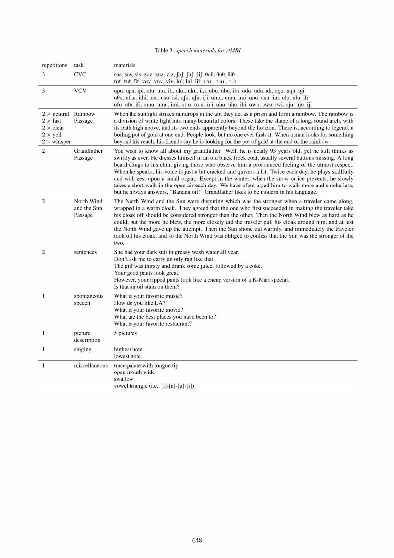
repetitions task materials
3 CVC sAs, sus, sis, zAz, zuz, ziz, SAS, SuS, SiS, TAT, TuT, TiTfAf, fuf, fif, vAv, vuv, viv, lAl, lul, lil, ô Aô , ô uô , ô iô
3 VCV ApA, upu, ipi, AtA, utu, iti, AkA, uku, iki, AbA, ubu, ibi, AdA, udu, idi, AgA, ugu, igiATA, uthu, ithi, AsA, usu, isi, ASA, uSu, iSi, AmA, umu, imi, AnA, unu. ini, AlA, ulu, iliAfA, ufu, ifi, AmA, umu, imi, Aô A, uô u, iô i, AhA, uhu, ihi, AwA, uwu, iwi, AjA, uju, iji
2× neutral2× fast2× clear2× yell2×whisper
RainbowPassage
When the sunlight strikes raindrops in the air, they act as a prism and form a rainbow. The rainbow isa division of white light into many beautiful colors. These take the shape of a long, round arch, withits path high above, and its two ends apparently beyond the horizon. There is, according to legend, aboiling pot of gold at one end. People look, but no one ever finds it. When a man looks for somethingbeyond his reach, his friends say he is looking for the pot of gold at the end of the rainbow.
2 GrandfatherPassage
You wish to know all about my grandfather. Well, he is nearly 93 years old, yet he still thinks asswiftly as ever. He dresses himself in an old black frock coat, usually several buttons missing. A longbeard clings to his chin, giving those who observe him a pronounced feeling of the utmost respect.When he speaks, his voice is just a bit cracked and quivers a bit. Twice each day, he plays skillfullyand with zest upon a small organ. Except in the winter, when the snow or ice prevents, he slowlytakes a short walk in the open air each day. We have often urged him to walk more and smoke less,but he always answers, “Banana oil!” Grandfather likes to be modern in his language.
2 North Windand the SunPassage
The North Wind and the Sun were disputing which was the stronger when a traveler came along,wrapped in a warm cloak. They agreed that the one who first succeeded in making the traveler takehis cloak off should be considered stronger than the other. Then the North Wind blew as hard as hecould, but the more he blew, the more closely did the traveler pull his cloak around him, and at lastthe North Wind gave up the attempt. Then the Sun shone out warmly, and immediately the travelertook off his cloak, and so the North Wind was obliged to confess that the Sun was the stronger of thetwo.
2 sentences She had your dark suit in greasy wash water all year.Don’t ask me to carry an oily rag like that.The girl was thirsty and drank some juice, followed by a coke.Your good pants look great.However, your ripped pants look like a cheap version of a K-Mart special.Is that an oil stain on them?
1 spontaneousspeech
What is your favorite music?How do you like LA?What is your favorite movie?What are the best places you have been to?What is your favorite restaurant?
1 picturedescription
5 pictures
1 singing highest notelowest note
1 miscellaneous trace palate with tongue tipopen mouth wideswallowvowel triangle (i.e., [i]-[a]-[u]-[i])
Table 3: speech materials for rtMRI
648
[4] S. G. Lingala, A. Toutios, J. Toger, Y. Lim, Y. Zhu, Y.-C. Kim,C. Vaz, S. S. Narayanan, and K. S. Nayak, “State-of-the-art mriprotocol for comprehensive assessment of vocal tract structureand function,” in Interspeech 2016, 2016, pp. 475–479. [Online].Available: http://dx.doi.org/10.21437/Interspeech.2016-559
[5] A. D. Scott, M. Wylezinska, M. J. Birch, and M. E. Miquel,“Speech mri: morphology and function,” Physica Medica,vol. 30, no. 6, pp. 604–618, 2014. [Online]. Available:http://dx.doi.org/10.1016/j.ejmp.2014.05.001
[6] A. Toutios and S. S. Narayanan, “Advances in real-time magneticresonance imaging of the vocal tract for speech science andtechnology research,” APSIPA Transactions on Signal andInformation Processing, vol. 5, p. e6, 2016. [Online]. Available:http://dx.doi.org/10.1017/ATSIP.2016.5
[7] Y.-C. Kim, S. S. Narayanan, and K. S. Nayak, “Acceleratedthree-dimensional upper airway mri using compressed sensing,”Magnetic Resonance in Medicine, vol. 61, no. 6, pp. 1434–1440,2009. [Online]. Available: http://dx.doi.org/10.1002/mrm.21953
[8] ——, “Accelerated 3d mri of vocal tract shaping usingcompressed sensing and parallel imaging,” in Acoustics, Speechand Signal Processing, 2009. ICASSP 2009. IEEE InternationalConference on. IEEE, 2009, pp. 389–392. [Online]. Available:http://dx.doi.org/10.1109/ICASSP.2009.4959602
[9] ——, “Flexible retrospective selection of temporal resolution inreal-time speech mri using a golden-ratio spiral view order,”Magnetic resonance in medicine, vol. 65, no. 5, pp. 1365–1371,2011. [Online]. Available: http://dx.doi.org/10.1002/mrm.22714
[10] S. Narayanan, K. Nayak, S. Lee, A. Sethy, and D. Byrd, “Anapproach to real-time magnetic resonance imaging for speechproduction,” The Journal of the Acoustical Society of America,vol. 115, no. 4, pp. 1771–1776, 2004. [Online]. Available:http://dx.doi.org/10.1121/1.1652588
[11] J. M. Santos, G. A. Wright, and J. M. Pauly, “Flexible real-timemagnetic resonance imaging framework,” in Engineering inMedicine and Biology Society, 2004. IEMBS’04. 26th AnnualInternational Conference of the IEEE, vol. 1. IEEE, 2004,pp. 1048–1051. [Online]. Available: http://dx.doi.org/10.1109/IEMBS.2004.1403343
[12] E. Bresch, J. Nielsen, K. Nayak, and S. Narayanan, “Synchro-nized and noise-robust audio recordings during realtime magneticresonance imaging scans,” The Journal of the Acoustical Societyof America, vol. 120, no. 4, pp. 1791–1794, 2006. [Online].Available: http://dx.doi.org/10.1121/1.2335423
[13] V. Toro-Ibacache, V. Z. Munoz, and P. OHiggins, “Therelationship between skull morphology, masticatory muscle forceand cranial skeletal deformation during biting,” Annals ofAnatomy-Anatomischer Anzeiger, vol. 203, pp. 59–68, 2016.[Online]. Available: http://dx.doi.org/10.1016/j.aanat.2015.03.002
[14] B. R. Chrcanovic, M. H. N. G. Abreu, and A. L. N. Custodio,“Morphological variation in dentate and edentulous humanmandibles,” Surgical and radiologic anatomy, vol. 33, no. 3, pp.203–213, 2011. [Online]. Available: http://dx.doi.org/10.1007/s00276-010-0731-4
[15] N. Fakhry, L. Puymerail, J. Michel, L. Santini, C. Lebreton-Chakour, D. Robert, A. Giovanni, P. Adalian, and P. Dessi,“Analysis of hyoid bone using 3d geometric morphometrics: ananatomical study and discussion of potential clinical implica-tions,” Dysphagia, vol. 28, no. 3, pp. 435–445, 2013. [Online].Available: http://dx.doi.org/10.1007/s00455-013-9457-x
[16] R. J. Schwab, M. Pasirstein, R. Pierson, A. Mackley,R. Hachadoorian, R. Arens, G. Maislin, and A. I. Pack,“Identification of upper airway anatomic risk factors forobstructive sleep apnea with volumetric magnetic resonanceimaging,” American journal of respiratory and critical caremedicine, vol. 168, no. 5, pp. 522–530, 2003. [Online]. Available:http://dx.doi.org/10.1164/rccm.200208-866OC
[17] H. K. Vorperian, S. Wang, M. K. Chung, E. M. Schimek, R. B.Durtschi, R. D. Kent, A. J. Ziegert, and L. R. Gentry, “Anatomicdevelopment of the oral and pharyngeal portions of the vocaltract: An imaging study a,” The Journal of the Acoustical Societyof America, vol. 125, no. 3, pp. 1666–1678, 2009. [Online].Available: http://dx.doi.org/10.1121/1.3075589
[18] A. Lammert, M. Proctor, and S. Narayanan, “Morphologicalvariation in the adult hard palate and posterior pharyngealwall,” Journal of Speech, Language, and Hearing Research,vol. 56, no. 2, pp. 521–530, 2013. [Online]. Available:http://dx.doi.org/10.1044/1092-4388(2012/12-0059)
[19] ——, “Interspeaker variability in hard palate morphology andvowel production,” Journal of Speech, Language, and HearingResearch, vol. 56, no. 6, pp. S1924–S1933, 2013. [Online].Available: http://dx.doi.org/10.1044/1092-4388(2013/12-0211)
[20] M. Li, A. Lammert, J. Kim, P. Ghosh, and S. Narayanan, “Au-tomatic classification of palatal and pharyngeal wall shape cat-egories from speech acoustics and inverted articulatory signals,”in ISCA Workshop on Speech Production in Automatic SpeechRecognition, Lyon, France, August 2013.
[21] T. Sorensen, A. Toutios, L. Goldstein, and S. S. Narayanan,“Characterizing vocal tract dynamics across speakers usingreal-time mri,” in Interspeech 2016, 2016, pp. 465–469. [Online].Available: http://dx.doi.org/10.21437/Interspeech.2016-583
[22] E. Godoy, A. Dumas, J. Melot, N. Malyska, and T. F.Quatieri, “Relating estimated cyclic spectral peak frequency tomeasured epilarynx length using magnetic resonance imaging,”in Interspeech 2016, 2016, pp. 948–952. [Online]. Available:http://dx.doi.org/10.21437/Interspeech.2016-1362
649