Embed Size (px)
Citation preview
---- .LIBRARY -INTERNATIONAL tSLAMIC UNIVERSITY MALAYSIA
MIC.ROENCAPSULATION AND CHARACTERIZATION OF GENTAMICIN-PLGA
MICROSPHERE INTENDED FOR ORTHOPAEDIC INFECTION
BY
AHMAD FAHMI BIN HARUN @ ISMAIL
A dissertation in fulfilment of the requirement for the degree of Master in Pharmaceutical Science
(Pharmaceutical Technology)
Kulliyyah of Pharmacy International Islamic University
Malaysia
AUGUST2012
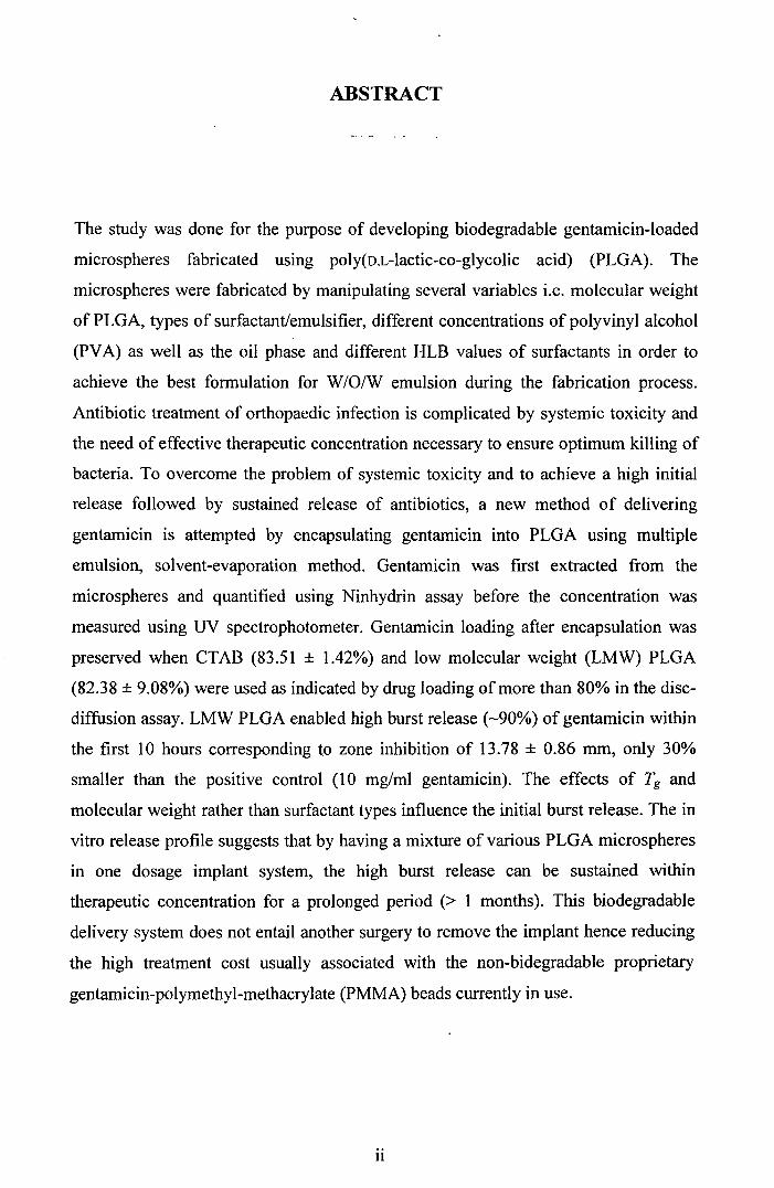
ABSTRACT
The study was done for the purpose of developing biodegradable gentamicin-loaded
microspheres fabricated using poly(D.L-lactic-co-glycolic acid) (PLGA). The
microspheres were fabricated by manipulating several variables i.e. molecular weight
of PLGA, types of surfactant/emulsifier, different concentrations of polyvinyl alcohol
(PVA) as well as the oil phase and different HLB values of surfactants in order to
achieve the best formulation for W /0/W emulsion during the fabrication process.
Antibiotic treatment of orthopaedic infection is complicated by systemic toxicity and
the need of effective therapeutic concentration necessary to ensure optimum killing of
bacteria. To overcome the problem of systemic toxicity and to achieve a high initial
release followed by sustained release of antibiotics, a new method of delivering
gentamicin is attempted by encapsulating gentamicin into PLGA using multiple
emulsion, solvent-evaporation method. Gentamicin was first extracted from the
microspheres and quantified using Ninhydrin assay before the concentration was
measured using UV spectrophotometer. Gentamicin loading after encapsulation was
preserved when CTAB (83.51 ± 1.42%) and low molecular weight (LMW) PLGA
(82.38 ± 9.08%) were used as indicated by drug loading of more than 80% in the disc
diffusion assay. LMW PLGA enabled high burst release (-90%) of gentamicin within
the first 10 hours corresponding to zone inhibition of 13.78 ± 0.86 mm, only 30%
smaller than the positive control (10 mg/ml gentamicin). The effects of Tg and
molecular weight rather than surfactant types influence the initial burst release. The in
vitro release profile suggests that by having a mixture of various PLGA microspheres
in one dosage implant system, the high burst release can be sustained within
therapeutic concentration for a prolonged period (> 1 months). This biodegradable
delivery system does not entail another surgery to remove the implant hence reducing
the high treatment cost usually associated with the non-bidegradable proprietary
gentamicin-polymethyl-methacrylate (PMMA) beads currently in use.
11
~.J-='31 f,~4 ~t..~4 ~ ',7~, JblU ~Li ~,~~ .J:!P t..i1.;~1 o~ u~
POL Y(D,L-LACTIDE-CO-GL YCOLIDE) l_;~l..9 PLGA ~ ~ •
04 J:!Al4 ~I~ 04 ~I~~ ~W:,..JI u1:bi,,Loll ~~ ul~..9_;S;-JI
''F~'il J.oWl/~I <>- ~WI J.,WI e.~ ,~;.Jl ~~I wJJll ~.J ,ul~I
J~I J.: i;9 ~.J-='31 04 4 ili; 0 J:!SIJiPV A A u;; o.11 ~I.J ~jll ~I c)! u~~4
..llHLB r;;'ii ~ .~oill ~ 9,~j 9,I.A/~.J/9,I.A •.1bi11Lo.ll ~ ~i c)! ~_,ll
t'l~4 ~I ~-.,1.J'.JJA ~ ~ u43~ o~ ~ ~ 4t.l,.4o.ll ul~..9_;S;-JI
u~, ~.tt, • f. .. l~La uUi ~- t1 Juui '-' • .. ~i Lo.11 • .-:(tvl ,A_ At' • ._)j.t*' 9'-""° ~ t' • ....)'1'- • ~..)_J,I .. .J ~ e,,r.JJ'**' f ~
. & L- _t, . I.A~I ~.(.. Lall • ,.A~~tL, 0 \Lo.11 A.:L .t. o:: .. t.-: .. ,~j t1 '?'" ~ ~ • ~JJ U"' .. ,.J:U~ • _;; .. J- ~ ~ c,s-.
~ ~ ~1.A~I ~ld .~l..9-lll ~I ~ld ~u.; c>, J~ ul~..9_;S;-JI
u4..9~ JWMll _»iCUI l,.,,1_;.ll ~.)JI ~ } ,~ti..a~I ~~ 1+,o;;'ii ~ ul_~..9_;S;-JI
~.lll A.:Jjija.11 u1_;µ1 ~S. aureus ~I~ ~li 4:~! i_»ib ~wll u~i Ji..9
,•.e.. J.a .... t.---: 4.Jld IA .t ... _ a.ai~I ~ Ji • 1.A~l 4.Jld ~~ ·i t1 " w ,._,- .. JJ ~ .. -~ ~ • .. u .. . (.J ~ll ~
.JI t'l~4 4t.l,.o.o.ll ul~..9.~ 14'i~hi ~ 0/o80CTAB ~I c>, ~\.9 J.ola.S
~ .u.,:illJ 14000 ~~' wJJll (jj ~.J-='31 t'1~4 4t.l..o.o.11 ul~..9_;S;-JI ~i.J
.lb_,I J!..9 o/020 ~ J:!j:i ~ ~ld ~ ~I ul~.J~ _;~I [ j~ _;~I
· t-..1.:. u .. Ji · 1.A~I • o/ 80 • i ~ • I.Al.:wJJ .Jj · • t1 ·~1 ·- -:t1 ~ _)~ ~ • (.)A /0 U .. ~ • ~-..>- '-r • _)J-1"'
u.,:illJ 14000 ~.J-='31 t'l~4 o~I ul~..9.A!,,oll 04 c)..9~1 ~\.wJI PLGA ~ •
.. I ui...... .( ··'I • ul • .. · · ·· • .:. t La 30 .. • 1.Al.:wJJ J.a.\.o.11 ·- -:t1 · t ·- · .. -: '-,I ..n--.JJ'*:l"'""' (.)A ~ u-- ~ ~ ~ . .. _)J-1"' ~
t'l~4 4t.l..o.o.11CTAB .J TRITON X-100 °/06.J 0/o5 ,~I c>, ~tj J.ol.Jl,S
..1l J:!SJi c)! U~} ~jll ~I J:!Sljll ~/6J.JPVA ul~.J~ .~/6J.J 0/ol
.JIPLGA lAJJ~.J lf:l,..,al_;J c) ~jill ~ ~ o~ ~ti.a! u~i ~I.A~4 Uu.oll
.9'1..9.lll ~~I 9'U:i~I ~ J'+4 c) _;~~I¥ 0$.l..9 ~JAS
11l
ABSTRAK
Kajian itu dilakukan b&gi tujuan membangunkan gentamicin yang dimuatkan ke
dalam mikrosfera terbiodegradasi menggunakan poli (D.L-laktik-co Glycolik asid)
(PLGA). Mikrosfera telah direka oleh memanipulasi beberapa pembolehubah iaitu
berat molekul PLGA, jenis surfaktan / pengemulsi yang berbeza kepekatan polivinil
alkohol (PVA) serta fasa minyak dan nilai-nilai yang berbeza HLB surfaktan untuk
mencapai rumusan terbaik untuk W / 0 / W emulsi ketika proses fabrikasi. Rawatan
ortopedik bagi jangkitan antibiotik rumit oleh ketoksikan sistemik dan memerlukan
kepekatan terapeutik yang berkesan diperlukan untuk memastikan pembasmian
bacteria yang optimum. Untuk mengatasi masalah ketoksikan sistemik dan untuk
mencapai kadar antibiotik awal yang tinggi yang diikuti oleh kadar antibiotic yang
berterusan, satu kaedah baru telah digunakan dengan memberikan gentamicin
menggunakan emulsi berganda, pelarut-kaedah penyejatan. Gentamicin diekstrak dari
mikrosfera dan dengan menggunakan ninhydrin kepekatan gentamicin diukur
menggunakan spektrofotometer UV. Muatan gentamicin selepas pengkapsulan telah
dikekalkan apabila CTAB (83,51 ± 1.42%) dan berat molekul yang rendah (LMW)
PLGA (82,38 ± 9,08%) telah digunakan seperti yang ditunjukkan oleh jumlah
antibiotik lebih daripada 80% dalam cerakin cakera-resapan. LMW PLGA
membolehkan pembebasan gentamicin yang tinggi (- 90%) dalam tempoh 10 jam
pertama sepadan dengan zon perencatan daripada 13, 78 ± 0,86 mm, hanya 30% lebih
kecil daripada kawalan positif (10 mg / ml gentamicin). Kesan Tg dan berat molekul
dan bukannya jenis surfaktan mempengaruhi pelepasan pecah awal. Dalam profil
pelepasan vitro mencadangkan bahawa dengan mempunyai campuran pelbagai PLGA
mikrosfera dalam satu sistem dos implan, pelepasan pecah yang tinggi boleh
dikekalkan dalam kepekatan terapeutik untuk tempoh yang lama(> 1 bulan). Sistem
penyampaian terbiodegradasi ini tidak melibatkan pembedahan untuk membuang
implan seterusnya mengurangkan kos rawatan yang tinggi yang biasanya dikaitkan
dengan mamc bukan-biodegradasi proprietari gentamicin-polimetil-metakrilat
(PMMA) yang kini dalam penggunaan di hospital-hospital.
iv
APPROVAL PAGE
I certify that I have supervised and read this study and that in my opinion, it conforms to acceptable standards of scholarly presentation and is fully adequate, in scope and quality, as a thesis for the degree of Master in Pharmaceutical Science
(Pharmaceutical Technology). . ... ~~ ............................... . F~4~d~h
1Mohamed
Supervisor
I certify that I have read this thesis and that in my opinion, it conforms to acceptable standards of scholarly presentation and is fully adequate, in scope and quality, as a thesis for the degree of Master in Pharmaceutical Science (Pharmaceutical
Technology). . .... k ........................................ . Kausar Ahmad Internal Examiner
Wong Tin Wui External Examiner
This dissertation was submitted to the Department of Pharmaceutical Technology and is accepted as a partial fulfilment of the requirements for the degree of Master in Pharmaceutical Science (Pharmaceutical Technology) .
. . .. ·:;;;i;;;;;~ Md.j:ii~ ...................... . Head Department of Pharmaceutical Technology
This dissertation was submitted to the Kulliyah of Pharmacy and is accepted as a partial fulfilment of the requirements for the degree aster in Pharmaceutical Science (Pharmaceutical Technology).
Dean, Kulliyyah of Pharmacy
V
DECLARATION PAGE
I hereby declare that this dissertation is the result of my own investigations, except
where otherwise stated. I also declare that it has not been previously or concurrently
submitted as a whole for any other degrees at IIUM or other institutions.
Ahmad Fahrni Bin Harun@Ismail
Signature ......................................... . Date ......................... .
VI
INTERNATIONAL ISLAMIC UNIVERSITY MALAYSIA
DECLARATION OF COPYRIGHT AND AFFIRMATION OF FAIR USE OF UNPUBLISHED RESEARCH
Copyright ©2011 by International Islamic University Malaysia. All rights reserved.
MICROENCAPSULATION AND CHARACTERIZATION OF GENTAMICIN-PLGA MICROSPHERE INTENDED FOR ORTHOPAEDIC
INFECTION
I hereby affirm that The International Islamic University Malaysia (IIUM) hold all
rights in the copyright of this Work and henceforth any reproduction or use in any
form or by means whatsoever is prohibited without the written consent of IIUM. No
part of this unpublished research may be reproduced, stored in a retrieval system, or
transmitted, in any form or by means, electronic, mechanical, photocopying,
recording or otherwise without prior written permission of the copyright holder.
Affirmed by Ahmad Fahrni Bin Harun @ Ismail
Signature Date
Vll
.. To My Beloved Wife and Daughter .. .. The Apple of My Eye ..
Vlll
ACKNOWLEDGEMENTS
Alhamdulillah. As all praises are belong to Almighty God. Peace and blessing be upon our beloved Prophet SAW. First and foremost, I would like to thank to ALLAH as by HIS permission and will, I have completed my research and this thesis successfully. It was indeed a great experience to complete such an important task in order for me to graduate. May ALLAH provide benefits from this research to those who seek knowledge and make this research as a reason for others to increase their Iman towards HIM.
Here I would like to thank my parents, Hj. Harun @ Ismail bin Yusof and Hjh. Juwariah bt Mustafa and also all my siblings for supporting me during my studies here and also for giving me many ideas on how to improve my research. I appreciated all the ideas and suggestions.
And also a very big thank to my supervisor, Asst. Prof. Dr. Farahidah Mohamed for helping me and giving me guidance throughout the research and giving me endless advises in completing this thesis.
I would like to dedicate this thesis to my beloved wife, Nurul Aziyana Kamaruddin for her supports and for being with me through thick and thin, looked after our lovely daughter Nurhusna when I had to do my lab work. May ALLAH shower us with happiness and blessing till the end. InsyaALLAH.
Next, I want to express my gratitude to all lab assistants that were involved in this research especially Sis. Haryanti, Bro. Dzadil, and our science officer of Pharmaceutical Technology Department, Sis. Zaililah for their assistances during the lab works.
Not to forget, the important persons throughout the experiment, my project colleagues; AbdAlmonem, Aimen and Mulham. They had been helping me a lot during the research and providing me with many ideas to make our research into reality. Special gratitude also goes to Bro. Saadi for being my SPSS expert, and also to my good friend, Usop, for all those lame jokes when I needed one. Thank you.
To all my friends, Bro. Suhaib, Bro. Anung, Pak Dedi, Bro. Firdaus, Bro. Muhammad and Bro. Abdurrahman, I thank all of you and may ALLAH repay you with HIS blessing and HIS mercy, here and hereafter. JnsyaALLAH
Thank you, Wassalam.
IX
TABLE OF CONTENTS
Abstract ......................................................................................................................... ii Abstract in Arabic .............................................................................. iii Abstract in Malay ........................................................................................................ iv Approval Page .............................................................................................................. v Declaration Page .......................................................................................................... vi Copyright Page ........................................................................................................... vii Dedication ................................................................................................................. viii Acknowledgements ............................................................................. .ix List of Tables .................................................................................. xiii List of Figures ............................................................................................................ xvi List of Abbreviations ................................................................................................. xix
CHAPTER 1: .............................. ............................................. .......... 1 1.1 Introduction .............................................................................................. 1 1 .2 Research Background .............................................................................. 2 1.3 Literature Review .................................................................................... 6
1.3.1 Poly (D,L-Lactic-Co-Glycolic Acid) (PLGA) ............................... 6 1.3.1.1 The Overview ............................................................... 6 1.3.1.2 Glass Transition Temperature (T g) ............................. l 0 1.3.1.3 Related Work .............................................................. 12
1.3.2 Gentamicin ............................... '. ................................................... 14 1.3.2.1 An Overview .............................................................. 14 1.3.2.2 Gentamicin Toxicity ................................................... 15 1.3.2.3 Pharmacokinetic ......................................................... 17 1.3.2.4 PMMA Beads ............................................................. 18
1.3.3 Double Emulsion Solvent Evaporation Method of Microsphere Fabrication ................................................................................... 20
1.3 .3 .1 The Overview ............................................................. 20 1.3.3.2 Surfactants .................................................................. 2 I 1.3 .3 .3 Chitosan Microspheres ............................................... 24 1.3.3.4 Drug Release Mechanism ........................................... 25
1.4 Objectives and Scope of Study .............................................................. 29 1.5 Hypotheses ............................................................................................. 30
CHAPTER 2: DEVELOPMENT OF GENTAMICIN-LOADED PLGA MICROSPEHERES ................................................................................................. 31
2.1 Introduction ............................................................................................ 31 2.2 Materials and Chemicals ........................................................................ 32 2.3 Methodology .......................................................................................... 33
2.3.1 Fabrication OfGentamicin-Loaded Microspheres ...................... 33 2.4 Characterization of Microspheres .......................................................... 41
2.4.1 Particle Morphology .................................................................... 41
X
2.4.2 Particle Size Analysis ................................................................. .41 2.4.3 Drug Loading ............................................................................... 42 2.4.4 Gentamicin Stock Preparation .................................................... .43 2.4.5 Construction Of Gentamicin Standard Curve ............................. .44 2.4.6 Ninhydrin Assay .......................................................................... 44 2.4.7 HLB Preparation .......................................................................... 45 2.4.8 Statistical Analysis ....................................................................... 46
2.5 Result and Discussion ............................................................................. 47 2.5.1 Particle Size Distribution, Morphology and Drug Loading
Efficiency ..................................................................................... 4 7 2.5.1.1 Surfactant Study ........................................................ .47 2.5.1.2 HLB Value ................................................................. 55 2.5.1.3 Molecular Weight of PLGA ....................................... 60 2.5 .1.4 PV A Concentration .................................................... 63 2.5.1.5 PLGA Concentration in Oil Phase ............................. 68
CHAPTER 3: IN-VITRO RELEASE AND ANTIMICROBIAL STUDY .......... 74 3.1 Introduction ............................................................................................ 74 3 .2 Material .................................................................................................. 7 5 3.3 Method ................................................................................................... 75
3.3.1 Fabrication ofMicrospheres ........................................................ 75 3.3.2 In-Vitro Release Profile ............................................................... 76 3.3.3 Antimicrobial Study ..................................................................... 77
3.3.3.1 Nutrient Agar Preparation .......................................... 77 3.3.3.2 Nutrient Broth Preparation ......................................... 77 3.3.3.3 Preparation of Bacterial Culture ................................. 77 3.3.3.4 McFarland Standard for Bacterial Culture ................. 78 3.3.3.5 Extraction of Gentamicin From Microspheres ........... 78 3.3.3.6 Disk Diffusion Assay ................................................. 79
3.4 Result & Discussion .............................................................................. 80 3.4.1 In-Vitro Release Profile ............................................................... 80 3 .4.2 Antimicrobial Study ..................................................................... 86
CHAPTER 4: GENTAMICIN-LOADED CHITOSAN/PLGA MICROSPHERES .................................................................................................................................... 91
4.1 Introduction ............................................................................................ 91 4 .2 Material ......................................................................... , ........................ 92 4.3 Methodology .......................................................................................... 93
4.3.1 Fabrication of Gentamicin-Loaded Chitosan/PLGA Microspheres ..................................................................................................... 93
4.3.2 Microspheres Characterization .................................................... 95 4.3.2.1 Surface Morphology ................................................... 95 4.3.2.2 Particle Size Distribution ........................................... 95 4.3.2.3 Drug Loading Efficiency ............................................ 95
4.4 Results & Discussion ............................................................................. 96 4.4.1 Morphology ................................................................................. 96 4.4.2 Particle Size ................................................................................. 96 4.4.3 Drug Loading ............................................................................... 98
Xl
CHAPTER 5: GLASS TRANSITION OF PLGA MICROSPHERES .............. 102 5 .1 Introduction .......................................................................................... 102 5.2 Material ................................................................................................ 103 5.3 Method ................................................................................................. 103 5.4 Results & Discussion ........................................................................... 104
5.4.1 Effect of PLGA Molecular Weight on Tg .................................. 106 5.4.2 Effect of Surfactant Types on Tg of the Gentamicin-Loaded
Microspheres .............................................................................. 108 5.4.3 Effect of Different PVA Concentrations Used In Aqueous 2 and
Hardening Tank on Tg···· ............................................................ 110 5.4.4 Effect of Concentration of PLGA in Oil Phase on Tg ................ 112 5.4.5 Effect ofHLB Values on Tg .................................... : .................. 113
CHAPTER 6: GENERAL DISCUSSION ............................................................ 115
CHAPTER 7: CONCLUSION .............................................................. 122 7.1 Future Study ......................................................................................... 122
BIBLIOGRAPHY ............................................................................. 124
PUBLICATIONS AND PRESENTATIONS ............................................ 135
APPENDIX A .................................................................................... 136
Xll
Table No.
1.1
2.1
2.2
2.3
2.4
2.5
2.6
2.7
2.8
2.9
LIST OF TABLES
Decreasing order of nephrotoxic aminoglycosides used systemically. 1 is the highly toxic, 5 is the least toxic (Decker & Molitoris, 2008).
Composition of microsphere fabrication process for the various types of surfactant.
Composition of microspheres fabrication process for different concentration of PLGA.
Composition of microsphere fabrication process with different HLB values of surfactants used.
Composition of microsphere fabrication process to study the effect of molecular weight of PLGA employed to synthesize the microspheres.
Composition of microsphere fabrication process with different concentrations of PV A for aqueous 2 and hardening tank.
The HLB value for the nonionic surfactants used provided by the (Manual Safety Data Sheet) MSDS from the supplier.
Particle size distribution of gentamicin-loaded microspheres synthesized with selected surfactants. Data were expressed as mean± standard deviation (S.D); n=3.
Particle size distribution of gentamicin-loaded microspheres synthesized with selected HLB values of surfactant blends. Data are expressed as the mean ± standard deviation (S.D); n=3.
Particle size distribution of gentamicin-loaded microspheres synthesized with selected molecular weight of PLGA. Data are expressed as the mean ± standard deviation (S.D); n=3 and analyzed by ANOV A Tukey multiple comparison test.
Xlll
Page No.
16
35
36
38
39
40
45
47
57
61
Table No.
2.10
2.11
3.1
4.1
4.2
5.1
5.2
5.3
5.4
Particle size distribution of gentamicin-loaded microspheres with selected PV A concentration for aqueous 2 and hardening tank. Data are expressed as the mean± standard _deviation (S.D); n=3.
Particle size distribution of gentamicin-loaded microspheres synthesized with selected PLGA concentration. Data are expressed as the mean ± standard deviation (S.D); n=3
The ingredients of four formulations out of six formulations of gentamicin-loaded PLGA microspheres that achieved more than 20% drug loading efficiency and more. I% w/v aqueous PV A were employed in aqueous 2 and in hardening tank for all formulations.
The chitosan concentration used m formulation of gentamicin-loaded chitosan/PLGA microspheres.
Particle size distribution of gentamicin-loaded microspheres synthesized with selected chitosan concentration. Data expressed as the mean ± standard deviation (S.D); n=3.
The onset, midpoint and endpoint of glass transition for the gentamicin-loaded microspheres fabricated using different PLGA molecular weight. The data tabulated was collected post quench cool.
The onset, midpoint and endpoint of glass transition for the gentamicin-loaded microspheres (100 kDa PLGA) fabricated using different types of surfactant. The data tabulated was collected post quench-cooled.
The onset, midpoint and endpoint of glass transition for the gentamicin-loaded microspheres (100 kDa PLGA) fabricated using different concentrations of PV A m Aquoues 2 and hardening tank. The data tabulated was collected post quench cool.
The onset, midpoint and endpoint of glass transition for the gentamicin-loaded microspheres (100 kDa PLGA) fabricated using different concentrations of oil phase. The data tabulated was collected post quench cool
xiv
Page No.
65
70
75
94
97
106
108
111
112
Table No.
5.5 The onset, midpoint and endpoint of glass transition for the gentamicin-loaded microspheres (100 kDa PLGA) fabricated using different concentrations of oil phase. The data tabulated was collected post quench cool.
xv
Page No.
113
Figure No.
1.1
1.2
1.3
2.1
2.2
2.3
2.4
2.5
LIST OF FIGURES
Examples of hydrolytic biodegradable polymers (Hoffman, 2006).
(a) Formation of PLGA polymer from lactic acid and glycolic acid and (b) degradation of PLGA chain into its monomers via hydrolysis (Bouissou & Walle, 2006).
The schematic diagram drawn shows how the drug released from the microenvironment of small particle (S. Yang & Washington, 2006).
Schematic diagram to prepare gentam1cm PLGA Microspheres. Oil Phase (#), Aqueous I (€), Primary Emulsion (Q) and Secondary Emulsion (~).
Surfaces morphologies of the microspheres fabricated using PV A in the secondary emulsion and the following surfactants in the primary emulsion: (A) PVA; (B) Tween 20; (C) Tween 80; (D) Span 80; (E) Span 85; (F) SDS; (G) CTAB and (H) Triton X-100.
The bar chart illustrates the data for drug loading efficiency based on types of surfactant used. The group marked with asterisks showed significantly higher with 95% confidence level (P<0.05) when compared with the control group (PV A).
Surfaces morphologies of the microspheres fabricated using PV A in the secondary emulsion and surfactant blends with the following HLBs in the primary emulsion: (A) HLB 4; (B) HLB 10; (C) HLB 13.5 and (D) HLB 16.
Drug loading of gentamicin microspheres with difference HLB of surfactant. All formulations showed statistically no significant different compared to control (PV A). The HLB consisted of the surfactant blends mentioned in Table 2.7.
XVI
Page No.
7
8
26
34
50
52
56
58
Figure No.
2.6
2.7
2.8
2.9
2.10
2.11
2.12
Surfaces morphologies of the microspheres fabricated using Tween 80 in primary emulsion, PV A in the secondary emulsion and the following PLGA molecular weight in the primary emulsion: (A) PLGA 14 kDa; (B) PLGA 34 kDa and (C) PLGA 100 kDa.
Drug loading of gentamicin in microspheres fabricated based on different molecular weight of PLGA. Data was analyzed using multiple comparison Tukey Test.
Surfaces morphologies of the microspheres fabricated using the following PV A concentration for Aquoues 2 and hardening tank: (A) 1 % w/v PV A; (B) 3% w/v PV A and (C) 5% w/v PV A. All microspheres were fabricated employing PLGA 100 kDa, Triton X-100 as surfactant in primary emulsion, 1 % PV A in aqueous 2 and in the hardening tank
Drug loading of gentamicin in microspheres fabricated based on different concentrations of PV A. Asterisk indicates the drug loading is significantly higher than control (PVA) based on One-way ANOVA (Dunnett's Test).
Surfaces morphologies of the microspheres fabricated using PV A in the secondary emulsion and the following oil phase concentrations in the primary emulsion: (A) 5% w/v; (B) 10% w/v and (C) 20% w/v
Drug loading of gentamicin in microspheres fabricated based on different concentrations of PLGA in oil phase. Asterisks showed drug loading significantly higher than the control group (microspheres fabricated with PV A).
The bar chart illustrates the 6 formulations of gentamicin microspheres with more than 20% drug loading efficiency and a control group. The figures were the average taken from triplicate measurements (n=3). All 6 formulations showed significantly higher drug loading efficiency (marked with asterisk) with 95% confidence level (P<0.05) when compared against microspheres fabricated using PV A as surfactant in the primary emulsion ( control group)
xvu
Page No.
61
62
64
66
68
71
73
Figure No .:.
3.1
3.2
3.3
3.4
4.1
4.2
4.3
5.1
Configuration of the four points (1 - 4) for measurement of the diameters of inhibition zone. The centre point (C) corresponds to the paper disc centre.
The cumulative data (%) for the release profile of gentamicin microspheres plotted as mean values with error bar; n=3.
The zone of inhibition of microspheres fabricated using (A) Fl formulation, (B) F2 formulation, (C) F3 formulation, (D) F4 formulation, (E) naked gentamicin at 10 mg/ml and (F) positive control marked with ([+ve] ctrl) and negative control marked with ([-ve] ctrl) and PBS.
The average zone of inhibition (mm); n=3, based on the four formulations (Fl-F4) and 'naked' gentamicin (10 mg/ml) as positive control. The negative control was not included as the value for zone of inhibition was zero. Data for four formulations were analyzed using multiple comparison Tukey test.
The SEM images of gentamicin microspheres fabricated by using three different concentrations of chitosan added in aqueous 2 and hardening tank labeled with (A) Formulation 1, (B) Formulation 2 and (C) Formulation 3.
The bar charts represent mean data for drug loading with error bar for triplicate samples of gentamicin microspheres for chitosan concentration study. One-way ANOVA (Dunnett's Test) showed significantly higher (P<0.05) with 95% confidence level (marked with asterisks) compared with control group.
The chemical structure of (A) acetylated chitin and (B) chitosan (Aranaz et al., 2009).
Simplified schematic diagram illustrates the process of determining glass transition temperature (Tg). The quench cool step marked with *.
xvm
Page No .
79
83
87
88
96
99
101
104
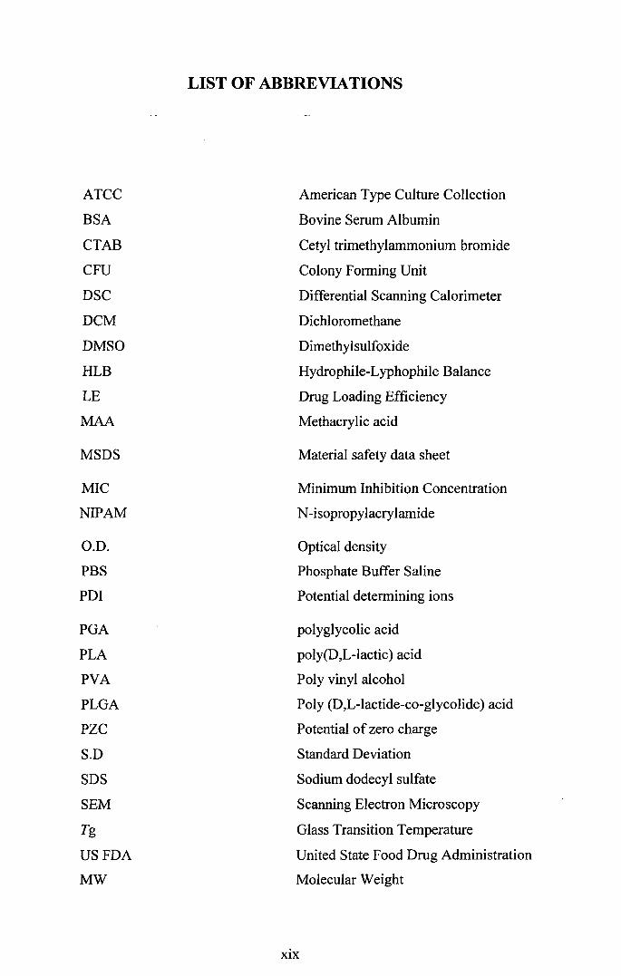
LIST OF ABBREVIATIONS
ATCC American Type Culture Collection
BSA Bovine Serum Albumin
CTAB Cetyl trimethylammonium bromide
CFU Colony Forming Unit
DSC Differential Scanning Calorimeter
DCM Dichloromethane
DMSO Dimethylsulfoxide
HLB Hydrophile-Lyphophile Balance
LE Drug Loading Efficiency
MAA Methacrylic acid
MSDS Material safety data sheet
MIC Minimum Inhibition Concentration
NIPAM N-isopropylacrylamide
O.D. Optical density
PBS Phosphate Buffer Saline
PDI Potential determining ions
PGA polyglycolic acid
PLA poly(D,L-lactic) acid
PVA Poly vinyl alcohol
PLGA Poly (D,L-lactide-co-glycolide) acid
PZC Potential of zero charge
S.D Standard Deviation
SDS Sodium dodecyl sulfate
SEM Scanning Electron Microscopy
Tg Glass Transition Temperature
USFDA United State Food Drug Administration
MW Molecular Weight
xix
CHAPTER ONE
1.1 INTRODUCTION
The science of drug delivery can be defined as the control of the in vivo temporal and
spatial location of drug molecules for clinical purposes by the biological and chemical
applications (Uchegbu, 2006). As a drug is being administered, only small fraction of
the dosage will reach the intended site and give the pharmaceutical effect. The rest
will be wasted in a few ways either being taken up by the wrong tissue, being
removed by the tissue too fast before the drug can exhibit the effect or being
metabolized on its way to the target site by many possible enzymatic processes
(Uchegbu, 2006). There are two main reasons for researchers to persistently continue
explore in the field of drug delivery which are to maximize the drug activities and to
minimize the side effects (Allen & Cullis, 2004).
Nowadays research on advanced drug delivery has become more and more
demanding in the scientific world as there are several reasons for this booming
phenomenon. This is due to increase use of biological materials with poorly
understood physical properties or questionable shelf life, emergence of more
challenging low-molecular-weight molecules and biomacromolecules with either poor
aqueous solubility or poor tissue permeation and realization of having to control the
release of some portion of drug on the target sites to reduce the toxicity and to give
higher therapeutic index of that drug itself (Uchegbu, 2006). In addition, some clinical
advancements have also been the driving force for such researches to take place,
especially those treatment which are better using the implant system rather than using
the conventional way.
1
1.2 RESEARCH BACKGROUND
Clinical problems arise from infection of the bone due to fractures or post surgery
complications are amongst the problems faced by the general practitioners nowadays
and yet the best possible solutions are still under debate amongst the scholars. A
common, debilitating infection in orthopaedics setting is osteomyelitis, mainly caused
by Staphylococcal aureus (Balmayor et al., 2011). It is an inflammatory bone disease
with deep bone involvement infiltrating the medullary cavity, cortex and periosteum
(Chung, 2001). Osteomyelitis normally arises as a nosocomial infection due to post
operative orthopaedic surgeries, introduced during implantation of a prosthesis or
carried to the biomaterial surface by a temporary bacteraemia where they adhere and
grow to form a biofilm (Neut et al., 2001). Osteomyelitis is especially complicated if
patients are immunocompromised (Brin et al., 2008; Chung, 2001 ).
In addition, osteomyelitis is one of the inflammatory bone diseases caused by
infectious bacteria which can induce microbial infection of the medullary cavity of the
bone, the cortex as well as the periosteum (Chung, 2001). The infections are either
introduced during implantation of a prosthesis or are carried to the biomaterial surface
by a temporary bacteraemia, where they adhere and grow to form a biofilm (Neut et
al., 2001). But the most common cause of this bone disease is the post-operative
sepsis following orthopaedic procedures which has become a major concern amongst
the orthopedic surgeons (Chung, 2001).
In the case of osteomyelitis, the treatment is sometimes quite complicated
since the optimum concentration level of the antibiotic at the site of infection is
difficult to be achieved via systemic administration. Many contributing factors have
been identified which lead to this particular problem in treating such infection.
Successful treatment is difficult to achieve without causing irreversible side effects to
2
the patient i.e. the systemic toxicity of the antibiotic which in return prohibits a high
systemic dose of the drug administrated (Stephens et al., 2000). Besides, inherent
problems of the drug such as short half-life of the antibiotic and poor circulation to the
infected area can slow down the rate of therapeutic outcome. However, the problems
associated with the drug itself can be mitigated by re-formulating the drug or creating
a carrier system that can directly deliver the drug to the site of infection.
One of the conventional treatments for osteomyelitis which is still being
practiced clinically is by local administration of antibiotic either by spraying or by
injecting to the infected site followed by oral administration to optimize the
therapeutic effect of the drug given (Xue & Shi, 2004). Although injection
(intravenous) can give adequate blood level of gentamicin, the long-term indwelling
catheter as well as the daily dose of administration remain the drawbacks of the
conventional method (Brin et al., 2008). This method will give rise to the typical drug
concentration in the blood that shows peaks and valleys wherein below the therapeutic
level the clinical effect of the drug is ineffective while above the minimum toxic
concentration (MTC), the side effects can be greatly manifested clinically.
Hence, the toxicity and the minimum effective levels give a very narrow
window for any mistakes to happen in administrating any drugs. The toxicity of
gentamicin to the patient which can cause ototoxic and nephrotoxic effects is the main
concern whenever the patient is administered with gentamicin (Chaisri, Ghassemi,
Hennink & Okonogi, 2011). The toxicity of gentamicin in treating bone infections for
instance, researchers have been considering alternative ways of administrating the
drug by using polymers to get a controlled release effect. Besides, it has been proven
that by using an implant system to administer gentamicin, the risk of ototoxicity and
nephrotoxicity can be minimized (Chaisri et al., 2011; Chang, Perrie & Coombes,
3
2006). That is why amongst the treatment options for osteomylistis is by using a local
implant containing polymethyl-methacrylate (PMMA) beads or calcium phosphate
bone cements. PMMA by itself delivers several advantages as vehicle for gentamicin
(Brin et al., 2008). However, a major problem associated with this PMMA beads; is its
non-biodegradability which requires second surgery to remove them.
The progression of scientific findings nowadays have further shown a few
biodegradable polymers can be used in medical aspects due to their hydrolysable
backbone linkages such as orthoester, anhydride, ester, carbonate, urea and urethane.
These polymers have undergone many scientific tests and to date, two of them seem to
get the spotlight in medical field. These two biodegradable and biocompatible
polymers namely poly(glycolic acid) or PGA and poly(o,L-lactic acid) or PLA have
been showing promising biological natures and have received approval by the US
FDA to be used on human (Di Toro, Betti & Spampinato, 2004)
One of the most commonly employed co-polymers in drug delivery is Poly(o.L
lactic-co-glycolic acid) (PLGA) a combination of PLA and PGA. Having the property
to degrade naturally in the human body without creating any harmful by products
systemically, PLGA is a promising candidate to be used as micro carrier system for
gentamicin for several reasons which are (1) to protect the drug from being
metabolised prematurely; (2) to deliver the drug safely to the infection site; (3) to
provide a controlled release of the gentamicin which can be extended to a significant
period oftime depending on the PLGA molecular weight used (Fischer, Foerg, Merkle
& Gander, 2004 ). Hence, gentamicin-loaded PLGA microsphere is envisaged to solve
the problem of having multiple administrations via the conventional way as it can
replace it with a single dose which can give a sustained-release over several weeks to
months.
4
In this study, gentamicin-loaded PLGA microspheres were fabricated by using
water-in-oil-in-water (w/o/w) double emulsion solvent evaporation method. Even
though there were some extensive studies on gentamicin microspheres, a lot more
variables and parameters are waiting to be manipulated in order to fabricate the best
microspheres with the best formulation. The background of this study is to manipulate
several parameters such as different molecular weight of PLGA, different
concentrations of poly-vinyl alcohol (PV A), different Hydrophile Lipophile balance
(HLB) values of the surfactants, different types of surfactant or emulsifier used such
as PV A, Tween 20, Span 80, Tween 80, Span 85, Triton XI 00 and combinations of
the surfactants and last but not least, the concentration of oil phase used ranging from
5% up to 20%. The influences of different concentrations of chitosan in fabricating the
microspheres were also studied. 0.1 %, 0.3% and 0.5% of chitosan concentrations were
used during the fabrication process.
The fabricated microspheres were characterized based on the particle size
distributions, the external morphologies, the encapsulation efficiency of gentamicin
inside the microspheres and the profiling of the release patterns of gentamicin-loaded
PLGA microspheres over time. The outcome of the study is intended to broaden the
knowledge under this field apart from giving an alternative way of administrating
small molecule, water-soluble drugs in the future.
5